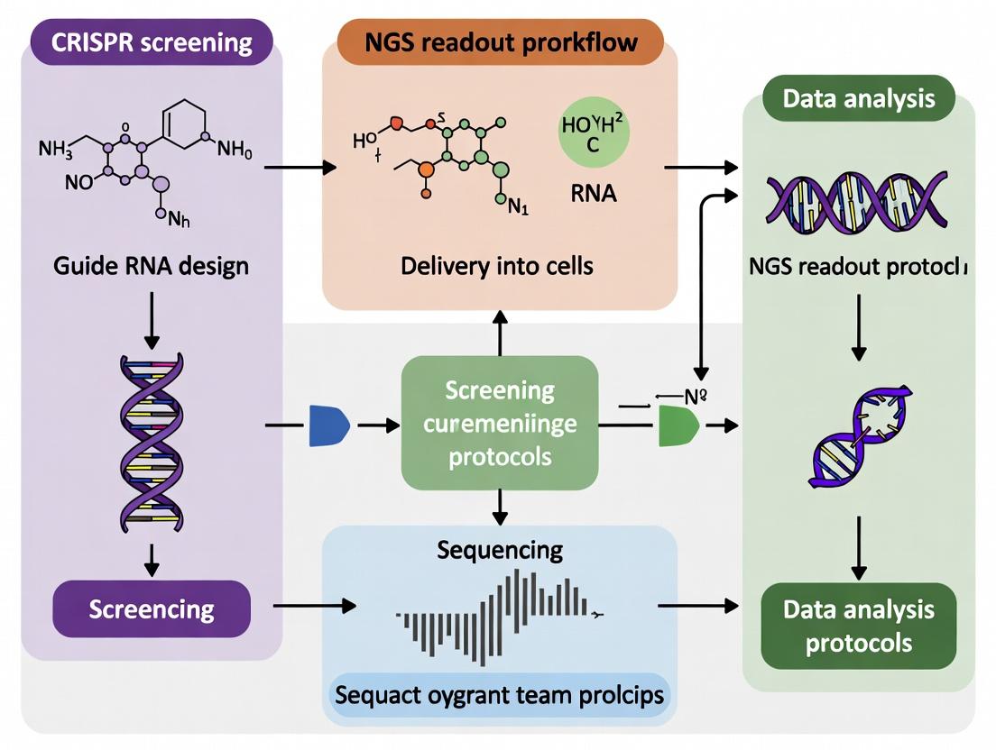
A Comprehensive Guide to CRISPR Screening with NGS Readout: From Library Design to Data Analysis
This article provides a complete roadmap for implementing CRISPR screening with Next-Generation Sequencing (NGS) readout, tailored for researchers, scientists, and drug development professionals.
A Comprehensive Guide to CRISPR Screening with NGS Readout: From Library Design to Data Analysis
Abstract
This article provides a complete roadmap for implementing CRISPR screening with Next-Generation Sequencing (NGS) readout, tailored for researchers, scientists, and drug development professionals. We cover the foundational principles of pooled and arrayed screening, detail step-by-step protocols for library design, lentiviral production, infection, and sequencing preparation. The guide addresses common troubleshooting and optimization challenges, and critically evaluates validation strategies and comparative analysis of different screening approaches and computational tools. By integrating all four intents, this resource aims to empower the design of robust, high-quality functional genomics screens to accelerate target discovery and validation.
CRISPR Screening Essentials: Understanding Pooled vs. Arrayed Screens and NGS Fundamentals
Functional genomics aims to understand the relationship between genotype and phenotype on a genome-wide scale, moving beyond static sequence data to dynamic gene function. Within the broader thesis on CRISPR screening with NGS readout protocols, this field provides the conceptual framework for systematically linking genes to biological processes, disease mechanisms, and therapeutic targets. CRISPR-based screening has emerged as the preeminent tool for forward and reverse genetic screens due to its precision, scalability, and flexibility. This document details application notes and protocols central to this research.
Core Quantitative Data and Performance Metrics
Table 1: Comparison of Major CRISPR Screening Modalities
| Screening Modality | Typical Library Size (guides) | Primary Readout | Key Applications | Typical Hit Rate* |
|---|---|---|---|---|
| CRISPR Knockout (CRISPRko) | 50,000 - 200,000 | NGS (Indel frequency) | Essential gene identification, fitness screens | 0.5 - 5% |
| CRISPR Interference (CRISPRi) | 50,000 - 100,000 | NGS (Transcript/protein abundance) | Loss-of-function, non-coding element screens | 1 - 10% |
| CRISPR Activation (CRISPRa) | 50,000 - 100,000 | NGS (Transcript/protein abundance) | Gain-of-function, suppressor/enhancer screens | 1 - 5% |
| Base Editing Screens | 20,000 - 80,000 | NGS (Variant frequency) | Functional variant analysis, saturation mutagenesis | 0.1 - 2% |
| Prime Editing Screens | 20,000 - 50,000 | NGS (Precise edit frequency) | Precise sequence alteration studies | 0.05 - 1% |
*Hit rate defined as percentage of guides showing significant phenotype beyond thresholds (e.g., |log2 fold change| > 1, FDR < 0.05). Data compiled from recent literature (2023-2024).
Table 2: Key NGS Metrics for CRISPR Screen Readout
| Metric | Typical Value/Range | Importance for Screen Analysis |
|---|---|---|
| Sequencing Depth (per sample) | 50 - 100 million reads | Ensures sufficient coverage for guide quantification |
| Average Reads per Guide | 200 - 500 | Minimizes Poisson noise in guide count data |
| PCR Duplication Rate | < 20% | High rates indicate low complexity, biasing counts |
| Guide Dropout Rate (T0 vs Library) | < 10% | Indicates poor library representation or amplification bias |
| Pearson Correlation (Replicates) | > 0.9 | Essential for assessing technical reproducibility |
Detailed Experimental Protocols
Protocol 3.1: Lentiviral Production for CRISPR Library Delivery
Objective: Produce high-titer, low-variance lentivirus for transducing a pooled CRISPR guide RNA library.
Materials: See "Scientist's Toolkit" (Section 5). Procedure:
- Day 1: Plate Cells. Seed HEK293T cells (or similar packaging line) at 8x10^6 cells per 15-cm dish in 20 mL complete DMEM. Incubate overnight (37°C, 5% CO2).
- Day 2: Transfection. Ensure cell confluence is 70-80%. Prepare transfection mix for each dish:
- Solution A: 1.5 mL Opti-MEM + 36 µL of 1 µg/µL library plasmid (e.g., lentiGuide-Puro) + 24 µg psPAX2 + 12 µg pMD2.G.
- Solution B: 1.5 mL Opti-MEM + 108 µL PEI MAX (1 mg/mL). Combine Solutions A and B, vortex briefly, incubate 15 min at RT. Add dropwise to cells.
- Day 3: Media Change. 6-8 hours post-transfection, replace media with 20 mL fresh complete DMEM.
- Day 4 & 5: Harvest Virus. Collect supernatant 48 and 72 hours post-transfection. Filter through a 0.45 µm PES filter. Pool harvests. Concentrate using Lenti-X Concentrator (1:3 ratio) per manufacturer's instructions. Aliquot and store at -80°C.
- Titer Determination: Perform a puromycin kill curve on target cells to determine optimal viral volume for ~30% transduction efficiency (Multiplicity of Infection ~0.3).
Protocol 3.2: Pooled CRISPR Knockout Screen with NGS Readout
Objective: Perform a negative selection (fitness) screen to identify genes essential for cell proliferation/survival under a specific condition.
Workflow Overview:
- Library Transduction & Selection:
- Day 0: Seed 2x10^7 target cells (e.g., HAP1, K562) in appropriate medium.
- Transduce cells at MOI ~0.3 with the pooled CRISPRko lentiviral library (e.g., Brunello, 77,441 guides) in the presence of 8 µg/mL polybrene. Spinoculate (1000g, 90 min, 32°C).
- Day 1: Replace transduction media with fresh complete media.
- Day 2: Begin selection with appropriate antibiotic (e.g., 2 µg/mL puromycin). Maintain selection for 5-7 days until >90% of non-transduced control cells are dead.
- Screen Passage & Harvest:
- Maintain a minimum of 500 cells per guide (e.g., for 77k-guide library, maintain >38.5 million cells) throughout the screen to prevent stochastic guide dropout.
- Passage cells every 2-3 days, never allowing confluence >80%.
- Harvest a representative sample (50-80 million cells) at the end of selection (Time Point T0). Pellet, wash with PBS, and freeze pellet at -80°C for genomic DNA (gDNA) extraction.
- Continue culturing the remaining population. Harvest experimental samples (Tfinal) after ~14 population doublings (e.g., 21 days) and a matched control (if applicable) at the same time.
- Genomic DNA Extraction & Guide Amplification:
- Extract gDNA from cell pellets using a maxi-prep kit (e.g., Qiagen Blood & Cell Culture DNA Maxi Kit). Aim for >200 µg gDNA per sample.
- Perform a two-step PCR to amplify integrated sgRNA sequences and attach Illumina adapters and sample barcodes.
- PCR1: Amplify sgRNA cassette from 100-200 µg gDNA per sample using Herculase II polymerase. Use 25-30 cycles. Pool reactions per sample.
- Purify PCR1 product (AMPure XP beads).
- PCR2: Add Illumina flow cell adapters and dual-index barcodes using 8-10 cycles.
- Purify final library, quantify by qPCR, and validate size (~280 bp) on a Bioanalyzer.
- Sequencing & Analysis:
- Sequence on an Illumina platform (e.g., NovaSeq 6000) to achieve >200x average reads per guide.
- Process FASTQ files with a pipeline (e.g., MAGeCK, BAGEL2) to count guides, normalize counts, and calculate log2 fold changes and statistical significance (FDR) between T0 and Tfinal or treatment vs. control.
Title: Workflow for a Pooled CRISPRko Fitness Screen
Protocol 3.3: CRISPR Screening Data Analysis with MAGeCK
Objective: Analyze NGS read counts from a CRISPR screen to identify significantly enriched/depleted sgRNAs and genes.
Procedure:
- Guide Count Quantification:
- Use
mageck countto process demultiplexed FASTQ files. - Command example:
mageck count -l library.csv -n sample_name --sample-label T0,Tfinal --fastq sample_R1.fastq.gz - Outputs a count table with raw and normalized read counts for each guide in each sample.
- Use
- Test for Significant Enrichment/Depletion:
- Use
mageck testto compare conditions (e.g., Tfinal vs T0). - Command example:
mageck test -k count_table.txt -t Tfinal -c T0 -n output_name --norm-method median - The algorithm (RRA) ranks sgRNAs by log2 fold change and tests for coordinated enrichment/depletion at the gene level, outputting p-values and FDRs.
- Use
- Quality Control and Visualization:
- Use
mageck mlefor more complex designs (multiple time points, doses). - Generate QC plots: Guide count distributions, Gini index for library uniformity, and gene ranking plots (volcano, rank).
- Perform pathway enrichment analysis (e.g., using g:Profiler, Enrichr) on significant hit genes.
- Use
Title: CRISPR Screen Data Analysis Pipeline
Key Signaling Pathways Interrogated by CRISPR Screens
CRISPR screens are frequently deployed to map components of critical signaling pathways involved in disease and treatment response.
Title: Oncogenic Signaling Pathway for CRISPR Screening
The Scientist's Toolkit: Key Research Reagent Solutions
Table 3: Essential Materials for CRISPR Screening with NGS
| Item | Function & Description | Example Vendor/Product |
|---|---|---|
| CRISPR sgRNA Library | Pooled, lentiviral-ready plasmid library targeting the genome (e.g., whole-genome, kinase subset). Defines screen scope. | Broad Institute GPP (Brunello, Calabrese), Addgene pooled libraries. |
| Lentiviral Packaging Plasmids | Required for producing replication-incompetent lentivirus (2nd/3rd generation systems). | psPAX2 (packaging), pMD2.G (VSV-G envelope). |
| Packaging Cell Line | HEK293-derived cell line optimized for high-titer lentivirus production. | HEK293T, Lenti-X 293T (Takara). |
| Transfection Reagent | For delivering library and packaging plasmids into producer cells. | PEI MAX (Polysciences), Lipofectamine 3000. |
| Polybrene | Cationic polymer that enhances viral transduction efficiency. | Hexadimethrine bromide, 8 µg/mL working concentration. |
| Selection Antibiotic | Selects for cells successfully transduced with the library vector. | Puromycin, Blasticidin, depending on vector resistance marker. |
| Genomic DNA Extraction Kit | High-yield, high-purity gDNA extraction from millions of screen cells. | Qiagen Blood & Cell Culture DNA Maxi Kit. |
| High-Fidelity Polymerase | For accurate, unbiased amplification of sgRNA sequences from gDNA. | Herculase II Fusion (Agilent), KAPA HiFi. |
| Next-Generation Sequencer | Platform for high-throughput sequencing of the amplified sgRNA pool. | Illumina NovaSeq 6000, NextSeq 2000. |
| Analysis Software | Computational tools for guide counting, normalization, and hit calling. | MAGeCK, BAGEL2, CRISPRcleanR. |
| Validated Control sgRNAs | Positive (essential gene) and negative (non-targeting) controls for screen QC. | e.g., PLKO anti-GFP sgRNA, core essential gene targeting sgRNAs. |
CRISPR screening, coupled with Next-Generation Sequencing (NGS) readout, is a cornerstone of modern functional genomics. Within the broader thesis of optimizing NGS-based screening protocols, this document details the core principles, application notes, and protocols for three primary screening modalities: CRISPR Knockout (KO), CRISPR Interference (CRISPRi), and CRISPR Activation (CRISPRa). Each method enables genome-wide interrogation of gene function but operates through distinct molecular mechanisms to achieve loss-of-function or gain-of-function phenotypes.
Molecular Mechanisms
CRISPR-KO: Utilizes the CRISPR-Cas9 nuclease (commonly Streptococcus pyogenes Cas9) to create targeted double-strand breaks (DSBs) in the coding region of a gene. Repair via error-prone non-homologous end joining (NHEJ) leads to small insertions or deletions (indels) that can disrupt the open reading frame, resulting in a permanent, null knockout.
CRISPRi: Employs a catalytically "dead" Cas9 (dCas9) fused to a transcriptional repressor domain, such as KRAB. The dCas9-KRAB complex is guided to the transcription start site (TSS) or promoter of a target gene, where it sterically hinders RNA polymerase binding or recruitment and mediates epigenetic silencing through chromatin modification, leading to robust, reversible gene knockdown.
CRISPRa: Uses a dCas9 fused to transcriptional activator domains, such as VP64, p65, and Rta (e.g., VPR). The dCas9-activator complex is guided to enhancer regions or promoters upstream of the TSS. It recruits co-activators and the basal transcriptional machinery to drive increased transcription of the target gene, enabling gain-of-function studies.
Quantitative Comparison of Screening Modalities
Table 1: Key Characteristics of CRISPR Screening Platforms
| Feature | CRISPR-KO | CRISPRi | CRISPRa |
|---|---|---|---|
| Cas9 Form | Wild-type, nuclease-active | dCas9 (H840A, D10A mutations) | dCas9 (H840A, D10A mutations) |
| Fusion Effector | None | Repressor (e.g., KRAB) | Activator (e.g., VPR, SAM) |
| Primary Effect | Indels, frameshift mutations → protein truncation/loss | Epigenetic repression → reduced transcription | Transcriptional recruitment → increased transcription |
| Gene Targeting Region | Early exons (coding sequence) | TSS / Promoter ( -50 to +300 bp relative to TSS) | Enhancer or proximal promoter upstream of TSS |
| Reversibility | Permanent | Reversible (upon sgRNA/dCas9 withdrawal) | Reversible (upon sgRNA/dCas9 withdrawal) |
| On-Target Efficacy | High (but variable by indel outcome) | High, consistent knockdown (typically 70-95%) | Moderate to high activation (often 5-50x induction) |
| Key Off-Target Concerns | DNA DSB at off-target sites; NHEJ repair | Transcriptional repression at off-target sites | Transcriptional activation at off-target sites |
| Optimal for Screening | Essential gene identification, tumor suppressor discovery | Essential gene ID (hypomorphic), synthetic lethality, tunable knockdown | Gain-of-function, drug resistance, suppressor screens |
Application Notes
CRISPR-KO Screening
Best for: Identifying essential genes for cell proliferation/survival, tumor suppressors, and genes involved in DNA repair pathways. The binary, permanent nature of KO makes it ideal for positive selection screens (e.g., identifying genes whose loss confers resistance to a toxin) and negative selection screens (e.g., identifying essential genes).
CRISPRi Screening
Best for: Studying essential genes where complete KO is lethal to the cell pool, enabling hypomorphic analysis. Excellent for studying gene dosage effects, synthetic lethal interactions, and in contexts where reversibility is desired. Superior specificity compared to RNAi.
CRISPRa Screening
Best for: Identifying genes whose overexpression drives a phenotype, such as drug resistance, cellular differentiation, or escape from immunotherapy. Crucial for mapping regulatory networks and uncovering oncogenes in a pooled format.
Detailed Experimental Protocols
Universal Workflow: Pooled Library Screening with NGS Readout
Table 2: General Workflow Steps
| Step | Duration | Key Outcome |
|---|---|---|
| 1. Library Design & Cloning | 2-3 weeks | A plasmid pool encoding the Cas9/dCas9 system and the sgRNA library. |
| 2. Lentiviral Production | 1 week | High-titer, infectious lentiviral particles carrying the sgRNA library. |
| 3. Cell Transduction & Selection | 1-2 weeks | A population of cells stably expressing Cas9/dCas9, each with a single sgRNA. |
| 4. Screening Experiment | 1-6 weeks | Application of selective pressure (e.g., drug, time in culture). |
| 5. Genomic DNA Extraction & sgRNA Amplification | 1 week | PCR-amplified sgRNA cassette ready for sequencing. |
| 6. NGS & Bioinformatic Analysis | 1-2 weeks | Identification of enriched or depleted sgRNAs/genes. |
Protocol: CRISPR-KO Positive Selection Screen (e.g., Drug Resistance)
Aim: Identify genes whose knockout confers resistance to a chemotherapeutic agent.
Materials: See "The Scientist's Toolkit" below.
Procedure:
- Cell Preparation: Generate a stable Cas9-expressing polyclonal cell line. Confirm Cas9 activity via surveyor or T7E1 assay on a control target.
- Viral Transduction: Transduce cells with the pooled sgRNA lentiviral library at a low Multiplicity of Infection (MOI ~0.3) to ensure most cells receive a single sgRNA. Include a non-transduced control.
- Selection: 48 hours post-transduction, add puromycin (or relevant antibiotic) for 5-7 days to select for successfully transduced cells.
- Screen Initiation: Harvest a pre-selection sample (~50M cells, T0). Split the remaining population into two arms: Treatment (containing the drug at a predetermined lethal concentration, e.g., IC90) and Control (vehicle only). Culture cells for 14-21 days, maintaining library coverage (>500 cells per sgRNA) and drug selection.
- Endpoint Harvest: Collect ~50M cells from each arm at the endpoint (T_end).
- gDNA Extraction & PCR: Isolate genomic DNA from T0 and T_end samples using a mass-prep kit. Perform a two-step PCR:
- PCR1: Amplify the integrated sgRNA cassette from 100-200 µg of gDNA per sample using primers containing partial Illumina adapters.
- PCR2: Add full Illumina adapters and sample barcodes.
- NGS & Analysis: Pool and sequence PCR products on an Illumina platform. Align reads to the sgRNA library reference. Normalize sgRNA counts between samples. Use statistical packages (MAGeCK, BAGEL) to compare sgRNA abundance in Treatment vs. Control, identifying significantly enriched sgRNAs/genes in the drug-treated arm.
Protocol: CRISPRi Knockdown for Essential Gene Identification
Aim: Identify genes essential for cell proliferation in a specific cell line.
Key Modifications from CRISPR-KO Protocol:
- Use a cell line stably expressing dCas9-KRAB.
- Target sgRNAs to the TSS of genes (library design is distinct from KO libraries).
- The screen is a negative selection assay with no external drug pressure besides the selection for the sgRNA construct.
- Procedure: Follow the general workflow. The selective pressure is simply propagation in culture over ~14 population doublings. Essential gene sgRNAs will deplete from the population over time (T_end vs. T0). Bioinformatic analysis identifies significantly depleted sgRNAs/genes.
Visualizations
Diagram Title: Molecular Mechanisms of CRISPR-KO, i, and a
Diagram Title: Pooled CRISPR Screen Workflow with NGS Readout
The Scientist's Toolkit
Table 3: Essential Research Reagents & Materials
| Item | Function in CRISPR Screens | Example/Note |
|---|---|---|
| Cas9/dCas9 Expression System | Provides the effector protein (nuclease or transcriptional modulator). | Lentiviral vector for stable integration (e.g., lentiCas9-Blast, lenti-dCas9-KRAB-Blast). |
| Pooled sgRNA Library | Contains thousands of unique sgRNAs targeting genes genome-wide or in a subset. | Genome-wide human Brunello (KO), Dolcini (CRISPRi), or Calabrese (CRISPRa) libraries. |
| Lentiviral Packaging Plasmids | Required for production of replication-incompetent lentivirus to deliver sgRNAs. | psPAX2 (packaging) and pMD2.G (VSV-G envelope) are standard 2nd generation. |
| HEK293T Cells | Standard cell line for high-titer lentiviral production due to high transfectability. | Often used at ~70-80% confluency for calcium phosphate or PEI transfection. |
| Polybrene / Protamine Sulfate | Cationic agents that enhance viral infection efficiency by neutralizing charge repulsion. | Typically used at 4-8 µg/mL during transduction. |
| Selection Antibiotics | Select for cells that have stably integrated the Cas9/dCas9 or sgRNA vector. | Puromycin (for sgRNA vector), Blasticidin (for Cas9 vector). Critical to determine kill curve. |
| High-Yield gDNA Extraction Kit | Isolate microgram quantities of high-quality genomic DNA from millions of pooled cells. | Qiagen Blood & Cell Culture DNA Maxi Kit or similar. Yield is critical for representation. |
| High-Fidelity PCR Master Mix | Accurately amplify the integrated sgRNA cassette from gDNA with minimal bias. | KAPA HiFi HotStart ReadyMix or Q5 Hot Start. Essential for maintaining library diversity. |
| Illumina Sequencing Platform | Perform deep sequencing of amplified sgRNA pools to quantify their abundance. | HiSeq 2500/4000, NovaSeq 6000, or NextSeq 550. Need >100 reads per sgRNA. |
| Bioinformatics Software | Analyze NGS data to identify significantly enriched or depleted genes. | MAGeCK, BAGEL, CRISPResso2. Require sgRNA count files and library annotation. |
Within the broader scope of CRISPR screening with NGS readout protocols, the selection of screening format is a foundational decision. Pooled and arrayed formats represent two distinct experimental philosophies, each with unique advantages, limitations, and optimal applications in functional genomics and drug discovery. This note provides a detailed comparison and protocols to guide researchers in selecting and implementing the appropriate strategy.
Core Comparison of Formats
Table 1: Fundamental Characteristics of Pooled vs. Arrayed CRISPR Screening
| Parameter | Pooled Screening | Arrayed Screening |
|---|---|---|
| Library Format | All sgRNAs/cells in one vessel (e.g., a single flask). | Each sgRNA/perturbation in a separate well (e.g., 96-/384-well plate). |
| Throughput (Scale) | Very high (10,000s to 100,000s of genes/sgRNAs). | Moderate to high (100s to 10,000s of targets). |
| Phenotype Readout | Typically survival/proliferation (enrichment/depletion) measured by NGS of sgRNA barcodes. | Multiplexed: High-content imaging, cytometry, luminescence, transcriptomics (scRNA-seq). |
| Key Advantage | Cost-effective per target, scalable for genome-wide screens. | Enables complex, time-resolved phenotypic measurements (e.g., morphology, signaling). |
| Primary Limitation | Limited to simple, scalable phenotypes (e.g., viability). Requires deconvolution by NGS. | Higher reagent cost, more complex logistics (liquid handling automation required). |
| CRISPR Modality | Primarily CRISPR-KO (Cas9). CRISPRi/a also common. | All: KO, i, a, base editing, prime editing. |
| Data Output | Relative sgRNA abundance from bulk NGS. | Rich, multi-parametric data per well (e.g., cell count, intensity, shape). |
| Typical Application | Genome-wide loss-of-function screens to identify essential genes. | Target-focused screens with complex phenotypes (e.g., synthetic lethality, biomarker discovery). |
Table 2: Quantitative Comparison of Resource Requirements and Output
| Aspect | Pooled Screening | Arrayed Screening |
|---|---|---|
| Starting Cells | ~1e3 cells per sgRNA (e.g., 100M cells for 100k library). | ~1e3 - 5e3 cells per well (e.g., 1M cells for a 384-well plate). |
| Library Cost (per gene) | Very Low ($0.01 - $0.10) | High ($10 - $50) |
| Screen Duration | 2-4 weeks (including selection, phenotype induction, and sample prep). | 1-2 weeks (direct phenotypic measurement). |
| NGS Requirement | High-depth sequencing of the sgRNA locus (1 sample = entire population). | Lower depth, but more samples if sequencing per well (e.g., for scRNA-seq). |
| Automation Need | Low (bulk cell culture). | High (plate-based liquid handling, imaging systems). |
| Data Complexity | Lower (count tables). | Very High (multi-TB imaging data, complex analysis pipelines). |
Experimental Protocols
Protocol A: Basic Workflow for a Genome-Wide Pooled CRISPR-KO Screen
Objective: Identify genes essential for cell proliferation under standard culture conditions. Materials: See "The Scientist's Toolkit" below. Workflow:
- Library Amplification & Lentivirus Production:
- Transform the plasmid sgRNA library (e.g., Brunello) into competent E. coli and amplify to maintain >500x representation. Purify plasmid DNA.
- Co-transfect HEK293T cells with the library plasmid and packaging plasmids (psPAX2, pMD2.G) using PEI. Harvest lentiviral supernatant at 48 and 72 hours.
- Concentrate virus via ultracentrifugation and titrate on target cells.
- Cell Infection and Selection:
- Infect target cells (e.g., Cas9-expressing cell line) at a low MOI (<0.3) to ensure most cells receive ≤1 sgRNA. Include a non-targeting control sgRNA condition.
- At 48 hours post-infection, add puromycin (or relevant antibiotic) for 3-7 days to select transduced cells.
- Screen Passage and Harvest:
- Maintain cells at a minimum coverage of 500 cells per sgRNA for the entire screen. Passage cells every 2-3 days.
- Harvest a sample of cells at the start (Day 0, reference time point) and at the end of the screen (e.g., Day 14, or after sufficient phenotypic divergence).
- Pellet cells and extract genomic DNA using a maxi-prep scale kit.
- NGS Library Preparation & Sequencing:
- Amplify the integrated sgRNA cassette from gDNA using a two-step PCR protocol.
- PCR1: Use primers that add partial Illumina adapters and sample barcodes. Use 5-10 µg gDNA per reaction, split across multiple tubes.
- PCR2: Add full Illumina flow cell binding sequences and dual-index barcodes.
- Purify PCR products, quantify, pool, and sequence on an Illumina platform (Minimum: 100-200 reads per sgRNA for the initial pool).
- Amplify the integrated sgRNA cassette from gDNA using a two-step PCR protocol.
- Data Analysis:
- Demultiplex sequencing reads and align to the sgRNA library reference file.
- Count sgRNA reads for the T0 and Tfinal samples.
- Use a dedicated analysis tool (e.g., MAGeCK, BAGEL2) to calculate sgRNA depletion/enrichment and identify significantly essential genes.
Title: Pooled CRISPR Screen Workflow
Protocol B: Workflow for an Arrayed CRISPRi Screen with High-Content Imaging Readout
Objective: Identify genes modulating a specific morphological phenotype (e.g., mitochondrial fragmentation). Materials: See "The Scientist's Toolkit" below. Workflow:
- Arrayed sgRNA Plate Preparation:
- Source an arrayed library (e.g., a sub-library of nuclear-encoded mitochondrial genes in lentiviral format) in 384-well plates.
- Thaw and briefly centrifuge library plates. Using an acoustic liquid handler (e.g., Echo), transfer 20-50 nL of viral supernatant per well into black, clear-bottom assay plates.
- Reverse-Transfection and Cell Seeding:
- Prepare a suspension of inducible CRISPRi (dCas9-KRAB) cells in antibiotic-free medium.
- Add a transfection reagent (e.g., Lipofectamine HD) to the cell suspension and immediately dispense into the assay plates containing virus (e.g., 1000 cells/well in 30 µL).
- Centrifuge plates gently to mix. Incubate for 72h to allow transduction and gene repression.
- Phenotypic Induction and Staining:
- Add a stimulus or stressor to induce the phenotype of interest (e.g., a mitochondrial uncoupler).
- After 24h, stain cells with live-cell dyes for mitochondria (e.g., MitoTracker Red) and nuclei (Hoechst 33342).
- High-Content Imaging and Analysis:
- Image plates using a high-content microscope (e.g., ImageXpress Micro) with a 20x objective. Acquire 4-9 fields per well.
- Use image analysis software (e.g., CellProfiler, IN Carta) to segment nuclei and cytoplasm, identify mitochondria, and extract >100 morphological features (e.g., mitochondrial area, branching, intensity).
- Normalize data per plate (Z-score). For each sgRNA, aggregate features into a phenotypic signature and compare to non-targeting control wells using robust statistical methods (e.g., Z-prime, strictly standardized mean difference - SSMD).
Title: Arrayed Screen with Imaging Readout
The Scientist's Toolkit: Key Research Reagent Solutions
| Item | Function in CRISPR Screening |
|---|---|
| Cas9-Expressing Cell Line | Provides the CRISPR nuclease constitutively, ensuring uniform editing capability. Essential for pooled screens. |
| dCas9-KRAB/i/a Cell Line | Enables CRISPR interference or activation screens. Often used in arrayed formats for precise transcriptional modulation. |
| Validated sgRNA Library (Pooled/Arrayed) | Pre-designed, high-confidence collection of sgRNAs targeting the genome. The core screening reagent (e.g., Brunello, Calabrese). |
| Lentiviral Packaging Plasmids (psPAX2, pMD2.G) | Third-generation system for producing replication-incompetent lentivirus to deliver sgRNAs into target cells. |
| Polybrene or Protamine Sulfate | Polycations that enhance viral transduction efficiency by neutralizing charge repulsion between virus and cell membrane. |
| Puromycin/Blasticidin/Other Antibiotics | Selection agents to eliminate non-transduced cells, ensuring a pure population of sgRNA-expressing cells. |
| High-Capacity gDNA Extraction Kit | For pooled screens: to purify sufficient, high-quality genomic DNA from millions of cells for PCR amplification of sgRNAs. |
| Illumina-Compatible PCR Primers with Indexes | To amplify and barcode the sgRNA region from gDNA for multiplexed NGS. |
| Automated Liquid Handler (e.g., Echo, Biomek) | For arrayed screens: essential for precise, non-contact transfer of viruses/reagents to 384/1536-well plates. |
| High-Content Imager (e.g., ImageXpress, Opera) | For arrayed screens: automated microscope to capture high-resolution, multi-channel images for complex phenotypic analysis. |
| CellProfiler / IN Carta Software | Open-source or commercial software to analyze high-content images and extract quantitative phenotypic data. |
| MAGeCK / BAGEL2 Software | Computational pipelines specifically designed for analyzing count-based data from pooled CRISPR screens to identify hit genes. |
This application note, situated within a broader thesis on CRISPR screening with NGS readout protocols, details critical parameters for ensuring robust and interpretable results from pooled CRISPR screens. The accurate quantification of single-guide RNA (sgRNA) abundance via Next-Generation Sequencing (NGS) is the fundamental readout for determining gene phenotype. This document provides a comprehensive guide to the core considerations of sgRNA library amplification, determining requisite sequencing depth, and assessing library complexity, along with detailed protocols to implement these analyses.
Table 1: Recommended Sequencing Depth for Pooled CRISPR Screens
| Screen Type | Library Size (sgRNAs) | Minimum Reads per sgRNA (Coverage) | Total Recommended Sequencing Depth | Notes |
|---|---|---|---|---|
| Genome-wide (GeCKO, Brunello) | ~70,000 - 100,000 | 200-500x | 20 - 50 million reads | Ensures detection of modest phenotype effects. |
| Sub-library (Kinase, Epigenetic) | 5,000 - 20,000 | 500-1000x | 5 - 20 million reads | Higher per-sgRNA coverage increases statistical power for smaller libraries. |
| Arrayed Validation | < 100 | >10,000x | 1 - 5 million reads | Deep sequencing for precise individual sgRNA activity measurement. |
Table 2: Impact of PCR Cycle Number on Library Complexity and Bias
| PCR Amplification Cycles | Relative Library Complexity | Risk of Over-amplification Bias | Recommended Use Case |
|---|---|---|---|
| 12-15 cycles | High | Low | Initial library generation from ample starting material. |
| 16-20 cycles | Moderate | Moderate | Typical amplification from genomic DNA or plasmid pools. |
| 21+ cycles | Low | High | Avoid; leads to skewed sgRNA representation and loss of rare clones. |
Table 3: Metrics for Assessing Library Quality Pre- and Post-Sequencing
| Metric | Calculation / Method | Target Value | Indicates |
|---|---|---|---|
| Pre-Seq Library Complexity | Unique sgRNA molecules identified in pre-sequencing QC (e.g., Bioanalyzer, qPCR). | >80% of expected sgRNAs | Cloning efficiency and initial representation. |
| Post-Seq Read Distribution | Percentage of sgRNAs with read counts > 20% of median. | >90% | Evenness of amplification and sequencing. |
| Population Evenness | Gini Coefficient (0=perfect equality, 1=perfect inequality). | < 0.2 | Low skew in sgRNA abundance distribution. |
| PCR Bottleneck Coefficient | Ratio of reads from PCR duplicates to total reads. | < 0.5 | Level of over-amplification artifact. |
Detailed Experimental Protocols
Protocol 3.1: Two-Step PCR Amplification of sgRNA Libraries from Genomic DNA
Objective: To amplify the integrated sgRNA cassette from genomic DNA of screened cells for NGS library preparation while minimizing bias.
Materials: See "The Scientist's Toolkit" (Section 5).
Procedure:
- Primary PCR (Add Sequencing Adaptors):
- Set up a 50 µL reaction for each sample using a high-fidelity polymerase.
- Use 1-2 µg of purified genomic DNA as template.
- Use forward and reverse primers that anneal to the constant regions of the sgRNA vector (e.g., U6 promoter and sgRNA scaffold) and contain overhangs with partial Illumina adaptor sequences (P5 and P7).
- Thermocycler conditions: Initial denaturation: 98°C for 30s; 12-18 cycles of: 98°C for 10s, 60°C for 15s, 72°C for 15s; Final extension: 72°C for 2 min. Optimize cycles to use the minimum number yielding sufficient product.
- Purify the PCR product using SPRI beads (e.g., 1.0x ratio).
- Secondary PCR (Add Full Indexes and Flow Cell Sequences):
- Set up a 50 µL reaction using 5-50 ng of purified primary PCR product as template.
- Use forward and reverse primers containing the full Illumina P5/P7 flow cell binding sites, unique dual index (i5 and i7) sequences for multiplexing, and sequences complementary to the overhangs added in the primary PCR.
- Thermocycler conditions: Initial denaturation: 98°C for 30s; 8-12 cycles of: 98°C for 10s, 65°C for 15s, 72°C for 15s; Final extension: 72°C for 2 min.
- Purify the final library using SPRI beads (e.g., 0.8x ratio). Quantify by fluorometry and assess size distribution by Bioanalyzer/TapeStation.
Protocol 3.2: Determining Sequencing Depth via Saturation Analysis
Objective: To empirically determine the minimum sequencing depth required for phenotype calling in a specific screen.
Procedure:
- Sequence your initial library to a very high depth (e.g., >100 million reads for a genome-wide library).
- Downsampling: Use bioinformatics tools (e.g.,
seqtk) to randomly subsample your sequenced reads to progressively lower fractions (e.g., 10%, 20%, 30%...100% of total). - Phenotype Calculation: For each downsampled dataset, align reads to the sgRNA reference library and count reads per sgRNA. Perform phenotype analysis (e.g., calculate log2 fold-change and p-value for each gene using MAGeCK or similar).
- Saturation Plotting: For each depth level, plot the number of significantly hit genes (e.g., FDR < 0.1) against the total number of reads sequenced.
- Determination: Identify the point where the curve plateaus (adding more reads yields minimal new hits). The depth just past the plateau's inflection point is the recommended minimum depth for future screens of similar design and complexity.
Protocol 3.3: Assessing Library Complexity via PCR Duplicate Removal
Objective: To calculate the fraction of sequencing reads derived from PCR duplicates, a key indicator of over-amplification and loss of complexity.
Procedure:
- Preprocessing: After demultiplexing, align reads to the sgRNA reference library. Retain only perfectly matching reads.
- Identify Unique Molecules: For each sgRNA sequence, examine the start and end coordinates of the alignment. Reads with identical sgRNA identity and identical start/end positions are considered PCR duplicates stemming from the same original molecule.
- Calculate Metrics:
- PCR Bottleneck Coefficient (PBC): PBC = Number of unique genomic locations / Number of total mapped reads.
- Non-Redundant Fraction (NRF): NRF = Number of unique (deduplicated) reads / Total number of reads.
- Interpretation: A PBC > 0.5 or NRF > 0.5 is generally acceptable. Lower values indicate excessive PCR duplication, suggesting the initial PCR used too many cycles or input material was too limited.
Visualizations
Diagram Title: Two-Step PCR for sgRNA NGS Library Prep
Diagram Title: Impact of Key Parameters on Screen Results
The Scientist's Toolkit
Table 4: Essential Research Reagent Solutions for sgRNA NGS Readout
| Item | Function & Explanation | Example Vendor/Product |
|---|---|---|
| High-Fidelity PCR Master Mix | Enzymatic blend for high-accuracy, low-bias amplification of sgRNA sequences from complex genomic DNA templates. Critical for maintaining representation. | NEB Q5, KAPA HiFi, IDT AccuPrime |
| SPRI (Solid Phase Reversible Immobilization) Beads | Magnetic beads for size-selective purification and cleanup of PCR products. Used to remove primers, dNTPs, and short fragments between amplification steps. | Beckman Coulter AMPure, Sigma MagBind |
| Dual-Indexed PCR Primers | Primer sets containing unique i5 and i7 index sequences. Allow multiplexing of many samples in a single NGS run by assigning a unique "barcode" to each. | Illumina TruSeq, IDT for Illumina |
| Fluorometric Quantification Kit | Accurate quantification of final NGS library concentration by measuring fluorescence of dsDNA. Essential for pooling libraries at equimolar ratios. | Thermo Fisher Qubit dsDNA HS, Invitrogen |
| High-Sensitivity Nucleic Acid Analyzer | Microfluidic capillary electrophoresis for assessing library fragment size distribution and detecting adapter dimers or other contaminants. | Agilent Bioanalyzer, Agilent TapeStation |
| sgRNA Reference Library FASTA File | Digital reference file containing all sgRNA sequences used in the screen. Mandatory for read alignment and counting. | Public repositories (Addgene) or custom design. |
| Read Counting Software | Bioinformatics pipeline to align NGS reads to the reference and generate a count table (sgRNAs x samples). | MAGeCK, CRISPResso2, custom BWA/featureCounts scripts |
Application Notes
CRISPR screening, coupled with Next-Generation Sequencing (NGS) readout, has become a cornerstone of functional genomics. Within a broader thesis on CRISPR-NGS protocol optimization, these applications represent the primary translational endpoints that drive methodological advancements.
1. Essential Gene Discovery: Genome-wide CRISPR knockout (CRISPRko) screens identify genes critical for cellular survival or proliferation under specific conditions. Quantitative data from these screens, represented as log-fold changes (LFC) in sgRNA abundance and associated statistical scores, pinpoint non-redundant cellular functions.
2. Synthetic Lethality (SL) Screening: This application identifies gene pairs where co-inhibition is lethal, but inhibition of either alone is not. CRISPR-based SL screens, often using focused libraries targeting genes involved in DNA repair or specific pathways, are pivotal for identifying tumor-specific therapeutic targets, especially in cancers with known driver mutations (e.g., BRCA1/2 mutations).
3. Drug Resistance & Mechanism of Action (MoA) Studies: CRISPR gain-of-function (CRISPRa) or knockout screens performed in the presence of a therapeutic compound reveal genes whose modulation confers resistance or sensitivity. This data elucidates drug MoA, predicts potential resistance mechanisms in patients, and identifies candidate combination therapies.
Table 1: Quantitative Metrics for CRISPR Screen Analysis
| Metric | Description | Typical Threshold | Primary Application |
|---|---|---|---|
| Log2 Fold Change (LFC) | Change in sgRNA abundance between conditions. | LFC < -1 (Depletion); LFC > 1 (Enrichment) | All screens |
| p-value | Significance of sgRNA/gene depletion/enrichment. | p < 0.05 (after correction) | All screens |
| False Discovery Rate (FDR) | Corrected probability of false positive. | FDR < 0.05 (for hit selection) | All screens |
| RSA Score | Redundant siRNA Activity score; ranks genes. | Score > 1 (Enrichment) | Pooled screens |
| MAGeCK Score | Model-based analysis score from MAGeCK algorithm. | p < 0.05; FDR < 0.05 | Essential/SL screens |
| β-score | Gene effect score from CERES/Chronos algorithms. | β < -0.5 (Essential); β > 0.5 (Positive selection) | Essential screens |
Detailed Protocols
Protocol 1: Genome-wide Essentiality Screen with CRISPRko
Objective: Identify genes essential for proliferation in a cancer cell line. Workflow: 1) Library Production: Amplify Brunello genome-wide sgRNA library (4 sgRNAs/gene, ~76k guides). 2) Viral Production: Lentivirally package library in HEK293T cells. 3) Cell Infection & Selection: Infect target cells at low MOI (0.3) to ensure single guide integration. Select with puromycin for 7 days. 4) Sample Collection: Harvest cells at initial timepoint (T0) and after ~14 population doublings (Tfinal). 5) NGS Prep: PCR-amplify integrated sgRNA cassettes from genomic DNA, adding Illumina adapters and sample barcodes. 6) Sequencing: Pool samples and sequence on Illumina NextSeq (≥50 reads/guide). 7) Analysis: Align reads to library reference, count guides, and use MAGeCK or CERES to calculate essentiality scores (β).
Protocol 2: Synthetic Lethality Screen with a Focused Library
Objective: Find genes synthetically lethal with a mutant BRCA1 background. Workflow: 1) Isogenic Cell Lines: Use paired cell lines: wild-type BRCA1 and homozygous BRCA1 mutant. 2) Library Design: Use a sub-library targeting DNA damage response (DDR) genes and controls. 3) Parallel Screening: Conduct Protocol 1 steps 2-6 in parallel for both cell lines. 4) Comparative Analysis: Calculate differential essentiality (e.g., Δβ = βmutant - βWT). Genes with significant depletion (Δβ < -0.8, FDR<0.05) only in the BRCA1 mutant background are candidate synthetic lethal interactors (e.g., PARP1).
Protocol 3: Drug Resistance Screen with CRISPRa
Objective: Identify genes whose overexpression confers resistance to Drug X. Workflow: 1) CRISPRa System: Use dCas9-VPR SAM (Synergistic Activation Mediator) system. 2) Library: Use a focused CRISPRa sgRNA library targeting known drug target pathway genes and transcription factors. 3) Screen: Transduce library into cells, select, and split into Vehicle and Drug X-treated arms. Treat for 14+ days. 4) Analysis: Harvest genomic DNA, sequence, and identify sgRNAs significantly enriched (LFC > 1, FDR<0.05) in the Drug X arm vs. Vehicle. Enriched genes point to potential resistance drivers or alternative survival pathways.
Diagrams
Title: CRISPR Pooled Screen Core Workflow (76 chars)
Title: Synthetic Lethality Conceptual Model (51 chars)
Title: Drug Resistance Mechanisms from CRISPR Screens (70 chars)
The Scientist's Toolkit
Table 2: Essential Research Reagent Solutions for CRISPR-NGS Screens
| Reagent / Material | Function / Purpose | Example/Notes |
|---|---|---|
| Validated sgRNA Library | Targets genes of interest; determines screen scope. | Genome-wide (Brunello), focused (DDR), or custom libraries. Cloned in lentiviral backbone. |
| Lentiviral Packaging Mix | Produces infectious viral particles to deliver sgRNA library. | 2nd/3rd gen systems (psPAX2, pMD2.G, pCMV-VSV-G). Essential for high-titer, safe production. |
| Polybrene (Hexadimethrine Bromide) | Enhances viral transduction efficiency by neutralizing charge repulsion. | Used at 4-8 µg/mL during infection. Critical for hard-to-transduce cells. |
| Puromycin (or other selectable marker) | Selects for cells successfully transduced with the sgRNA vector. | Kill curve must be established for each cell line. Selection typically lasts 5-7 days. |
| PCR Additives for GC-rich amplicons | Enables robust amplification of sgRNA sequences from gDNA for NGS. | Q5 Hot Start HiFi polymerase, DMSO, or Betaine improve yield and specificity. |
| Dual-Indexed NGS Primers | Amplifies and barcodes sgRNA inserts for multiplexed sequencing. | Must be compatible with library design. Adds sample-specific indices and Illumina adapters. |
| NGS Analysis Pipeline Software | Processes raw sequencing data into gene-level scores. | MAGeCK, PinAPL-Py, CRISPRAnalyzeR, or custom R/Python scripts. |
| Positive & Negative Control sgRNAs | Assesss screen performance and data normalization. | Non-targeting controls (NTCs) and essential (e.g., RPA3) and non-essential (e.g., AAVS1) gene targets. |
Step-by-Step Protocol: Executing a CRISPR Screen from Library to NGS Data
Application Notes
The initial phase of a CRISPR screen is critical, determining its scope, specificity, and success. This phase involves selecting a library optimized for the screening paradigm (e.g., knockout, activation, inhibition) and the biological question. The design principles revolve around maximizing on-target efficacy while minimizing off-target effects. Key public libraries have been developed as community standards.
Key Library Comparisons:
| Library Name | Type | Target Species | # of sgRNAs/Gene | Total Size | Key Features & Design Principles | Primary Use Case |
|---|---|---|---|---|---|---|
| GeCKO v2 (2014) | Knockout (KO) | Human, Mouse | 3-6 | ~123,000 (Human, 2 sub-libs) | One of first genome-scale libs. Uses first-gen sgRNA design rules. Two-library format reduces cloning bias. | Early proof-of-concept, broad identification of essential genes. |
| Brunello (2016) | Knockout (KO) | Human | 4 | 77,441 | Improved on-target efficacy prediction (Rule Set 2). Fewer, higher-quality sgRNAs/gene reduces library size. | High-confidence dropout screens with reduced noise. |
| CRISPRi v2 (2016) | Interference (i) | Human | 10 (TSS-targeting) | 137,411 sgRNAs | Targets transcriptional start sites (TSS) with dCas9-KRAB. Uses truncated sgRNAs (tru-sgRNAs) for specificity. | Repression of non-coding & essential genes, finely tuned knockdown. |
| CRISPRa v2 (2016) | Activation (a) | Human | 10 (TSS-targeting) | 137,411 sgRNAs | Targets TSS with dCas9-VPR activator. Uses tru-sgRNAs. | Gain-of-function screens, identification of drug resistance genes. |
| Mouse GeCKO v2 | Knockout (KO) | Mouse | 3-6 | ~130,000 | Adapted from human GeCKO for mouse genome. | In vitro and in vivo screening in mouse models. |
| miniLibCas9 (2022) | Knockout (KO) | Human | 2 | 17,032 | Focuses on ~5,000 core fitness genes. Ultra-small size enables complex assays (single-cell, spatial). | High-complexity perturbation screens with multi-modal readouts. |
Selection Criteria:
- Screen Goal: KO for loss-of-function, CRISPRa for gain-of-function, CRISPRi for essential gene knockdown or fine-tuned repression.
- Library Size: Larger libraries require greater sequencing depth and cell numbers. Compact libraries (e.g., Brunello, miniLib) are more cost-effective for deep coverage or complex assays.
- Design Algorithm: Newer libraries (Brunello, later CRISPRi/a) use improved efficacy scores (e.g., Rule Set 2, DeepHF/Specter) for higher activity.
- Validation Status: Established libraries have published performance metrics (e.g., recall of known essential genes).
Detailed Protocol: Library Selection and Cloning Verification
This protocol outlines the steps for selecting a CRISPR library and performing essential quality control before proceeding to virus production.
I. Materials & Reagents
Research Reagent Solutions Toolkit:
| Item | Function |
|---|---|
| Plasmid Library (e.g., pLCKO, lentiCRISPRv2 backbone) | The vector containing the pooled sgRNA library. Typically obtained from Addgene as a high-concentration stock. |
| Endura ElectroCompetent Cells | High-efficiency cells for large, complex plasmid library transformation to maintain diversity. |
| LB Agar Plates + Selection Antibiotic (e.g., Ampicillin) | For titering transformation and assessing colony count (library coverage). |
| NucleoBond Xtra Maxi Prep Kit | For high-yield, high-quality plasmid DNA isolation from large bacterial cultures. |
| Sanger Sequencing Primers (U6 forward) | For verifying individual sgRNA clone sequences. |
| Next-Generation Sequencing (NGS) Library Prep Kit (e.g., Illumina) | For deep sequencing of the plasmid pool to verify sgRNA representation. |
| Qubit Fluorometer & dsDNA HS Assay Kit | Accurate quantification of low-concentration plasmid DNA. |
| Agarose Gel Electrophoresis System | Check plasmid size and integrity. |
II. Methodology
Step 1: Library Selection and Acquisition
- Align library choice with experimental goals using the table above.
- Order the library plasmid (e.g., lentiCRISPRv2-Brunello) from a reputable depository (Addgene). Request the plasmid as transformation-ready, electrocompetent bacteria or as high-concentration plasmid DNA.
- If received as DNA, proceed to Step 3. If received as bacteria, proceed to Step 2.
Step 2: Library Expansion & Plasmid Recovery (If received as bacteria)
- Thaw Electrocompetent Library: Rapidly thaw the vial of electrocompetent cells containing the library on ice.
- Electroporation: Transform the entire contents into prepared Endura cells via electroporation (1.8 kV, 200Ω, 25µF). Immediately add 1 mL recovery medium.
- Recovery: Recover cells at 37°C for 1 hour with shaking.
- Titering: Perform a 1:10,000 dilution of the culture. Plate 100 µL on LB+Amp plates. Incubate overnight at 37°C.
- Mass Culture: Dilute the remaining recovery culture into 500 mL of LB+Amp broth. Grow overnight (12-16 hrs) at 37°C with vigorous shaking.
- Plasmid Maxiprep: Harvest cells by centrifugation. Isolate plasmid DNA using the NucleoBond Xtra Maxi kit according to the manufacturer's protocol. Elute in sterile TE buffer or nuclease-free water.
- Quantification: Measure DNA concentration using Qubit. Expect yields of 100-300 µg. Check integrity on a 0.8% agarose gel.
Step 3: Library Representation Analysis by NGS (Critical QC) Objective: Confirm the library contains all sgRNAs without major dropouts or skewing.
- PCR Amplification of sgRNA Cassettes: Set up a 50 µL PCR reaction using ~100 ng of the plasmid library as template. Use primers that amplify the sgRNA scaffold region and add partial Illumina adapter sequences.
- Primer Example (Forward): AATGATACGGCGACCACCGAGATCTACAC[i5]ACACTCTTTCCCTACACGACGCT
- Primer Example (Reverse): CAAGCAGAAGACGGCATACGAGAT[i7]GTGACTGGAGTTCAGACGTGTGCT
- Purify PCR Product: Use a PCR purification kit. Size-select for the correct band (~200-300 bp) if needed.
- Indexing PCR: Perform a second, limited-cycle PCR to add full Illumina P5/P7 adapters and unique dual indices (i5 and i7) for multiplexing.
- Sequencing: Pool and purify the final library. Sequence on an Illumina MiSeq or NextSeq platform using a 75-150 bp single-end run. Aim for 50-100 reads per sgRNA as a minimum.
- Data Analysis: Demultiplex reads. Map the sequenced sgRNAs to the library reference file using a short-read aligner (Bowtie2). >90% of sgRNAs should be detected with relatively even representation (e.g., most sgRNAs within 100-fold range of the median count).
Step 4: Validation of Individual Clones (Optional but Recommended)
- Transform a small aliquot of the plasmid library into standard competent DH5α cells. Plate for single colonies.
- Pick 20-30 colonies, inoculate mini-cultures, and perform plasmid minipreps.
- Sanger sequence the sgRNA insert using the U6 forward primer.
- Align sequences to the expected library list. This confirms correct cloning and absence of major sequence errors.
Visualization of Key Concepts
Title: CRISPR Library Selection Decision Workflow
Title: Library Structure & Functional Outputs
Application Notes
Within a CRISPR screening research thesis utilizing Next-Generation Sequencing (NGS) readout, the quality and consistency of the lentiviral library directly determine screening success. Phase 2 focuses on generating a high-titer, functional lentiviral library and validating the target cell line's transduction and screening fitness. Key parameters include achieving a high viral titer (>1x10^8 TU/mL) to maintain library complexity, ensuring a low Multiplicity of Infection (MOI ~0.3) to enforce single-guide RNA (sgRNA) integration per cell, and validating robust cell viability and proliferation post-transduction. The data from this phase establishes the foundation for a reproducible and interpretable screen.
Table 1: Key Quantitative Benchmarks for Phase 2
| Parameter | Target Value | Purpose & Rationale |
|---|---|---|
| Lentiviral Titer | >1 x 10^8 TU/mL | Ensures sufficient viral volume to transduce entire cell population at low MOI without library bottlenecking. |
| Transduction MOI | 0.2 - 0.4 | Limits to ~1 viral integration per cell, ensuring single sgRNA per cell for clear phenotype-genotype linkage. |
| Transduction Efficiency | 30-50% (at MOI=0.3) | Validates functional titer calculation and confirms cell line susceptibility. |
| Cell Viability (Post-Transduction) | >90% (vs. untransduced) | Confirms lack of acute cytotoxicity from transduction reagents or viral components. |
| Puromycin Kill Curve EC100 | Determined empirically (e.g., 1-5 µg/mL) | Identifies minimum antibiotic concentration that kills all non-transduced cells within 3-5 days. |
| Library Coverage (Post-Selection) | >500 cells/sgRNA | Maintains library representation for statistical power in NGS readout. |
Detailed Protocols
Protocol 2.1: Lentiviral Production via HEK293T Transfection Objective: Produce high-titer lentiviral particles encoding the CRISPR sgRNA library.
- Day 0: Seed HEK293T cells in poly-L-lysine coated 10-cm dishes at 60-70% confluency in complete DMEM (10% FBS, 1% Pen/Strep).
- Day 1 (Morning): Replace medium with 8 mL fresh complete DMEM.
- Day 1 (Afternoon): Prepare transfection mix in two tubes:
- Tube A (DNA): 9 µg Library Plasmid (psgRNA), 6.75 µg psPAX2 (packaging), 2.25 µg pMD2.G (envelope) in 1.5 mL serum-free DMEM.
- Tube B (Reagent): 54 µL PEI MAX (1 mg/mL) in 1.5 mL serum-free DMEM. Incubate Tube B with Tube A for 15-20 min at RT. Add mixture dropwise to cells. Gently swirl.
- Day 2 (Morning): Replace medium with 10 mL fresh complete DMEM.
- Day 3 & 4 (48 & 72h post-transfection): Harvest viral supernatant, filter through a 0.45 µm PES filter, and store at 4°C. Pool harvests.
- Concentration (Optional): Concentrate pooled supernatant using Lentivirus Concentration Solution (e.g., Lenti-X) per manufacturer's protocol. Aliquot and store at -80°C.
Protocol 2.2: Functional Titer Determination via Puromycin Selection Objective: Quantify functional viral titer (Transducing Units per mL, TU/mL) on the target cell line.
- Day 0: Seed target cells in a 12-well plate at 2x10^5 cells/well in 1 mL complete growth medium.
- Day 1: Prepare serial dilutions of virus (e.g., 10 µL, 2 µL, 0.4 µL) in fresh medium containing 8 µg/mL polybrene. Replace cell medium with 1 mL of virus-polybrene mix. Include a no-virus control.
- Day 2: Replace medium with 1 mL fresh complete growth medium.
- Day 3: Trypsinize and reseed all wells into 6-well plates with appropriate puromycin selection medium (concentration from kill curve, Protocol 2.3).
- Day 6-7: Stain colonies with crystal violet and count. Calculate titer:
- TU/mL = (Number of colonies) / (Volume of virus in mL x Dilution Factor).
- Use a dilution yielding 20-200 colonies for accuracy.
Protocol 2.3: Cell Line Validation: Puromycin Kill Curve & Proliferation Objective: Determine optimal puromycin concentration and validate cell fitness post-transduction.
- Kill Curve: Seed cells in a 24-well plate at a density to be ~30% confluent the next day. Apply a range of puromycin concentrations (e.g., 0, 0.5, 1, 2, 4, 8 µg/mL) in triplicate. Refresh antibiotic every 2-3 days. Monitor daily. The minimal concentration that results in 100% cell death within 5 days is the EC100 for selection.
- Proliferation Assay: Transduce cells at MOI=0.3 (using titer from 2.2) and mock transduce a control. After puromycin selection, seed equal numbers of surviving cells. Count cells every 24h for 3-5 days using an automated cell counter or MTT assay. Compare doubling times to ensure no significant impact from transduction/selection.
Visualizations
The Scientist's Toolkit: Research Reagent Solutions
| Item | Function & Rationale |
|---|---|
| HEK293T/17 Cells | Production cell line for lentivirus; highly transfectable, provides necessary transcriptional machinery for high-titer virus. |
| psPAX2 Packaging Plasmid | Provides gag, pol, rev, and tat genes necessary for viral particle assembly and RNA packaging. |
| pMD2.G (VSV-G) Envelope Plasmid | Encodes the Vesicular Stomatitis Virus G glycoprotein, providing broad tropism for infecting most mammalian cell lines. |
| Polyethylenimine (PEI MAX) | Cationic polymer transfection reagent for efficient co-delivery of three plasmids into HEK293T cells. |
| Polybrene (Hexadimethrine Bromide) | Cationic polymer that reduces charge repulsion between virus and cell membrane, enhancing transduction efficiency. |
| Puromycin Dihydrochloride | Antibiotic selection agent; kills non-transduced cells as the sgRNA vector contains a puromycin resistance gene. |
| Lentivirus Concentration Solution | PEG-based solution that concentrates viral particles via precipitation, increasing functional titer for difficult-to-transduce cells. |
| 0.45 µm PES Filter | Sterile-filters viral supernatant to remove producer cell debris while allowing lentiviral particles to pass through. |
Application Notes
Phase 3 represents the critical experimental execution of a CRISPR screen. Following library cloning and amplification, this phase involves delivering the sgRNA library to the target cell population, applying selection pressure based on the phenotypic outcome of interest, and harvesting genomic DNA for NGS library preparation. Success hinges on maintaining library representation and achieving sufficient phenotypic separation between control and experimental populations.
Key Quantitative Parameters for Screen Execution
| Parameter | Typical Target / Range | Rationale & Impact |
|---|---|---|
| Cell Coverage (Library Representation) | 500-1000x cells per sgRNA | Ensures each sgRNA is present in sufficient starting cells to mitigate stochastic dropout. |
| Transduction Multiplicity of Infection (MOI) | 0.3 - 0.6 | Aims for <1 viral integration per cell to ensure most positive cells contain only one sgRNA. |
| Transduction Efficiency | 30-70% (lentivirus) | High efficiency is critical but must be balanced with low MOI. Efficiency is assayed via fluorescence or antibiotic markers. |
| Selection Antibiotic (e.g., Puromycin) Duration | 3-7 days post-transduction | Complete elimination of non-transduced cells is required to ensure a pure edited population. Kill curve validation is essential. |
| Phenotypic Duration / Passaging | Varies: 5-21+ days | Must be optimized for phenotype (e.g., proliferation, resistance, differentiation). Longer durations increase signal but may exacerbate bottlenecks. |
| Final Cell Harvest Coverage | ≥ 500x cells per sgRNA | Ensures sufficient gDNA for PCR and maintains library representation at endpoint. |
Experimental Protocols
Protocol 1: Lentiviral Transduction for CRISPR Library Delivery
Objective: To deliver the pooled sgRNA library to the target cell line at low MOI while maintaining high complexity. Materials: Packaging cells (HEK293T), target cells, lentiviral transfer plasmid (e.g., lentiCRISPRv2, lentiGuide-Puro), packaging plasmids (psPAX2, pMD2.G), polybrene, puromycin.
- Day 0: Seed HEK293T cells in 10-cm dishes for 70-80% confluency the next day.
- Day 1: Transfect cells using polyethylenimine (PEI). For one dish: mix 10 µg library plasmid, 7.5 µg psPAX2, and 2.5 µg pMD2.G in 500 µL serum-free media. Add 60 µL PEI (1 mg/mL), vortex, incubate 15 min, and add dropwise to cells.
- Day 2: Replace transfection media with fresh growth media.
- Day 3 & 4: Harvest viral supernatant at 48h and 72h post-transfection. Filter through a 0.45 µm PVDF filter, aliquot, and store at -80°C or use immediately.
- Day 4 (Titration): Perform a pilot transduction on target cells with serial dilutions of virus in the presence of 8 µg/mL polybrene. Assess transduction efficiency (e.g., via fluorescence) after 48h to calculate functional titer.
- Day 4 (Library Transduction): Seed a large quantity of target cells (to achieve 500-1000x coverage). Transduce at the pre-determined MOI of 0.3-0.6 in the presence of polybrene.
- Day 5: Replace media 24h post-transduction.
- Day 6: Begin puromycin selection (concentration and duration determined by prior kill curve). Maintain cells under selection for 3-7 days until all non-transduced control cells are dead. This is the T0 timepoint; harvest a cell pellet (~50-100 million cells) for gDNA as a reference.
Protocol 2: Phenotypic Enrichment/Depletion via Competitive Proliferation
Objective: To apply selection pressure that enriches or depletes sgRNAs based on their effect on cell fitness. Materials: Transduced and selected cell pool (from Protocol 1), appropriate growth media.
- After puromycin selection, expand the cell population. This is the start of the screen (Day 0 post-selection).
- Passage cells continuously, maintaining a minimum of 500x coverage for each sgRNA at all times. Do not let cells become over-confluent.
- For Positive Selection (e.g., drug resistance): At a defined passage, split the population and treat one arm with the drug of interest (e.g., a chemotherapeutic) and maintain a parallel vehicle-treated control arm. Continue passaging both populations.
- For Negative Selection (fitness screens): Simply passage the single population. sgRNAs targeting essential genes will be depleted over time relative to non-targeting controls.
- Harvest cell pellets at defined experimental endpoints (e.g., Day 14, Day 21, or when a clear phenotypic shift in the control population is observed). Always snap-freeze pellets for gDNA extraction.
Protocol 3: Genomic DNA Harvest and sgRNA Amplification for NGS
Objective: To isolate high-quality gDNA and amplify the integrated sgRNA cassette for sequencing. Materials: Cell pellets, gDNA extraction kit (e.g., Qiagen Blood & Cell Culture Maxi Kit), Herculase II Fusion DNA Polymerase, PCR purification kit, NGS indexing primers.
- Extract gDNA from frozen cell pellets using a Maxi-scale kit. Elute in nuclease-free water. Quantify using a fluorometer (e.g., Qubit).
- Perform the 1st PCR (sgRNA recovery): Set up 100 µL reactions per sample, using enough reactions to keep per-reaction gDNA input constant (e.g., 5 µg per 100 µL reaction). Use primers that anneal to the constant regions of the lentiviral sgRNA expression backbone.
- Cycle Conditions: 95°C 2min; [98°C 20s, 60°C 20s, 72°C 20s] x 25 cycles; 72°C 5min.
- Pool all 1st PCR reactions for each sample. Purify the pooled product using a PCR cleanup kit. Quantify.
- Perform the 2nd PCR (NGS index addition): Use 50-100ng of purified 1st PCR product as template. Use primers that add full Illumina adapters (P5/P7) and sample-specific dual indices.
- Cycle Conditions: 95°C 2min; [98°C 20s, 65°C 20s, 72°C 20s] x 10-12 cycles; 72°C 5min.
- Purify the final PCR product, validate size (~250-300bp) on a bioanalyzer, quantify, and pool equimolar amounts of all indexed samples for sequencing on an Illumina HiSeq or NextSeq (minimum 75bp single-end).
Mandatory Visualizations
Title: CRISPR Screen Phase 3 Workflow
Title: 2-Step PCR for sgRNA NGS Library Prep
The Scientist's Toolkit: Research Reagent Solutions
| Item | Function & Rationale |
|---|---|
| Lentiviral Packaging Plasmids (psPAX2, pMD2.G) | Second-generation packaging system; psPAX2 provides gag/pol, pMD2.G provides VSV-G envelope for broad tropism and particle stability. |
| Polybrene (Hexadimethrine Bromide) | A cationic polymer that neutralizes charge repulsion between viral particles and cell membrane, enhancing transduction efficiency. |
| Puromycin Dihydrochloride | Aminonucleoside antibiotic that inhibits protein synthesis. Common selectable marker (PAC gene) in lentiviral vectors for eliminating non-transduced cells. |
| Polyethylenimine (PEI), Linear | High-efficiency, low-cost cationic polymer transfection reagent for producing lentivirus in HEK293T packaging cells. |
| Herculase II Fusion DNA Polymerase | A high-fidelity, high-processivity polymerase ideal for evenly amplifying complex sgRNA libraries from gDNA with minimal bias. |
| Dual-Indexed NGS Primers (i5/i7) | Primer sets containing unique combinatorial indices for each sample, enabling multiplexed sequencing and accurate demultiplexing post-run. |
| gDNA Extraction Maxi Kit | Scalable, column-based purification for obtaining high-molecular-weight, PCR-quality gDNA from large cell pellets (≥50 million cells). |
| Fluorometric DNA Quantification Kit (e.g., Qubit) | Essential for accurate quantification of low-concentration or fragmented DNA (like PCR products) without interference from RNA or contaminants. |
Within the broader context of optimizing CRISPR screening workflows with NGS readout, the sample preparation phase is critical. This phase bridges the phenotypic selection in a pooled screen to the quantitative sequencing data that identifies hits. Robust, high-yield gDNA extraction, specific and uniform sgRNA amplification, and precise barcoding are essential to minimize batch effects and technical noise, ensuring the final data accurately reflects biological variance.
Genomic DNA (gDNA) Extraction from Pelleted Cells
The quality and yield of extracted gDNA directly impact the sensitivity and dynamic range of the screen. Degraded or low-yield gDNA can lead to skewed sgRNA representation and loss of statistical power.
Detailed Protocol: Column-Based gDNA Extraction from Mammalian Cell Pellets
Reagents & Materials:
- Cell pellet from screened population (typically 1x10^7 to 1x10^8 cells, frozen).
- PBS, ice-cold.
- Proteinase K.
- RNase A (optional but recommended).
- Lysis Buffer (containing chaotropic salts, e.g., from commercial kits).
- Wash Buffers (typically two different ethanol-containing buffers).
- Elution Buffer (10 mM Tris-HCl, pH 8.5, or nuclease-free water).
- Microcentrifuge and swing-bucket rotor for 2 mL tubes.
- DNA-binding silica spin columns and collection tubes.
- Heated block or water bath (56°C).
Procedure:
- Cell Lysis: Resuspend the frozen cell pellet in 200 µL of PBS. Add 20 µL of Proteinase K and mix thoroughly. Add 200 µL of Lysis Buffer and vortex vigorously for 15 seconds. Incubate at 56°C for 10 minutes. Optional: Add 4 µL of RNase A, mix, and incubate at room temperature for 2 minutes.
- Binding: Add 200 µL of 100% ethanol to the lysate and mix by vortexing. Transfer the entire mixture to the spin column. Centrifuge at ≥11,000 x g for 1 minute. Discard the flow-through.
- Washing: Place the column back in the collection tube. Add 500 µL of Wash Buffer 1. Centrifuge at 11,000 x g for 1 minute. Discard flow-through. Add 500 µL of Wash Buffer 2. Centrifuge at 11,000 x g for 1 minute. Discard flow-through.
- Drying & Elution: Perform an additional centrifugation at 11,000 x g for 2 minutes to dry the column membrane. Transfer the column to a clean 1.5 mL microcentrifuge tube. Apply 50-100 µL of pre-warmed (65°C) Elution Buffer directly to the center of the membrane. Incubate at room temperature for 2 minutes. Centrifuge at 11,000 x g for 1 minute to elute the purified gDNA.
- Quantification: Measure gDNA concentration using a fluorometric assay (e.g., Qubit dsDNA HS Assay). Assess purity and integrity by measuring A260/A280 ratio (~1.8) and by agarose gel electrophoresis.
Table 1: gDNA Yield and Quality Metrics from Different Cell Inputs
| Cell Input Number | Average Yield (µg) | A260/A280 Ratio | Average Fragment Size (by gel) | Sufficient for 1st PCR? (Goal: ≥2.5 µg) |
|---|---|---|---|---|
| 1 x 10^7 cells | 25 - 40 µg | 1.75 - 1.85 | >20 kb | Yes |
| 5 x 10^6 cells | 12 - 20 µg | 1.75 - 1.85 | >20 kb | Yes |
| 1 x 10^6 cells | 2 - 5 µg | 1.70 - 1.85 | >15 kb | Yes (lower limit) |
sgRNA Library Amplification and Barcoding via Two-Step PCR
Sequencing a pooled sgRNA library requires the addition of platform-specific adapters and sample-specific barcodes (indices) via PCR. A two-step approach minimizes bias and allows for flexible indexing.
Detailed Protocol: Two-Step PCR Amplification of sgRNA Cassettes
Step 1 PCR (sgRNA Amplification): Amplifies the sgRNA cassette (~150-200 bp region) from the genomic locus using primers with partial adapter overhangs.
- Primers: Forward primer:
5'-[Partial i5 adapter]-[Library-specific sequence]-3'. Reverse primer:5'-[Partial i7 adapter]-[Library-specific sequence]-3'. - Reaction Setup (50 µL): 2.5 µg gDNA, 10 µL 5X High-Fidelity Buffer, 1 µL 10 mM dNTPs, 2.5 µL 10 µM Forward Primer, 2.5 µL 10 µM Reverse Primer, 0.5 µL High-Fidelity DNA Polymerase, nuclease-free water to 50 µL.
- Thermocycling:
- 98°C for 30 sec (initial denaturation)
- 20-25 cycles of:
- 98°C for 10 sec (denaturation)
- 60-65°C for 20 sec (annealing; optimize per library)
- 72°C for 20 sec (extension)
- 72°C for 5 min (final extension)
- Hold at 4°C.
- Purification: Clean up the PCR product using magnetic beads (e.g., SPRIselect) at a 0.8x bead-to-sample ratio to remove primers, primer dimers, and large genomic DNA. Elute in 20-30 µL of EB buffer.
Step 2 PCR (Indexing & Adapter Completion): Adds full-length Illumina adapters and unique dual indices (i5 and i7) to each sample.
- Primers: Use commercial index primers (e.g., Illumina Nextera XT Index Kit v2).
- Reaction Setup (50 µL): 50 ng purified Step 1 product, 10 µL 5X HF Buffer, 1 µL dNTPs, 5 µL i5 Primer, 5 µL i7 Primer, 0.5 µL DNA Polymerase, water to 50 µL.
- Thermocycling: Run for 8-12 cycles using a similar profile as Step 1, with an annealing temperature of 55-60°C.
- Purification & Pooling: Clean up each reaction with a 0.8x SPRIselect bead ratio. Quantify each indexed library by fluorometry. Pool libraries equimolarly based on concentration. Perform a final 0.8x SPRI cleanup on the pool and quantify by qPCR for accurate sequencing loading concentration.
Table 2: Two-Step PCR Protocol Parameters and Optimization
| Step | Template | Cycle Number Goal | Purification (SPRI Ratio) | Key Quality Control Check |
|---|---|---|---|---|
| PCR 1 | gDNA (2.5 µg) | Minimal cycles to reach sufficient yield (20-25) | 0.8x | Check fragment size (~250-300 bp with overhangs) on Bioanalyzer. |
| PCR 2 | Purified PCR1 (50 ng) | 8-12 cycles | 0.8x | Verify final library size (~350-450 bp) and confirm absence of primer dimer peak. |
Visualizations
Workflow for NGS Library Prep from CRISPR Screen
Two-Step PCR for sgRNA Amplification and Barcoding
The Scientist's Toolkit: Key Reagents and Materials
| Item | Function in Protocol | Key Considerations |
|---|---|---|
| High-Fidelity DNA Polymerase | PCR amplification of sgRNA cassettes. Essential for low-error, unbiased amplification. | Use polymerases with proofreading activity to minimize PCR-induced mutations. |
| Silica-Membrane Spin Columns | Bind, wash, and elute purified gDNA during extraction. | Compatible with lysis buffer chemistry. Higher binding capacity columns needed for large cell inputs. |
| Magnetic SPRI Beads | Size-selective purification of PCR products. Removes primers, dimers, and salts. | Bead-to-sample ratio (e.g., 0.8x) is critical for optimal size selection and yield. |
| Dual-Indexed PCR Primers | Adds unique i5 and i7 indices during Step 2 PCR for multiplexing samples. | Ensure index compatibility with sequencer and balance index diversity to prevent demultiplexing errors. |
| Fluorometric DNA Assay (e.g., Qubit) | Accurate quantification of dsDNA for gDNA and final libraries. | More accurate for quantifying PCR products than spectrophotometry (A260), which is sensitive to contaminants. |
| Library Quantification qPCR Kit | Accurate quantification of amplifiable sequencing library fragments for pooling. | Essential for determining the molarity of the final pool for balanced sequencing loading. |
Within the broader research thesis on CRISPR screening with NGS readout protocols, the sequencing phase is critical for accurate hit identification. The choice of sequencing platform, optimal read length, and sufficient coverage depth directly determine the sensitivity, specificity, and statistical power of the screen. This application note details the considerations and protocols for this decisive phase.
Platform Choice: Comparative Analysis
The selection of a sequencing platform balances cost, throughput, read length, and accuracy. For CRISPR screening, where quantifying guide RNA abundance is paramount, key platform attributes are compared below.
Table 1: NGS Platform Comparison for CRISPR Screen Readout
| Platform | Typical Read Length | Max Output per Run | Key Strengths for CRISPR Screens | Key Limitations for CRISPR Screens |
|---|---|---|---|---|
| Illumina NovaSeq 6000 | 50-300 bp (PE) | Up to 6000 Gb | Very high throughput for genome-wide screens; low error rates. | Higher initial cost; overkill for smaller, focused libraries. |
| Illumina NextSeq 550 | 75-300 bp (PE) | Up to 400 Gb | Ideal for mid-size projects; good balance of throughput and cost. | Lower multiplexing capacity than NovaSeq. |
| Illumina MiSeq | 75-600 bp (PE) | Up to 15 Gb | Long reads useful for complex amplicons; rapid turnaround. | Low throughput; suitable for pilot or small-scale screens only. |
| MGI DNBSEQ-G400 | 50-300 bp (PE) | Up to 1440 Gb | Cost-effective alternative to Illumina; high data quality. | Ecosystem and reagent access may be limited in some regions. |
| Ion Torrent Genexus | Up to 400 bp | Up to 100 Gb | Fast, integrated workflow from library to report. | Lower throughput; higher error rates in homopolymers. |
Recommendation: For a genome-wide CRISPR knockout screen (e.g., ~90,000 gRNAs), the Illumina NextSeq 550/2000 or NovaSeq 6000 systems are most appropriate due to their high multiplexing capacity and output. For focused library validation, the MiSeq is sufficient.
Read Length Requirements
Read length must be tailored to the library design. A standard CRISPR sgRNA is 20nt, but flanking constant regions and sample barcodes require additional length.
Table 2: Read Length Specifications by Library Type
| Library Component | Minimum Length (nt) | Recommended Read Length (Single-End) | Paired-End Recommendation |
|---|---|---|---|
| sgRNA core (variable) | 20 | 20 | Read 1: 20-30 |
| Constant Region (e.g., U6 tail) | 5-15 | Included in 30 | Included in Read 1 |
| Sample Index (i7) | 6-10 | Separate read | Read 2 (if short) or i7 index read |
| i5 Index | 0-10 | N/A | i5 index read |
| Total Minimum Read | 30-40 | 75 bp | 2x 75 bp |
Protocol 3.1: Validating Read Length Sufficiency
- Design Check: Map the full expected amplicon sequence for your library (including all adapter regions) in silico.
- Sequencing Run: Perform a pilot sequencing run (e.g., MiSeq Nano) using the intended read length.
- Analysis: Use a tool like
CRISPResso2or a custom alignment script.- Input: FastQ files from the pilot run.
- Command (example):
cutadapt -a YOUR_ADAPTER_SEQ -m 20 input.fastq | bowtie2 -x sgRNA_lib_index -U -
- Success Criterion: ≥95% of reads should align perfectly to the reference library over the full guide sequence. If alignment fails at the 3' end, increase read length.
Coverage Requirements and Calculations
Adeverage coverage ensures statistical confidence in gRNA depletion/enrichment measurements. Insufficient coverage leads to false negatives.
Table 3: Recommended Sequencing Coverage for CRISPR Screens
| Screen Type | Minimum Coverage per gRNA (T0) | Recommended Coverage per gRNA (T0) | Total Reads Required (Example: 90k lib) |
|---|---|---|---|
| Genome-wide Knockout (e.g., Brunello) | 200-300x | 500x | 45 - 90 Million reads |
| Focused/Sub-library Knockout | 500x | 1000x | Scales with library size |
| CRISPR Activation/Inhibition | 500x | 1000x | Higher due to subtler phenotypes |
| Paired Screening (e.g., Dual guide) | 1000x | 2000x | Double for two guides per construct |
Protocol 4.1: Calculating and Achieving Required Coverage
- Define Parameters:
- N = Total number of unique gRNAs in the library.
- C = Desired average coverage per gRNA (e.g., 500).
- R = Total number of samples to be multiplexed in one sequencing lane (including T0 and replicates).
- Calculate Total Reads Needed:
Total Reads = N * C * R- Example: N=90,000, C=500, R=12 (T0 + 11 conditions) → 90,000 * 500 * 12 = 540 Million reads.
- Select Platform and Lane: Consult Table 1. A NovaSeq S4 flow cell (~4000M reads/lane) can accommodate this. A NextSeq High output kit (~800M reads/run) would also suffice.
- Demultiplexing Yield: Account for a 10-15% loss during demultiplexing and quality filtering. Increase the planned output accordingly.
Integrated Experimental Protocol for NGS Library Sequencing
Protocol 5.1: From Purified PCR Amplicon to Sequenced Data Input: Purified PCR-amplified library from the CRISPR screen, quantified via Qubit dsDNA HS Assay.
Part A: Library Pool Normalization and Denaturation (Illumina Platform)
- Pooling: Combine equal molar amounts of each sample library (from different time points/conditions) into a single tube.
- Final Quantification: Use qPCR with a library quantification kit (e.g., KAPA Biosystems) for the most accurate concentration measurement of the pool.
- Dilution: Dilute the pooled library to the loading concentration specified in the platform-specific sequencing guide (e.g., 1.2-1.8 nM for Illumina).
- Denaturation: Denature the diluted pool with fresh 0.1N NaOH following the manufacturer's protocol. Neutralize and further dilute in pre-chilled hybridization buffer to the final loading concentration (e.g., 8-10 pM for MiSeq).
Part B: Sequencing Run Setup
- Sample Sheet Creation: Prepare the sample sheet CSV file accurately listing each sample's index sequences (i7 and i5).
- Flow Cell Loading: Prime and load the flow cell according to the system manual.
- Run Parameters: Set the cycle numbers for Read 1, Index Read(s), and Read 2 (if paired-end) as determined in Section 3.0.
- Monitoring: Initiate the run and monitor cluster density and Q30 scores via the instrument's software dashboard.
Visualizations
Diagram 1: NGS Sequencing Workflow Decision Path
Diagram 2: NGS Read Structure for CRISPR gRNA Libraries
The Scientist's Toolkit
Table 4: Essential Research Reagent Solutions for NGS Sequencing
| Item | Function | Example Product |
|---|---|---|
| Library Quantification Kit (qPCR-based) | Accurately measures concentration of amplifiable library fragments for precise pooling. | KAPA Library Quantification Kit for Illumina |
| Sequencing Platform-Specific Kit | Contains all flow cells, reagents, and buffers required to perform a sequencing run. | Illumina NovaSeq 6000 S4 Reagent Kit (300 cycles) |
| 0.1N NaOH Fresh Dilution | For denaturing double-stranded DNA libraries into single strands for clustering. | Freshly diluted from 10N NaOH stock |
| PhiX Control v3 | A spiked-in control library to monitor sequencing performance, cluster density, and error rate. | Illumina PhiX Control Kit |
| High-Sensitivity DNA Analysis Kit | Validates final library fragment size distribution prior to pooling/sequencing. | Agilent Bioanalyzer 2100 HS DNA kit |
| Post-Sequencing Analysis Software | Aligns reads, quantifies gRNA counts, and performs statistical analysis for hit calling. | CRISPResso2, MAGeCK-VISPR |
Solving Common Challenges: Optimization and Troubleshooting for Robust Screens
Optimizing MOI and Ensuring High Library Representation to Avoid Bottlenecks
Abstract Within CRISPR-Cas9 screening, the optimization of Multiplicity of Infection (MOI) and preservation of high library representation are critical pre-sequencing bottlenecks determining statistical power and hit identification validity. This application note details quantitative frameworks and protocols for MOI titration, representation analysis, and bottleneck mitigation, framed within next-generation sequencing (NGS) readout workflows for pooled screens.
1. Introduction: The Representation Bottleneck in CRISPR Screening A pooled CRISPR screen's success hinges on maintaining a complex, representative population of guide RNA (gRNA)-bearing cells from transduction through NGS library preparation. Two primary failure points exist: 1) Skewed Transduction: An incorrectly optimized MOI leads to an overabundance of cells with multiple gRNAs or, conversely, insufficient infected cells, distorting library representation. 2) Population Bottlenecks: Insufficient cell numbers at screening initiation or excessive population contraction during selection pressures (e.g., drug treatment) stochastically deplete gRNAs, creating noise and false positives/negatives. This protocol addresses these points through empirical titration and calculated cell number thresholds.
2. Core Protocols and Data Analysis
2.1. Protocol: Empirical Determination of Optimal MOI Objective: Achieve a high percentage of infected cells with a minimal fraction containing multiple viral integrations.
Materials:
- Target cells in log-phase growth.
- CRISPR lentiviral library (e.g., Brunello, GeCKO v2) and packaging plasmids.
- Polybrene (8 µg/mL final concentration) or equivalent enhancer.
- Puromycin or appropriate selection agent.
- Flow cytometer with FITC/GFP channel.
Procedure:
- Virus Production: Produce lentivirus for a non-targeting control (NTC) gRNA co-expressing a fluorescent marker (e.g., GFP).
- Titration Plate Setup: Seed 5e4 cells per well in a 24-well plate. Prepare a dilution series of viral supernatant (e.g., undiluted, 1:2, 1:5, 1:10) in culture medium containing polybrane.
- Transduction: Replace medium on cells with diluted virus. Spinoculate at 1000 × g for 1-2 hours at 32°C (optional but recommended).
- Incubation: After 24h, replace with fresh medium.
- Analysis: 72-96h post-transduction, analyze GFP+ percentage via flow cytometry. Include non-transduced cells as a negative control.
MOI Calculation & Interpretation: The Poisson distribution predicts the relationship between the percentage of transduced cells (T) and the fraction with a single integration. Formula: P(0) = e^(-m), where m = MOI. Therefore, T = (1 - e^(-m)) * 100%. The fraction of cells with exactly one integration is: P(1) = m * e^(-m).
Table 1: Poisson-Distributed Outcomes for Variable MOI
| Target Transduction (% GFP+ Cells) | Inferred MOI | Cells with 0 Integrations | Cells with 1 Integration | Cells with >1 Integration |
|---|---|---|---|---|
| 40% | 0.51 | 60.0% | 30.6% | 9.4% |
| 60% | 0.92 | 40.0% | 36.7% | 23.3% |
| 80% | 1.61 | 20.0% | 32.3% | 47.7% |
| 90% | 2.30 | 10.0% | 23.0% | 67.0% |
| 95% | 3.00 | 5.0% | 14.9% | 80.1% |
Recommendation: Aim for 30-50% transduction efficiency for arrayed screens (maximizing single-integration events). For pooled screens, target 20-40% transduction (MOI ~0.2-0.5) to overwhelmingly avoid >1 gRNA/cell, accepting lower initial infection for cleaner representation.
2.2. Protocol: Assessing and Maintaining Library Representation Objective: Ensure the screening population maintains >500x coverage of the gRNA library to prevent stochastic loss.
Calculating Minimum Cell Numbers: Formula: N = (G * C) / F * N = Minimum number of cells at each stage (transduction, selection, harvest). * G = Number of distinct gRNAs in the library (e.g., 100,000 for a human genome-wide library). * C = Desired coverage (typically 500-1000x). * F = Fraction of cells surviving the preceding step (estimate; e.g., transduction efficiency).
Table 2: Minimum Cell Number Guide for a 100,000 gRNA Library
| Desired Coverage | 200x | 500x | 1000x |
|---|---|---|---|
| At Transduction (30% eff.) | 6.67e7 cells | 1.67e8 cells | 3.33e8 cells |
| Post-Selection (80% surv.) | 2.50e7 cells | 6.25e7 cells | 1.25e8 cells |
| For Genomic DNA Extraction | >2.50e7 cells | >6.25e7 cells | >1.25e8 cells |
Procedure for Maintaining Representation:
- Scale Transduction: Calculate the total cells needed based on Table 2. Use large format vessels (e.g., hyperflasks, cell factories).
- Pooled Selection: Apply selection (e.g., puromycin) 24-48h post-transduction for 5-7 days. Maintain cell numbers well above the minimum threshold; do not let cultures become over-confluent.
- Harvest Baseline (T0) Sample: At the end of selection, harvest a minimum of 1e7 cells (for 500x coverage on a 100k library, this provides 100x coverage for baseline NGS). Pellet, wash with PBS, and store at -80°C for gDNA extraction.
- Passage Screening Population: Split cells as needed for the screen duration, always maintaining population size above the minimum (N). For negative selection screens (e.g., viability), increased coverage (1000x) is critical.
3. The Scientist's Toolkit: Essential Research Reagent Solutions Table 3: Key Reagents for CRISPR Screen Bottleneck Mitigation
| Reagent / Material | Function & Rationale |
|---|---|
| Lentiviral Packaging Plasmids (psPAX2, pMD2.G) | 2nd/3rd generation systems for production of high-titer, replication-incompetent virus essential for consistent MOI. |
| Polybrene or Hexadimethrine Bromide | Cationic polymer that neutralizes charge repulsion between virus and cell membrane, enhancing transduction efficiency. |
| Screened Fetal Bovine Serum (FBS) | Reduces batch-to-batch variability in cell growth and transduction, critical for reproducible library representation. |
| Puromycin Dihydrochloride | Selectable antibiotic for lentiviral vectors; rapid and effective selection of transduced cells to establish uniform library population. |
| DNeasy Blood & Tissue Kit (or equivalent) | Robust, scalable gDNA extraction method with high yield and purity, essential for high-quality NGS library prep from millions of cells. |
| KAPA HiFi HotStart PCR Kit | High-fidelity polymerase for accurate, minimal-bias amplification of gRNA cassettes from genomic DNA during NGS library construction. |
| Unique Dual-Index (UDI) Adapters | For multiplexed NGS, prevents index hopping and allows precise demultiplexing of multiple screening arms or replicates. |
4. Workflow and Pathway Visualization
Diagram 1: MOI Titration and Optimization Workflow
Diagram 2: Library Representation Bottleneck Points
Addressing Low Viral Titer, Poor Transduction Efficiency, and Selection Issues
Within the framework of CRISPR screening research using Next-Generation Sequencing (NGS) readouts, the quality of the initial pooled library transduction is the most critical determinant of screening success. Low viral titer, poor transduction efficiency, and ineffective selection directly compromise library representation, introduce severe bottlenecks, and generate confounding noise that is often irrecoverable during NGS data analysis. This application note details protocols to diagnose, troubleshoot, and overcome these fundamental challenges.
Quantitative Assessment and Benchmarking
Establishing baseline metrics is essential for diagnosing issues. Key parameters must be quantified prior to large-scale screening.
Table 1: Key Performance Indicators (KPIs) for Lentiviral Transduction
| Parameter | Target Range | Measurement Method | Implication for Screen Quality |
|---|---|---|---|
| Viral Titer (TU/mL) | > 1 x 10^8 | qPCR (p24) or Functional Titration | Dictates MOI and library coverage. |
| Transduction Efficiency | 30-50% (for MOI~0.3-0.4) | Flow cytometry (GFP/mCherry) | Ensures single-copy integrations and minimizes multiple integrations. |
| Cell Viability Post-Transduction | > 80% | Trypan Blue or ATP-based assay | Maintains population diversity; avoids selection bias. |
| Selection Efficiency | > 95% kill of non-transduced cells | Puromycin/Kill Curve Analysis | Ensures pure population of guide RNA-containing cells. |
| Library Coverage | > 500x | NGS of plasmid vs. genomic DNA | Minimizes stochastic guide loss. |
Protocols for Optimization
Protocol 2.1: High-Titer Lentivirus Production (Lenti-X 293T System)
Objective: Generate consistent, high-titer lentiviral supernatants (>1x10^8 TU/mL).
- Day 0: Seed Lenti-X 293T cells in 10-cm dishes at 5x10^6 cells/dish in 10 mL DMEM + 10% FBS (no antibiotics). Target 70-80% confluency for transfection.
- Day 1 (Transfection): Prepare transfection mix in two tubes.
- Tube A (DNA): 10 µg transfer plasmid (e.g., lentiCRISPRv2), 7.5 µg psPAX2, 2.5 µg pMD2.G in 500 µL Opti-MEM.
- Tube B (Reagent): 60 µL Polyethylenimine (PEI, 1 mg/mL) in 500 µL Opti-MEM. Incubate Tube B with Tube A for 15 min at RT. Add dropwise to cells. Gently rock.
- Day 2 (Media Change): 8-16 hrs post-transfection, replace media with 8 mL fresh, pre-warmed complete media.
- Day 3 & 4 (Harvest): Collect supernatant at 48h and 72h post-transfection. Pool harvests, filter through a 0.45 µm PES filter. Aliquot and store at -80°C. Do not freeze-thaw repeatedly.
Protocol 2.2: Functional Viral Titer Determination by qPCR
Objective: Accurately quantify transducing units (TU) via genomic integration.
- Day 1: Seed target cells (e.g., HEK293T) in a 24-well plate at 1x10^5 cells/well.
- Day 2: Prepare serial dilutions (e.g., 10^-3 to 10^-5) of viral supernatant in fresh media containing 8 µg/mL polybrene. Infect cells.
- Day 3: Replace media with fresh media (no virus).
- Day 5: Extract genomic DNA from infected cells.
- qPCR: Perform qPCR using primers specific to the lentiviral backbone (e.g., WPRE) and a reference gene (e.g., RPP30). Use a standard curve from serially diluted plasmid to calculate vector copies per cell.
- Calculation: TU/mL = (Vector copies per cell) x (Number of cells at transduction) x (Dilution Factor) / (Volume of virus in mL).
Protocol 2.3: Optimizing Transduction for Difficult Cells
Objective: Achieve 30-50% efficiency in low-susceptibility cell lines (e.g., primary cells, suspension cells).
- Centrifugation Enhancement (Spinoculation): Plate cells, add virus + polybrane (4-8 µg/mL), and centrifuge plate at 800-1000 x g for 60-90 min at 32°C. Return to incubator.
- Reagent Optimization: Test transduction enhancers (e.g., ViroMag, LentiBOOST) per manufacturer's instructions alongside polybrane controls.
- Surface Coating: For adherent cells, pre-coat plates with RetroNectin (10 µg/mL in PBS, 2h at RT) before seeding cells and adding virus.
- Critical: Perform a pilot MOI sweep (e.g., 0.1, 0.3, 0.6, 1.0) using a small-scale reporter virus to determine the volume yielding ideal efficiency without cytotoxicity.
Protocol 2.4: Definitive Selection Kill Curve
Objective: Establish the minimal antibiotic concentration and duration for 100% kill of non-transduced cells.
- Plate untransduced cells in a 12-well plate at ~20% confluency.
- Apply a range of antibiotic concentrations (e.g., Puromycin: 0.5, 1.0, 2.0, 4.0, 8.0 µg/mL) in triplicate. Include a no-drug control.
- Refresh media + antibiotic every 2-3 days.
- Monitor cell death daily. The ideal concentration achieves >95% kill within 3-5 days. Use this concentration and duration for library selection.
The Scientist's Toolkit: Research Reagent Solutions
Table 2: Essential Reagents for Robust CRISPR Library Transduction
| Reagent / Material | Function / Purpose | Example Product |
|---|---|---|
| Lentiviral Packaging Plasmids | Provides structural (Gag/Pol) and envelope proteins for virus production. | psPAX2 (Gag/Pol), pMD2.G (VSV-G) |
| Polyethylenimine (PEI) | High-efficiency, low-cost cationic polymer for transient transfection of packaging cells. | Linear PEI, MW 25,000 |
| Polybrene (Hexadimethrine Bromide) | Cationic polymer that neutralizes charge repulsion between virus and cell membrane. | Standard for most adherent lines. |
| LentiBOOST / ViroMag | Enhances transduction efficiency in sensitive or hard-to-transduce cells. | Commercial chemical enhancers. |
| RetroNectin | Recombinant fibronectin fragment; co-localizes virus and cell, enhancing integration. | Critical for primary T cells, stem cells. |
| Puromycin / Blasticidin | Selection antibiotics for eliminating non-transduced cells post-infection. | Common resistance markers in CRISPR vectors. |
| qPCR Kit for WPRE/RPP30 | Enables precise, functional viral titer measurement via genomic integration. | Commercial probe-based kits. |
Visualizing Workflows and Relationships
Title: CRISPR Screen Transduction Troubleshooting Workflow
Title: Impact of Transduction Issues on NGS Readout
Troubleshooting PCR Bias and Non-Uniform Amplification in sgRNA Library Prep
Within the broader thesis on optimizing CRISPR screening with NGS readout protocols, achieving uniform amplification of pooled sgRNA libraries is paramount. PCR bias during library preparation leads to skewed representation, where some sgRNAs are over-represented while others are under-represented or lost. This compromises screen sensitivity and statistical power, producing false negatives and distorting phenotype-genotype linkages. This application note details the sources of bias and provides validated protocols to mitigate them, ensuring quantitative NGS data.
Key factors contributing to non-uniform amplification are summarized in the table below.
Table 1: Primary Sources of PCR Bias and Their Impact
| Source of Bias | Mechanism | Impact on Library |
|---|---|---|
| GC Content Variation | High-GC sequences form stable secondary structures, hindering polymerase processivity; low-GC sequences denature more easily. | Over-amplification of low-GC sgRNAs; under-representation of high-GC sgRNAs. |
| Early-Cycle Stochasticity | Stochastic primer binding and extension in early PCR cycles (<10 cycles) are exponentially amplified. | Large variance in final read counts unrelated to biological effect. |
| Polymerase Choice | Different polymerases have varying fidelity, processivity, and ability to handle complex templates. | Enzyme-specific bias patterns; some are more prone to sequence-dependent bias. |
| PCR Cycle Number | Excessive cycles (>20) amplify minute initial differences and reach plateau phase. | Exacerbates all other sources of bias; reduces library complexity. |
| Primer Design & Concentration | Non-optimized primers with mismatches or low concentration favor certain templates. | Systematic under-amplification of subsets of sgRNAs. |
Protocols for Uniform Amplification
Protocol 3.1: Two-Step Limited-Cycle PCR with High-Fidelity Polymerase
This protocol minimizes bias by separating the amplification of the sgRNA insert from the addition of full NGS adapters.
I. Materials & Reagents (Research Reagent Solutions) Table 2: Essential Reagents for Bias-Minimized PCR
| Reagent | Function & Rationale |
|---|---|
| KAPA HiFi HotStart ReadyMix | High-fidelity polymerase with strong processivity for high-GC content and minimal sequence bias. |
| Q5 Hot Start High-Fidelity DNA Polymerase | Alternative high-fidelity enzyme with robust performance on complex templates. |
| Proofreading Polymerase (e.g., Pfu) | Can be used in mix with Taq to improve fidelity and reduce errors. |
| Nuclease-Free Water (PCR-grade) | Prevents enzyme inhibition and RNase/DNase contamination. |
| Low-Bias Adapters & Primers | HPLC-purified primers with balanced nucleotide composition; avoid long homopolymer stretches. |
| SPRIselect Beads | For precise size selection and cleanup, removing primer dimers and large concatemers. |
| D1000 ScreenTape (Agilent) | For accurate quantification and size assessment of amplicons. |
II. Step-by-Step Procedure
- Step 1 PCR (Amplify sgRNA Insert):
- Reaction Setup: In a 50 µL reaction: 10-100 ng plasmid or genomic DNA library, 0.5 µM forward and reverse primers containing gene-specific sequences and partial adapter overhangs, 1X KAPA HiFi HotStart ReadyMix.
- Cycling Conditions:
- 95°C for 3 min (initial denaturation)
- Cycle 10-12 times: 98°C for 20s, 60°C for 15s, 72°C for 20s.
- 72°C for 5 min (final extension).
- Cleanup: Purify amplicons with 1X SPRIselect beads. Elute in 25 µL nuclease-free water.
- Step 2 PCR (Add Full Adapters and Indexes):
- Reaction Setup: In a 50 µL reaction: 5 µL purified Step 1 product, 0.5 µM forward and reverse primers containing full Illumina adapter sequences and unique dual indexes (UDIs), 1X KAPA HiFi HotStart ReadyMix.
- Cycling Conditions:
- 95°C for 3 min.
- Cycle 8-10 times: 98°C for 20s, 65°C for 15s, 72°C for 30s.
- 72°C for 5 min.
- Cleanup & Size Selection: Purify with 0.8X SPRIselect beads to remove primer dimers. Quantify with Qubit and profile with TapeStation.
Protocol 3.2: Optimization and Bias Assessment QC Protocol
A mandatory parallel experiment to validate library uniformity.
- Setup: Perform the Protocol 3.1 Step 1 PCR using the same template but varying cycle numbers (e.g., 10, 12, 14, 16 cycles) in separate reactions.
- Quantification: Quantify each product with fluorometry. Plot yield vs. cycle number. The optimal cycle number is within the exponential phase, typically where yield doubles per cycle.
- Sequencing QC: Sequence all conditions on a MiSeq or iSeq. Calculate the coefficient of variation (CV) of sgRNA read counts and the Pearson correlation between replicates for each condition.
- Acceptance Criteria: A uniform library should have a CV < 0.4 and inter-replicate correlation R² > 0.95. Choose the lowest cycle number that meets these criteria.
Visualizing Workflows and Strategies
Title: PCR Bias Sources and Mitigation Strategies
Title: Two-Step Limited-Cycle PCR Workflow
Within the broader thesis on optimizing CRISPR screening with NGS readout protocols, a central challenge is distinguishing true biological signal from pervasive screen noise. Two predominant sources of this noise are: (1) High Essential Gene Dropout, where the lethality of targeting core cellular genes dominates the screening results, masking subtler phenotypes; and (2) Off-Target Effects, where sgRNAs cleave unintended genomic loci, inducing false-positive or false-negative results. This document provides application notes and detailed protocols to identify, quantify, and mitigate these confounding factors, thereby enhancing the reliability and dynamic range of CRISPR screening data.
Table 1: Common Sources of Screen Noise and Their Impact
| Noise Source | Typical Cause | Primary Impact on Screen | Estimated False Discovery Rate Increase* |
|---|---|---|---|
| High Essential Gene Dropout | Targeting housekeeping genes (e.g., ribosomal proteins) | High false-negative rate for non-essential gene hits; compressed dynamic range. | 15-25% in negative selection screens |
| On-Target, Off-Phenotype | Gene essentiality in specific cell line/context | Context-dependent false positives/negatives. | Variable (5-40%) |
| True Off-Target Cleavage | sgRNA seed region homology | False positives in positive selection; false negatives in negative selection. | 10-50% for sgRNAs with >3 mismatches |
| Variable sgRNA Efficiency | Chromatin state, local sequence features | Increased variance, reduced screen sensitivity. | N/A (increases needed library size) |
| Toxic sgRNAs | Unknown sequence-specific effects | False positives in negative selection screens. | 5-15% |
Estimates compiled from recent literature (2023-2024) including Replogle et al., *Cell, 2022; Michlits et al., Nature Communications, 2023.
Table 2: Comparison of Off-Target Prediction and Validation Tools
| Tool/Method | Principle | Throughput | Key Metric/Output | Best Use Case |
|---|---|---|---|---|
| In Silico Prediction (e.g., CFD, MIT) | Sequence homology & scoring algorithms | High | Cutting Frequency Determination (CFD) score | sgRNA design & pre-screening filter |
| GUIDE-seq | Capture dsDNA breaks via integration of oligo | Low-Medium | All detected off-target sites | Comprehensive, unbiased in vitro validation |
| CIRCLE-seq | In vitro cleavage of genomic DNA & NGS | High | Cleavage read counts per site | Genome-wide, cell-type-agnostic profile |
| SITE-seq | Biotinylated sgRNA capture of cleaved DNA | Medium | Off-target sites with read counts | Sensitive detection from cellular material |
| BLISS | Direct labeling of dsDNA breaks in situ | Medium | Genomic coordinates of breaks | Single-cell & spatial context |
Experimental Protocols
Protocol 3.1: Identification and Counter-Selection of Constitutively Toxic sgRNAs
Objective: Pre-filter sgRNAs that cause cell death regardless of target context (e.g., via p53 activation) to reduce high background dropout.
Materials: See "Scientist's Toolkit" (Section 5).
Method:
- Cloning & Transduction: Clone your candidate sgRNA library into a lentiviral vector with a rapid turnover fluorescent marker (e.g., d2GFP). Produce lentivirus at low MOI (<0.3).
- Negative Selection Passage: Transduce your target cell line at >500x coverage. 24h post-transduction, split cells and maintain in culture for 14 days, passaging every 3-4 days.
- FACS Sorting & NGS: At days 3 (reference) and 14, harvest cells. Sort the lowest 10% fluorescent population (representing cells that lost the sgRNA construct due to toxicity). Extract genomic DNA from sorted pools and the day 3 reference.
- Sequencing & Analysis: Amplify sgRNA regions for NGS. Calculate depletion score for each sgRNA (log2 fold-change: Day14lowGFP / Day3reference). sgRNAs with significant depletion (FDR < 0.01, log2FC < -3) are flagged as constitutively toxic and recommended for removal from primary screening libraries.
Protocol 3.2: Experimental Off-Target Profiling Using CIRCLE-seq
Objective: Empirically determine the off-target landscape of top-hit sgRNAs from a primary screen.
Method:
- Genomic DNA Isolation & Shearing: Isolate high-molecular-weight gDNA (≥10 µg) from the cell line of interest. Shear gDNA to ~300 bp using a focused-ultrasonicator.
- Circularization: Repair sheared ends, add A-overhangs, and ligate using a high-concentration splint oligo to promote intramolecular circularization. Purify circularized DNA.
- In vitro Cleavage Reaction: Incubate purified circular DNA with pre-complexed SpCas9 (or relevant nuclease) and sgRNA of interest (100 nM each) for 16h at 37°C.
- Adapter Ligation & Enrichment: Repair ends of linearized DNA fragments (resulting from off-target cleavage) and ligate sequencing adapters. Perform PCR to enrich only linearized fragments.
- NGS & Analysis: Sequence on an Illumina platform. Map reads to the reference genome. Off-target sites are identified as genomic locations where read ends cluster, with significant enrichment over a background control (no sgRNA). Validate top 3-5 off-target sites via targeted amplicon sequencing in screen cells.
Visualizations
Title: Hit Triage and Validation Workflow to Mitigate Screen Noise
Title: Two Major Noise Sources and Primary Mitigation Strategies
The Scientist's Toolkit: Research Reagent Solutions
Table 3: Essential Materials for Noise Mitigation in CRISPR Screens
| Item | Function/Description | Example Product/Catalog |
|---|---|---|
| Second-Generation sgRNA Libraries | Pre-designed libraries filtered for off-targets and toxic sgRNAs, improving signal-to-noise. | Brunello (Addgene #73179), TKOv3 (Addgene #90294) |
| High-Fidelity Cas9 Variants | Engineered nucleases with reduced off-target cleavage while maintaining on-target activity. | SpCas9-HF1 (Addgene #72247), HiFi Cas9 (IDT) |
| "Dead" Cas9 (dCas9) Fusions | Catalytically inactive Cas9 fused to transcriptional repressors (KRAB) for CRISPRi screens, which have minimal off-target effects. | dCas9-KRAB (Addgene #71237) |
| CIRCLE-seq Kit | Streamlined reagents for empirical, high-throughput off-target profiling. | CIRCLE-seq Kit (ToolGen) |
| Next-Generation Sequencing Reagents | For sgRNA library amplification and quantification. | Illumina Nextera XT, Q5 High-Fidelity DNA Polymerase (NEB) |
| Cell Viability Assay Kits | To confirm essential gene dropout phenotypes in validation. | CellTiter-Glo (Promega) |
| Bioinformatics Pipelines | For essential gene analysis and off-target calling. | MAGeCK, CRISPResso2, Cas-OFFinder |
Best Practices for Cell Population Maintenance and Avoiding Phenotype Masking.
Within the context of CRISPR-Cas9 screening coupled with Next-Generation Sequencing (NGS) readout, data integrity hinges on phenotypic penetrance. A core challenge is the maintenance of a representative, healthy, and uniformly edited cell population throughout the screen. Suboptimal culture or rapid phenotypic drift can mask true gene knockout effects, leading to false negatives, reduced screen sensitivity, and compromised hit identification. This application note details protocols and best practices to maintain cell population integrity and minimize phenotype masking in pooled CRISPR-NGS screens.
Key Challenges and Quantitative Impact
Phenotype masking arises from multiple technical and biological factors. The table below summarizes key contributors and their potential impact on screen outcomes.
Table 1: Sources of Phenotype Masking and Their Impact in CRISPR Screens
| Source | Mechanism | Quantitative Impact & Evidence |
|---|---|---|
| Over-confluence & Nutrient Depletion | Induction of stress responses, altered cell cycle, and increased cell death. | >80% confluence can reduce proliferation phenotypes by 30-50%. Lactate/ammonia buildup alters global gene expression. |
| Insufficient Library Representation | Stochastic loss of gRNAs/sgRNAs from the population due to bottlenecks. | Minimum of 500 cells per gRNA is standard; <200x leads to significant loss of low-abundance guides (p<0.01). |
| Rapid Phenotype Development | Early, strong fitness effects cause guide dropout before sampling, missing genes with later phenotypes. | Sampling at <5 population doublings may miss >40% of late-acting essential genes. |
| Heterogeneous Editing Efficiency | Mixed population of wild-type, heterozygous, and homozygous knockout cells dilutes phenotype. | A 50% editing efficiency can reduce observed phenotypic strength by >70% compared to a pure knockout pool. |
| Cellular Adaptation/Drift | Long-term culture selects for subpopulations with fitness advantages unrelated to the edit. | Karyotypic and transcriptomic shifts detectable after ~10 passages, confounding endpoint analysis. |
Core Protocols for Population Maintenance
Protocol 3.1: Calculating and Maintaining Library Representation Objective: Ensure each gRNA in the pooled library is represented in sufficient copies to avoid stochastic loss.
- Determine Minimum Cell Number: Multiply the total number of gRNAs in the library by the desired coverage (e.g., 500 cells/gRNA). For a 10,000-gRNA library: 10,000 * 500 = 5,000,000 cells.
- At Transduction: Use a high MOI (>0.3) to ensure most cells receive one gRNA, but aim for <30% infection efficiency to minimize multiple integrations. Use a large cell pool (≥5x the minimum number from Step 1).
- During Passaging: Always maintain cell counts far above the minimum. Calculate the population doubling level (PDL) at each passage. Never allow the total cell count to drop below the minimum representation threshold.
- Harvesting for Genomic DNA: For endpoint NGS, harvest pellets with cell counts exceeding the minimum representation (e.g., ≥5M cells for the example library). Split into aliquots to avoid freeze-thaw cycles.
Protocol 3.2: Optimized Passaging Schedule to Avoid Over-confluence Objective: Maintain cells in mid-log phase to prevent nutrient stress and phenotype dampening.
- Determine Doubling Time: For your cell line under screen conditions (e.g., + antibiotics, + selection agents), establish an accurate population doubling time.
- Set Confluence Limits: Passage cells when they reach 60-70% confluence. Never exceed 80-85% confluence.
- Calculation for Passage:
- Harvest and count cells.
- Calculate the seeding density required to reach 60-70% confluence in a timeframe equal to the doubling time. Example: For a 24h doubling time in a T225 flask, seed 3-4 million cells to reach ~60% in 24 hours.
- Always reseed at the calculated density, maintaining total cell numbers above the library representation minimum.
- Media Refresh: If passaging is not required, refresh 50-80% of the media every 2-3 days to remove metabolic waste.
Protocol 3.3: Multi-Timepoint Sampling for Dynamic Phenotypes Objective: Capture both early and late phenotypic effects to avoid masking.
- Design Timepoints: Plan harvests at multiple PDLs post-selection. A typical scheme includes an initial timepoint (T0) after selection, and subsequent harvests at T5, T10, and T15 PDLs.
- Parallel Culture Flasks: At the start of the screen, split the transduced/selected pool into multiple parallel culture vessels. Harvest entire flasks at each predetermined timepoint to avoid sampling bias.
- gDNA Extraction & NGS Library Prep: Perform gDNA extraction (using a scalable method like phenol-chloroform or commercial maxi-prep kits) and NGS library preparation for each timepoint independently.
- Analysis: Analyze gRNA abundance changes dynamically. Early timepoints reveal strong fitness genes, while later timepoints unveil genes involved in slow-adaptive processes.
Signaling Pathways in Phenotype Masking
Phenotype masking is often mediated by stress-responsive signaling pathways activated by poor culture conditions.
Title: Stress Pathways Leading to Phenotype Masking
Experimental Workflow for Robust Screening
A robust screening workflow integrates the protocols above to preserve phenotype integrity.
Title: CRISPR-NGS Workflow Minimizing Phenotype Masking
The Scientist's Toolkit: Essential Reagent Solutions
Table 2: Key Research Reagents for Cell Population Integrity
| Reagent / Material | Function & Rationale |
|---|---|
| High-Complexity Pooled CRISPR Library | Pre-designed, array-synthesized libraries ensure uniform gRNA distribution and reduce bottleneck risk. |
| Validated High-Titer Lentivirus | Essential for achieving high, consistent transduction efficiency with low multiplicity of infection (MOI). |
| Puromycin (or appropriate selection agent) | For effective selection of transduced cells, creating a pure population for phenotype observation. |
| Phenol-Chloroform-Isoamyl Alcohol (25:24:1) | Scalable, cost-effective gDNA extraction from large cell pellets (>10^7 cells) for NGS. |
| NGS Library Prep Kit for gDNA Amplicons | Optimized kits for amplifying and indexing gRNA regions from genomic DNA with high fidelity. |
| Cell Culture Media with High Buffering Capacity (e.g., HEPES) | Mitigates pH swings from metabolic waste, maintaining a more stable microenvironment. |
| Automated Cell Counter (or Hemocytometer) | For precise, frequent cell counting to adhere to strict passaging and representation thresholds. |
| Cryopreservation Medium (DMSO-based) | For archiving aliquots of the selected pool at early passages (T0) as a reference and backup. |
Validating Hits and Comparing Tools: Ensuring Reproducibility and Biological Relevance
Application Notes
The integration of CRISPR-Cas9 screening with Next-Generation Sequencing (NGS) readout has revolutionized functional genomics, enabling genome-scale interrogation of gene function. Primary data analysis is the critical step that translates raw sequencing reads into meaningful biological insights. Within a thesis on CRISPR screening with NGS readout protocols, the selection and application of appropriate computational tools directly impact the validity and depth of the conclusions drawn. This overview details three cornerstone tools for different stages of primary analysis: MAGeCK for screen hit identification, pinAPL for pooled screen analysis with dual-sgRNA constructs, and CRISPResso2 for quantifying genome editing efficiency at target loci. Their combined use forms a robust pipeline from raw data to validated hits and characterization.
MAGeCK (Model-based Analysis of Genome-wide CRISPR-Cas9 Knockout) is a comprehensive algorithm designed for analyzing both positive and negative selection screens from NGS data. It ranks genes based on the distribution of sgRNA abundance changes between experimental conditions (e.g., initial plasmid library vs. post-selection population). Its robust statistical model accounts for sgRNA efficiency and variance, making it a standard for hit calling in knockout, activation (CRISPRa), and inhibition (CRISPRi) screens.
pinAPL (pooled in vitro and in vivo negative selection Analysis with the PinAPL-Py software) specializes in analyzing negative selection screens, particularly those utilizing dual-guide RNA libraries. Its key strength is in correcting for the "dagger" effect, where the loss of one effective sgRNA in a pair can lead to the false classification of its partner as ineffective. This provides a more accurate assessment of gene essentiality, which is crucial for drug target identification in oncology and infectious disease research.
CRISPResso2 operates downstream of hit identification, focusing on the precise quantification of editing outcomes at specific genomic loci from amplicon sequencing data. It aligns reads to a reference amplicon sequence, precisely identifies the cut site, and characterizes the spectrum of insertions, deletions (indels), and homology-directed repair (HDR) events. This tool is indispensable for validating screening hits and characterizing the molecular consequences of CRISPR-mediated edits in follow-up experiments.
Quantitative Tool Comparison
Table 1: Core Feature Comparison of Primary Analysis Tools
| Feature | MAGeCK | pinAPL | CRISPResso2 |
|---|---|---|---|
| Primary Purpose | Genome-wide hit identification and ranking | Analysis of dual-guide RNA negative selection screens | Quantification of editing efficiency & outcomes |
| Screen Type | Knockout, CRISPRa, CRISPRi (positive/negative) | Negative selection (specialized) | Validation & characterization (amplicon-seq) |
| Key Innovation | Robust Rank Estimation (RRA) & α-RRA algorithms | Correction for "dagger effect" in paired guides | Precise alignment around cut site; batch analysis |
| Input Data | sgRNA count tables (from FASTQ) | sgRNA read counts per condition | FASTQ files from amplicon sequencing |
| Primary Output | Gene ranking, p-values, FDR | Gene essentiality scores, dagger-corrected stats | % Indels, editing efficiency, allele plots |
| Quantitative Readout | Log2 fold change of sgRNA abundance | Normalized gene fitness score | Percentage of reads with indels (or HDR) |
Table 2: Typical Output Metrics from a Negative Selection Screen Analysis
| Metric | Description | Typical Value for Essential Gene |
|---|---|---|
| Gene RRA Score (MAGeCK) | Rank of the gene based on sgRNA depletion (lower = more essential). | < 0.05 |
| FDR (q-value) | False Discovery Rate-adjusted p-value for gene essentiality. | < 0.25 (commonly < 0.1) |
| Gene Fitness Score (pinAPL) | Normalized score representing gene essentiality (lower = more essential). | < -1.0 (context-dependent) |
| Log2 Fold Change | Average log2 depletion of sgRNAs targeting the gene between Time T and T0. | < -1.0 |
| % Indels (CRISPResso2) | Percentage of sequencing reads containing insertions/deletions at the target locus in validation. | 50-90% (efficient knockout) |
Experimental Protocols
Protocol 1: MAGeCK Workflow for Hit Calling from a Negative Selection Screen
Objective: To identify essential genes from a genome-wide CRISPR-Cos9 knockout screen using NGS readouts of sgRNA abundance.
Materials:
- Paired-end FASTQ files from sequencing of the sgRNA library at Day 0 (T0) and post-selection (e.g., Day 21, T21).
- Reference file mapping each sgRNA sequence to its corresponding gene.
- MAGeCK software installed (via Conda:
conda install -c bioconda mageck).
Method:
- sgRNA Count Quantification:
The library.csv file contains sgRNA IDs, sequences, and target genes.
Quality Control (QC):
- Inspect the
sample_output.countsummary.txtfile. Key metrics include: Gini index (< 0.2 indicates good library uniformity), percentages of mapped and zero-count reads.
- Inspect the
Test for Essential Genes:
This compares T21 (treatment) against T0 (control) using the default Robust Rank Aggregation (RRA) algorithm.
Output Interpretation:
- Primary results are in
mageck_result.gene_summary.txt. Key columns:pos|score(RRA score),neg|score,pos|p-value,neg|p-value,pos|fdr,neg|fdr. - Genes with a negative selection (
neg|fdr< 0.1 andneg|score< 0) are candidate essential genes.
- Primary results are in
Protocol 2: pinAPL Analysis for Dual-Guide RNA Screen
Objective: To analyze data from a negative selection screen performed with a dual-sgRNA library, correcting for paired-guide effects.
Materials:
- Read count tables for all sgRNA pairs for T0 and Tfinal conditions.
- Annotation file linking each sgRNA pair to its target gene.
- pinAPL-Py software (available from GitHub repository).
Method:
- Data Preparation:
- Format count data into a tab-separated file with columns:
sgRNA_pair_ID,gene,count_T0,count_Tfinal. - Normalize counts to counts per million (CPM) within each sample.
- Format count data into a tab-separated file with columns:
Fitness Score Calculation:
The core script calculates normalized gene fitness scores using its internal dagger-effect correction model.
Output Analysis:
- The main output file contains fitness scores for each gene. A more negative fitness score indicates stronger essentiality.
- Compare the ranked gene list from pinAPL to a standard single-guide analysis to identify genes whose essentiality was masked by the dagger effect.
Protocol 3: CRISPResso2 Analysis for Editing Validation
Objective: To quantify the indel frequency and spectrum at a specific genomic locus following CRISPR-Cas9 editing.
Materials:
- FASTQ files from amplicon sequencing of the target region (from PCR on genomic DNA of edited cells).
- Reference amplicon sequence (wild-type, ~300bp surrounding cut site).
- sgRNA sequence used for editing.
- CRISPResso2 installed (via Conda:
conda install -c bioconda crispresso2).
Method:
- Run CRISPResso2:
The --amplicon_seq is the ~300bp reference sequence, and --guide_seq is the 20nt sgRNA spacer.
Output Interpretation:
- Navigate to the results folder and open
CRISPResso2_report.html. - Key quantitative data: '% Readsefficiently edited' (primary editing efficiency), breakdown of 'Insertions' and 'Deletions'.
- Visualize the distribution of indel sizes and sequences around the cut site from the interactive plots.
- Navigate to the results folder and open
Batch Analysis (for multiple amplicons):
Prepare a configuration table specifying amplicon and guide for each sample.
Visualizations
Diagram 1: CRISPR Screen Primary Analysis Workflow
Diagram 2: CRISPResso2 Analysis Pipeline
The Scientist's Toolkit
Table 3: Key Research Reagent Solutions for CRISPR Screening & Analysis
| Item | Function / Purpose |
|---|---|
| Genome-wide sgRNA Library | Pre-designed pooled library targeting all human/mouse genes. Essential for screen initiation. |
| Lentiviral Packaging Mix | For generating high-titer lentivirus to deliver the sgRNA library into target cells. |
| Puromycin/Blasticidin | Selection antibiotics for cells transduced with the sgRNA vector (contains resistance cassette). |
| NGS Library Prep Kit (for sgRNA) | Reagents to amplify and barcode the integrated sgRNA region from genomic DNA for sequencing. |
| High-Fidelity PCR Master Mix | For accurate amplification of sgRNA loci or validation amplicons prior to NGS. |
| Genomic DNA Extraction Kit | To purify high-quality, high-molecular-weight DNA from screened or edited cell populations. |
| Amplicon-EZ Service (or similar) | Outsourced NGS sequencing service specifically for amplicon libraries (used with CRISPResso2 validation). |
| Reference Genome File (FASTA) | Genome sequence file for alignment tools used upstream of count generation (e.g., BWA, Bowtie2). |
| sgRNA-to-Gene Annotation File | Crucial tab-separated file linking each sgRNA sequence to its target gene for MAGeCK/pinAPL. |
In the context of CRISPR screening with NGS readout, the primary goal is to identify genes that are essential (or non-essential) for a specific phenotype, such as cell viability or drug resistance. Following the sequencing of guide RNAs (gRNAs) from pre- and post-selection samples, a robust statistical framework is required to distinguish true "hits" from background noise. This document details the core concepts of Log2 Fold Change (LFC), p-values, and False Discovery Rate (FDR), providing application notes and protocols for their implementation in CRISPR screen analysis.
Core Statistical Concepts & Quantitative Data
Key Metrics Defined
- Log2 Fold Change (LFC): A measure of the magnitude of gRNA depletion or enrichment. It quantifies the change in gRNA abundance between conditions (e.g., post-selection vs. initial plasmid library). A negative LFC suggests gene depletion (potential essentiality), while a positive LFC suggests enrichment (e.g., in a positive selection screen).
- p-value: The probability of observing the measured LFC (or a more extreme value) under the null hypothesis that the gene has no effect. A small p-value (< 0.05) indicates the observed change is unlikely due to random chance.
- False Discovery Rate (FDR): The expected proportion of false positives among all genes called as hits. Controlling the FDR (e.g., at 5%) is crucial in high-throughput experiments to manage the trade-off between discovery and false positives, as opposed to the more stringent family-wise error rate (FWER).
The following table summarizes key quantitative aspects and use cases for prevalent analytical frameworks.
Table 1: Comparison of Statistical Models for CRISPR Screen Hit Calling
| Model/Method | Core Statistical Approach | Key Outputs | Primary Use Case in CRISPR Screening |
|---|---|---|---|
| MAGeCK | Robust Rank Aggregation (RRA) & Negative Binomial | Gene score, LFC, p-value, FDR | Genome-wide knockout/activation screens; robust to outliers. |
| DESeq2 | Negative Binomial Generalized Linear Model (GLM) | LFC, p-value, FDR (adjusted p-value) | Screens with complex designs (multiple timepoints, conditions). |
| edgeR | Negative Binomial Models with Empirical Bayes | LFC, p-value, FDR | Similar to DESeq2; often used for precision and flexibility. |
| SSREA | Signal-to-Noise ratio & Gene Set Enrichment | Normalized Enrichment Score (NES), FDR | Gene set/pathway-level analysis from single-guide readings. |
| CRISPRcleanR | Correction of LFC values using genomic patterns | Corrected LFC | Corrects LFC for screen-specific biases (e.g., copy-number effect). |
Experimental Protocol: End-to-End CRISPR Screen Analysis Workflow
This protocol assumes completion of a pooled CRISPR screen (e.g., for cell fitness) through NGS library preparation and sequencing.
A. Pre-processing and Alignment
- Demultiplex: Assign raw NGS reads to their respective samples based on index/barcode sequences.
- gRNA Extraction: Use pattern matching (e.g., regular expressions) to identify the 20bp gRNA sequence from each read.
- Alignment & Counting: Align extracted gRNA sequences to the reference library file (FASTA). Count the number of reads per gRNA for each sample (initial plasmid and post-selection).
- Quality Control: Generate a count table. Apply a minimum read count threshold (e.g., 30 reads across all samples) to filter out low-count gRNAs.
B. Statistical Analysis with MAGeCK (Example Protocol)
- Installation: Install MAGeCK via conda:
conda install -c bioconda mageck. - Normalization & LFC Calculation: Run
mageck testto normalize count data (median normalization) and calculate LFC for each gRNA and gene.
- Hit Calling via RRA: The same command performs statistical testing. The RRA algorithm ranks gRNAs by LFC, aggregates ranks across all gRNAs targeting a gene, and calculates a p-value and FDR for each gene.
- Output Interpretation: Key output file
output_prefix.gene_summary.txtcontains columns for gene, LFC, p-value, and FDR. Genes with FDR < 0.05 (or a user-defined threshold) and a strong negative LFC are candidate essential hits.
C. Visualization & Validation
- Generate a volcano plot (LFC vs. -log10[p-value]) to visualize hits.
- Rank genes by LFC or FDR to generate a hit list.
- Validate top hits using orthogonal assays (e.g., siRNA, individual knockout validation).
Visualizing the Statistical Framework
Title: Statistical Hit Calling Workflow for CRISPR Screens
Title: End-to-End CRISPR Screen Analysis Pipeline
The Scientist's Toolkit: Essential Research Reagents & Materials
Table 2: Key Reagent Solutions for CRISPR Screening with NGS Readout
| Item | Function/Benefit |
|---|---|
| Validated Pooled CRISPR Library (e.g., Brunello, GeCKO) | Pre-designed, synthesized, and QC'd library of gRNAs targeting the genome of interest, ensuring comprehensive coverage and minimal off-target effects. |
| Lentiviral Packaging Plasmids (psPAX2, pMD2.G) | For production of lentiviral particles to deliver the CRISPR library into target cells at low MOI. |
| Puromycin or other Selection Antibiotic | To select for cells that have successfully integrated the CRISPR construct, ensuring a uniform starting population. |
| NGS Library Prep Kit (e.g., Illumina Nextera XT) | For efficient preparation of sequencing libraries from amplified gRNA cassette PCR products. |
| SPRIselect Beads (Beckman Coulter) | For accurate size selection and clean-up during NGS library preparation, removing primer dimers and large fragments. |
| High-Fidelity PCR Polymerase (e.g., KAPA HiFi) | For accurate amplification of gRNA regions from genomic DNA prior to sequencing, minimizing PCR errors. |
| Negative Control (Non-targeting) gRNAs | Scrambled or non-targeting guides integrated into the library to establish the null distribution for statistical modeling. |
| Positive Control (Core Essential) gRNAs | Guides targeting genes essential for cell survival (e.g., ribosomal proteins) to monitor screen performance and dynamic range. |
| Cell Line with High Transduction Efficiency | A robust, relevant biological model (e.g., HeLa, K562) that can be efficiently transduced to ensure high library representation. |
| Bioinformatics Software (MAGeCK, DESeq2, R/Python) | Essential tools for executing the statistical frameworks described to translate raw counts into biological insights. |
Within the context of CRISPR-Cas9 screening coupled with Next-Generation Sequencing (NGS) readout, primary hit identification is only the first step. The high-throughput nature of these screens can introduce noise from off-target effects, clonal variation, and assay-specific artifacts. Therefore, rigorous validation using orthogonal methods—techniques based on distinct physicochemical principles—is paramount to confirm phenotypic causality and gene function. This Application Note details protocols for three core orthogonal validation approaches: RT-qPCR for transcriptional assessment, Western Blot for protein-level verification, and Secondary Cell-Based Assays for functional reconfirmation in a different assay format.
RT-qPCR for Transcriptional Validation
Purpose: To validate that CRISPR-mediated genetic perturbation (knockout, knockdown, activation) leads to the expected change in mRNA expression of the target gene and its downstream effectors.
Protocol: cDNA Synthesis and qPCR
- Total RNA Isolation: Harvest cells (≥5x10^5) from your CRISPR-edited and control populations 72-96 hours post-transduction/transfection. Use a silica-membrane column-based kit with on-column DNase I digestion to eliminate genomic DNA contamination.
- RNA Quantification & Integrity Check: Measure RNA concentration using a spectrophotometer (e.g., Nanodrop). Ensure A260/A280 ratio is ~2.0. For critical samples, assess integrity via agarose gel electrophoresis or Bioanalyzer (RIN > 8).
- cDNA Synthesis: Using 500 ng – 1 µg of total RNA, perform reverse transcription with a mix of random hexamers and oligo(dT) primers. Include a no-reverse transcriptase (-RT) control for each sample to detect residual genomic DNA.
- qPCR Setup: Prepare reactions in triplicate using a SYBR Green or TaqMan probe-based master mix.
- Primers/Probes: Design primers to amplify a 80-150 bp amplicon spanning an exon-exon junction. Required Controls: Target gene, a housekeeping gene (e.g., GAPDH, ACTB), and a positive control gene known to be affected.
- Cycling Conditions: 95°C for 3 min; 40 cycles of 95°C for 10 sec, 60°C for 30 sec (with plate read).
- Data Analysis: Calculate ∆Ct [Ct(Target) – Ct(Housekeeping)]. Use the ∆∆Ct method to determine fold-change relative to the control sample (e.g., non-targeting sgRNA).
Table 1: Example RT-qPCR Validation Data for a Putative Tumor Suppressor Gene Hit
| Sample (sgRNA) | Target Gene Ct (Mean ± SD) | GAPDH Ct (Mean ± SD) | ∆Ct | ∆∆Ct | Fold-Change (2^-∆∆Ct) |
|---|---|---|---|---|---|
| Non-Targeting | 22.3 ± 0.2 | 19.1 ± 0.1 | 3.2 | 0 | 1.0 |
| Gene A #1 | 19.8 ± 0.3 | 19.2 ± 0.1 | 0.6 | -2.6 | 6.0 |
| Gene A #2 | 20.1 ± 0.2 | 19.0 ± 0.2 | 1.1 | -2.1 | 4.3 |
| Gene B (Neg) | 22.5 ± 0.4 | 19.3 ± 0.2 | 3.2 | 0 | 1.1 |
Title: RT-qPCR Validation Workflow from Cells to Data
Western Blot for Protein-Level Validation
Purpose: To confirm that changes at the mRNA level translate to corresponding changes in target protein abundance and/or phosphorylation state.
Protocol: Protein Extraction and Immunoblotting
- Cell Lysis: Lyse 1-2x10^6 cells in RIPA buffer supplemented with protease and phosphatase inhibitors. Incubate on ice for 30 min, vortex intermittently.
- Centrifugation & Quantification: Clear lysates by centrifugation (16,000 x g, 15 min, 4°C). Quantify protein concentration using a BCA assay. Normalize all samples to the same concentration (e.g., 2 µg/µL) in Laemmli buffer.
- Gel Electrophoresis: Load 20-40 µg of protein per lane on a 4-20% gradient SDS-PAGE gel. Include a pre-stained protein ladder. Run at 120-150V until the dye front reaches the bottom.
- Membrane Transfer: Transfer proteins to a PVDF membrane using a wet or semi-dry transfer system. Activate PVDF in methanol first.
- Blocking and Antibody Incubation:
- Block membrane in 5% non-fat milk in TBST for 1 hour at RT.
- Incubate with primary antibody (e.g., anti-target protein, anti-β-Actin loading control) diluted in blocking buffer overnight at 4°C.
- Wash 3x with TBST, 5 min each.
- Incubate with appropriate HRP-conjugated secondary antibody for 1 hour at RT.
- Wash 3x with TBST.
- Detection: Use a chemiluminescent substrate. Image the blot on a digital imager, ensuring non-saturating exposure.
Table 2: Key Controls for Western Blot Validation
| Control Type | Purpose | Example |
|---|---|---|
| Loading Control | Normalize for total protein loaded | β-Actin, GAPDH, Vinculin |
| Positive/Negative CRISPR Control | Confirm editing efficiency | sgRNA against a known essential gene |
| Specificity Control | Verify antibody specificity | Use of a knockout cell line if available |
| Phospho-Specific | Confirm signaling changes | Total vs. phospho-protein antibodies |
Title: Key Steps in Western Blot Protein Validation
Secondary Cell-Based Assays for Functional Validation
Purpose: To reconfirm the phenotypic hit in an assay format distinct from the primary CRISPR screen, ideally measuring a more proximal or mechanistic readout.
Protocol: Apoptosis Assay via Caspase-3/7 Activity (Example) For validating a pro-apoptotic hit from a survival screen.
- Cell Plating: Seed CRISPR-edited and control cells in a white-walled, clear-bottom 96-well plate at a density optimized for 24-48 hour growth (e.g., 3,000-5,000 cells/well).
- Treatment (Optional): If relevant, add a cytotoxic agent or vehicle control to induce apoptotic stress.
- Caspase-3/7 Assay: At the desired endpoint, add a Caspase-Glo 3/7 reagent (or equivalent luminescent substrate) directly to each well. Mix on an orbital shaker for 30 sec.
- Incubation and Measurement: Incubate at room temperature for 30-60 min. Measure luminescence on a plate reader.
- Data Analysis: Normalize luminescence of test wells to control wells (non-targeting sgRNA). Express data as fold-change in caspase activity.
Table 3: Common Secondary Cell-Based Assays for Functional Validation
| Assay Type | Primary Screen Context | Orthogonal Readout |
|---|---|---|
| Caspase 3/7 Activity | Positive Selection / Survival | Apoptosis Induction |
| Incucyte Live-Cell Imaging | Proliferation | Confluence, Cytotoxicity |
| Flow Cytometry (Cell Cycle) | Cell Cycle Regulators | DNA Content (PI staining) |
| Mitochondrial Stress Test (Seahorse) | Metabolic Dependencies | OCR/ECAR Rates |
| Colony Formation | Clonogenic Survival | Crystal Violet Staining |
Title: Orthogonal Validation Path from CRISPR Screen Hit
The Scientist's Toolkit: Research Reagent Solutions
| Item | Function & Application in Validation |
|---|---|
| DNase I (RNase-free) | Eliminates genomic DNA during RNA prep, critical for accurate RT-qPCR. |
| High-Capacity cDNA Reverse Transcription Kit | Provides consistent, efficient cDNA synthesis from diverse RNA inputs. |
| TaqMan Gene Expression Assays | Probe-based qPCR assays offering high specificity and multiplexing capability. |
| RIPA Lysis Buffer | Comprehensive buffer for total protein extraction from mammalian cells. |
| Phosphatase/Protease Inhibitor Cocktails | Preserves labile protein modifications and prevents degradation during lysis. |
| HRP-Conjugated Secondary Antibodies | Enables sensitive chemiluminescent detection of target proteins on blots. |
| Caspase-Glo 3/7 Assay | Homogeneous, luminescent assay for quantifying apoptosis in cell-based formats. |
| CRISPR Validated Control sgRNAs | Non-targeting (negative) and targeting (positive) controls for editing efficiency. |
| β-Actin (HRP-conjugate) Antibody | Allows direct detection of loading control without a secondary antibody step. |
This application note, framed within a broader thesis on CRISPR screening with NGS readout protocols, provides a systematic comparison of prevalent CRISPR libraries and screening platforms. The objective is to equip researchers with data and standardized protocols to select optimal tools for large-scale functional genomics and drug target discovery.
Benchmarking CRISPR Knockout (KO) Libraries
Table 1: Comparison of Popular Genome-Wide Human CRISPR KO Libraries
| Library Name | Core Developer | Approx. # of sgRNAs | Gene Coverage | Design Philosophy | Key Feature |
|---|---|---|---|---|---|
| Brunello | Doench et al. | ~77,400 | 19,114 genes | 4 sgRNAs/gene; improved on-target/off-target rules | High efficiency, minimal off-target. Broadly adopted. |
| TKOv3 | Hart et al. | ~71,090 | 17,661 protein-coding genes | 4 sgRNAs/gene; targets constitutive exons | Context-specific; includes non-targeting controls. |
| Human CRISPR Knockout (GeCKO) v2 | Zhang Lab / Sanjana et al. | ~123,411 | 19,050 genes | 6 sgRNAs/gene; mixed designs (2 libraries) | Early benchmark; extensive validation data. |
| Brie | Doench et al. | ~78,637 | 19,674 genes | 4 sgRNAs/gene; includes alternate designs | "Brunello improved”; includes sub-pools. |
Protocol 1.1: Titering Lentiviral CRISPR Libraries
- Aim: Determine the volume of lentiviral supernatant required to transduce target cells at a low Multiplicity of Infection (MOI ~0.3).
- Materials:
- HEK293T cells (for production) or target screening cells (e.g., A549, HeLa).
- CRISPR library lentiviral stock (titer unknown).
- Polybrene (8 µg/mL final concentration).
- Puromycin or appropriate selection antibiotic.
- Method:
- Seed target cells in a 12-well plate.
- Prepare serial dilutions of the lentiviral stock (e.g., 1 µL, 2 µL, 5 µL, 10 µL) in culture medium containing Polybrene.
- Infect cells. After 24h, replace with fresh medium.
- 48h post-infection, apply selection antibiotic. Maintain for 5-7 days.
- Calculate titer:
Titer (TU/mL) = (Cell number at seeding * % infection * dilution factor) / Volume of virus (mL). Use the well with ~30% cell survival for calculation.
- Analysis: The volume yielding 30-50% survival is used for the large-scale screen to ensure most cells receive a single sgRNA.
Screening Platform Comparison: Arrayed vs. Pooled
Table 2: Arrayed vs. Pooled Screening Platforms
| Parameter | Pooled Screening | Arrayed Screening |
|---|---|---|
| Format | All sgRNAs in one heterogeneous pool. | Each sgRNA/well in a multi-well plate. |
| Readout | NGS of sgRNA amplicons from population. | High-content imaging, luminescence, absorbance per well. |
| Primary Cost | Lower upfront reagent cost. | Higher upfront reagent/automation cost. |
| Phenotype Flexibility | Limited to bulk survival or FACS-based sorting. | Enables complex, time-resolved, multi-parametric assays. |
| Data Analysis | Complex; requires statistical deconvolution (MAGeCK, BAGEL). | Simpler; direct well-to-phenotype linkage. |
| Best For | Positive/Negative selection screens (e.g., drug resistance/sensitivity). | Complex phenotypes (morphology, synergy, kinetics). |
Protocol 2.1: Pooled Screen Workflow – Positive Selection for Drug Resistance
- Large-Scale Transduction: Infect >1e7 cells at MOI=0.3, ensuring >500x coverage of the sgRNA library.
- Selection & Expansion: Apply puromycin (2-5 days). Allow all cells to recover and expand for 10-14 population doublings.
- Challenge: Split cells into vehicle (DMSO) and drug-treated arms. Maintain drug pressure for 14-21 days.
- Harvest & Genomic DNA (gDNA) Extraction: Harvest ≥1e7 cells per arm at endpoint. Use a column-based or liquid-handling automated gDNA extraction.
- sgRNA Amplification & NGS: Perform a two-step PCR.
- PCR1: Amplify sgRNA cassette from gDNA (20-25 cycles). Use a single forward primer and a reverse primer containing a partial Illumina adapter.
- PCR2: Add full Illumina adapters and sample indices (10-12 cycles).
- Sequencing: Pool libraries and sequence on an Illumina platform (MiSeq for QC, HiSeq/NextSeq for full screen).
Title: Pooled CRISPR Screen with NGS Workflow
The Scientist's Toolkit: Key Research Reagent Solutions
| Item | Function & Application |
|---|---|
| Lentiviral Packaging Mix (3rd Gen.) | Plasmid mix (psPAX2, pMD2.G) for producing replication-incompetent lentivirus with high biosafety. |
| Polybrene (Hexadimethrine bromide) | A cationic polymer that neutralizes charge repulsion between virus and cell membrane, enhancing transduction efficiency. |
| Puromycin Dihydrochloride | Selection antibiotic for cells transduced with vectors containing a puromycin resistance gene. Typical working concentration: 1-5 µg/mL. |
| NGS Library Prep Kit (for amplicons) | Optimized enzyme mixes and buffers for efficient, high-fidelity amplification of sgRNA sequences from gDNA. |
| MAGeCK (Model-based Analysis of Genome-wide CRISPR-Cos) | Key open-source computational tool for identifying positively/negatively selected sgRNAs and genes from pooled screen NGS data. |
| Bovine Serum Albumin (BSA), Molecular Grade | Additive in PCR reactions to reduce gDNA inhibition and improve amplification efficiency from complex genomic samples. |
Protocol 3.1: Two-Step PCR for NGS Library Preparation from Pooled Screens
- Aim: Generate barcoded Illumina libraries for sequencing sgRNA abundance.
- Materials:
- gDNA (500 ng - 1 µg per sample).
- High-fidelity DNA Polymerase (e.g., KAPA HiFi).
- PCR1 Primer Mix: Forward: 5'-AATGATACGGCGACCACCGAGATCTACAC-3'. Reverse: 5'-CAAGCAGAAGACGGCATACGAGAT-3' + sample-specific 8-nt index.
- PCR2 Primer Mix: Standard Illumina dual-indexing primers (i5 and i7).
- Method:
- PCR1 (Amplify sgRNA): 50 µL reaction: 500 ng gDNA, 0.5 µM each primer, 1X polymerase buffer, 1U polymerase. Cycle: 98°C 2min; [98°C 20s, 60°C 30s, 72°C 30s] x 25; 72°C 5min.
- Purification: Clean up PCR1 product with magnetic beads (0.8x ratio).
- PCR2 (Add Adapters): 50 µL reaction: 5 µL purified PCR1 product, 0.5 µM i5/i7 primers, 1X polymerase buffer, 1U polymerase. Cycle: 98°C 2min; [98°C 20s, 65°C 30s, 72°C 30s] x 12; 72°C 5min.
- Final Purification & QC: Purify with magnetic beads (0.8x ratio). Quantify by qPCR or Bioanalyzer. Pool equimolar amounts for sequencing.
Analysis & Hit Validation Workflow
Title: NGS Data Analysis & Validation Pipeline
The ongoing thesis research on optimizing CRISPR screening with NGS readout protocols necessitates rigorous benchmarks for reproducibility. Historical shRNA screening datasets provide a critical validation resource. This application note details protocols for cross-validating new CRISPR-NGS screen hits against legacy shRNA data and published datasets, assessing concordance to filter high-confidence candidates and refine novel CRISPR screening parameters.
Table 1: Concordance Metrics Between CRISPR and shRNA Screens (Hypothetical Data)
| Metric | Value | Interpretation |
|---|---|---|
| Gene-Level Overlap (Top 100 hits) | 30-40% | Moderate overlap; highlights context-specific differences. |
| Pearson Correlation (Gene Scores) | 0.45 - 0.60 | Significant positive correlation but not identity. |
| False Discovery Rate (FDR) < 0.1 Overlap | 25% | Core essential genes show highest reproducibility. |
| Pathway Enrichment Concordance | 70% | Higher agreement at pathway level than individual gene level. |
Table 2: Published Dataset Sources for Cross-Validation
| Database/Resource | Screen Type | Key Feature | Utility in Validation |
|---|---|---|---|
| Project DRIVE | shRNA | Genome-wide shRNA, viability scores. | Benchmark for essential gene discovery. |
| Achilles Genome | CRISPR-Cas9 | Public DepMap Avana scores. | Primary CRISPR benchmark. |
| GenomeRNAi | RNAi/shRNA | Curated gene phenotypes. | Orthogonal evidence aggregation. |
| DepMap Portal | Multi-modal | Integration of CRISPR, RNAi, drug response. | Systems-level consistency check. |
Experimental Protocols
Protocol 3.1: Cross-Validation Workflow for Hit Confirmation Objective: To validate hits from a new CRISPR-NGS screen using historical shRNA data. Materials: List of candidate genes from CRISPR screen (ranked by statistical significance, e.g., MAGeCK RRA score), publicly available shRNA dataset (e.g., Project DRIVE).
Steps:
- Data Normalization: Normalize gene scores from both datasets to a common scale (e.g., Z-scores or percentile ranks) to enable comparison.
- Rank Correlation Analysis:
- For each gene in the CRISPR hit list, retrieve its corresponding score/rank in the shRNA dataset.
- Calculate Spearman's rank correlation coefficient between the two ranked lists for overlapping genes.
- Overlap Significance Assessment:
- Define a hit threshold for each dataset (e.g., FDR < 0.1, top 10% of genes).
- Identify the overlapping gene set.
- Perform a hypergeometric test to determine if the overlap is significantly greater than expected by chance.
- Pathway/GO Term Concordance:
- Perform Gene Ontology (GO) enrichment analysis separately on the top hits from the CRISPR and shRNA screens.
- Compare the significantly enriched terms. Calculate the Jaccard index for the top 10 enriched pathways.
Protocol 3.2: Meta-Analysis with Published Datasets Objective: To integrate multiple external datasets for robust hit prioritization. Materials: Internal CRISPR screen results, 2-3 curated public screening datasets.
Steps:
- Dataset Curation:
- Download processed gene dependency scores from selected public repositories (e.g., DepMap CRISPR, Project DRIVE).
- Align gene identifiers (e.g., Ensembl ID) across all datasets.
- Evidence Scoring:
- For each gene, record its significance metric (p-value, FDR) in each dataset.
- Assign a binary or tiered evidence score (e.g., 1 if FDR < 0.1 in a dataset, 0 otherwise).
- Consensus Hit Generation:
- Sum the evidence scores for each gene across all analyzed datasets (internal + external).
- Rank genes by total evidence score. Genes scoring positively in multiple independent screens are high-confidence hits.
- Visualization: Generate an UpSet plot or consensus heatmap to display the overlap of hits across the integrated datasets.
Visualizations
Diagram Title: Cross-Validation and Meta-Analysis Workflow
Diagram Title: Hit Triage Logic for Reproducibility Assessment
The Scientist's Toolkit
Table 3: Essential Research Reagent Solutions & Materials
| Item | Function/Application in Validation |
|---|---|
| Validated shRNA Library Clones (e.g., TRC) | For direct orthogonal experimental validation of CRISPR hits via lentiviral knockdown. |
| CRISPR Knockout/Knockdown Pooled Libraries | Primary screening tool (e.g., Brunello, GeCKO). Serves as the baseline dataset for comparison. |
| NGS Library Prep Kits (Illumina-compatible) | For generating sequencing-ready amplicons from both CRISPR and shRNA screen samples. |
| Pooled Lentiviral Production System | Essential for generating both CRISPR and shRNA screening reagents. |
| Cell Line Authentication Kit | Critical to ensure reproducibility; validates cell identity used in internal vs. published studies. |
| Viability/Phenotypic Assay Reagents | Functional validation post-screening (e.g., ATP-based viability, apoptosis markers). |
| Bioinformatics Pipelines (e.g., MAGeCK, HiTSelect) | Software for analyzing screen NGS data and generating gene ranks/scores for comparison. |
| Public Data Portal Access | Subscription or login to resources like DepMap, GenomeRNAi for dataset retrieval. |
Conclusion
CRISPR screening coupled with NGS readout has revolutionized systematic functional genomics, offering unparalleled scale and precision. Success hinges on a solid foundational understanding, meticulous execution of protocols, proactive troubleshooting, and rigorous statistical and orthogonal validation of hits. Future directions point towards integrating single-cell transcriptomic readouts (Perturb-seq), in vivo screening models, and more sophisticated base-editing screens. As libraries and analytical tools continue to evolve, these approaches will become even more integral to deconvoluting complex disease biology and identifying novel, druggable targets, ultimately accelerating the pipeline from basic research to clinical therapeutics.