ATAC-seq: A Comprehensive Guide to Mapping Chromatin Accessibility for Epigenetic Insights
This comprehensive article provides a detailed exploration of the Assay for Transposase-Accessible Chromatin using sequencing (ATAC-seq), a pivotal technology for mapping the epigenetic landscape.
ATAC-seq: A Comprehensive Guide to Mapping Chromatin Accessibility for Epigenetic Insights
Abstract
This comprehensive article provides a detailed exploration of the Assay for Transposase-Accessible Chromatin using sequencing (ATAC-seq), a pivotal technology for mapping the epigenetic landscape. Designed for researchers, scientists, and drug development professionals, it covers foundational principles, advanced methodologies, and practical applications. The content systematically addresses the underlying biology of chromatin accessibility, step-by-step experimental and computational protocols, common troubleshooting strategies, and comparative analyses with complementary techniques like ChIP-seq and DNase-seq. This guide serves as a critical resource for leveraging ATAC-seq to uncover gene regulatory mechanisms, identify biomarkers, and drive innovation in therapeutic development.
ATAC-seq Fundamentals: Decoding Chromatin Accessibility and Epigenetic Regulation
Chromatin accessibility, the degree to which nuclear macromolecules can physically interact with genomic DNA, is a fundamental determinant of cellular identity and function. It serves as the primary gatekeeper of the epigenetic landscape, dynamically regulating gene expression programs without altering the underlying DNA sequence. Within the broader thesis of ATAC-seq (Assay for Transposase-Accessible Chromatin using sequencing) as a premier tool for epigenetic mapping, understanding this landscape is critical for elucidating mechanisms in development, disease, and therapeutic intervention.
The Biochemical and Structural Basis of Chromatin Accessibility
Chromatin is organized as repeating nucleosome core particles (147 bp of DNA wrapped around an octamer of histone proteins) connected by linker DNA. Accessibility is governed by:
- Nucleosome Positioning & Occupancy: Stable nucleosomes block access to transcription factors (TFs) and machinery.
- Histone Post-Translational Modifications (PTMs): Acetylation (e.g., H3K27ac) typically reduces histone-DNA affinity and recruits chromatin "readers," promoting openness. Methylation (e.g., H3K4me3 at promoters, H3K27me3 for repression) has context-dependent effects.
- ATP-Dependent Chromatin Remodeling Complexes: Enzymes (e.g., SWI/SNF, ISWI families) that slide, evict, or restructure nucleosomes.
- Transcription Factor Binding: Pioneer factors (e.g., FOXA1, PU.1) can bind closed chromatin and initiate local opening, facilitating subsequent TF binding.
Regions of high accessibility, known as Open Chromatin Regions (OCRs) or DNase I Hypersensitive Sites (DHSs), are enriched for regulatory elements like promoters, enhancers, insulators, and silencers. The precise mapping of these regions provides a functional annotation of the genome, revealing the cis-regulatory code.
ATAC-seq: The Core Methodology for Mapping Accessibility
ATAC-seq has become the dominant method due to its simplicity, low cell input requirements, and speed.
Detailed Experimental Protocol
Principle: A hyperactive mutant Tn5 transposase is pre-loaded with sequencing adapters. It simultaneously fragments accessible genomic DNA and tags the cleavage sites with these adapters in a process called "tagmentation." The tagged fragments are then PCR-amplified and sequenced.
Step-by-Step Workflow:
Cell/Nuclei Preparation:
- Cells: Harvest and wash cells with cold PBS. Count and assess viability (>90% recommended).
- Nuclei Isolation (Critical for intact cells): Lyse cells in ice-cold lysis buffer (10 mM Tris-HCl pH 7.4, 10 mM NaCl, 3 mM MgCl2, 0.1% IGEPAL CA-630). Immediately pellet nuclei (500 rcf, 10 min, 4°C) and resuspend in cold PBS or transposase reaction buffer.
Tagmentation Reaction:
- Combine nuclei (50,000 - 100,000 ideal) with TD Buffer and loaded Tn5 Transposase (e.g., from Illumina Nextera or commercial kits).
- Incubate at 37°C for 30 minutes with gentle mixing.
- Immediately purify DNA using a MinElute or SPRI bead cleanup system.
Library Amplification & Indexing:
- Amplify the tagmented DNA with limited-cycle PCR (typically 5-12 cycles) using a high-fidelity polymerase and index primers.
- Determine optimal cycle number via qPCR or by monitoring amplification with fluorescent dyes.
- Purify the final library using double-sided SPRI bead size selection (e.g., 0.5x left-side to remove large fragments, then 1.2x right-side to recover ~100-1000 bp fragments).
Quality Control & Sequencing:
- Assess library quality and size distribution using a Bioanalyzer or Tapestation.
- Quantify via qPCR or fluorometry.
- Sequence on an Illumina platform (typically 2x50 bp or 2x150 bp), aiming for 50-100 million non-duplicate reads per mammalian sample.
ATAC-seq Experimental Workflow
Key Signaling Pathways Regulating Chromatin State
Chromatin accessibility is an endpoint of multiple signaling cascades. Two primary pathways are detailed below.
MAPK/ERK Pathway to Chromatin Remodeling
MAPK Signaling to Chromatin Opening
TGF-β/SMAD Pathway to Chromatin Repression
TGF-β Signaling to Chromatin Closing
Data Presentation: Quantitative Insights from Recent Studies
Table 1: Impact of Chromatin Accessibility Perturbations in Disease Models
| Disease/Model | Perturbed Gene/Pathway | Change in Accessible Regions | Key Functional Outcome | Citation (Year) |
|---|---|---|---|---|
| Acute Myeloid Leukemia | DNMT3A mutation | ~15,000 new accessible regions gained | Ectopic activation of stem cell and lineage-affiliated enhancers | Spencer et al., Nature (2023) |
| Alzheimer's Disease (Glial) | APOE4 risk allele | 2,949 differential OCRs in microglia | Enriched for immune response & lipid metabolism genes | Gurusamy et al., Cell Genom. (2024) |
| Cardiac Hypertrophy | BET Bromodomain Inhibition | 8,102 peaks significantly decreased | Repression of hypertrophy-associated transcriptional programs | Tiede et al., Circ. Res. (2023) |
| T-cell Exhaustion | PD-1 signaling | 3,250 regions more accessible in exhausted T-cells | Stabilization of exhausted phenotype, impaired effector function | Khan et al., Immunity (2023) |
Table 2: Comparative Performance of Epigenomic Profiling Methods
| Method | Principle | Minimum Cells | Resolution | Key Advantage | Key Limitation |
|---|---|---|---|---|---|
| ATAC-seq | Tn5 tagmentation | ~500 (bulk) Single-cell | ~1 bp (footprint) | Fast, simple, low input, can footprint | Sequence bias of Tn5, mitochondrial reads |
| DNase-seq | DNase I cleavage | ~1 million | ~1 bp (footprint) | Gold standard, excellent for footprinting | High cell input, complex protocol |
| MNase-seq | MNase digestion | ~1 million | Nucleosome | Maps nucleosome positions | Cleaves accessible DNA first, requires titration |
| FAIRE-seq | Phenol-chloroform extraction | ~1 million | 100-500 bp | Simple biochemical separation | Lower signal-to-noise, poor for low-input |
The Scientist's Toolkit: Essential Research Reagents & Solutions
Table 3: Key Reagents for ATAC-seq and Chromatin Accessibility Research
| Item | Function | Example Product/Component |
|---|---|---|
| Hyperactive Tn5 Transposase | Enzyme that simultaneously fragments and tags accessible chromatin with sequencing adapters. | Illumina Nextera Tn5, EasyTag Tn5 |
| Cell Permeabilization/Lysis Buffer | Gently lyses the plasma membrane while leaving nuclear membrane intact for clean tagmentation. | 10mM Tris-HCl, 10mM NaCl, 3mM MgCl2, 0.1% IGEPAL CA-630, in nuclease-free water. |
| SPRI (Solid Phase Reversible Immobilization) Beads | Magnetic beads for post-tagmentation clean-up and size selection of libraries. | AMPure XP, SPRIselect |
| Indexed PCR Primers | Primers containing unique dual indices (i5 and i7) for multiplexing samples and P5/P7 flow cell sequences. | Illumina Nextera Index Kit, custom i5/i7 primers. |
| High-Fidelity PCR Master Mix | Amplifies tagmented DNA with low error rates and minimal bias during limited-cycle library PCR. | NEBNext High-Fidelity 2X PCR Master Mix, KAPA HiFi HotStart ReadyMix. |
| Nucleosome Positioning Standard | Synthetic nucleosome-covered DNA standard to assess Tn5 digestion efficiency and fragment size distribution. | ARTseq Nucleosome Standard (Diagenode). |
| Chromatin Remodeler/Writer Inhibitors | Small molecule probes to perturb accessibility (e.g., CBP/p300, BET bromodomain, HDAC inhibitors). | JQ1 (BETi), A-485 (CBP/p300i), Trichostatin A (HDACi). |
| Next-Generation Sequencer | Platform for high-throughput sequencing of the generated ATAC-seq libraries. | Illumina NovaSeq, NextSeq; PacBio Revio (for long-read ATAC). |
Advanced Analysis: From Peaks to Biological Insight
Primary ATAC-seq data analysis involves:
- Read Alignment & Processing: Alignment to reference genome (e.g., using BWA-MEM), filtering for duplicates, mitochondrial reads, and low-quality mappings.
- Peak Calling: Identification of statistically significant regions of enrichment (accessible regions) using tools like MACS2 or Genrich.
- Differential Accessibility Analysis: Comparing peak intensities across conditions with tools like DESeq2 or edgeR.
- Integration & Interpretation:
- Motif Analysis: (HOMER, MEME-ChIP) to identify TF binding motifs enriched in OCRs.
- Footprinting: (TOBIAS, HINT-ATAC) to infer precise TF binding sites within OCRs from Tn5 cleavage bias patterns.
- Integration with RNA-seq: Correlating changes in accessibility with changes in gene expression.
- Enhancer-Gene Linking: Using correlation (e.g., Cicero for scATAC-seq) or chromatin conformation data (Hi-C) to connect distal OCRs to target genes.
This integrated, multi-modal approach transforms a map of open chromatin into a dynamic, mechanistic understanding of gene regulatory networks, providing a powerful framework for discovering novel drug targets and biomarkers in human disease.
Assay for Transposase-Accessible Chromatin with high-throughput sequencing (ATAC-seq) has become the premier method for probing the epigenetic landscape, specifically for mapping open chromatin regions genome-wide. The core innovation enabling this technique is the engineered Tn5 transposase. This whitepaper details the mechanistic principle of how Tn5 transposase acts as a direct molecular sensor of chromatin accessibility, making it the fundamental engine of ATAC-seq and related epigenetic mapping technologies.
The Mechanistic Principle of Tn5 Transposase
Tn5 is a bacterial transposase enzyme that has been engineered for in vitro use. Its core function is to simultaneously cut and paste ("tagment") double-stranded DNA. The hyperactive mutant form used in ATAC-seq is pre-loaded with oligonucleotide adapters ("mosaic ends") that serve as sequencing primers.
Key Principle: In ATAC-seq, the transposase complex can only insert its adapters into genomic regions where the DNA is nucleosome-free and not bound by other proteins—i.e., open chromatin. Regions tightly wrapped around nucleosomes or bound by transcription factors are protected from tagmentation. This selective insertion provides a direct, high-resolution readout of chromatin accessibility.
Table 1: Comparative Performance Metrics of Chromatin Accessibility Assays
| Assay | Typical Reads per Sample (Millions) | Resolution (bp) | Primary Cells Required | Hands-on Time (Hours) | Key Advantage |
|---|---|---|---|---|---|
| ATAC-seq (Tn5) | 25 - 100 | <10 | 50 - 50,000 | 4 - 6 | Speed, low cell input, high resolution |
| DNase-seq | 30 - 100 | 10 - 100 | 50,000 - 1,000,000 | 2 - 3 days | Well-established, sensitive |
| FAIRE-seq | 30 - 100 | 100 - 1000 | 1,000,000 - 10,000,000 | 2 - 3 days | Simplicity of protocol |
| MNase-seq | 30 - 100 | 1 - 10 | 1,000,000+ | 2 - 3 days | Maps nucleosome positions directly |
Table 2: Tn5 Tagmentation Efficiency Under Different Conditions
| Condition | Insert Size Mode (bp) | Duplicate Rate (%) | Fraction of Reads in Peaks (FRiP) |
|---|---|---|---|
| Optimal (High Access.) | ~190 (nucleosome-free) | 15 - 30 | 30 - 60% |
| Suboptimal (Low Access.) | Variable, larger fragments | 40 - 70 | 10 - 20% |
| Over-tagmented | < 100 | > 50 | < 10% |
| Under-tagmented | > 500 | < 10 | Low complexity |
Detailed Experimental Protocol for ATAC-seq
Protocol: Omni-ATAC-seq for Challenging Cell Types (Adapted from Corces et al., 2017)
A. Cell Lysis and Tagmentation
- Cell Preparation: Isolate 50,000 - 100,000 viable cells. Wash with cold PBS.
- Lysis: Resuspend cell pellet in 50 µL of cold ATAC-seq Lysis Buffer (10 mM Tris-HCl pH 7.4, 10 mM NaCl, 3 mM MgCl2, 0.1% IGEPAL CA-630, 0.1% Tween-20, 0.01% Digitonin). Incubate on ice for 3 minutes.
- Wash: Immediately add 1 mL of Wash Buffer (10 mM Tris-HCl pH 7.4, 10 mM NaCl, 3 mM MgCl2, 0.1% Tween-20). Invert to mix.
- Pellet Nuclei: Centrifuge at 500 RCF for 10 minutes at 4°C. Carefully aspirate supernatant.
- Tagmentation: Prepare 50 µL Tagmentation Mix: 25 µL 2x TD Buffer (Illumina), 16.5 µL PBS, 0.5 µL 1% Digitonin, 0.5 µL 10% Tween-20, 2.5 µL H2O, and 5 µL Tn5 Transposase (Illumina). Resuspend the nuclei pellet in this mix by pipetting. Incubate at 37°C for 30 minutes in a thermomixer with shaking (1000 rpm).
B. DNA Purification and Library Amplification
- Cleanup: Add 250 µL of Buffer PB (Qiagen) with 5% volume of 3M Sodium Acetate, pH 5.5, to the tagmentation reaction. Purify using a MinElute column (Qiagen). Elute in 21 µL Elution Buffer.
- PCR Setup: To the purified DNA, add 2.5 µL of a 25 µM custom Primer 1 (Ad2.1), 2.5 µL of a 25 µM custom Primer 2 (Ad2.2), and 25 µL of 2x NEBnext High-Fidelity PCR Master Mix.
- Amplify with QC: Run PCR with the following program:
- 72°C for 5 min (gap filling)
- 98°C for 30 sec
- Cycle 5x: 98°C for 10 sec, 63°C for 30 sec, 72°C for 1 min.
- Pause. Remove 5 µL of the reaction for qPCR side-reaction to determine additional cycles.
- Resume cycling for the remaining number of cycles (typically 3-7), as determined by qPCR curve to avoid saturation.
- Final extension at 72°C for 5 min.
C. Final Cleanup and QC
- Purify the final PCR product using a 1.8x ratio of SPRIselect beads (Beckman Coulter). Elute in 20 µL TE buffer.
- Assess library quality and fragment distribution using a Bioanalyzer or TapeStation (typical nucleosome ladder pattern: ~200 bp, ~400 bp, ~600 bp fragments).
Visualizing the Core Principle and Workflow
Diagram 1: Tn5 selectively tags open chromatin.
Diagram 2: ATAC-seq core workflow.
The Scientist's Toolkit: Key Research Reagent Solutions
Table 3: Essential Materials for ATAC-seq Experiments
| Item | Example Product/Supplier | Function & Critical Note |
|---|---|---|
| Hyperactive Tn5 Transposase | Illumina Tagment DNA TDE1, Diagenode HyperActive Tn5 | Engineered enzyme pre-loaded with sequencing adapters. The core reagent. Batch variability can affect efficiency. |
| Cell Permeabilization Reagent | Digitonin (Sigma), IGEPAL CA-630 | Gently lyses plasma membrane while keeping nuclei intact. Digitonin concentration is critical for clean nuclei prep. |
| Magnetic Beads for SPRI | SPRIselect (Beckman Coulter), AMPure XP (Beckman Coulter) | Size-selective purification of DNA fragments post-tagmentation and PCR. Ratios determine size selection. |
| High-Fidelity PCR Master Mix | NEBNext High-Fidelity 2X PCR Master Mix, KAPA HiFi HotStart ReadyMix | Amplifies tagmented DNA with low error rates and minimal bias. Essential for low-input samples. |
| Dual-Size DNA Standard | High Sensitivity D1000 (Agilent), Bioanalyzer HS DNA Kit | Quality control to check the characteristic nucleosome ladder pattern (~200, 400, 600 bp peaks). |
| Nuclei Counter | Trypan Blue, Countess II FL (Invitrogen) | Accurate quantification of intact nuclei before tagmentation is vital for consistency. |
| Barcoded i5/i7 Primers | Illumina Indexing Primers, Nextera Index Kit | Enables multiplexing of samples. Must be compatible with Tn5 adapter sequences. |
| PCR Cleanup Kit | MinElute PCR Purification Kit (Qiagen), Zymo DNA Clean Columns | For cleanup post-tagmentation before PCR to remove salts and transposase. |
Within the broader thesis of utilizing ATAC-seq (Assay for Transposase-Accessible Chromatin using sequencing) to map the epigenetic landscape, a central goal is to elucidate how chromatin accessibility dictates regulatory element function. This guide details the mechanistic links between nucleosome-depleted regions identified by ATAC-seq, the binding of transcription factors (TFs), and the subsequent functional outputs of enhancers and silencers. Understanding these relationships is fundamental for interpreting disease-associated non-coding genetic variants and for developing targeted epigenetic therapies in drug development.
Table 1: Core Quantitative Relationships in Chromatin Accessibility and Regulation
| Relationship Measured | Typical Experimental Method | Representative Quantitative Finding (Range) | Key Implication |
|---|---|---|---|
| Correlation between ATAC-seq signal & TF binding | ATAC-seq + ChIP-seq correlation | Pearson's r: 0.6 - 0.9 for active TFs | High accessibility strongly predicts, but does not guarantee, TF occupancy. |
| Accessibility at functional enhancers vs. background | ATAC-seq signal intensity | 2- to 10-fold higher signal at validated enhancers | Accessibility is a primary biomarker for enhancer discovery. |
| Effect of pioneer TF binding on local accessibility | ATAC-seq pre- and post-TF perturbation | 1.5- to 4-fold increase in peak intensity/width | Pioneer TFs actively open closed chromatin, creating new ATAC-seq peaks. |
| Nucleosome positioning around TF motifs | ATAC-seq fragment size analysis | ~10 bp periodicity of protected fragments flanking motif | Successful binding requires precise nucleosome remodeling. |
| Silencer-associated accessibility profile | ATAC-seq + H3K27me3/H3K9me3 overlay | Accessible region embedded within broad repressive domain | "Poised" accessible silencers exist, challenging simple open/closed dichotomy. |
Key Experimental Protocols
Protocol 3.1: Integrated ATAC-seq and TF Perturbation Analysis
Objective: To establish causality between a specific TF and observed chromatin accessibility changes.
- Cell Culture & Perturbation: Culture target cells. Perform TF knockout (CRISPR-Cas9), knockdown (siRNA/shRNA), or chemical inhibition.
- ATAC-seq Library Preparation (Standard Protocol): a. Harvest 50,000 viable cells. Pellet and wash with cold PBS. b. Lyse cells in cold lysis buffer (10 mM Tris-HCl pH 7.4, 10 mM NaCl, 3 mM MgCl2, 0.1% Igepal CA-630) for 3 min on ice. c. Immediately pellet nuclei and resample in transposition reaction mix (25 µL 2x TD Buffer, 2.5 µL Tn5 Transposase (Illumina), 22.5 µL nuclease-free water). Incubate at 37°C for 30 min. d. Purify DNA using a MinElute PCR Purification Kit (Qiagen). e. Amplify library with indexed primers (5-10 cycles) and purify.
- Sequencing & Analysis: Sequence on an Illumina platform. Align reads (e.g., using BWA-MEM). Call peaks (e.g., using MACS2). Perform differential accessibility analysis (e.g., using DESeq2 or diffBind) between control and perturbed samples.
- Validation: Motif enrichment analysis in lost peaks confirms direct targets. Integrate with RNA-seq to link accessibility changes to expression.
Protocol 3.2: Validating Enhancer/Silencer Function of ATAC-seq Peaks
Objective: To assign functional activity to accessible regions identified by ATAC-seq.
- Candidate Selection: Select ATAC-seq peaks in putative regulatory regions (e.g., using Cicero for co-accessibility).
- Reporter Assay Cloning: PCR-amplify genomic region (~200-500 bp) and clone into a minimal promoter-driven luciferase vector (e.g., pGL4.23).
- Cell Transfection: Transfect reporter construct into relevant cell line alongside a Renilla luciferase control.
- Luciferase Assay: After 48h, measure firefly and Renilla luciferase activity (e.g., Dual-Glo Luciferase Assay, Promega). Activity relative to empty vector control identifies enhancer (fold increase >2) or silencer (fold decrease >2) function.
- CRISPR-based Validation: For endogenous validation, use CRISPRi (dCas9-KRAB) to target the region and measure downregulation of associated gene, or CRISPRa (dCas9-VPR) to measure upregulation.
Visualizations
Title: From Chromatin Opening to Regulatory Element Fate
Title: ATAC-seq Data Analysis Workflow for TF Insights
The Scientist's Toolkit: Essential Research Reagents & Materials
Table 2: Key Reagent Solutions for ATAC-seq and Functional Validation Experiments
| Item | Function in Research | Example Product/Kit |
|---|---|---|
| Tn5 Transposase | Enzymatically fragments accessible DNA and simultaneously adds sequencing adapters. Core reagent for ATAC-seq. | Illumina Tagment DNA TDE1 Enzyme, Nextera DNA Library Prep Kit. |
| Nuclei Isolation & Lysis Buffer | Gently lyses plasma membrane while keeping nuclear membrane intact for clean tagmentation. Critical for signal-to-noise ratio. | Homemade (IGEPAL-based) or commercial (e.g., 10x Genomics Nuclei Isolation Kit). |
| Dual-Luciferase Reporter Assay System | Quantifies transcriptional activity of cloned candidate enhancers/silencers in a high-throughput format. | Promega Dual-Glo Luciferase Assay System. |
| dCas9-KRAB/dCas9-VPR Expression Systems | Enables CRISPR interference (CRISPRi) or activation (CRISPRa) for endogenous validation of regulatory element function. | Addgene plasmids (e.g., pHR-dCas9-KRAB, pHR-dCas9-VPR). |
| ChIP-grade Antibodies (for TFs/Histone Marks) | Validates TF occupancy (ChIP-seq) and defines enhancer (H3K27ac) or silencer (H3K27me3) states at ATAC-seq loci. | Abcam, Cell Signaling Technology, Diagenode antibodies. |
| Cell-Permeable Small Molecule Inhibitors | Rapidly perturbs specific TF or chromatin regulator function to study acute effects on accessibility. | e.g., JQ1 (BRD4 inhibitor), Tazemetostat (EZH2 inhibitor). |
| Magnetic Beads for DNA Clean-up | Provides efficient size selection and purification of ATAC-seq libraries post-amplification. | SPRIselect beads (Beckman Coulter). |
| Indexed PCR Primers | Allows multiplexing of samples during ATAC-seq library amplification, reducing cost per sample. | Illumina Nextera Index Kit, IDT for Illumina UD Indexes. |
Within the broader thesis of mapping the epigenetic landscape for therapeutic discovery, the evolution of chromatin accessibility assays represents a pivotal technological narrative. The journey from foundational enzymatic tools like Micrococcal Nuclease (MNase) to the contemporary, high-throughput Assay for Transposase-Accessible Chromatin using sequencing (ATAC-seq) has fundamentally reshaped our ability to decipher the regulatory genome. This whitepaper provides an in-depth technical guide to this evolution, detailing the methodologies, quantitative advancements, and reagent solutions that empower modern epigenetic research in drug development.
Historical Foundations: Micrococcal Nuclease (MNase) Assays
Micrococcal Nuclease, an endo-exonuclease from Staphylococcus aureus, was the cornerstone of early chromatin studies. It preferentially digests linker DNA between nucleosomes, leaving protected nucleosomal cores.
Core MNase-Seq Protocol
- Cell Lysis & Nuclei Isolation: Cells are lysed in a hypotonic buffer (e.g., 10 mM Tris-HCl pH 7.4, 15 mM NaCl, 60 mM KCl, 0.15 mM spermine, 0.5 mM spermidine, 1 mM EDTA) with 0.1% NP-40. Nuclei are pelleted.
- MNase Digestion: Isolated nuclei are resuspended in digestion buffer (with 1-2 mM CaCl₂, which activates MNase). A titration of MNase (e.g., 0.5 to 20 U/µg DNA) is added and incubated at 37°C for 5-15 minutes. The reaction is stopped with EGTA.
- DNA Purification: Chromatin is treated with Proteinase K and RNAse A, followed by phenol-chloroform extraction and ethanol precipitation of DNA.
- Analysis: The purified DNA fragments are analyzed by gel electrophoresis (showing a "nucleosomal ladder") or used to construct sequencing libraries.
Limitations: MNase has sequence bias (preference for AT-rich regions) and cannot provide single-cell resolution. It defines protected regions but is less direct for mapping accessible regions compared to modern methods.
The Paradigm Shift: The Advent of ATAC-seq
Developed in 2013, ATAC-seq revolutionized the field by using a hyperactive Tn5 transposase to simultaneously fragment and tag accessible chromatin regions with sequencing adapters.
Standard ATAC-seq Protocol (Bulk)
- Cell Preparation & Lysis: 50,000-100,000 cells are washed in cold PBS, then lysed in cold lysis buffer (10 mM Tris-HCl pH 7.4, 10 mM NaCl, 3 mM MgCl₂, 0.1% IGEPAL CA-630). Nuclei are immediately pelleted.
- Tagmentation: The pelleted nuclei are resuspended in a transposition reaction mix containing the Tn5 transposase preloaded with adapters (Illumina Nextera-style). Reaction is incubated at 37°C for 30 minutes.
- DNA Purification: The tagmented DNA is purified using a silica-membrane column or SPRI beads.
- PCR Amplification & Library QC: The DNA is amplified with 10-12 PCR cycles using compatible index primers. Library size distribution is checked via Bioanalyzer/TapeStation (typical nucleosomal ladder pattern: ~200bp mononucleosome, ~400bp dinucleosome).
- Sequencing: Typically performed on Illumina platforms, with paired-end sequencing recommended.
Quantitative Evolution: A Comparative Analysis
The table below summarizes key quantitative metrics highlighting the technological evolution.
Table 1: Quantitative Comparison of Chromatin Accessibility Techniques
| Feature | MNase-Seq | DNase-Seq | Modern ATAC-seq (Bulk) | High-Throughput ATAC-seq (Single-Cell/Multiome) |
|---|---|---|---|---|
| Starting Material | 1-10 million cells | 1-50 million cells | 50,000-100,000 cells | 500-100,000+ individual cells |
| Assay Time | 3-5 days | 3-5 days | ~1 day | 2-3 days (library prep) |
| Primary Enzyme | Micrococcal Nuclease | DNase I | Tn5 Transposase | Tn5 Transposase |
| Readout | Protected nucleosomal DNA | Cleaved accessible DNA | Tagmented accessible DNA | Tagmented DNA per cell barcode |
| Signal-to-Noise | High for nucleosomes | Moderate | High | Variable (per cell) |
| Resolution | Bulk, ~150bp (nucleosome) | Bulk, ~10bp (footprint) | Bulk, single-base | Single-cell, cluster-level |
| Primary Application | Nucleosome positioning | DHS mapping, footprinting | Genome-wide accessibility | Cellular heterogeneity, cis-regulatory logic |
Modern High-Throughput ATAC-seq: Single-Cell & Multiomic Integrations
The current state-of-the-art involves scaling ATAC-seq to thousands of single cells and pairing it with other modalities.
The dominant method uses a microfluidics-based platform (e.g., 10x Genomics Chromium).
- Nuclei Preparation: Cells are nuclei-isolated, counted, and viability-checked.
- Barcoding & Tagmentation: Nuclei are co-encapsulated with gel beads in droplets. Each bead contains unique barcoded oligonucleotides. The Tn5 transposase enters the droplets and performs tagmentation, linking a unique cell barcode to each accessible fragment.
- Breakage & Library Prep: Droplets are broken, and barcoded DNA is purified and amplified via PCR.
- Sequencing & Analysis: Libraries are sequenced deeply. Computational tools (e.g., CellRanger-ATAC, ArchR, Signac) demultiplex cells, call peaks, and create chromatin accessibility landscapes per cell type.
Multiomic ATAC-seq Protocols
- ATAC + RNA (Multiome): Uses a shared gel bead with two sets of barcodes for chromatin and RNA from the same single nucleus.
- ATAC + Antibody-derived Tags (ADT): Tags surface proteins alongside chromatin accessibility.
Diagram 1: High-Throughput Single-Cell Multiome ATAC-seq Workflow (97 chars)
The Scientist's Toolkit: Essential Research Reagent Solutions
Table 2: Key Reagents and Materials for Modern ATAC-seq
| Item | Function & Critical Notes |
|---|---|
| Hyperactive Tn5 Transposase | Engineered enzyme for simultaneous fragmentation and adapter tagging. Commercial loaded versions (e.g., Illumina Tagment DNA TDE1, Diagenode Hyperactive Tn5) are standard. |
| Digitonin or IGEPAL CA-630 | Detergent for cell membrane lysis during nuclei isolation. Concentration is critical: IGEPAL for standard lysis, digitonin for more gentle or difficult lysates. |
| SPRI (Solid Phase Reversible Immobilization) Beads | Magnetic beads for size selection and purification of tagmented DNA. Used to remove small fragments and optimize library size distribution. |
| PCR Index Kits (i5/i7) | Unique dual-index primers for multiplexing samples. Essential for reducing index hopping and enabling pooled sequencing of hundreds of samples. |
| Nuclei Isolation Kits | Pre-optimized buffers and protocols for specific sample types (e.g., frozen tissue, blood, cultured cells). Improve reproducibility. |
| Cell Viability Dye (e.g., DAPI, Trypan Blue) | For assessing nuclei integrity and counting post-lysis. High viability is crucial for single-cell applications. |
| Microfluidic Chip & Gel Beads (10x Genomics) | Commercial solution for partitioning single cells/nuclei and delivering barcodes for scATAC-seq and multiome protocols. |
| Next-Generation Sequencing Kit | Platform-specific sequencing chemistry (e.g., Illumina NovaSeq S4 flow cell for high-throughput). |
Advanced Applications & Data Interpretation in Drug Development
Mapping the epigenetic landscape via ATAC-seq informs multiple drug discovery stages.
Diagram 2: ATAC-seq Data Analysis Pipeline for Target ID (86 chars)
Key Analysis Workflow:
- Peak Calling: Using tools like MACS2 or Genrich to identify statistically significant regions of chromatin accessibility.
- Differential Analysis: Tools like DESeq2 or edgeR (on count matrices) identify DARs between conditions (e.g., treated vs. untreated, disease vs. healthy).
- Motif Enrichment: HOMER or chromVAR scans DARs for enrichment of transcription factor binding motifs, predicting regulatory drivers.
- Integration: Linking DARs to nearby gene expression changes (from RNA-seq) or histone marks (ChIP-seq) to build causal regulatory networks.
- Validation: Candidate regulatory elements are functionally validated using CRISPRi/a or reporter assays, confirming their role in disease pathophysiology and their potential as therapeutic targets.
The evolution from MNase to high-throughput ATAC-seq epitomizes the trajectory of genomic technology: towards higher sensitivity, lower input, greater throughput, and multimodal integration. For researchers and drug developers, modern ATAC-seq is not merely an assay but a foundational tool for deconvoluting the epigenetic heterogeneity of disease and discovering the next generation of therapeutic targets within the non-coding genome.
ATAC-seq (Assay for Transposase-Accessible Chromatin using sequencing) is a pivotal technique for mapping the epigenetic landscape, revealing regions of open chromatin associated with regulatory activity. The interpretation of ATAC-seq data hinges on understanding four essential, interrelated terminologies: Peaks, Footprints, Nucleosome Positioning, and Insertion Size Distribution. This guide provides an in-depth technical exploration of these concepts, forming the analytical core of ATAC-seq-based epigenomic research.
Peaks: Identifying Accessible Chromatin Regions
Definition: Peaks are genomic intervals with a statistically significant enrichment of sequencing reads, corresponding to regions of open chromatin accessible to the Tn5 transposase. Biological Significance: Peaks map putative regulatory elements such as promoters, enhancers, insulators, and locus control regions. Analysis Workflow: Peak calling involves aligning sequencing reads, generating a coverage track, and using statistical models to distinguish true signal from background noise.
Table 1: Common Peak-Calling Algorithms for ATAC-seq
| Algorithm | Primary Model | Key Features | Best For |
|---|---|---|---|
| MACS2 | Poisson distribution | Accounts for local biases, provides summit location, robust for broad/narrow peaks. | General ATAC-seq peak calling. |
| Genrich (v0.6) | Negative binomial | No input control needed, removes mitochondrial reads, includes PCR duplicate filtering. | ATAC-seq without a control sample. |
| HMMRATAC | Hidden Markov Model | Integrates insertion size information to distinguish nucleosomal from nucleosome-free reads. | Nucleosome-aware peak calling. |
Detailed Protocol: Peak Calling with MACS2
- Input: Aligned, filtered, duplicate-marked BAM file (single-end or paired-end).
- Command:
- Output:
_peaks.narrowPeak(BED format),_summits.bed(precise summit locations). - Downstream Analysis: Annotate peaks to nearest genes, intersect with known regulatory elements, perform motif analysis for transcription factor binding.
Diagram Title: ATAC-seq Peak Calling and Analysis Workflow
Footprints: Inferring Transcription Factor Binding
Definition: Footprints are short (~6-12 bp) regions of protection within an ATAC-seq peak, characterized by a dip in cleavage/insertion events caused by a bound transcription factor (TF) blocking Tn5 access. Biological Significance: Footprints pinpoint the exact binding site of a TF, allowing inference of active regulatory complexes. Analytical Challenge: The signal is subtle and requires high-depth sequencing and specialized tools for detection.
Table 2: Footprint Detection Tools and Key Metrics
| Tool/Method | Underlying Principle | Required Input | Output |
|---|---|---|---|
| TOBIAS | Corrects Tn5 insertion bias, calculates footprint score via Wilcoxon rank-sum test. | ATAC-seq BAM + peak regions. | Corrected signals, footprint scores, bound/unbound sites. |
| HINT-ATAC | Integrates sequence bias correction and a hidden Markov model to segment footprint regions. | ATAC-seq BAM file. | BED file with predicted footprint regions. |
| Footprint Depth | Average read depth in the protected region. | Mapped insertion sites. | Quantitative measure of protection strength. |
| Footprint Score | Statistical significance of the depletion (e.g., -log10(p-value)). | Tool-specific (e.g., TOBIAS). | Confidence metric for footprint call. |
Detailed Protocol: Footprinting Analysis with TOBIAS
- Input: BAM file and consensus peak set (BED) from multiple samples/conditions.
- Bias Correction:
Footprint Scoring:
Footprint Calling & TF Binding Inference:
Diagram Title: The Relationship Between TF Binding and Footprint Signal
Nucleosome Positioning: Chromatin Architecture
Definition: The pattern of nucleosome occupancy and spacing in open chromatin regions. In ATAC-seq, nucleosomes protect ~147 bp of DNA, causing a periodic absence of Tn5 insertions. Biological Significance: The positioning of nucleosomes relative to transcription start sites (TSS) and TF binding sites regulates accessibility. A nucleosome-free region (NFR) flanked by regularly spaced nucleosomes is a hallmark of active promoters. Data Source: Inferred from the insertion size distribution of paired-end reads.
Detailed Protocol: Assessing Nucleosome Positioning
- Generate Insert Size Histogram: Extract the fragment length (TLEN field) from properly paired, high-quality BAM file entries mapping to nuclear (non-mitochondrial) DNA.
- Plot the Distribution: The histogram typically shows periodicity: ~200 bp (nucleosome-free), ~400 bp (mononucleosome), ~600 bp (dinucleosome), etc.
- Nucleosome Calling: Use tools like
NucleoATACorHMMRATACto identify positioned nucleosomes.
Insertion Size Distribution: The Foundational Metric
Definition: The frequency distribution of the sequenced fragment lengths (distance between paired-end reads) generated by ATAC-seq. Biological Interpretation: It directly encodes information about chromatin compaction: * < 100 bp: Tn5 insertions in open, nucleosome-free DNA. * ~ 200 bp: Fragments protected by a single nucleosome core particle (~147 bp DNA + linkers). * ~ 400 bp, ~600 bp: Di- and tri-nucleosome fragments. Utility: Used for quality control, nucleosome positioning analysis, and is integral to peak/footprint callers like HMMRATAC.
Table 3: Quantitative Interpretation of Insertion Size Distribution
| Fragment Size Range | Chromatin State Inferred | Typical % of Reads (Healthy Sample) | Significance |
|---|---|---|---|
| < 100 bp | Nucleosome-Free Region (NFR) | 30-50% | Open chromatin accessible to TFs. |
| ~ 180-220 bp | Mononucleosome | 20-40% | Protection by one nucleosome. |
| ~ 360-440 bp | Dinucleosome | 10-20% | Two adjacent nucleosomes. |
| > 600 bp | Higher-order chromatin | < 10% | Technically accessible but compacted regions. |
Diagram Title: How Insertion Size Distribution Reveals Chromatin State
The Scientist's Toolkit: Essential Reagents & Materials
Table 4: Key Research Reagent Solutions for ATAC-seq
| Item | Function in Experiment | Example Product/Kit |
|---|---|---|
| Cell Permeabilization Detergent | Creates pores in the cell membrane to allow Tn5 transposase entry. | Digitonin (preferred for ATAC-seq) or NP-40 alternative. |
| Tn5 Transposase (Loaded) | Engineered enzyme that simultaneously fragments and tags accessible DNA with sequencing adapters. | Illumina Tagmentase TDE1, Nextera Tn5, or homemade Tagmentase. |
| Magnetic Beads (SPRI) | Size-selective purification of DNA fragments, e.g., to enrich for sub-nucleosomal fragments. | AMPure XP Beads, KAPA Pure Beads. |
| Library Amplification Master Mix | High-fidelity PCR enzyme to amplify tagged fragments with index primers for multiplexing. | KAPA HiFi HotStart ReadyMix, NEBNext High-Fidelity 2X PCR Master Mix. |
| Dual-Size Selection Beads | For precise isolation of library fragments within a specific size range (e.g., removing primer dimers and large fragments). | SPRISelect Beads. |
| High-Sensitivity DNA Assay Kit | Accurate quantification of low-concentration ATAC-seq libraries prior to sequencing. | Agilent Bioanalyzer HS DNA kit, Qubit dsDNA HS Assay. |
| Indexed Sequencing Primers | Enables multiplexing of samples during sequencing. | Illumina sequencing primers (P5, P7). |
ATAC-seq Protocol: From Sample Preparation to Data Analysis and Diverse Applications
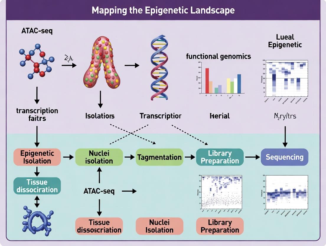
This technical guide details the critical wet-lab phase of the Assay for Transposase-Accessible Chromatin using sequencing (ATAC-seq), a cornerstone method in modern epigenetic landscape research. Within the broader thesis, this protocol enables the genome-wide mapping of open chromatin regions, which are indicative of active regulatory elements. The reproducibility of this step directly impacts downstream data quality, influencing analyses of transcription factor binding, nucleosome positioning, and chromatin dynamics in development and disease—key insights for drug target discovery.
Detailed Experimental Protocols
Cell Lysis and Nuclei Isolation
Objective: To obtain intact, clean nuclei free of cytoplasmic contaminants that can inhibit transposition. Detailed Protocol (for adherent cells):
- Harvesting: Culture cells to 50-80% confluency. Wash once with cold 1x PBS.
- Trypsinization & Quenching: Detach cells using Trypsin-EDTA (e.g., 0.25%) at 37°C for 3-5 min. Neutralize with complete growth medium.
- Washing: Centrifuge cell suspension at 500 RCF for 5 min at 4°C. Aspirate supernatant and gently resuspend pellet in 1 mL of cold 1x PBS. Repeat centrifugation.
- Hypotonic Lysis: Resuspend cell pellet in 50 µL of cold ATAC-seq Lysis Buffer (10 mM Tris-HCl pH 7.4, 10 mM NaCl, 3 mM MgCl2, 0.1% IGEPAL CA-630, 0.1% Tween-20, 0.01% Digitonin). Critical: Digitonin concentration may require optimization per cell type.
- Incubation: Incubate on ice for 3-10 minutes (monitor under microscope for lysed plasma membrane and intact nuclei).
- Washing & Resuspension: Immediately add 1 mL of cold Wash Buffer (10 mM Tris-HCl pH 7.4, 10 mM NaCl, 3 mM MgCl2, 0.1% Tween-20) to stop lysis. Centrifuge at 500 RCF for 10 min at 4°C. Carefully aspirate supernatant.
- Quantification: Resuspend nuclei pellet in 50 µL of PBS with 0.1% Tween-20. Count using a hemocytometer with Trypan Blue exclusion. Adjust concentration.
Transposition Reaction
Objective: To simultaneously fragment accessible chromatin and insert sequencing adapters using a hyperactive Tn5 transposase. Detailed Protocol:
- Reagent Assembly: In a 0.2 mL PCR tube, combine the following on ice:
- Nuclei suspension (containing 50,000 - 100,000 nuclei): variable volume
- 2x Tagmentation DNA (TD) Buffer: 25 µL
- Tn5 Transposase (commercial kit enzyme): variable, typically 2.5 µL
- Nuclease-free water to a final volume of 50 µL.
- Gently mix by pipetting, avoid bubbles.
- Tagmentation: Immediately place the tube in a pre-heated thermal cycler and incubate at 37°C for 30 minutes.
- Clean-up: Add 250 µL of DNA Binding Buffer (from a commercial DNA clean-up kit) directly to the tagmentation reaction. Follow the kit's standard silica-membrane column protocol for DNA purification. Elute in 20-30 µL of Elution Buffer (10 mM Tris-HCl, pH 8.0).
Library Preparation by PCR Amplification
Objective: To amplify the tagmented DNA fragments and add full-length sequencing adapters and sample indexes. Detailed Protocol:
- PCR Master Mix: In a PCR tube, combine:
- Purified tagmented DNA: 20 µL
- 2x High-Fidelity PCR Master Mix (with GC-rich buffer): 25 µL
- Custom PCR Primer 1 (with i5 index): 2.5 µL
- Custom PCR Primer 2 (with i7 index): 2.5 µL
- Total volume: 50 µL.
- Amplification: Run the following thermocycling program:
- 72°C for 5 min (gap filling)
- 98°C for 30 sec (initial denaturation)
- Cycle N times: 98°C for 10 sec, 63°C for 30 sec, 72°C for 1 min.
- Note: N must be determined by a qPCR side reaction or pre-defined based on starting cell number (see Table 1).
- Double-Sided SPRI Bead Clean-up:
- Add 1.0x volume of SPRIselect beads to the PCR reaction. Incubate 5 min at RT.
- Place on magnet, discard supernatant after 5 min.
- Wash beads twice with 80% ethanol.
- Air dry for 5 min. Elute in 20 µL of Elution Buffer.
- Perform a second cleanup with 0.55x volume of beads to remove primer dimers and large fragments. Elute final library in 15-20 µL.
Data Presentation: Critical Parameters
Table 1: Quantitative Optimization Guidelines for ATAC-seq Workflow
| Parameter | Recommended Range | Impact of Deviation | Source/Reference |
|---|---|---|---|
| Input Cell Number | 50,000 - 100,000 viable cells | Low: High background noise. High: Overly dense nuclei, poor tagmentation. | Buenrostro et al., 2015; Corces et al., 2017 |
| Tagmentation Time | 30 min at 37°C | Short: Low library complexity. Long: Over-fragmentation, loss of nucleosome signal. | Omni-ATAC Protocol, 2017 |
| PCR Cycles (N) | 8-12 cycles (for 50K nuclei) | Too few: Low yield. Too many: Over-amplification, duplication artifacts. | Determined by qPCR or SYBR Green add-on |
| Final Library Size Distribution | Majority of fragments < 1,000 bp; Mononucleosome peak ~200 bp, Dinucleosome ~400 bp. | Skew to large fragments: Incomplete tagmentation or lysis issues. | Bioanalyzer/TapeStation profile |
| Final Library Concentration (Qubit) | > 5 nM for Illumina sequencing | Low concentration may lead to poor cluster generation on sequencer. | Standard NGS library QC |
Visualization of Core Workflow
Diagram Title: ATAC-seq Core Wet-Lab Workflow
The Scientist's Toolkit: Essential Research Reagent Solutions
Table 2: Key Reagents and Their Functions in ATAC-seq
| Reagent/Category | Example Product/Component | Critical Function | Notes for Selection |
|---|---|---|---|
| Cell Lysis Detergent | Digitonin, IGEPAL CA-630 | Permeabilizes plasma membrane while keeping nuclear envelope intact. | Digitonin concentration is cell-type sensitive; critical for clean nuclei. |
| Hyperactive Tn5 Transposase | Illumina Tagment DNA TDE1, Diagenode HyperTagment | Simultaneously fragments DNA and ligates sequencing adapters in open chromatin. | Pre-loaded with adapters; major determinant of library complexity. |
| Tagmentation Buffer | 2x TD Buffer (Mg2+ containing) | Provides optimal ionic conditions (Mg2+) for Tn5 transposase activity. | Supplied with commercial Tn5 kits. |
| High-Fidelity PCR Mix | NEBNext Q5, KAPA HiFi | Amplifies tagmented fragments with low error rate and handles GC-rich regions. | Essential for minimizing PCR artifacts and bias. |
| Dual-Indexed PCR Primers | Nextera XT Index Kit v2, IDT for Illumina | Adds full-length adapters and unique sample indexes for multiplexing. | Enables pooling of >96 samples in one sequencing run. |
| Size Selection Beads | SPRIselect, AMPure XP | Clean up reactions and perform size selection to remove primers and large fragments. | The 0.55x SPRI ratio is critical for removing primer dimers. |
| Nuclei Staining Dye | DAPI, Trypan Blue | Visualize nuclei integrity and count after lysis. | Quality control step before expensive tagmentation. |
Within the framework of ATAC-seq (Assay for Transposase-Accessible Chromatin using sequencing) for mapping the epigenetic landscape, the initial quality of the cellular input is the single greatest determinant of experimental success. This technical guide details the critical pre-sequencing parameters—cell number, viability, and handling—that define the robustness, reproducibility, and biological validity of downstream epigenetic data. Compromised cellular input leads to artifacts in chromatin accessibility profiles, confounding biological interpretation and threatening drug development pipelines.
Quantitative Specifications for ATAC-seq Input
The following table summarizes the current consensus and empirical data on cellular input requirements for various ATAC-seq modalities.
Table 1: Cell Input Specifications for ATAC-seq Protocols
| Protocol Type | Recommended Cell Number | Minimum Cell Number | Critical Viability Threshold | Key Considerations |
|---|---|---|---|---|
| Standard Bulk ATAC-seq | 50,000 - 100,000 cells | 5,000 - 10,000 cells | >90% | Higher numbers ensure library complexity and reproducibility. |
| Low-Input/Bulk | 500 - 5,000 cells | 100 cells | >95% | Requires specialized protocols (e.g., modified tagmentation buffer, post-tagmentation cleanup). |
| Single-Cell ATAC-seq (scATAC-seq) | 10,000 - 50,000 cells (for loading) | N/A | >90% (with high membrane integrity) | Input defines cell recovery; viability critical to reduce background from dead cells. |
| Frozen Nuclei | 50,000 - 100,000 nuclei | 10,000 nuclei | N/A (Intact nuclei) | Integrity post-thaw is key; assess via microscopy. Avoid repeated freeze-thaws. |
Experimental Protocols for Assessment and Preparation
Protocol 2.1: Accurate Determination of Cell Viability and Count
Objective: To precisely quantify live cell concentration prior to ATAC-seq. Materials: Single-cell suspension, hemocytometer or automated cell counter (e.g., Countess II), Trypan Blue dye (0.4%) or AO/PI stains, PBS. Procedure:
- Prepare Cell Suspension: Ensure a single-cell suspension by gentle pipetting or filtering through a 35-40 µm cell strainer.
- Mix with Dye: Combine 10 µL of cell suspension with 10 µL of Trypan Blue. For automated counters, use appropriate fluorescent stains (Acridine Orange/Propidium Iodide).
- Load and Count: Transfer to a hemocytometer chamber. Count live (unstained) and dead (blue-stained) cells in all four quadrants.
- Calculate: Viability (%) = [Live Cells / (Live + Dead Cells)] x 100. Concentration (cells/mL) = Average count per quadrant x Dilution Factor x 10^4.
Protocol 2.2: Nuclei Isolation from Cryopreserved Cells/Tissues for ATAC-seq
Objective: To obtain intact, high-quality nuclei from archived samples. Materials: Frozen cell pellet or tissue piece, Ice-cold Lysis Buffer (10 mM Tris-HCl pH 7.4, 10 mM NaCl, 3 mM MgCl2, 0.1% IGEPAL CA-630, 1% BSA, 0.1 U/µL RNase Inhibitor), Wash Buffer (PBS + 1% BSA), Dounce homogenizer (for tissue). Procedure:
- Thaw Rapidly: Place frozen sample on wet ice.
- Lyse Cells: Resuspend pellet or minced tissue in 1 mL ice-cold Lysis Buffer. For tissue, dounce 10-15 strokes. Incubate on ice for 5-10 minutes.
- Verify Lysis: Check under microscope; >95% of cells should be lysed, releasing intact nuclei.
- Pellet Nuclei: Centrifuge at 500 x g for 5 min at 4°C. Carefully discard supernatant.
- Wash: Gently resuspend nuclei pellet in 1 mL Wash Buffer. Centrifuge at 500 x g for 5 min at 4°C. Repeat.
- Resuspend and Count: Resuspend in desired buffer (e.g., ATAC-seq Resuspension Buffer). Count nuclei using hemocytometer (DAPI stain) or automated counter.
Visualizing Workflows and Logical Relationships
Title: Quality Control Workflow for ATAC-seq Sample Prep
Title: Impact of Input Quality on ATAC-seq Data Artifacts
The Scientist's Toolkit: Essential Research Reagents & Materials
Table 2: Key Reagent Solutions for ATAC-seq Cell Preparation
| Item | Function in ATAC-seq Context | Example/Key Component |
|---|---|---|
| Viability Stain (AO/PI or Trypan Blue) | Distinguishes live/dead cells for accurate input normalization. Prevents dead cell chromatin from contributing to background. | Acridine Orange/Propidium Iodide (AO/PI) for automated counters. |
| Nuclei Lysis Buffer | Gently lyses plasma membrane while leaving nuclear envelope intact. Critical for transposase access to chromatin. | Tris-HCl, NaCl, MgCl2, Detergent (e.g., IGEPAL CA-630). |
| Transposase (Tn5) | Enzyme that simultaneously fragments and tags accessible chromatin with sequencing adapters. Core enzyme of ATAC-seq. | Loaded Tn5 transposase complex (commercial kits available). |
| Magnetic Beads (SPRI) | For size selection and purification of tagmented DNA fragments. Removes small fragments and enzyme contaminants. | AMPure XP or similar SPRI (Solid Phase Reversible Immobilization) beads. |
| RNase Inhibitor | Prevents RNA degradation during nuclei isolation, which can release ribonucleoproteins that stick to chromatin. | Recombinant RNase Inhibitor. |
| BSA (Bovine Serum Albumin) | Acts as a stabilizer and carrier protein in buffers, reducing nonspecific adhesion of nuclei/tags to tubes. | Molecular biology grade, nuclease-free BSA. |
| Cell Strainer | Ensures a single-cell or single-nucleus suspension by removing clumps and debris. Essential for accurate counting. | 35-40 µm nylon mesh strainers. |
| Cryopreservation Medium | For archiving cells pre-ATAC-seq. Must maintain high viability post-thaw. Often contains FBS and DMSO. | 90% FBS + 10% DMSO or commercial alternatives. |
This technical guide details the computational framework essential for analyzing Assay for Transposase-Accessible Chromatin with high-throughput sequencing (ATAC-seq) data. Within the broader thesis of mapping epigenetic landscapes, this pipeline transforms raw sequencing reads into interpretable maps of chromatin accessibility, which serve as proxies for regulatory element activity. The accurate execution of read alignment, peak calling, and rigorous quality control (QC) is foundational for downstream analyses such as differential accessibility testing, motif discovery, and integration with other epigenomic datasets.
Experimental Protocols for ATAC-seq
2.1. Key Wet-Lab Protocol (Summarized)
- Cell Lysis & Transposition: Isolated nuclei are treated with the engineered Tn5 transposase, which simultaneously fragments accessible DNA and inserts sequencing adapters.
- PCR Amplification: Transposed DNA fragments are amplified with indexed primers for multiplexing. The cycle number is minimized (typically 5-12 cycles) to prevent amplification biases.
- Size Selection: Libraries are purified using SPRI beads to selectively retain fragments primarily below 1kb, enriching for nucleosome-free regions (NFRs) and mononucleosome fragments.
- Sequencing: Paired-end sequencing (commonly 2x50 bp or 2x75 bp) is performed on Illumina platforms to capture both ends of each transposed fragment.
Core Bioinformatics Pipeline
3.1. Quality Control of Raw Reads
- Tool: FastQC is used for initial assessment.
- Metrics: Per-base sequence quality, adapter contamination, GC content, and sequence duplication levels. Adapters are trimmed using Trimmomatic or Cutadapt.
3.2. Read Alignment to Reference Genome
- Methodology: Pre-processed reads are aligned to a reference genome (e.g., GRCh38, mm10) using aligners optimized for genomic data.
- Protocol:
- Alignment: Use Bowtie2 or BWA-MEM with options to retain paired-end information (
-X 2000for large fragment sizes). - Filtering: Remove unmapped, low-quality (MAPQ < 30), duplicate (marked with Picard Tools), and mitochondrial reads.
- Post-alignment QC: Assess alignment statistics (total reads, duplicate rate, mitochondrial proportion).
- Alignment: Use Bowtie2 or BWA-MEM with options to retain paired-end information (
3.3. Peak Calling
- Methodology: Identifies genomic regions with a statistically significant enrichment of aligned fragment ends (cut sites), corresponding to open chromatin regions.
- Protocol:
- BAM File Preparation: Use alignment BAM files from individual or pooled replicates.
- Peak Calling Execution: Run MACS2 (
macs2 callpeak) with the--nomodel --shift -75 --extsize 150parameters tailored for ATAC-seq cut-site signals. - Blacklist Filtering: Remove peaks falling within ENCODE-defined blacklist regions (e.g., high-signal artifacts).
3.4. Advanced QC Metrics Beyond initial FastQC, ATAC-seq-specific metrics are critical.
- Insert Size Distribution: Calculated from paired-end BAM files, showing periodicity corresponding to nucleosome positioning.
- Transcription Start Site (TSS) Enrichment: Measures signal strength at annotated TSSs; high enrichment indicates high-quality library.
- Fraction of Reads in Peaks (FRiP): Proportion of all reads falling within called peak regions; indicates signal-to-noise ratio.
Table 1: Key QC Metrics and Their Interpretation
| Metric | Target / Ideal Outcome | Indication of Problem |
|---|---|---|
| Reads Aligned | > 80% of total reads | Poor library prep or contamination |
| Mitochondrial Reads | < 20% (cell type dependent) | Excessive cell death during prep |
| Duplication Rate | < 50% (library complexity) | Insufficient starting material or over-amplification |
| FRiP Score | > 0.2 - 0.3 | Low signal-to-noise; poor experiment |
| TSS Enrichment | > 5 - 10 (higher is better) | Low quality; insufficient accessible chromatin |
| NFR Fragment Peak | Clear peak ~50-100 bp in insert size plot | Poor transposase activity or size selection |
The Scientist's Toolkit: Research Reagent Solutions
Table 2: Essential Materials for ATAC-seq Experiments
| Item | Function | Example/Note |
|---|---|---|
| Tn5 Transposase | Enzyme for simultaneous fragmentation and adapter tagging. | Illumina Nextera or homemade loaded Tn5. |
| AMPure XP Beads | SPRI beads for post-transposition and post-PCR size selection and cleanup. | Critical for removing large fragments and primers. |
| Qubit dsDNA HS Assay | Fluorometric quantification of low-concentration DNA libraries. | More accurate than spectrophotometry for lib prep. |
| High-Sensitivity DNA Bioanalyzer Chip | Assess library fragment size distribution prior to sequencing. | Confirms enrichment for sub-nucleosomal fragments. |
| Indexed PCR Primers | Amplify transposed DNA and add unique sample indexes for multiplexing. | Illumina P5/P7 or custom i5/i7 indexed primers. |
| Cell Permeabilization Buffer | Lyse cells while keeping nuclei intact for transposition. | Contains detergent (e.g., IGEPAL CA-630). |
Visualization of Core Pipeline and Quality Signals
ATAC-seq Bioinformatics Pipeline Workflow
Key QC Signal Profiles for ATAC-seq Data
This guide details the critical downstream analysis phase following ATAC-seq (Assay for Transposase-Accessible Chromatin with high-throughput sequencing) experimentation. Within the broader thesis of mapping the epigenetic landscape, this phase transforms raw chromatin accessibility data into biologically interpretable insights. It enables researchers to pinpoint genomic regions with significant accessibility changes between conditions (e.g., disease vs. healthy, treated vs. untreated) and to infer the transcription factor (TF) networks driving these epigenetic alterations. This is fundamental for understanding gene regulation mechanisms in development, disease pathogenesis, and drug response.
Identifying Differential Accessibility
Differential accessibility (DA) analysis identifies genomic regions where chromatin openness statistically differs between biological conditions.
Core Methodology & Tools
The process typically involves:
- Peak Calling & Count Matrix Generation: Consolidated peaks from all samples are defined as regions of interest. Reads mapping to each peak in each sample are counted to create a quantitative matrix.
- Normalization: Counts are normalized to account for differences in library size and sequencing depth (e.g., using TMM, DESeq2, or median-of-ratios methods).
- Statistical Testing: Generalized linear models (GLMs) are applied to test for significant differences, often incorporating appropriate dispersion estimates and controlling for relevant covariates (e.g., batch effects).
Key Software Packages
| Tool Name | Statistical Core | Key Features | Best For |
|---|---|---|---|
| DESeq2 | Negative binomial GLM with shrinkage estimators. | Robust to over-dispersion, includes hypothesis testing with Wald test or LRT, excellent for complex designs. | Most ATAC-seq DA analyses, especially with biological replicates. |
| edgeR | Negative binomial models with quasi-likelihood tests. | Highly flexible, efficient with many samples, offers both GLM and exact test routes. | Experiments with many replicates or groups. |
| diffReps | Sliding window with statistical tests (e.g., χ²). | Peak-free, identifies differential sites without pre-defined peaks, useful for broad domains. | Discovery of novel, unannotated differential regions. |
| limma-voom | Linear modeling with precision weights. | Applies experience from microarray/RNA-seq to ATAC-seq counts after voom transformation. | Experiments with very large sample sizes. |
Table 1: Typical Output Metrics from a Differential Accessibility Analysis (Hypothetical Data).
| Condition Comparison | Total DA Peaks | Up-Accessible | Down-Accessible | Adj. p-value < 0.05 | Typical log2FC Range |
|---|---|---|---|---|---|
| Disease vs. Control | 5,247 | 2,891 (55.1%) | 2,356 (44.9%) | 5,247 | -4.5 to +5.2 |
| Drug-Treated vs. Untreated | 1,843 | 1,102 (59.8%) | 741 (40.2%) | 1,843 | -3.8 to +4.1 |
| Timepoint 2 vs. Timepoint 1 | 3,569 | 1,785 (50.0%) | 1,784 (50.0%) | 3,569 | -3.2 to +3.9 |
Experimental Protocol: DESeq2 for DA
Protocol: Differential Peak Analysis with DESeq2. Input: A consensus peak set (BED file) and aligned BAM files for all samples. Steps:
- Generate Count Matrix: Use
featureCounts(from Subread package) or similar to count fragments overlapping each peak for each BAM file.
DESeq2 Analysis in R:
Output Interpretation: The primary outputs are
log2FoldChange(magnitude/direction of change) andpadj(adjusted p-value). Peaks withpadj < 0.05andabs(log2FoldChange) > 0.58(∼1.5-fold) are typically considered significant.
Motif Discovery & TF Inference
Following DA analysis, motif discovery identifies over-represented transcription factor binding motifs within differential peaks, linking accessibility changes to potential regulatory drivers.
Core Methodology & Tools
The workflow involves:
- Sequence Extraction: Obtain DNA sequences for significant DA peaks.
- De Novo Motif Discovery: Finds novel, over-represented sequence patterns without prior assumptions.
- Motif Enrichment Analysis: Tests for enrichment of known TF motifs from databases (e.g., JASPAR, CIS-BP) within DA peaks compared to a background set.
Key Software Packages
| Tool Name | Primary Function | Key Features | Output |
|---|---|---|---|
| HOMER | De novo discovery & known motif enrichment. | Comprehensive, integrates with genomic annotations, user-friendly. | Motif files, enrichment statistics, TF assignment. |
| MEME-ChIP | De novo discovery (MEME) & refinement (DREME). | Suite of tools, good for short, peaked ChIP/ATAC-seq data. | HTML report with motifs, E-values, logos. |
| AME (MEME-Suite) | Known motif enrichment analysis. | Uses statistical tests (Fisher's exact, rank-sum) against motif databases. | Table of enriched motifs, p-values. |
| RSAT | De novo and known motif analysis via web or CLI. | Peak-motifs tool tailored for ATAC/ChIP-seq, uses oligo analysis. | Motifs, matrices, genome tracks. |
Table 2: Example Results from HOMER Motif Enrichment Analysis on Up-Accessible Peaks.
| Motif Name (TF) | p-value | log P-value | % of Target Peaks | % of Background Peaks |
|---|---|---|---|---|
| NFκB (RelA) | 1e-25 | -57.6 | 28.5% | 8.2% |
| AP-1 (Fos-Jun) | 1e-22 | -50.7 | 32.1% | 12.5% |
| RUNX1 | 1e-18 | -41.4 | 18.7% | 5.8% |
| SPI1 (PU.1) | 1e-15 | -34.5 | 22.4% | 9.1% |
Experimental Protocol: HOMER for Motif Analysis
Protocol: De Novo and Known Motif Discovery with HOMER. Input: A BED file of significant differential peaks (e.g., up-accessible peaks). Steps:
- Install and Set Up HOMER: Follow instructions at http://homer.ucsd.edu/homer/.
- Run De Novo Motif Discovery:
- Run Known Motif Enrichment:
- Output Interpretation: Key results are in
knownResults.txtandhomerResults.html. The% of Targetvs.% of Backgroundand thelog P-valueindicate enrichment significance. HOMER provides a likely TF name for each motif.
Visualizing Workflows and Relationships
Diagram 1: Core downstream ATAC-seq analysis workflow.
Diagram 2: Statistical framework for differential accessibility.
The Scientist's Toolkit: Research Reagent Solutions
Table 3: Essential Reagents and Materials for ATAC-seq Downstream Analysis.
| Item | Function in Downstream Analysis | Example/Notes |
|---|---|---|
| High-Fidelity PCR Master Mix | Amplification of libraries post-tagmentation. Critical for maintaining complexity and avoiding biases. | NEBNext Ultra II Q5 Master Mix. |
| Dual-Size Selection Beads | Precise selection of library fragments (e.g., 150-500 bp) to optimize sequencing of mononucleosomal fragments. | SPRIselect (Beckman Coulter) or equivalent. |
| Indexing Primers (Unique Dual Indexes) | Multiplexing samples. UDIs are essential to minimize index hopping in paired-end sequencing on patterned flow cells. | Illumina IDT for Illumina UD Indexes. |
| High-Sensitivity DNA Assay Kits | Accurate quantification of library concentration and size distribution prior to sequencing. | Agilent Bioanalyzer High Sensitivity DNA kit or TapeStation D1000/High Sensitivity D1000. |
| qPCR Quantification Kit | Precise, amplification-based quantification of adapter-ligated fragments for accurate pooling and cluster generation. | KAPA Library Quantification Kits for Illumina. |
| High-Output Sequencing Reagents | Generation of sufficient sequencing depth (typically 50-100 million paired-end reads per sample). | Illumina NovaSeq 6000 S4 Reagent Kit (300 cycles) or equivalent. |
| Positive Control Chromatin | Validating the entire ATAC-seq wet-lab and analysis pipeline. | Commercially available reference chromatin (e.g., from cell lines with well-characterized open regions). |
| Bioinformatics Software Suites | Executing the analysis pipelines described in Sections 2 & 3. | Galaxy platform, Anaconda/Python/R environments with Bioconductor packages. |
| High-Performance Computing (HPC) Resources | Essential for storage, alignment, and intensive computational analysis of sequencing data. | Local cluster or cloud computing (AWS, Google Cloud, Azure). |
The broader thesis of ATAC-seq research is to map the dynamic, accessible chromatin landscape that defines cellular identity and function. While bulk ATAC-seq provides population-averaged views, single-cell ATAC-seq (scATAC-seq) represents a paradigm shift, enabling the deconvolution of epigenetic heterogeneity within tissues. This whitepaper details advanced scATAC-seq methodologies, their integration with other omics layers, and their transformative application in deciphering disease mechanisms and identifying therapeutic targets.
Core scATAC-seq Technologies and Quantitative Benchmarks
Current platforms differ in throughput, data quality, and multiomic capabilities. The following table summarizes key quantitative metrics from recent benchmarking studies (2023-2024).
Table 1: Comparison of Primary High-Throughput scATAC-seq Platforms
| Platform | Principle | Cells per Run (Typical) | Median Fragments per Cell | TSS Enrichment | Key Multiomic Pairing | Cost per 10k Cells (USD) |
|---|---|---|---|---|---|---|
| 10x Chromium | Microfluidics, Tn5 | 10,000 | 20,000 - 50,000 | 10 - 20 | scRNA-seq (ATAC + GEX) | ~$4,500 |
| sci-ATAC-seq | Combinatorial Indexing | 50,000 - 100,000 | 1,000 - 5,000 | 4 - 8 | sci-RNA-seq | ~$2,000 |
| mtscATAC-seq | Nuclear Hashing, Pooling | 100,000+ | 5,000 - 15,000 | 8 - 15 | Not native | ~$1,500 |
| SHARE-seq | Split-pool, Linker Capture | 10,000 - 20,000 | 8,000 - 20,000 | 8 - 12 | scRNA-seq, chromatin state | ~$3,500 |
| Paired-Tag | Antibody-guided Indexing | 1,000 - 10,000 | 5,000 - 15,000 | 6 - 10 | Histone modification (CUT&Tag) | ~$3,000 |
Detailed Protocol: Multiomic scATAC-seq + scRNA-seq (10x Genomics)
This protocol enables simultaneous profiling of chromatin accessibility and gene expression from the same single nucleus/cell.
Day 1: Nuclei Isolation and Multiomic Library Preparation
- Tissue Dissociation & Lysis: Minced tissue is homogenized in cold lysis buffer (10mM Tris-HCl pH 7.4, 10mM NaCl, 3mM MgCl2, 0.1% IGEPAL CA-630, 1% BSA, 0.2 U/µl RNase inhibitor). Incubate on ice for 3-5 minutes.
- Nuclei Purification: Filter lysate through a 40µm flow-through strainer. Pellet nuclei (500 rcf, 5 min, 4°C). Resuspend in wash buffer (PBS + 1% BSA + 0.2 U/µl RNase inhibitor). Count with Trypan Blue.
- Tagmentation & GEM Generation: Use the Chromium Next GEM Chip K. Combine nuclei, Tn5 transposase, and Master Mix. Load into the chip with Gel Beads containing barcoded oligos for both ATAC and RNA, and Partitioning Oil. This generates Gel Bead-in-Emulsions (GEMs) where transposition and reverse transcription occur.
- Post-GEM Incubation: Perform tagmentation (53°C for 45 min) followed by reverse transcription of RNA (53°C for 45 min).
- Cleanup & Amplification: Break GEMs, recover barcoded cDNA and tagmented DNA. Perform PCR amplification (12 cycles) to add P5/P7 handles and sample indices.
Day 2: Library Construction & QC
- Size Selection & QC: For ATAC library, perform double-sided SPRIselect bead cleanup (0.55x and 1.5x ratios) to select 100-700 bp fragments. For GEX library, perform a 0.6x SPRI cleanup. Assess libraries on a Bioanalyzer (High Sensitivity DNA kit). Expected peak: ~300 bp for ATAC; broad distribution for GEX.
- Sequencing: Pool libraries and sequence on an Illumina platform. Recommended sequencing: ATAC: 25,000 paired-end reads/nucleus (50+8+16+0+50); GEX: 20,000 reads/cell (28+10+10+0+90).
Multiomics Integration: Methods and Computational Workflows
Integration leverages joint embedding or graph-based methods to link peaks, genes, and regulatory elements.
Figure 1: Workflow for Integrating scATAC-seq and scRNA-seq Data.
Application in Disease Research: Uncovering Mechanisms
Integrated multiomics reveals disease-specific cell states and causal regulatory circuits.
Table 2: Key Disease Insights from Recent scATAC-seq Multiomics Studies (2023-2024)
| Disease Context | Key Finding | Method Used | Therapeutic Implication |
|---|---|---|---|
| Alzheimer's Disease | Microglia subpopulation with APOE-linked accessible sites driving pro-inflammatory state. | snATAC-seq + snRNA-seq (post-mortem brain) | Targeting PU.1 or SPI1 transcription factor. |
| Autoimmunity (RA, SLE) | CD4+ T cell subset with co-accessible motifs for BATF and IRF4, linked to IL21 expression. | scATAC-seq + scRNA-seq (PBMCs) | Disrupting the BATF-IRF4 complex. |
| Cardio-Oncology | Cardiomyocyte chromatin remodeling post-doxorubicin treatment, preceding apoptosis. | scMultiome (Heart tissue) | Early epigenetic intervention to prevent damage. |
| Clonal Hematopoiesis | TET2-mutant clones show distinct chromatin landscape in monocytes, priming for inflammation. | scATAC-seq with genotyping. | Demethylating agents to restore regulation. |
| Solid Tumors (e.g., GBM) | Recurrent tumor-specific chromatin loops connecting enhancers (H3K27ac) to oncogenes (MYC). | scATAC-seq + HiChIP (patient-derived xenografts) | BET bromodomain inhibitors to disrupt loops. |
Figure 2: Disease Mechanism and Epigenetic Targeting Pathway.
The Scientist's Toolkit: Essential Reagents & Materials
Table 3: Key Research Reagent Solutions for scATAC-seq Multiomics
| Item | Function | Example Product/Catalog |
|---|---|---|
| Nuclei Isolation Buffer | Gentle lysis of plasma membrane while preserving nuclear envelope and chromatin state. | 10x Genomics Nuclei Isolation Kit (CG000365) |
| Tn5 Transposase | Engineered enzyme that simultaneously fragments and tags accessible DNA with sequencing adapters. | Illumina Tagment DNA TDE1 Enzyme |
| Barcoded Gel Beads | Microbeads containing oligonucleotides with cell barcode, UMI, and primers for both ATAC and RNA. | 10x Chromium Next GEM Chip K (1000269) |
| Dual Index Kit | Provides unique sample indices for multiplexing libraries during the final PCR. | 10x Dual Index Kit TT Set A (1000215) |
| SPRIselect Beads | Magnetic beads for size selection and cleanup of libraries, critical for removing adapter dimers. | Beckman Coulter SPRIselect (B23318) |
| RNase Inhibitor | Protects RNA from degradation during nuclei isolation and subsequent steps. | Protector RNase Inhibitor (3335402001) |
| Cell Hashing Antibodies | For multiplexing samples, using TotalSeq-C antibodies with barcoded oligonucleotides. | BioLegend TotalSeq-C Hashtag Antibodies |
| Chromatin Immunoprecipitation Kits | For integrated methods like Paired-Tag, profiling histone modifications alongside accessibility. | Cell Signaling Technology CUTANA Kits |
Solving ATAC-seq Challenges: Expert Tips for Optimization and Troubleshooting
ATAC-seq (Assay for Transposase-Accessible Chromatin using sequencing) is a cornerstone method for mapping the epigenetic landscape, revealing regions of open chromatin indicative of regulatory activity. Its integration into broader theses on gene regulation and disease mechanisms is now standard. However, several pervasive technical pitfalls can compromise data integrity, leading to misinterpretation. This guide details three critical challenges—low library complexity, high mitochondrial reads, and background noise—providing diagnostic criteria, mitigation protocols, and analytical solutions.
Low Library Complexity
Library complexity refers to the number of unique, non-PCR-duplicate fragments in a library. Low complexity reduces statistical power and confounds peak calling.
Diagnosis and Quantitative Benchmarks
Low complexity is indicated by high PCR duplication rates. Metrics are calculated from alignment files using tools like picard MarkDuplicates.
Table 1: Library Complexity Metrics and Interpretation
| Metric | Optimal Range | Problematic Range | Primary Tool for Calculation |
|---|---|---|---|
| Non-Redundant Fraction (NRF) | > 0.8 | < 0.7 | Picard Tools |
| PCR Bottlenecking Coefficients (PBC1, PBC2) | PBC1 > 0.9, PBC2 > 3 | PBC1 < 0.7, PBC2 < 1 | ENCODE ChIP-seq guidelines |
| Estimated Library Size | > 20 million unique fragments | < 10 million unique fragments | Preseq |
Experimental Protocol for Complexity Rescue
- Sample Input Optimization: For cultured cells, ensure a starting input of 50,000-100,000 viable, nuclei. Avoid over-tagmentation.
- PCR Cycle Minimization: Perform a qPCR side-reaction prior to final library amplification to determine the minimum number of PCR cycles (Cq) required. Typically, aim for ½ to ¾ of the total reaction volume to reach plateau.
- Protocol: Set up a 10 µL qPCR reaction with 2 µL of pre-amplified tagmented DNA, SYBR Green, and library amplification primers. Run on a real-time cycler. The Cq value indicates the required cycles for the main reaction.
- Cleanup Strategy: Use SPRI bead-based size selection (e.g., 0.5x left-side cleanup to remove large fragments, then 1.3x right-side cleanup to isolate library) to narrow insert size distribution and improve sequencing efficiency.
High Mitochondrial Reads
A high proportion of reads mapping to the mitochondrial genome (mtDNA) consumes sequencing depth and originates from accessible mitochondrial DNA or cytoplasmic contamination.
Diagnosis and Quantitative Benchmarks
Mitochondrial read percentage is calculated from aligned reads (e.g., using samtools idxstats).
Table 2: Mitochondrial Read Percentages by Sample Type
| Sample Type / Condition | Expected Range | High (Requires Action) | Likely Cause |
|---|---|---|---|
| Healthy Mammalian Cell Lines | 5% - 20% | > 30% | Inefficient lysis or nuclei isolation |
| Primary Tissues (e.g., liver, muscle) | 20% - 50% | > 60% | High mitochondrial content in tissue |
| Apoptotic / Stressed Cells | Variable, often high | > 50% | Mitochondrial outer membrane permeabilization |
Experimental Protocol for Mitochondrial Depletion
- Density Gradient Purification: Isolate nuclei via sucrose gradient centrifugation prior to tagmentation.
- Reagents: Homogenization buffer (10 mM Tris-HCl pH 8.0, 1 mM EDTA, 0.1% NP-40), 1.6 M Sucrose cushion.
- Protocol: Lyse cells in homogenization buffer on ice for 5 min. Layer lysate over a 1.6 M sucrose cushion. Centrifuge at 12,000g for 30 min at 4°C. Pellet contains purified nuclei.
- Bioinformatic Filtering: If prevention fails, post-hoc filtering is necessary. Align reads to a concatenated genome (nuclear + mitochondrial). Use
samtools viewto filter out mtDNA reads (e.g.,chrM), or employ tools likeATACseqQCto subsample them computationally.
Background Noise
Background noise manifests as diffuse, low-signal regions or sporadic false-positive peaks, often from technical artifacts like adapter dimers, DNA contamination, or cryptic transcription start sites.
- Fragment Size Distribution: Visualize with
plotFingerprint(deepTools) orATACseqQC. A prominent peak < 100 bp indicates adapter dimer contamination. - Peak Distribution: An overabundance of peaks called in promoter regions vs. distal intergenic regions can indicate systematic noise.
Experimental & Analytical Mitigation
- Enhanced Size Selection: Use double-sided SPRI bead cleanup (e.g., 0.45x and 1.2x ratios) to aggressively remove both short (< 100 bp) and long (> 800 bp) fragments.
- Bioinformatic Background Subtraction: Employ peak callers with explicit noise models (e.g., MACS2 with
--nomodel --shift -100 --extsize 200parameters for ATAC-seq). Utilize control samples (e.g., using a Tn5 mutant without transposition activity) if available for differential peak calling. - Signal-to-Noise Thresholding: Apply a stringent FRiP (Fraction of Reads in Peaks) cutoff. For ATAC-seq, a FRiP score > 0.2 is generally acceptable, but cell-type-specific thresholds should be established.
The Scientist's Toolkit: Research Reagent Solutions
Table 3: Essential Reagents and Materials for Robust ATAC-seq
| Item | Function | Key Consideration |
|---|---|---|
| Tn5 Transposase (Loaded) | Simultaneously fragments and tags accessible DNA with sequencing adapters. | Commercial kits (Nextera) ensure consistent activity; in-house loading requires optimization. |
| Digitonin | Permeabilizes nuclear membrane for Tn5 access. | Concentration is critical (typically 0.01%-0.1%); overtreatment increases mitochondrial reads. |
| SPRI (Solid Phase Reversible Immobilization) Beads | Size selection and cleanup of DNA libraries. | Bead-to-sample ratio dictates size cutoffs; precise pipetting is essential for reproducibility. |
| NEBNext High-Fidelity 2X PCR Master Mix | Amplifies tagmented DNA with high fidelity and low bias. | Polymerase with low GC-bias and high processivity improves complex library representation. |
| Nuclei Counter (e.g., Trypan Blue, DAPI) | Accurate quantification of intact nuclei before tagmentation. | Ensures correct input; avoids over- or under-tagmentation. |
| DNas-free, RNAs-free Water | All dilution and reaction steps. | Prevents degradation of samples and reagents. |
Visualizing Workflows and Relationships
Diagram Title: ATAC-seq Pitfall Diagnostic & Mitigation Workflow
Diagram Title: Nuclei Isolation to Reduce Mitochondrial Reads
Within the broader thesis on mapping the epigenetic landscape using ATAC-seq, a critical bottleneck is sample quality and quantity. This technical guide details strategies for overcoming challenges posed by frozen tissues, low cell input, and rare cell populations, enabling robust chromatin accessibility profiling in translational and drug discovery research.
Section 1: Frozen Tissue Samples
Frozen tissues are a vital resource in biobanks but present challenges for ATAC-seq due to nuclear degradation, cross-linking, and ice crystal damage that obscure chromatin accessibility signals.
Key Optimization Strategies
- Nuclei Isolation Optimization: Gentle mechanical homogenization followed by optimized detergent-based lysis is crucial. The use of sucrose cushions during centrifugation improves nuclear integrity.
- Transposition Reaction Adjustment: Increasing the incubation time of the Tn5 transposase with fixed or compromised chromatin can improve tagmentation efficiency.
- Post-Fixation Protocols: For long-term frozen samples, a post-isolation formaldehyde fixation step can help preserve nuclear structure during subsequent washes.
| Strategy | Standard Protocol Metric | Optimized Protocol Metric | Key Outcome |
|---|---|---|---|
| Homogenization Buffer | 0.1% NP-40 | 0.05% IGEPAL CA-630 | 25% increase in intact nuclei yield |
| Tn5 Incubation Time | 30 min @ 37°C | 60 min @ 37°C | 40% higher library complexity |
| Centrifugation | 500 rcf, 5 min | 800 rcf through 1.6M sucrose cushion | 60% reduction in cytoplasmic contamination |
| Input Nuclei | 50,000 | 10,000 (with post-fix) | Comparable TSS enrichment achieved |
Detailed Protocol: Nuclei Isolation from Frozen Tissue
- Cryopulverization: Chill metal mortar and pestle with liquid N₂. Place 10-25 mg frozen tissue in mortar, cover with liquid N₂, and pulverize to fine powder.
- Homogenization: Transfer powder to a Dounce homogenizer containing 1 mL of pre-chilled Lysis Buffer (10 mM Tris-HCl pH 7.4, 10 mM NaCl, 3 mM MgCl₂, 0.05% IGEPAL CA-630, 0.1% Tween-20, 0.01% Digitonin, 1% BSA).
- Dounce Homogenize: Perform 15-20 strokes with the loose pestle (A), then 15-20 strokes with the tight pestle (B) on ice.
- Filtration & Cushion Centrifugation: Filter homogenate through a 40 µm cell strainer. Layer filtrate over 1 mL of 1.6M sucrose cushion in Wash Buffer. Centrifuge at 800 rcf for 20 min at 4°C.
- Nuclei Wash: Discard supernatant and gently resuspend pellet in 1 mL Wash Buffer (10 mM Tris-HCl pH 7.4, 10 mM NaCl, 3 mM MgCl₂, 1% BSA). Centrifuge at 500 rcf for 5 min.
- Count & QC: Resuspend in 50-100 µL of PBS + 0.1% BSA. Count using a hemocytometer with Trypan Blue. Assess integrity by DAPI staining if possible.
Section 2: Low Cell Input Protocols
Standard ATAC-seq requires 50,000-100,000 cells. Low-input protocols (500-5,000 cells) are essential for fine-needle aspirates, pediatric samples, or sorted cells, but suffer from high background noise and low library complexity.
Key Optimization Strategies
- Carrier-Assisted Tagmentation: The use of inert carrier DNA or nucleosomes during the Tn5 reaction improves kinetics without contributing to sequencing data.
- Post-Tagmentation Cleanup & Amplification: Optimized bead-based size selection and reduced-cycle, high-fidelity PCR are critical to minimize bias and retain rare fragments.
- Methylated Adapter Adoption: Using adapters resistant to exonuclease digestion allows for more stringent washes, reducing adapter dimer contamination.
| Cell Input | Protocol Modifications | Median Fragments per Cell | % of Reads in Peaks | TSS Enrichment |
|---|---|---|---|---|
| 50,000 (Standard) | Standard | 85,000 | 45% | 18 |
| 5,000 | 0.05% Digitonin, 1x Carrier | 42,000 | 38% | 15 |
| 500 | 2x Carrier, Methylated Adapters | 15,000 | 25% | 10 |
| 100 (Ultra-low) | Microfluidic Partitioning, Preamplification | 8,000 | 20% | 8 |
Detailed Protocol: ATAC-seq for 500-5,000 Cells
- Cell Lysis: Resuspend pelleted cells in 50 µL of cold Lysis Buffer (10 mM Tris-HCl pH 7.4, 10 mM NaCl, 3 mM MgCl₂, 0.1% IGEPAL CA-630, 0.01% Digitonin, 1% BSA). Incubate on ice for 3 min. Immediately add 1 mL of Wash Buffer (0.01% Tween-20 instead of IGEPAL/Digitonin) and invert to mix.
- Nuclei Pellet & Count: Centrifuge at 500 rcf for 10 min at 4°C. Resuspend nuclei in 50 µL of Tagmentation Buffer (33 mM Tris-acetate, 66 mM K-acetate, 11 mM Mg-acetate, 16% DMF, 0.01% Tween-20). Count a 2 µL aliquot.
- Tagmentation Reaction: To the remaining nuclei, add 1-2 µL of custom-loaded Tn5 transposase and 1 µL of 100 ng/µL carrier DNA (e.g., sheared salmon sperm DNA). Mix gently and incubate at 37°C for 45 min in a thermomixer with agitation.
- DNA Cleanup: Immediately add 20 µL of 40 mM EDTA + 1% SDS to stop the reaction. Purify DNA using SPRI beads at a 1.8x ratio. Elute in 20 µL EB buffer.
- Library Amplification: Perform a 12-14 cycle PCR using a high-fidelity polymerase and primers compatible with methylated adapters. Clean up final library with a double-sided SPRI selection (0.5x to remove large fragments, then 1.8x to recover the library).
Section 3: Rare Cell Population Analysis
Profiling rare cell types (e.g., circulating tumor cells, stem cells) requires upfront enrichment, which often yields low cell numbers and potential epigenetic perturbation from sorting.
Key Optimization Strategies
- Integrated Sort-ATAC Protocols: Performing tagmentation immediately after FACS sorting into plates containing lysis buffer minimizes handling loss and nuclear degradation.
- Bulk vs. Single-Cell Approach: For populations >1,000 cells, low-input bulk ATAC-seq provides deep coverage. For <1,000 cells or heterogeneous mixtures, single-cell ATAC-seq (scATAC-seq) is preferable.
- Pre-Enrichment Considerations: Gentle, epitope-preserving pre-enrichment methods like magnetic-activated cell sorting (MACS) prior to FACS can improve recovery.
| Method | Minimum Cell # | Key Requirement | Data Output | Cost per Sample |
|---|---|---|---|---|
| Low-Input Bulk ATAC | 500 | High viability post-sort | Aggregate profile | $$ |
| Plate-Based scATAC | 200-500 | Indexed FACS sorting | Cell-type specific peaks | $$$$ |
| Droplet-Based scATAC | 5,000+ (mixed) | Single-cell suspension | Heterogeneity maps | $$$ |
| ATAC with CUT&Tag | 1,000 | Target-specific antibody | Focused, ultra-low input | $$ |
Detailed Protocol: Integrated FACS-ATAC for Rare Populations
- Cell Preparation & Staining: Prepare a single-cell suspension using gentle dissociation. Stain with validated antibodies for surface markers. Include a viability dye (e.g., DAPI or PI).
- FACS Sorting Setup: Use a sorter equipped with a 100 µm nozzle and low pressure (20 psi). Prepare a 96-well PCR plate by adding 5 µL of Lysis Buffer (as in Low-Input Protocol) to each well designated for a cell. Seal and keep on ice.
- Direct Sort-Lyse: Sort single, live cells directly into the lysis buffer in each well. Immediately after sorting, seal the plate, vortex briefly, and centrifuge. Freeze at -80°C or proceed directly to tagmentation.
- In-Plate Tagmentation: Thaw plate on ice. Add 10 µL of Tagmentation Mix (2x Tagmentation Buffer, Tn5 enzyme, nuclease-free water) to each well. Mix by pipetting, then incubate at 37°C for 45 min in a thermal cycler.
- Pooled Cleanup & Amplification: Stop reactions by adding 20 µL of Stop Buffer (40 mM EDTA, 1% SDS) to each well. Pool all wells into a single tube. Purify DNA with SPRI beads and amplify for 14-18 cycles as a single library.
The Scientist's Toolkit: Essential Research Reagents & Materials
| Item | Function & Rationale | Example/Note |
|---|---|---|
| IGEPAL CA-630 | Non-ionic detergent for gentle cell membrane lysis during nuclei isolation. Preferred over NP-40 for frozen tissues. | Alternative: Triton X-100. |
| Digitonin | Sterol-based detergent for precise nuclear membrane permeabilization, critical for Tn5 entry in low-input protocols. | Titrate carefully (0.01-0.1%). |
| Sucrose (OptiPrep) | Forms density cushion for centrifugation, pelleting nuclei while leaving debris in supernatant; improves purity. | Used at 1.2M-1.6M concentration. |
| Carrier DNA | Inert DNA (e.g., sheared salmon sperm) improves Tn5 reaction kinetics in low-input samples by preventing enzyme loss. | Must be highly purified and RNA-free. |
| Methylated Adapters | Adapters resistant to exonuclease digestion allow stringent washes to remove adapter dimers in low-input preps. | Essential for ≤500 cell protocols. |
| High-Fidelity PCR Mix | Minimizes amplification bias during limited-cycle library PCR, preserving representation of rare fragments. | e.g., KAPA HiFi, NEB Next Ultra II. |
| Dual-Size SPRI Beads | Magnetic beads for selective binding of DNA fragments; used for post-tagmentation cleanup and final library size selection. | Ratios are critical (e.g., 0.5x, 1.8x). |
| Validated Antibody Panels | For pre-enrichment or FACS sorting of rare populations; must be titrated to avoid epitope damage. | Conjugation to rare earth metals for CyTOF is compatible. |
| Tn5 Transposase | Engineered transposase that simultaneously fragments and tags accessible chromatin with sequencing adapters. | Can be loaded in-house or purchased pre-loaded. |
Within the thesis on "ATAC-seq for Mapping the Epigenetic Landscape in Disease Models," rigorous quality control (QC) is paramount. The interpretation of fragment length distributions and correlation metrics forms the critical checkpoint that distinguishes high-quality, biologically interpretable data from technical noise. This guide details the technical standards and methodologies for these QC steps, ensuring robust downstream analysis of chromatin accessibility.
The Significance of Fragment Length Distributions in ATAC-seq
ATAC-seq utilizes the Tn5 transposase to fragment accessible DNA and insert sequencing adapters. The length of the resulting fragments is a direct readout of nucleosomal positioning. A high-quality ATAC-seq library exhibits a characteristic periodic pattern in its fragment size distribution.
Quantitative Benchmarks for Fragment Lengths
The table below summarizes the expected quantitative metrics from a successful ATAC-seq experiment.
Table 1: Expected Fragment Length Distribution Metrics in ATAC-seq
| Metric | Expected Value/Range | Biological Interpretation |
|---|---|---|
| Peak Periodicity | ~200 base pairs (bp) | Distance between nucleosome cores (Nucleosome Repeat Length). |
| Sub-nucleosomal Peak | < 100 bp | Tn5 insertion in nucleosome-free regions (NFRs). |
| Mononucleosome Peak | ~200 bp | DNA protected by a single nucleosome. |
| Dinucleosome Peak | ~400 bp | DNA protected by two nucleosomes. |
| Trinucleosome Peak | ~600 bp | DNA protected by three nucleosomes. |
| Ratio (NFR / Mono) | > 0.5 (Library dependent) | Indicates good signal-to-noise and transposition efficiency. |
| Fragment Size Mode | 50-100 bp | Most common fragment length, typically from NFRs. |
Protocol: Generating and Assessing the Fragment Length Distribution
Methodology:
- Alignment: Align sequencing reads (paired-end) to the reference genome using a splice-aware aligner (e.g., BWA-MEM, Bowtie2). Use parameters to properly handle paired-end data (
-X 2000for Bowtie2 allows larger fragment sizes). - Filtering: Remove reads mapping to mitochondrial DNA, low-mapping-quality reads (MAPQ < 30), duplicate reads (PCR artifacts), and reads mapping to blacklisted genomic regions (e.g., ENCODE DAC Blacklist).
- Fragment File Generation: Use tools like
samtoolsto extract properly paired, filtered alignments. The fragment length is calculated as the outer distance between the two read pairs. - Distribution Plotting: Using a programming environment (R/Python), plot a histogram of fragment lengths (x-axis: base pairs, y-axis: count/density) from 0 to 1000 bp.
Interpretation: A failed experiment will show a smooth, exponential decay from short fragments with no periodicity, indicating non-specific fragmentation or poor library complexity.
Correlation Metrics as a Measure of Replicability
Beyond fragment lengths, assessing the correlation between biological replicates is essential to confirm that observed signals are reproducible and not stochastic.
Key Correlation Metrics and Thresholds
Table 2: Correlation Metrics for ATAC-seq Replicate QC
| Metric | Calculation Method | QC Threshold | Interpretation |
|---|---|---|---|
| Pearson's r | Linear correlation of signal intensity (read counts) across genomic bins or peaks. | r ≥ 0.8 between true biological replicates. | Measures strength of linear relationship. Sensitive to outliers. |
| Spearman's ρ | Rank correlation of signal intensity. | ρ ≥ 0.8 between true biological replicates. | Measures monotonic relationship. Less sensitive to extreme values. |
| Irreproducible Discovery Rate (IDR) | Ranks peaks from replicates and measures consistency. | IDR < 0.05 for high-confidence peak sets. | Gold standard for assessing replicability in high-throughput data. |
Protocol: Calculating Correlation Metrics
Methodology for Genome-wide Correlation:
- Create Genome Bins: Tile the genome into non-overlapping bins (e.g., 500 bp or 1 kb) using
bedtools makewindows. - Count Reads per Bin: Count the number of fragments overlapping each bin for each sample/replicate using
bedtools coverageorfeatureCounts. - Normalize Counts: Perform read-depth normalization (e.g., Counts Per Million - CPM, or using median ratio methods like DESeq2).
- Calculate Correlation: On the normalized count matrix, compute pairwise Pearson and Spearman correlations between all samples.
- Visualize: Generate a scatterplot matrix or a hierarchically clustered heatmap of the correlation matrix.
Methodology for IDR on Peaks:
- Call Peaks per Replicate: Call peaks independently for each replicate using a peak caller (MACS2, Genrich).
- Rank Peaks: Sort peaks for each replicate by statistical significance (e.g., -log10(p-value) or -log10(q-value)).
- Run IDR: Use the
idrsoftware package to compare the ranked peak lists from two replicates. - Output: The software provides a set of high-confidence peaks passing a chosen IDR cutoff (e.g., 0.05).
Visualizing QC Workflows and Relationships
Diagram 1: ATAC-seq QC Decision Pathway
Diagram 2: Ideal ATAC-seq Fragment Length Profile
The Scientist's Toolkit: Essential Research Reagent Solutions
Table 3: Key Reagents & Materials for ATAC-seq QC
| Item | Function in QC Context | Example Product/Kit |
|---|---|---|
| High-Sensitivity DNA Assay | Accurate quantification of low-input ATAC-seq libraries prior to sequencing to ensure proper cluster density. | Agilent Bioanalyzer High-Sensitivity DNA Kit, Qubit dsDNA HS Assay. |
| Tn5 Transposase | The core enzyme. Batch-to-batch consistency is critical for reproducible fragment length distributions. | Illumina Tagment DNA TDE1, Nextera Tn5 (homemade or commercial). |
| SPRIselect Beads | For precise size selection and cleanup. Critical for removing short fragments and adapter dimers that distort the fragment profile. | Beckman Coulter SPRIselect. |
| PCR Amplification Kit | Limited-cycle PCR to add full adapters. Over-amplification reduces complexity and skews correlations. | KAPA HiFi HotStart ReadyMix, NEBNext High-Fidelity 2X PCR Master Mix. |
| Dual-Indexed Adapters | Enable multiplexing of samples. Proper balancing of indexes is necessary to avoid cross-sample contamination affecting correlation. | Illumina IDT for Illumina Nextera UD Indexes. |
| ENCODE Blacklist | A curated list of genomic regions with anomalous, unstructured signal. Filtering these regions is mandatory for accurate correlation metrics. | ENCODE DAC Exclusion List (species-specific). |
| Bioinformatics Tools | Software for generating fragment plots and calculating correlations. | samtools, bedtools, deepTools, MACS2, IDR package. |
Addressing Contamination and Technical Artifacts in Data Analysis
In the context of ATAC-seq (Assay for Transposase-Accessible Chromatin using sequencing) for mapping epigenetic landscapes, data integrity is paramount. Contamination and technical artifacts can obscure true biological signals, leading to erroneous conclusions about chromatin accessibility, transcription factor binding, and regulatory element activity. This whitepaper provides an in-depth technical guide for identifying, mitigating, and correcting these issues to ensure robust epigenetic research and subsequent drug development efforts.
The following table summarizes common artifacts, their sources, and their impact on data interpretation.
Table 1: Common Artifacts in ATAC-seq Data Analysis
| Artifact Type | Primary Source | Impact on Data | Typical Diagnostic Signature |
|---|---|---|---|
| Mitochondrial Read Contamination | Lysis of organelles during nuclei isolation; over-digestion by Tn5. | Can consume >50% of sequencing reads, drastically reducing usable data depth. | High percentage of reads aligning to mitochondrial genome (e.g., >20%). |
| Nuclear RNA Contamination | Co-purification of nuclear RNA with chromatin. | Reads mapping to intronic/exonic regions, mis-assigned as "accessible chromatin." | Significant peaks in gene bodies, especially in non-polyA selected protocols. |
| Tn5 Enzyme Bias | Sequence preference of the Tn5 transposase during insertion. | Uneven cleavage and amplification, creating false peaks or shadow peaks. | Periodicity of insert sizes around nucleosomes; sequence motif bias at cut sites. |
| PCR Duplicates | Over-amplification during library preparation. | Inflates read counts at specific loci, skewing peak calling and quantification. | High duplicate rate (>50%) not explained by sequencing depth. |
| Nuclear Contamination (Whole Cells) | Incomplete lysis of cytoplasmic membranes. | Very high fragment count from "open" cytoplasmic DNA, swamping signal. | Low fraction of reads in peaks (FRiP), high proportion of reads in <100bp fragments. |
| Background Noise | Non-specific Tn5 integration or DNA damage. | Diffuse, low-signal peaks across the genome, reducing specificity. | High number of low-magnitude peaks called in negative controls or input. |
Detailed Experimental Protocols for Mitigation
Protocol for High-Quality Nuclei Isolation (for Cell Lines)
This protocol minimizes mitochondrial and cytoplasmic contamination.
- Cell Collection: Harvest ~50,000-100,000 cells. Wash once with 1x PBS.
- Lysis: Resuspend cell pellet in 50 µL of cold Lysis Buffer (10 mM Tris-HCl pH 7.4, 10 mM NaCl, 3 mM MgCl2, 0.1% IGEPAL CA-630, 0.1% Tween-20, 0.01% Digitonin). Incubate on ice for 3 minutes.
- Wash: Immediately add 1 mL of Wash Buffer (10 mM Tris-HCl pH 7.4, 10 mM NaCl, 3 mM MgCl2, 0.1% Tween-20) and invert to mix. Centrifuge at 500 rcf for 5 minutes at 4°C.
- Nuclei Check: Discard supernatant. Gently resuspend nuclei pellet in 50 µL of Resuspension Buffer (10 mM Tris-HCl pH 7.4, 10 mM NaCl, 3 mM MgCl2). Count using a hemocytometer with Trypan Blue. Aim for >95% intact nuclei. Proceed immediately to transposition.
Protocol for Mitochondrial DNA Depletion (Post-Library)
A computational method for in silico depletion.
- Alignment: Align FASTQ reads to a concatenated reference genome (e.g., hg38 + rCRS mitochondrial genome) using a spliced-aware aligner like BWA-MEM or Bowtie2 with sensitive settings (
--very-sensitive). - Filtering: Use SAMtools to extract reads aligning primarily to the mitochondrial chromosome (
chrM).
- Reporting: Calculate the mitochondrial percentage:
(mt_reads / total_mapped_reads) * 100. If >20%, consider sample quality poor, but proceed withnuclear_reads.bamfor downstream analysis.
Protocol for Duplicate Marking with PICARD
Identifies and flags PCR duplicates.
- Sort by Coordinate:
samtools sort -o sorted.bam aligned.bam - Mark Duplicates: Run Picard MarkDuplicates.
- Filter: Use the
-F 1024flag insamtools viewto remove duplicates in downstream steps.
Visualization of Workflows and Relationships
Diagram 1: ATAC-seq Artifact Identification and Mitigation Workflow
Diagram 2: Tn5 Enzyme Bias Pathway and Correction
The Scientist's Toolkit: Key Research Reagent Solutions
Table 2: Essential Reagents for Robust ATAC-seq
| Reagent / Kit | Vendor Example | Critical Function | Role in Reducing Artifacts |
|---|---|---|---|
| Digitonin | Sigma-Aldrich, Thermo Fisher | A mild, cholesterol-dependent detergent. | Selectively permeabilizes plasma membrane while keeping nuclear membrane intact, minimizing cytoplasmic contamination. |
| Tagment DNA Enzyme (Tn5) | Illumina (Nextera), Diagenode | Engineered transposase for simultaneous fragmentation and adapter tagging. | Use of high-quality, pre-loaded enzyme reduces batch variability and non-specific integration. |
| AMPure XP Beads | Beckman Coulter | Solid-phase reversible immobilization (SPRI) magnetic beads. | Precise size selection removes primer dimers and large contaminants; clean-up reduces PCR inhibitors. |
| Dynabeads MyOne SILANE | Thermo Fisher | Magnetic beads for post-tagmentation clean-up. | Efficient removal of salts, enzymes, and detergents after tagmentation, improving library complexity. |
| KAPA HiFi HotStart ReadyMix | Roche | High-fidelity PCR polymerase mix. | Reduces PCR bias and over-amplification, lowering duplicate rates and improving evenness of coverage. |
| Nuclei Isolation Buffer (with IGEPAL/Tween) | Homemade or commercial (e.g., 10x Genomics) | Buffer system for cell lysis and nuclei washing. | Optimized detergent ratios ensure complete cytoplasmic lysis while preserving nuclear integrity. |
| DAPI or SYTOX Green Stain | Thermo Fisher | Fluorescent nucleic acid stains. | Enables flow cytometry or microscopy-based quantification and quality control of isolated nuclei. |
| RNase A | Qiagen, Thermo Fisher | Ribonuclease that degrades RNA. | Added during nuclei wash to degrade nuclear RNA, preventing RNA-DNA hybrid artifacts and spurious RNA-aligning reads. |
In the study of epigenetic landscapes via Assay for Transposase-Accessible Chromatin with sequencing (ATAC-seq), the pursuit of robust, reproducible findings is paramount. This guide details the essential practices—replicates, controls, and standardized protocols—that form the bedrock of reliable science, specifically within the context of an ATAC-seq-based thesis mapping epigenetic dynamics in disease models or drug response.
The Pillars of Reproducibility: Definitions and Quantitative Benchmarks
| Practice | Primary Function | Recommended Scope for ATAC-seq | Key Quantitative Metric |
|---|---|---|---|
| Technical Replicates | Assess variability from library prep & sequencing | 2-3 per biological sample | Pearson correlation (R) > 0.95 between fragment count distributions |
| Biological Replicates | Capture biological variation within a condition | ≥ 3 independent samples/condition (in vivo); ≥ 2 (in vitro) | FRIP (Fraction of Reads in Peaks) consistency (± 0.05); Consensus peak overlap > 70% |
| Positive Control | Verify assay sensitivity & functionality | Use well-characterized cell line (e.g., K562) in each run | Median TSS enrichment score > 10; Expected peak pattern at housekeeping genes |
| Negative Control | Identify background/artifactual signals | No-cells (buffer-only) or no-Tn5 control | < 1% of reads aligning to genome (no-cells control) |
| Spike-in Control | Normalize for technical variation (e.g., cell count) | Use foreign chromatin (e.g., D. melanogaster) at fixed ratio | Scaling factor derived from spike-in read count for cross-sample normalization |
Detailed Methodologies for Core ATAC-seq Experiments
Standardized ATAC-seq Protocol (Omni-ATAC)
- Cell Lysis & Transposition: Isolate 50,000-100,000 viable, nuclei. Resuspend pellet in 50 µL transposition mix (25 µL 2x TD Buffer, 2.5 µL Tn5 Transposase (Illumina), 22.5 µL PBS + 0.1% Tween-20 + 0.01% Digitonin). Incubate at 37°C for 30 min with shaking.
- DNA Purification: Immediately clean up reaction using a MinElute PCR Purification Kit. Elute in 21 µL Elution Buffer.
- Library Amplification: Amplify transposed DNA using 1x NEBnext PCR master mix and custom-indexed primers for 10-12 cycles, determined by qPCR side-reaction to avoid over-amplification.
- Size Selection & QC: Purify library using double-sided SPRI bead cleanup (0.5x and 1.5x ratios) to select fragments primarily < 700 bp. Validate profile on a Bioanalyzer (peak ~200 bp). Sequence on Illumina platform (paired-end, 42 bp x 42 bp recommended).
Implementing Spike-in Controls
- Protocol Integration: Add a fixed number of Drosophila melanogaster S2 cells (e.g., 500 cells) to each human sample prior to lysis. The ratio of human:Drosophila read counts (e.g., ~10:1) provides a scaling factor. Align reads separately to human (hg38) and Drosophila (dm6) genomes. Normalize human peak signals using the spike-in derived factor.
Reproducibility Assessment Workflow
- Peak Calling: Call peaks per replicate (e.g., using MACS2) and generate a consensus peak set.
- Overlap Analysis: Use BEDTools to calculate overlap between replicate peak sets. Jaccard index > 0.7 indicates high reproducibility.
- Correlation Analysis: Generate count matrix for consensus peaks across all replicates. Calculate Pearson/Spearman correlation between replicates. R > 0.9 is ideal.
Visualizing Workflows and Relationships
Title: ATAC-seq Reproducibility Workflow with Controls
Title: Data Convergence from Replicates to Consensus
The Scientist's Toolkit: Essential Research Reagent Solutions
| Item | Supplier Examples | Function in ATAC-seq |
|---|---|---|
| Tn5 Transposase | Illumina (Nextera), Custom (in-house) | Enzyme that simultaneously fragments and tags accessible chromatin with sequencing adapters. |
| Digitonin | Sigma-Aldrich, Thermo Fisher | Mild detergent used for cell permeabilization, allowing Tn5 access to nuclei while preserving integrity. |
| SPRI Beads | Beckman Coulter, Sigma-Aldrich | Magnetic beads for size selection and purification of DNA libraries, critical for removing adapter dimers. |
| Drosophila S2 Cells | Thermo Fisher, ATCC | Source of chromatin for spike-in controls, enabling quantitative normalization between samples. |
| Nuclei Isolation Kit | Miltenyi Biotec, Active Motif | Provides optimized buffers for clean nuclei extraction from difficult tissues (e.g., brain, heart). |
| High-Sensitivity DNA Assay | Agilent (Bioanalyzer/TapeStation), Thermo Fisher (Qubit) | Essential for accurate quantification and sizing of low-input DNA libraries prior to sequencing. |
| Dual-Indexed PCR Primers | Integrated DNA Technologies (IDT) | Unique dual indices allow for sample multiplexing, reducing batch effects and sequencing costs. |
| PCR Inhibition Relief Buffer | NEB (Next High-Fidelity), Qiagen | Specialized polymerase buffers that improve amplification efficiency from GC-rich or complex chromatin fragments. |
ATAC-seq vs. Other Epigenomic Tools: Validation, Integration, and Choosing the Right Assay
1. Introduction and Thesis Context
Within a broader thesis investigating ATAC-seq as a primary tool for mapping the epigenetic landscape, a comparative analysis of chromatin accessibility profiling techniques is foundational. The assessment of open chromatin regions is a cornerstone of functional genomics, revealing candidate cis-regulatory elements (cCREs) such as promoters, enhancers, and insulators. This technical guide provides an in-depth comparison of three core methodologies: Assay for Transposase-Accessible Chromatin with sequencing (ATAC-seq), DNase I hypersensitive sites sequencing (DNase-seq), and Micrococcal Nuclease sequencing (MNase-seq). Each method employs distinct biochemical principles to interrogate chromatin structure, leading to complementary strengths and specific limitations for epigenetic research and drug target discovery.
2. Methodological Principles & Protocols
2.1 ATAC-seq (Assay for Transposase-Accessible Chromatin with sequencing)
- Core Principle: Utilizes a hyperactive mutant Tn5 transposase pre-loaded with sequencing adapters. The transposase simultaneously cleaves open, nucleosome-depleted chromatin regions and inserts the adapters ("tagmentation") for direct PCR amplification and sequencing.
- Detailed Protocol (Key Steps):
- Cell Lysis & Nuclei Preparation: Cells are lysed with a cold hypotonic or detergent-based lysis buffer to isolate intact nuclei. Critical for ATAC-seq quality.
- Tagmentation Reaction: Isolated nuclei are incubated with the loaded Tn5 transposase (e.g., Illumina Nextera) at 37°C for 30 minutes. Reaction is stopped with EDTA and SDS.
- DNA Purification: Tagmented DNA is purified using a SPRI bead-based clean-up.
- PCR Amplification: Library is amplified with a limited number of PCR cycles (typically 5-12) using barcoded primers.
- Size Selection & QC: Libraries are purified, often with a double-SPRI size selection to enrich for sub-nucleosomal fragments (< 100 bp for nucleosome-free regions) and mononucleosomal fragments (~200 bp). Quantified by qPCR and bioanalyzer.
- Sequencing: Paired-end sequencing on platforms like Illumina NovaSeq.
2.2 DNase-seq (DNase I Hypersensitive Sites Sequencing)
- Core Principle: Employs the endonuclease DNase I, which preferentially cleaves nucleosome-depleted, accessible DNA. The resulting fragments are captured, size-selected, and sequenced.
- Detailed Protocol (Key Steps):
- Nuclei Isolation & Permeabilization: Cells are lysed to isolate nuclei, which are then permeabilized with a mild detergent.
- Titrated DNase I Digestion: Nuclei are treated with a carefully titrated amount of DNase I (e.g., 2-20 units) at 37°C for short periods (e.g., 3-5 minutes). Titration is critical to avoid over-digestion.
- Reaction Termination & DNA Extraction: Digestion is stopped with EDTA/SDS, and proteinase K is added for overnight digestion. DNA is extracted via phenol-chloroform.
- Fragment End-Repair & Size Selection: Recovered DNA is treated to repair ends. Fragments in the desired size range (typically 100-500 bp) are isolated from an agarose gel.
- Library Construction: Size-selected fragments undergo standard library prep steps: end-repair, A-tailing, adapter ligation, and PCR amplification.
- Sequencing: Single-end or paired-end sequencing.
2.3 MNase-seq (Micrococcal Nuclease Sequencing)
- Core Principle: Uses Micrococcal Nuclease (MNase), which cleaves linker DNA between nucleosomes. Digestion proceeds to completion, protecting nucleosome-bound DNA and revealing nucleosome positions, indirectly mapping accessible regions as the depleted signal.
- Detailed Protocol (Key Steps):
- Chromatin Digestion: Isolated nuclei are digested with a range of MNase (e.g., 0.5-20 units) at 37°C. Optimization is required to achieve a majority of mononucleosomal DNA (~80%).
- Reaction Stopping & Solubilization: Digestion is stopped with EDTA/SDS, and chromatin is solubilized.
- Nuclei Lysis & DNA Purification: Proteinase K treatment, followed by phenol-chloroform extraction and ethanol precipitation of DNA.
- Size Selection: The mononucleosomal DNA band (~147 bp) is excised from an agarose gel and purified. This step is essential for nucleosome positioning studies.
- Library Construction & Sequencing: Standard library prep and sequencing, similar to DNase-seq steps 5-6.
3. Quantitative Comparison of Strengths and Limitations
Table 1: Comparative Analysis of Technical Attributes
| Attribute | ATAC-seq | DNase-seq | MNase-seq |
|---|---|---|---|
| Primary Output | Direct map of open chromatin & inferred nucleosome positions. | Map of DNase I Hypersensitive Sites (DHS). | Map of nucleosome positions & occupancy; accessible regions as depletion. |
| Starting Material | 50K - 500K cells (standard), down to 50-500 cells (low-input). | 1M - 50M cells (bulk), high cell number required. | 1M - 10M cells (bulk). |
| Hands-on Time | ~4-5 hours (rapid, single-tube reaction post nuclei prep). | 2-3 days (multi-step, involves gel extraction). | 2-3 days (multi-step, involves gel extraction). |
| Resolution | Single-base pair (from insertion sites). | Single-base pair (from cleavage sites). | ~10-50 bp (defines nucleosome boundaries). |
| Signal-to-Noise | High in open regions; can have mitochondrial DNA contamination (>20% if not blocked). | High at DHS; low background noise. | High for nucleosome occupancy; low for direct accessibility. |
| Nucleosome Info | Yes. Inherently captures sub-nucleosomal and mono/di-nucleosomal fragments. | Indirect, via fragment size analysis (complex). | Yes. Primary purpose is nucleosome mapping. |
| Key Limitation | Sensitivity to mitochondrial DNA; transposase sequence bias. | High cell number; complex protocol; requires precise DNase I titration. | Does not directly label accessible regions; biased by MNase sequence preference (AT-rich). |
| Cost per Sample | $ (Lowest: minimal reagents, fast protocol). | $$$ (Highest: high cell number, more reagents, lengthy protocol). | $$ (Moderate). |
Table 2: Suitability for Research Applications
| Application | ATAC-seq | DNase-seq | MNase-seq | Rationale |
|---|---|---|---|---|
| Mapping cCREs (Enhancers/Promoters) | Excellent (Primary choice). | Excellent (Gold standard historically). | Poor (Indirect inference). | Direct labeling of open chromatin. MNase-seq identifies protected regions. |
| Low-input / Rare Cell Populations | Excellent (Optimized protocols for <1K cells). | Poor (Requires millions of cells). | Poor (Requires millions of cells). | High efficiency of Tn5 tagmentation. |
| Nucleosome Positioning & Phasing | Good (From fragment size distribution). | Fair (Complex analysis). | Excellent (Primary application). | MNase directly defines nucleosome boundaries. |
| TF Footprinting (In Vivo) | Good (Sensitive, requires high sequencing depth). | Excellent (Historically established). | Not applicable. | DNase I and Tn5 show cleavage biases at TF-bound sites, revealing footprints. |
| Large-Scale Epigenomic Screening | Excellent (Speed, cost, scalability). | Fair (Cost and throughput prohibitive). | Fair (Application-specific). | Fast protocol enables high-throughput profiling. |
4. Visualization of Experimental Workflows
Diagram 1: Comparative Workflows for Chromatin Accessibility Assays (76 chars)
Diagram 2: Assay Selection Decision Tree for Epigenetic Mapping (74 chars)
5. The Scientist's Toolkit: Key Reagent Solutions
Table 3: Essential Research Reagents and Materials
| Item | Function in Experiment | Key Considerations |
|---|---|---|
| Hyperactive Tn5 Transposase (Loaded) | Core ATAC-seq enzyme. Simultaneously fragments open chromatin and adds sequencing adapters. | Commercial kits (Illumina, Diagenode) ensure consistency. Aliquot to avoid freeze-thaw cycles. |
| DNase I, RNase-free | Core DNase-seq enzyme. Preferentially cleaves accessible DNA. | Requires careful titration for each cell type. Quality affects hypersensitivity. |
| Micrococcal Nuclease (MNase) | Core MNase-seq enzyme. Digests linker DNA, leaving nucleosome-protected DNA. | Must be titrated to achieve optimal mononucleosome yield. Calcium-dependent. |
| Digitonin or NP-40 | Cell membrane permeabilization agent for nuclei isolation and enzyme access. | Concentration is critical: too low leads to incomplete lysis, too high damages nuclei. |
| SPRI (Solid Phase Reversible Immobilization) Beads | Magnetic beads for DNA size selection and purification in ATAC-seq and other NGS lib preps. | Bead-to-sample ratio determines size selection cutoff (e.g., 0.5x removes large fragments). |
| PCR Amplification Kit with High-Fidelity Polymerase | Amplifies tagmented or size-selected DNA fragments to create sequencing libraries. | Use limited cycles to avoid PCR duplicates and bias. Index primers allow multiplexing. |
| Mitochondrial DNA Depletion Reagents (e.g., DpnII) | Optional for ATAC-seq. Digests mitochondrial DNA post-tagmentation to increase useful reads. | Significantly improves mapping efficiency and cost-effectiveness for nuclear genome analysis. |
| Nuclei Isolation/Cell Lysis Buffer | Provides osmotic and chemical environment to lyse cytoplasm while preserving nuclei integrity. | Often contains Tris, sucrose, MgCl2, and detergent. Must be ice-cold and freshly prepared. |
| Size Selection Agarose Gels | For DNase-seq and MNase-seq to isolate fragments of specific size ranges (e.g., 100-500 bp, ~147 bp). | Low-melt agarose preferred for high recovery. Critical for removing background noise. |
Integrating ATAC-seq with ChIP-seq and RNA-seq for a Holistic Regulatory View
This whitepaper addresses a pivotal chapter in a broader thesis on ATAC-seq for mapping epigenetic landscapes. While ATAC-seq alone reveals regions of open chromatin and putative regulatory elements, its integration with complementary epigenomic and transcriptomic assays is essential for constructing causal, mechanistic models of gene regulation. This guide details the technical rationale, methodologies, and analytical frameworks for combining ATAC-seq with ChIP-seq (for transcription factor binding and histone modification profiling) and RNA-seq (for gene expression quantification). The synergistic analysis of these datasets moves beyond correlation to infer the active regulatory grammar governing cellular states, with direct applications in understanding disease mechanisms and identifying novel therapeutic targets.
Core Quantitative Data and Experimental Outcomes
Table 1: Key Metrics from Integrated Multi-Omic Studies
| Metric / Observation | Typical ATAC-seq | Typical ChIP-seq | Typical RNA-seq | Integrated Insight |
|---|---|---|---|---|
| Primary Output | Accessible chromatin regions (peaks) | Protein-DNA binding sites (peaks) | Gene/isoform expression levels | Regulatory axis: TF binding → chromatin opening → gene expression |
| Resolution | ~100-500 bp (nucleosome-free regions) | 100-300 bp (binding site summit) | Single nucleotide (SNPs/allele-specific) | Base-pair overlap of TF motif, footprint, and accessible peak. |
| Sample Throughput | High (library prep < 1 day) | Moderate to Low (requires antibodies, crosslinking) | High (library prep < 1 day) | ATAC-seq can prioritize samples for deeper ChIP-seq analysis. |
| Key Quantitative Correlation | N/A | N/A | N/A | ATAC-seq signal at promoters/enhancers correlates positively with expression of linked genes (R ~0.6-0.8). |
| Differential Analysis Outcome | Differential Accessible Regions (DARs) | Differential Binding Sites (DBSs) | Differentially Expressed Genes (DEGs) | DARs overlapping DBSs of key TFs are strong candidates for causal regulatory elements driving DEGs. |
Table 2: Essential Bioinformatics Tools for Integration
| Tool Name | Primary Function | Input Data | Output |
|---|---|---|---|
| MACS2 | Peak calling | ATAC-seq/ChIP-seq aligned reads | BED files of confident peaks. |
| DESeq2 / edgeR | Differential analysis | Count matrices (peaks, genes) | Statistical significance of DARs/DEGs. |
| HOMER | De novo motif discovery & annotation | Genomic regions (peaks) | Enriched TF motifs, genomic annotations. |
| ChIPseeker | Peak annotation & visualization | Peak coordinates (BED) | Genomic feature distribution (promoter, intron, etc.). |
| MEME-ChIP | Advanced motif analysis | Peak sequences (FASTA) | Detailed motif models and comparisons. |
| R/Bioconductor (ChIPpeakAnno, diffBind) | Multi-omic peak overlap & correlation | Multiple peak/expression sets | Integrative genomic regions, correlation plots. |
Detailed Experimental Protocols
3.1. Paired Sample Preparation for Tri-Modal Analysis Principle: To minimize biological noise, use the same biological source (cell line, tissue aliquot) split for all three assays. Maintain consistent cell viability >90% for ATAC-seq.
A. Consecutive Assay Protocol from a Single Cell Population:
- Cell Harvest: Culture and harvest 1-2 million cells. Count and assess viability.
- Aliquot for RNA-seq: Snap-freeze 0.5-1 million cells in TRIzol or equivalent RNA stabilization buffer. Store at -80°C.
- Aliquot for ATAC-seq: Wash 50,000-100,000 cells in cold PBS. Perform transposition reaction using commercial kit (e.g., Illumina Tagment DNA TDE1 Enzyme and Buffer kits) immediately. Purify DNA for library PCR.
- Aliquot for ChIP-seq: Fix 1 million cells with 1% formaldehyde for 10 min at room temperature. Quench with glycine. Pellet cells, wash with PBS, and freeze pellet at -80°C for subsequent chromatin shearing (sonication or enzymatic) and immunoprecipitation with target-specific antibody.
B. Critical Controls:
- ATAC-seq: Include a "no transposase" control to identify artifact signals from mitochondrial DNA.
- ChIP-seq: Use an isotype control IgG and an antibody against a well-characterized histone mark (e.g., H3K4me3) as positive control.
- RNA-seq: Include biological replicates (n≥3) and spike-in RNA controls for normalization if needed.
3.2. Sequencing Depth and Quality Control Guidelines
- ATAC-seq: Aim for 50-100 million non-mitochondrial paired-end reads (2x50 bp or 2x75 bp). TSS enrichment score >10 and FRiP (Fraction of Reads in Peaks) >0.2 are quality indicators.
- ChIP-seq: Target 20-40 million aligned reads for broad histone marks (H3K27ac) and 10-20 million for transcription factors, with FRiP >0.01 for TFs and >0.1 for histone marks.
- RNA-seq: Sequence to a depth of 30-50 million paired-end reads (2x100 bp). Check RNA integrity number (RIN) >8 prior to library prep.
Visualization of Integrated Analysis Workflow
Diagram 1: Integrated Tri-Omics Analysis Workflow (93 chars)
Diagram 2: Logic of Multi-Omic Data Integration (87 chars)
The Scientist's Toolkit: Key Research Reagent Solutions
Table 3: Essential Materials for Integrated Epigenomic Profiling
| Item / Reagent | Function / Role | Example Product (Non-exhaustive) |
|---|---|---|
| Tn5 Transposase | Enzymatically fragments DNA and adds sequencing adapters in ATAC-seq. | Illumina Tagment DNA TDE1 Enzyme; DIY purified Tn5. |
| Magnetic Beads for Size Selection | Post-ATAC-PCR cleanup and selection of fragments (< 800 bp) to enrich for nucleosome-free regions. | SPRIselect beads (Beckman Coulter). |
| ChIP-Grade Antibody | Specific immunoprecipitation of target TF or histone modification for ChIP-seq. | Validated antibodies from CST, Abcam, Active Motif. |
| Protein A/G Magnetic Beads | Capture of antibody-bound chromatin complexes in ChIP-seq. | Dynabeads Protein A/G. |
| RNase Inhibitors & DNA-free RNA Kits | Preservation and purification of high-integrity total RNA for RNA-seq. | RNaseOUT, TRIzol, RNeasy Mini Kit (Qiagen). |
| Dual-SPRI Bead Cleanup | Simultaneous removal of short fragments and library purification for all three seq types. | AMPure XP Beads. |
| Indexed Sequencing Adapters | Multiplexing of samples from different assays on a single sequencing run. | Illumina TruSeq, Nextera, or IDT for Illumina kits. |
| Commercial Multi-Omic Kits | Streamlined, optimized protocols for specific sample types (e.g., nuclei, low input). | 10x Genomics Multiome ATAC + Gene Expression; Parse Biosciences kits. |
ATAC-seq (Assay for Transposase-Accessible Chromatin using sequencing) has become a cornerstone technique for mapping the dynamic epigenetic landscape, revealing regions of open chromatin indicative of regulatory activity. However, as with any high-throughput, discovery-based platform, its findings require rigorous orthogonal validation. This guide details the critical validation triad—qPCR, ChIP-qPCR, and functional assays—framed within the context of confirming ATAC-seq data to ensure robust, publication-ready conclusions in epigenetic research and drug target identification.
Quantitative PCR (qPCR) for Accessibility Validation
qPCR provides a rapid, cost-effective, and quantitative method to validate the differential chromatin accessibility identified in ATAC-seq peaks.
Experimental Protocol: qPCR on ATAC-seq DNA
- Sample Preparation: Use the same purified, non-amplified DNA library from your ATAC-seq protocol prior to sequencing. Include biological replicates.
- Primer Design: Design primers (amplicon size 80-150 bp) targeting the center of significantly differential ATAC-seq peaks. Include primers for positive control regions (e.g., promoter of active housekeeping gene) and negative control regions (e.g., heterochromatic region).
- qPCR Reaction: Use a SYBR Green-based master mix. Run reactions in triplicate.
- Typical 20 µL reaction: 10 µL 2X SYBR Green mix, 0.8 µL forward primer (10 µM), 0.8 µL reverse primer (10 µM), 2 µL ATAC-seq DNA (diluted 1:10), 6.4 µL nuclease-free water.
- Data Analysis: Calculate relative accessibility using the ΔΔCt method, normalizing target region Ct values to a stable, accessible reference genomic region and to an input control (genomic DNA).
Table 1: Example qPCR Validation Data from an ATAC-Seq Experiment
| Target Region (Gene Locus) | ATAC-Seq Fold Change (Condition B/A) | qPCR Fold Change (Condition B/A) | p-value (qPCR) | Validation Status |
|---|---|---|---|---|
| MYC Enhancer | +4.5 | +3.8 | 0.003 | Confirmed |
| P16 Promoter | -3.2 | -2.9 | 0.01 | Confirmed |
| Intergenic Region X | +1.5 | +1.1 | 0.35 | Not Confirmed |
| GAPDH Promoter (Pos Ctrl) | ~1.0 | ~1.0 | >0.5 | Control Valid |
Chromatin Immunoprecipitation qPCR (ChIP-qPCR) for Mechanistic Validation
ChIP-qPCR validates the functional consequence of accessibility changes by quantifying transcription factor (TF) binding or histone modification enrichment at regions of interest.
Experimental Protocol: ChIP-qPCR
- Crosslinking & Sonication: Fix cells with 1% formaldehyde. Quench with glycine. Lyse cells and shear chromatin via sonication to 200-500 bp fragments.
- Immunoprecipitation: Incubate sheared chromatin with antibody against your target TF or histone mark (e.g., H3K27ac for active enhancers). Include an isotype control IgG. Use protein A/G beads to capture antibody-chromatin complexes.
- Wash, Elute, Reverse Crosslinks: Wash beads stringently. Elute complexes and reverse crosslinks at 65°C with high salt.
- DNA Purification & qPCR: Purify DNA. Perform qPCR as in Section 1, using primers for validated ATAC-seq peaks. Calculate % Input or Fold Enrichment over IgG control.
Table 2: Essential Research Reagent Solutions for Validation
| Reagent / Material | Function in Validation | Example / Key Consideration |
|---|---|---|
| Validated Antibodies | Specific recognition of target antigen in ChIP. | Anti-H3K27ac, Anti-CTCF; Cite validation (knockout/RNAi proof). |
| SYBR Green Master Mix | Fluorescent detection of dsDNA in qPCR. | High specificity, low background; include ROX passive reference dye. |
| Magnetic Protein A/G Beads | Efficient capture of antibody-chromatin complexes. | Consistency in binding capacity reduces technical variability. |
| Nuclease-Free Water & Tubes | Prevent degradation of nucleic acids. | Essential for all molecular biology steps. |
| qPCR Primers | Specific amplification of target genomic loci. | Validate primer efficiency (90-110%); ensure single amplicon. |
| Cell Fixation Solution | Crosslink proteins to DNA for ChIP. | Fresh 1% formaldehyde in PBS; optimize fixation time. |
Title: Orthogonal Validation Workflow for ATAC-Seq Findings
Functional Assays for Causal Validation
Functional assays establish the biological consequence of altering a validated accessible region.
Experimental Protocol: Luciferase Reporter Assay
- Cloning: Clone the genomic region of interest (e.g., ~500 bp around ATAC-seq peak) into a luciferase reporter plasmid (e.g., pGL4.23).
- Transfection: Co-transfect the reporter construct and a Renilla control plasmid into relevant cell lines. Include experimental conditions (e.g., drug treatment, TF overexpression/knockdown) and controls (empty vector, mutated region).
- Measurement: After 24-48 hours, lyse cells and measure Firefly and Renilla luciferase activity using a dual-luciferase assay kit.
- Analysis: Normalize Firefly luminescence to Renilla. Compare activity across conditions and constructs to assess enhancer/promoter function.
CRISPR-based Functional Validation Protocol (e.g., Deletion)
- Design: Design two sgRNAs flanking the validated accessible region.
- Delivery: Co-deliver sgRNAs and Cas9 (as ribonucleoprotein or plasmid) into cells.
- Screening/Analysis: Isolate clones and genotype for deletion. Phenotype analysis (e.g., RNA-seq, proliferation, differentiation) establishes the functional role of the regulatory element.
A conclusive thesis on ATAC-seq mapping requires a multi-layered validation strategy. qPCR confirms the initial observation, ChIP-qPCR links accessibility to molecular mechanism, and functional assays establish causality. This triad moves beyond correlation to causation, providing the rigorous evidence required for target identification in drug development and high-impact publications.
Title: Causal Logic from Accessibility to Phenotype
Benchmarking Sensitivity, Resolution, and Cost-Effectiveness Across Platforms
1. Introduction The elucidation of the epigenetic landscape is central to understanding gene regulation, cellular differentiation, and disease pathogenesis. Within this broader thesis on employing ATAC-seq (Assay for Transposase-Accessible Chromatin using sequencing) for mapping epigenetic landscapes, the selection of a sequencing platform is a critical determinant of experimental success. This technical guide provides an in-depth comparison of current high-throughput sequencing platforms, benchmarking their sensitivity, resolution, and cost-effectiveness specifically for ATAC-seq applications. We focus on the needs of researchers and drug development professionals who must balance data quality with practical constraints.
2. Platform Overview & Key Metrics Three primary platforms dominate current ATAC-seq research. The following table summarizes their core characteristics and performance metrics relevant to chromatin accessibility profiling.
Table 1: Benchmarking of Sequencing Platforms for ATAC-Seq Applications
| Platform & Model | Read Length (bp) | Output per Run | Estimated Cost per Gb (USD) | Run Time | Key Strengths for ATAC-seq | Key Limitations for ATAC-seq |
|---|---|---|---|---|---|---|
| Illumina NovaSeq X Plus | 2x150 | 8-16 Tb | $3.5 - $5.0 | < 2 days | Ultra-high throughput for population-scale studies; low per-sample cost at scale. | High capital/infrastructure cost; overkill for low-sample-number projects. |
| Illumina NextSeq 2000 | 2x100 or 2x150 | 120-360 Gb | $12 - $18 | 11-48 hours | Ideal for mid-throughput labs; flexible output; good balance of speed and cost. | Lower per-run throughput than NovaSeq; higher per-Gb cost than NovaSeq. |
| MGI DNBSEQ-G400 | 2x100 or 2x150 | 144-360 Gb | $10 - $15 | 24-72 hours | Cost-effective alternative to Illumina; competitive data quality. | Less established ecosystem for some analysis tools; service and support variability by region. |
| PacBio Revio | HiFi reads: 15-20 kb | 120-360 Gb | $80 - $120 | < 24 hours | Very long reads for phased accessibility and structural variant detection in open chromatin. | High per-Gb cost; lower throughput; not ideal for peak calling alone. |
| Oxford Nanopore PromethION 2 | Ultra-long (>100 kb possible) | 100-200 Gb+ | $15 - $25 | Up to 72 hours (flexible) | Very long reads for direct detection of modifications and structural context. | Higher raw error rate requires specific basecalling; throughput can be variable. |
3. Experimental Protocols for Cross-Platform Validation To generate comparable data for benchmarking, a standardized ATAC-seq protocol must be followed before library multiplexing and platform-specific sequencing.
Protocol 3.1: Standardized ATAC-seq Library Preparation
- Cell Lysis: Isolate 50,000 viable nuclei from target cells using cold lysis buffer (10 mM Tris-HCl, pH 7.4, 10 mM NaCl, 3 mM MgCl2, 0.1% IGEPAL CA-630).
- Tagmentation: Resuspend nuclei in 25 µL transposase reaction mix (Illumina Tn5 or equivalent, 1x Tagmentation Buffer). Incubate at 37°C for 30 minutes with shaking.
- DNA Purification: Immediately clean up tagged DNA using a silica-column-based purification kit. Elute in 21 µL Elution Buffer.
- Library Amplification: Amplify purified DNA using 2x PCR Master Mix and 1.25 µM of unique dual-indexed primers (i5 and i7). Use a qPCR side-reaction to determine the optimal cycle number (usually 8-12 cycles) to avoid over-amplification.
- Final Purification & QC: Perform a double-sided SPRI bead cleanup (0.5x and 1.2x ratios). Quantity libraries using a fluorometric assay and assess fragment distribution on a High Sensitivity Bioanalyzer or TapeStation.
Protocol 3.2: Platform-Specific Sequencing Preparation
- For Illumina/MGI Platforms: Pool multiplexed libraries equimolarly. Denature and dilute to final loading concentration per the system's specifications (e.g., 200 pM for NextSeq 2000). Use standard PhiX spike-in (1-5%) for run quality monitoring.
- For PacBio Revio: For HiFi mode, generate a SMRTbell library from the amplified ATAC-seq product using the SMRTbell Prep Kit 3.0, ensuring a final size selection targeting the nucleosomal ladder fragments.
- For Oxford Nanopore: Perform a native barcoding protocol (e.g., Native Barcoding Kit 24 V14) on the amplified ATAC-seq product, followed by adapter ligation. Load the library onto a primed R10.4.1 flow cell.
4. Data Analysis & Comparative Visualization Sensitivity is measured by the number of unique, non-mitochondrial fragments aligning to the genome. Resolution is assessed by the sharpness of Tn5 insertion site signal at transcription start sites. Cost-effectiveness integrates consumable cost, labor, and data yield.
Title: ATAC-Seq Data Analysis Workflow for Platform Benchmarking
5. The Scientist's Toolkit: Essential Research Reagent Solutions
Table 2: Key Reagents and Materials for ATAC-seq Benchmarking Studies
| Item | Function | Example Product/Catalog |
|---|---|---|
| Tn5 Transposase | Enzyme that simultaneously fragments and tags accessible chromatin with sequencing adapters. | Illumina Tagment DNA TDE1 Enzyme, or home-made loaded Tn5. |
| Nuclei Isolation Buffer | Gently lyses the cellular membrane while keeping nuclei intact for tagmentation. | 10x Genomics Nuclei Buffer (10x Genomics, 1000153) or homemade buffer. |
| Dual-Indexed PCR Primers | Adds unique sample indices and full sequencing adapters during library amplification. | Illumina Nextera CD Indexes, IDT for Illumina UD Indexes. |
| SPRI Magnetic Beads | For size selection and clean-up of DNA fragments before and after PCR. | Beckman Coulter AMPure XP Beads (A63880). |
| High-Sensitivity DNA Assay | Accurate quantification of low-concentration libraries prior to sequencing. | Qubit dsDNA HS Assay Kit (Thermo Fisher, Q32851). |
| Fragment Analyzer | Quality control to visualize the characteristic nucleosomal ladder pattern. | Agilent High Sensitivity DNA Kit (5067-4626). |
| Sequencing Control | Phage or synthetic DNA control to monitor sequencing performance. | Illumina PhiX Control v3 (FC-110-3001). |
6. Conclusion No single platform is optimal for all ATAC-seq applications. For high-sensitivity, high-resolution mapping in large cohorts, Illumina's NovaSeq X Plus offers unparalleled throughput and cost-per-sample. For individual or mid-scale projects, the NextSeq 2000 and DNBSEQ-G400 provide excellent value. While long-read platforms (PacBio, Nanopore) currently have higher costs and lower throughput, they offer unique insights into haplotype-resolved accessibility and long-range chromatin interactions. The choice must align with the specific goals of the epigenetic mapping thesis—whether breadth, depth, or structural context is paramount.
Assay for Transposase-Accessible Chromatin with sequencing (ATAC-seq) has emerged as the cornerstone technique for mapping the dynamic epigenetic landscape due to its simplicity, low cell input requirements, and high resolution. Within the context of mapping epigenetic landscapes for disease mechanisms and therapeutic discovery, its integration into large-scale consortia and clinical pipelines represents the next frontier. This whitepaper details the technical roadmap, experimental standards, and analytical frameworks necessary for this transition.
Quantitative Landscape: Consortia Output and Clinical Trial Integration
The scale of data generation in contemporary epigenomics consortia is monumental. The following table summarizes key quantitative benchmarks from recent and ongoing initiatives.
Table 1: Scale and Output of Major Epigenomics Consortia Utilizing ATAC-seq
| Consortium / Initiative | Primary Focus | Target Sample Size (Cells/Tissues) | ATAC-seq Data Points Generated | Key Quantitative Finding from Data |
|---|---|---|---|---|
| ENCODE 4 | Element annotation across human, mouse | 1,000+ cell types/tissues | ~15,000 assays | ~2.8 million accessible chromatin regions defined in human genome. |
| IHEC (International Human Epigenome Consortium) | Reference epigenomes for health & disease | 10,000+ samples | ~5,000+ assays (subset) | >1 million disease-associated regulatory variants colocalize with ATAC-seq peaks. |
| Human Tumor Atlas Network (HTAN) | Single-cell multi-omics of cancer | 1,000+ tumors | ~5 million single cells (scATAC-seq) | Identified ~20 distinct chromatin accessibility programs predictive of tumor microenvironment states. |
| TOPMed (Trans-Omics for Precision Medicine) | Integrating omics with whole-genome sequencing | 100,000+ participants | ~5,000 bulk ATAC-seq profiles | >50,000 ATAC-seq QTLs (aqtls) discovered, linking variants to chromatin accessibility. |
| Clinical Trial: Checkpoint Inhibitor Response | Biomarker discovery in oncology | ~100-500 patients (pre/post-treatment) | ~1,000+ assays (bulk & single-cell) | ΔATAC-seq signal in T-cell regions correlates (AUC=0.82) with clinical response. |
Core Experimental Protocol: Standardized ATAC-seq for Consortia-Grade Data
This protocol is optimized for frozen tissue sections or isolated cell nuclei, ensuring reproducibility across collection sites.
Protocol: Omni-ATAC-seq for Frozen Clinical Specimens
I. Cell/Nuclei Isolation and Transposition
- Cryopreserved Tissue Dissociation: Mince 10-50 mg of frozen tissue in 1 mL ice-cold Homogenization Buffer (320 mM sucrose, 5 mM CaCl2, 3 mM MgAc2, 0.1 mM EDTA, 10 mM Tris-HCl pH 8.0, 0.1% NP-40, 1x protease inhibitor). Dounce homogenize (10-15 strokes). Filter through a 40 μm strainer.
- Nuclei Purification: Pellet nuclei (500 rcf, 5 min, 4°C). Resuspend in 1 mL Nuclei Wash Buffer (PBS, 1% BSA, 0.2 U/μL RNase inhibitor). Count using trypan blue.
- Transposition Reaction: Use 50,000 nuclei as input. Pellet and resuspend in 50 μL transposition mix: 25 μL 2x TD Buffer, 2.5 μL Transposase (Tn5), 16.5 μL PBS, 0.5 μL 1% Digitonin, 5 μL 10% Tween-20. Incubate at 37°C for 30 min with 1000 rpm shaking.
- DNA Purification: Immediately add 250 μL Buffer PB (Qiagen) with 1.2x volumes AMPure XP beads. Elute in 21 μL Elution Buffer (10 mM Tris-HCl, pH 8.0).
II. Library Amplification and Indexing
- PCR Setup: Combine 20 μL purified DNA with 2.5 μL of a uniquely barcoded i5 primer and 2.5 μL of a uniquely barcoded i7 primer, and 25 μL NEBNext High-Fidelity 2x PCR Master Mix.
- Cycling Conditions:
- 72°C for 5 min (gap filling)
- 98°C for 30 sec
- Cycle 5-12x: 98°C for 10 sec, 63°C for 30 sec, 72°C for 1 min.
- Size Selection: Cleanup with 1.2x AMPure XP beads to remove large fragments and excess primers. Perform a second cleanup with 0.55x beads to remove small fragments (<100 bp). Elute in 20 μL.
- Quality Control: Assess fragment distribution using a High Sensitivity DNA Kit on a Bioanalyzer/Tapestation. Expected profile shows nucleosomal periodicity (~200 bp, 400 bp, 600 bp fragments).
Visualization of Workflows and Analytical Pathways
- Diagram 1: ATAC-seq Wet-Lab to Data Workflow.
- Diagram 2: ATAC-seq in Consortia: From Collection to Insight.
The Scientist's Toolkit: Essential Reagents & Materials
Table 2: Key Research Reagent Solutions for Consortium-Grade ATAC-seq
| Item | Function & Rationale | Example/Note |
|---|---|---|
| Tn5 Transposase | Engine of the assay. Simultaneously fragments accessible DNA and adds sequencing adapters. | Use commercially available, pre-loaded, pre-qualified enzyme (e.g., Illumina Tagment DNA TDE1, or validated in-house). Critical for batch consistency. |
| Nuclei Isolation Buffers | Lyse cell membrane while keeping nuclear membrane intact, preserving chromatin state. | Omni-ATAC lysis buffer (with digitonin) is standard. For difficult tissues, optimized commercial kits (e.g., from 10x Genomics) are recommended. |
| Dual-Indexed PCR Primers | Allow multiplexing of hundreds of samples in a single sequencing run, essential for scale. | Use unique dual combinations (i5 & i7) to minimize index hopping artifacts. Illumina Nextera or IDT for Illumina sets. |
| SPRIselect / AMPure XP Beads | For post-transposition cleanup and precise library size selection. | Maintain strict bead-to-sample ratio (e.g., 1.2x, then 0.55x) to control fragment size distribution. |
| High-Sensitivity DNA Assay | QC of final library for nucleosomal periodicity and absence of adapter dimers. | Agilent Bioanalyzer HS DNA or Fragment Analyzer system. Peak at ~200 bp and multimers indicate success. |
| Single-Cell Partitioning System | For scATAC-seq, generates nanoliter-scale droplets containing single nuclei and barcoded beads. | 10x Genomics Chromium Controller is the current standard for high-throughput single-cell assays in consortia. |
| Cell Sorting Reagents | For pre-sequencing isolation of specific cell populations from complex tissues (e.g., tumor microenvironment). | Fluorescently labeled antibodies for cell surface markers (e.g., CD45, CD3, EpCAM) for FACS. |
Conclusion
ATAC-seq has revolutionized our ability to map the dynamic epigenetic landscape, providing unprecedented insights into gene regulation in health and disease. By mastering its foundational principles, methodological nuances, and optimization strategies, researchers can reliably decode chromatin accessibility patterns. When integrated with complementary omics data and validated through robust frameworks, ATAC-seq becomes a powerhouse for discovering novel regulatory elements, understanding disease mechanisms, and identifying therapeutic targets. As the field advances towards single-cell resolution, spatial context, and increased clinical application, ATAC-seq will remain a cornerstone technology, driving the next wave of discovery in precision medicine and drug development. Embracing its full potential requires not only technical proficiency but also a strategic approach to data integration and biological interpretation.