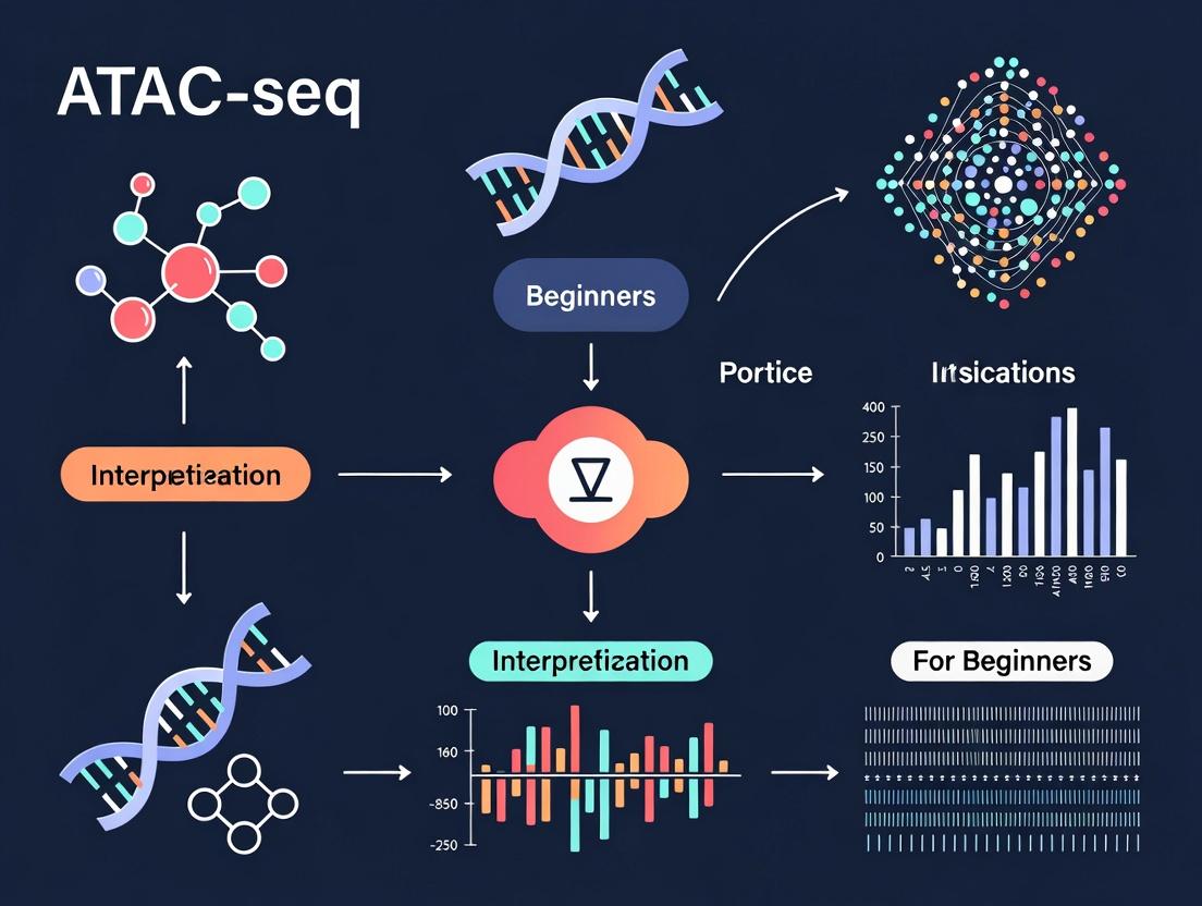
ATAC-seq Data Interpretation for Beginners: A Complete Guide for Researchers & Drug Developers
This comprehensive guide demystifies ATAC-seq data interpretation for researchers, scientists, and drug development professionals.
ATAC-seq Data Interpretation for Beginners: A Complete Guide for Researchers & Drug Developers
Abstract
This comprehensive guide demystifies ATAC-seq data interpretation for researchers, scientists, and drug development professionals. It begins by explaining core concepts—chromatin accessibility, peak calling, and quality control metrics—to build a foundational understanding. It then walks through practical workflows for analyzing and visualizing data, including differential accessibility and motif enrichment. A dedicated section addresses common pitfalls, troubleshooting low-quality data, and optimization strategies for experimental design. Finally, it covers critical validation techniques and comparative analysis with other epigenomic assays (e.g., ChIP-seq, RNA-seq). The article concludes by synthesizing key takeaways and highlighting the translational potential of ATAC-seq in identifying disease mechanisms and therapeutic targets.
ATAC-seq Fundamentals: Understanding Chromatin Accessibility and Your First Dataset
What is Chromatin Accessibility? The Link Between DNA Access and Gene Regulation
Chromatin accessibility, defined as the degree to which genomic DNA is physically open and available for protein binding, is a fundamental determinant of gene regulation. This whitepaper provides an in-depth technical guide to chromatin accessibility, framing its principles within the context of ATAC-seq (Assay for Transposase-Accessible Chromatin using sequencing) data interpretation for beginner researchers. We detail the quantitative features of accessible chromatin, provide standardized experimental protocols, and delineate the critical signaling pathways involved. This resource is tailored for researchers, scientists, and drug development professionals seeking a foundational and current understanding of this key epigenetic regulator.
The eukaryotic genome is packaged into a nucleoprotein complex called chromatin. The basic repeating unit is the nucleosome, consisting of ~147 base pairs of DNA wrapped around an octamer of histone proteins. This compaction inherently restricts access to the underlying DNA sequence. Chromatin accessibility refers to local regions where the chromatin structure is relaxed or "open," allowing transcription factors (TFs), RNA polymerase, and other regulatory complexes to bind and influence gene expression. These accessible regions are strong indicators of cis-regulatory elements, including promoters, enhancers, silencers, and insulators.
The dynamic regulation of accessibility is governed by chromatin remodeling complexes, histone modifications, and transcription factor binding—a process central to cellular differentiation, response to stimuli, and disease pathogenesis.
Quantitative Landscape of Chromatin Accessibility
Key quantitative metrics derived from assays like ATAC-seq characterize the chromatin accessibility landscape. The following table summarizes the core data types and their interpretations.
Table 1: Core Quantitative Metrics in Chromatin Accessibility Analysis
| Metric | Typical Value/Range | Biological Interpretation |
|---|---|---|
| Peak Number (per cell type) | 50,000 - 150,000 | Represents the total set of putative regulatory elements active in a given condition. |
| Peak Width | Median ~ 500 - 1000 bp | Indicates the span of an open chromatin region; broader peaks often associated with high-activity promoters/enhancers. |
| Insert Size Fragment Distribution (from ATAC-seq) | ~200 bp (nucleosome-free), ~400 bp (mono-nucleosome) | 200bp fragments indicate nucleosome-depleted (highly accessible) regions; ~400bp fragments indicate regions adjacent to a positioned nucleosome. |
| Read Depth / Sequencing Saturation | > 20-50 million reads per sample | Required for confident peak calling and detection of rare cell populations or low-activity elements. |
| Transcription Factor Motif Enrichment (-log10(p-value)) | 5 to >50 | Higher values indicate stronger statistical enrichment of a specific TF binding sequence within accessible peaks, suggesting potential regulator. |
| Differential Accessibility (log2 Fold Change) | >1 or <-1 | Signifies significant opening (positive) or closing (negative) of a region between conditions, linked to changes in regulatory potential. |
Methodological Deep Dive: The ATAC-seq Protocol
ATAC-seq is the current gold-standard method for profiling chromatin accessibility due to its simplicity, speed, and low cell number requirements. Below is a detailed protocol.
Detailed Experimental Protocol: ATAC-seq on Nuclei from Cultured Cells
Principle: A hyperactive mutant Tn5 transposase simultaneously cuts open chromatin regions and inserts sequencing adapters ("tagmentation").
Reagents & Equipment:
- Cell culture
- ATAC-seq kit (e.g., Illumina Tagment DNA TDE1 Kit) or purified Tn5 transposase loaded with adapters
- Cell lysis buffer (10 mM Tris-HCl pH 7.4, 10 mM NaCl, 3 mM MgCl2, 0.1% IGEPAL CA-630)
- PBS, Trypan Blue
- Magnetic bead-based DNA clean-up kit (e.g., SPRI beads)
- Qubit fluorometer, Bioanalyzer/TapeStation
- PCR thermocycler, qPCR (optional)
- High-sensitivity DNA reagents
- Sequencing platform (e.g., Illumina NovaSeq)
Procedure:
- Cell Harvest & Counting: Harvest ~50,000-100,000 viable cells. Wash once with cold PBS.
- Nuclei Isolation: Resuspend cell pellet in 50 µL of cold lysis buffer. Incubate on ice for 3-10 minutes. Immediately add 1 mL of cold wash buffer (PBS + 0.1% BSA + 2mM EDTA) to stop lysis.
- Nuclei Count & Quality Check: Pellet nuclei (500 x g, 10 min, 4°C). Resuspend in 50 µL PBS. Count using Trypan Blue on a hemocytometer. Adjust to desired nuclei concentration (typically ~1,000 nuclei/µL).
- Tagmentation: Combine 25 µL of nuclei suspension (~25,000 nuclei) with 25 µL of tagmentation mix (Tn5 transposase, Tagment DNA Buffer, nuclease-free water). Mix gently and incubate at 37°C for 30 minutes in a thermocycler with heated lid.
- DNA Purification: Immediately purify tagmented DNA using a DNA clean-up kit (e.g., 2X SPRI beads). Elute in 20-30 µL of Elution Buffer or 10 mM Tris pH 8.0.
- PCR Amplification & Barcoding: Amplify the purified DNA using a limited-cycle PCR program (e.g., 72°C 5 min; 98°C 30s; then 10-12 cycles of [98°C 10s, 63°C 30s, 72°C 1 min]). Use indexed primers to barcode samples for multiplexing.
- Library Purification & QC: Purify the final library using SPRI beads (1.2X ratio) to remove primer dimers and large fragments. Quantify using Qubit and assess fragment size distribution on a Bioanalyzer (High Sensitivity DNA chip). Expect a periodicity of ~200bp.
- Sequencing: Pool libraries and sequence on an Illumina platform. For standard analysis, paired-end 42bp x 42bp or 50bp x 50bp reads are sufficient.
Critical Considerations: All steps post-lysis should be performed on ice or at 4°C where possible to preserve nuclear integrity and prevent artefactual accessibility changes. Over-tagmentation (too much Tn5 or too long incubation) leads to small fragment bias; under-tagmentation yields low library complexity.
Visualizing Concepts and Workflows
Diagram 1: ATAC-seq Experimental Workflow
Diagram 2: Pathway to Chromatin Accessibility & Transcription
The Scientist's Toolkit: Essential Research Reagents
Table 2: Key Research Reagent Solutions for ATAC-seq Studies
| Item / Reagent | Function & Explanation |
|---|---|
| Hyperactive Tn5 Transposase | Engineered enzyme that simultaneously fragments ("tagments") accessible DNA and adds sequencing adapters. Core enzyme of ATAC-seq. |
| Digitonin or IGEPAL CA-630 | Mild, non-ionic detergents used for controlled cell membrane lysis to isolate intact nuclei, preserving chromatin state. |
| SPRI (Solid Phase Reversible Immobilization) Beads | Magnetic beads for size-selective purification and clean-up of DNA libraries, removing small primers/dimers and large contaminants. |
| Indexed PCR Primers | Oligonucleotides containing Illumina-compatible indices (barcodes) for multiplexing samples in a single sequencing run. |
| High-Sensitivity DNA Assay Kit (e.g., Agilent Bioanalyzer/TapeStation) | For precise quantification and quality assessment of final library fragment size distribution, critical for sequencing success. |
| Nextera Index Kit / Commercial ATAC-seq Kits (e.g., from Illumina, 10x Genomics) | Pre-optimized, standardized reagent sets ensuring reproducibility and reducing protocol development time. |
| Cell Viability Stain (e.g., Trypan Blue) | For accurate counting of viable cells or intact nuclei prior to tagmentation, essential for input normalization. |
| Dual-Size DNA Ladder | For calibrating fragment size selection during SPRI bead clean-up to retain nucleosomal fragments (~200-1000bp). |
Interpretation for Beginners: From Peaks to Biology
For the beginner interpreting ATAC-seq data, the primary output is a list of "peaks" (genomic coordinates of accessible regions). The critical next steps are:
- Annotation: Overlap peaks with known genomic features (promoters, introns, intergenic) using tools like ChIPseeker or HOMER.
- Motif Analysis: Identify enriched transcription factor binding motifs within peaks (e.g., using HOMER or MEME-ChIP) to predict regulating factors.
- Integration: Correlate accessibility changes with transcriptomic (RNA-seq) data to link regulatory element activity to gene expression changes.
- Visualization: Use genome browsers (IGV, UCSC) to inspect read coverage and nucleosomal periodicity at loci of interest.
Understanding that chromatin accessibility provides a permissive rather than instructive regulatory layer is key. An open region implies potential for regulation; the specific outcome is determined by the complement of TFs and co-factors recruited.
Chromatin accessibility is a fundamental and dynamic component of the epigenetic code, directly linking nuclear architecture to gene regulatory output. Techniques like ATAC-seq have democratized access to this information, enabling high-resolution mapping of regulatory landscapes across diverse cell types and disease states. For the beginner in genomics research, mastering the interpretation of chromatin accessibility data is a critical step towards unraveling the complex mechanisms of gene regulation in development, physiology, and pathology. Future directions include single-cell multi-omics, long-read sequencing for haplotype-resolved accessibility, and the integration of AI/ML models to predict regulatory logic from chromatin landscapes.
Within the broader thesis of ATAC-seq data interpretation for beginner researchers, understanding the fundamental assay mechanics is paramount. ATAC-seq (Assay for Transposase-Accessible Chromatin using sequencing) has become the premier method for profiling genome-wide chromatin accessibility. It enables researchers and drug development professionals to identify regulatory elements, such as enhancers and promoters, and infer transcription factor binding events, crucial for understanding gene regulation in development, disease, and drug response.
Core Principle
The assay leverages a hyperactive mutant Tn5 transposase pre-loaded with sequencing adapters (a "tagmentase"). This enzyme simultaneously cuts open chromatin regions and inserts the adapters in a single enzymatic step ("tagmentation"). These tagged fragments are then purified, amplified by PCR, and sequenced. The central hypothesis is that the frequency of sequenced fragments mapping to a genomic region correlates with its chromatin accessibility.
Step-by-Step Experimental Protocol
1. Cell Preparation and Lysis
- Input: 50,000 to 100,000 viable, nuclei for optimal signal-to-noise. Fewer cells lead to over-digestion; more cause under-tagmentation.
- Method: Cells are collected and washed in cold PBS. They are then lysed using a cold, hypotonic, detergent-containing lysis buffer (e.g., 10 mM Tris-HCl, pH 7.4, 10 mM NaCl, 3 mM MgCl2, 0.1% IGEPAL CA-630, 0.1% Tween-20, 0.01% Digitonin) to isolate nuclei while keeping chromatin intact. Nuclei are immediately pelleted and resuspended in transposase reaction mix.
2. Tagmentation Reaction
- Reagent: Commercially available Tagmentase (e.g., Illumina Nextera Tn5).
- Method: Resuspended nuclei are mixed with the Tagmentase reaction buffer and enzyme. The reaction is incubated at 37°C for 30 minutes. This critical step determines fragment size distribution. The reaction is stopped by adding EDTA and SDS.
3. DNA Purification
- Method: Tagmented DNA is purified using a silica membrane-based clean-up kit (e.g., MinElute PCR Purification Kit) with a binding buffer containing high-salt. Elution is performed in a low-volume, low-EDTA buffer to prepare for PCR.
4. Library Amplification (PCR)
- Method: Purified tagmented DNA is amplified using a limited-cycle (typically 5-12 cycles) PCR reaction. The primers contain Illumina P5 and P7 flow cell binding sequences, indexes for multiplexing, and sequences complementary to the adapters inserted by the Tn5. A qPCR side-reaction is often used to determine the optimal cycle number to avoid over-amplification.
5. Library Quality Control and Sequencing
- QC: The final library is assessed for fragment size distribution (typically a nucleosomal ladder pattern peaking below 1 kb) using a Bioanalyzer or TapeStation, and concentration is quantified via qPCR.
- Sequencing: Libraries are sequenced on Illumina platforms, typically paired-end (PE) to better map nucleosome positions.
ATAC-seq Experimental Workflow
Key Quantitative Parameters & Data Outputs
Table 1: Critical Experimental Parameters & Their Impact
| Parameter | Typical Value/Range | Impact on Data Quality |
|---|---|---|
| Cell Number | 50,000 - 100,000 nuclei | Too few: over-tagmentation & high duplicate rate. Too many: under-tagmentation & low complexity. |
| Tagmentation Time | 30 min at 37°C | Longer times increase fragment number but reduce average size. Optimized for nucleosomal ladder. |
| PCR Cycles | 5-12 cycles | Must be minimized to prevent GC bias and amplification artifacts. Determined by qPCR. |
| Read Configuration | Paired-end (PE) | PE (e.g., 2x50 bp) is standard for nucleosome positioning analysis. |
| Sequencing Depth | 25 - 50 million PE reads | Saturation for peak calling in mammalian genomes. Differential analysis may require more. |
Table 2: Expected Data Outputs from a Successful ATAC-seq Run
| Output Metric | Description & Significance |
|---|---|
| Fragment Size Distribution | Bioanalyzer plot showing periodicity of fragments ~200 bp apart (nucleosomal ladder). Key QC metric. |
| Fraction of Mitochondrial Reads | <20% for intact nuclei. High % indicates cytoplasmic contamination or damaged nuclei. |
| Fraction of Reads in Peaks (FRiP) | 20-40% in successful experiments. Measures signal-to-noise. Primary QC for bioinformatics. |
| Number of Accessible Peaks | ~50,000 - 150,000 in a human cell type. Varies by cell state and sequencing depth. |
| TSS Enrichment Score | Measures signal enrichment at transcription start sites. >5-10 indicates high-quality data. |
The Scientist's Toolkit: Essential Research Reagent Solutions
Table 3: Key Reagents and Materials for ATAC-seq
| Item | Function & Importance |
|---|---|
| Hyperactive Tn5 Transposase (Tagmentase) | Engineered enzyme that cleaves DNA and inserts sequencing adapters simultaneously. The core reagent. |
| Cell Permeabilization Buffer | Contains detergents (IGEPAL, Digitonin) to lyse plasma membrane while keeping nuclear membrane intact. |
| Nuclei Isolation & Storage Buffer | Sucrose- and glycerol-based buffer for cushioning nuclei during isolation and freezing. |
| Magnetic Bead-Based Cleanup Kits | For efficient purification of tagmented DNA and final library clean-up (e.g., SPRI beads). |
| Indexed PCR Primers | Contain Illumina adapter sequences and unique dual indices for sample multiplexing. |
| High-Sensitivity DNA Assay Kits | For accurate quantification of low-concentration libraries (e.g., Qubit dsDNA HS, qPCR kits). |
| Bioanalyzer/TapeStation Kits | For assessing library fragment size distribution and confirming nucleosomal ladder pattern. |
From Reads to Regulatory Insights: A Simplified Analysis Pathway
ATAC-seq Data Analysis Pipeline
A meticulous execution of the ATAC-seq wet-lab protocol, governed by the quantitative parameters outlined, is the foundation for generating high-quality chromatin accessibility data. For the beginner researcher, mastery of these steps—from precise nuclei isolation to controlled tagmentation and library amplification—is non-negotiable. This robust experimental data then feeds into the bioinformatic pipeline, enabling the identification of regulatory elements that can be linked to gene expression and, ultimately, phenotypic outcomes in basic research and drug discovery.
This guide serves as a core chapter in a broader thesis designed to demystify ATAC-seq (Assay for Transposase-Accessible Chromatin using sequencing) data interpretation for beginner researchers. Accurate comprehension of key metrics is foundational for robust analysis in epigenetics, translational research, and drug development. This whitepaper provides an in-depth technical exploration of four pivotal terminologies.
Peaks
In ATAC-seq, "peaks" refer to genomic regions with a significantly higher number of aligned sequencing reads compared to a background model, indicating areas of open chromatin. These regions are putative transcription factor binding sites or nucleosome-depleted regions.
Key Quantitative Metrics for Peak Calling:
| Metric | Typical Value/Range | Interpretation |
|---|---|---|
| q-value (FDR) | < 0.05 | Statistical significance threshold for peak calling. |
| Fold Enrichment | > 5-10x | Enrichment of reads in peak vs. background. |
| Peak Width | 100 - 2000 bp | Varies by regulatory element type. |
Experimental Protocol for Peak Calling (Typical Workflow):
- Alignment: Map sequencing reads to a reference genome (e.g., using
BWAorBowtie2). - Filtering: Remove mitochondrial reads, duplicate reads, and low-quality alignments.
- Peak Calling: Use specialized tools (e.g.,
MACS2) to identify statistically significant enrichments.- Command example:
macs2 callpeak -t treatment.bam -c control.bam -f BAM -g hs -n output --outdir peaks -q 0.05
- Command example:
- Annotation: Annotate peaks relative to genomic features (promoters, introns, etc.) using tools like
ChIPseekerorHOMER.
Insert Size
Insert size is the length of the original DNA fragment sequenced, measured from the start of the first read to the end of the second paired-end read. In ATAC-seq, it reveals nucleosome positioning.
Quantitative Data on Insert Sizes:
| Insert Size (bp) | Chromatin State Implication |
|---|---|
| < 100 | Transcription factor footprint or technical artifact. |
| ~ 200 | Nucleosome-free region (mononucleosome-sized protection). |
| ~ 400 | Fragment protected by a dinucleosome. |
| ~ 600 | Fragment protected by a trinucleosome. |
Methodology for Calculating Insert Size Distribution:
- After alignment, use
samtoolsto extract properly paired reads:samtools view -f 2 aligned.bam. - Calculate the insert size from the TLEN field in the SAM/BAM file, or use tools like
Picard CollectInsertSizeMetrics. - Plot the histogram of insert sizes to visualize periodicity.
TSS Enrichment Score
Transcription Start Site (TSS) Enrichment Score is a quality control metric that measures the signal-to-noise ratio by calculating the ratio of fragment coverage at transcription start sites versus flanking regions.
Interpretation of TSS Enrichment Scores:
| TSS Enrichment Score | Data Quality Assessment |
|---|---|
| < 5 | Poor quality, low signal-to-noise. |
| 5 - 10 | Moderate/acceptable quality. |
| > 10 | High-quality ATAC-seq library. |
Protocol for Calculating TSS Enrichment:
- Generate a BED file of known TSS locations (e.g., from RefSeq or Ensembl).
- Calculate read coverage ± 2 kb around each TSS using
deepTools computeMatrix. - Compute the ratio: (average read depth in the central region [e.g., -50 to +50 bp]) / (average read depth in the flanking regions [e.g., -1000 to -500 bp and +500 to +1000 bp]).
Fragment Length Distribution
Fragment length distribution is the genome-wide histogram of all sequenced fragment lengths (insert sizes). It provides a global snapshot of chromatin accessibility and nucleosome patterning.
Typical Distribution Profile:
| Distribution Peak (bp) | Biological Correlate | Approximate % of Fragments |
|---|---|---|
| ~ 50 | Subnucleosomal (TF-bound/open) | 10-25% |
| ~ 200 | Mononucleosomal | 50-70% |
| ~ 400 | Dinucleosomal | 10-20% |
| ~ 600 | Trinucleosomal | < 10% |
Method for Fragment Length Distribution Analysis:
- Follow the insert size calculation protocol for all fragments.
- Plot a density histogram of fragment lengths for the entire library.
- Periodicity of peaks (~200 bp intervals) indicates good library quality and nucleosome patterning.
Visualizing the ATAC-seq Analysis Workflow
Title: ATAC-seq Data Analysis Core Workflow
The Scientist's Toolkit: Essential Research Reagents & Materials
| Item | Function in ATAC-seq Experiment |
|---|---|
| Tn5 Transposase | Enzyme that simultaneously fragments and tags accessible DNA with sequencing adapters. |
| Nextera-style Adapters | Oligonucleotides bound to Tn5, serving as sequencing adapters for library construction. |
| AMPure XP Beads | Magnetic beads for size selection and cleanup of constructed libraries. |
| Qubit dsDNA HS Assay Kit | Fluorometric quantification of library DNA concentration. |
| Bioanalyzer/TapeStation | Capillary electrophoresis for assessing library fragment size distribution. |
| High-Fidelity PCR Mix | Amplifies adapter-ligated DNA for sequencing; low bias is critical. |
| SPRIselect Beads | Allow precise size selection to remove primer dimers and large fragments. |
| Indexing Primers (i5/i7) | Add unique barcodes to samples for multiplexed sequencing. |
| Nuclear Prep Buffer | (For cells) Gently lyses plasma membrane without disrupting nuclei. |
| Sequencing Reagents (e.g., Illumina SBS) | Chemicals for the sequencing-by-synthesis reaction on the flow cell. |
Within the broader thesis of ATAC-seq data interpretation for beginners, understanding the anatomy of its core data files is fundamental. This guide provides a technical walkthrough of ATAC-seq data transformation, from raw sequencing reads to aligned and interpreted genomic intervals. For researchers, scientists, and drug development professionals, mastering this pipeline is the first step towards unlocking insights into chromatin accessibility and gene regulation.
ATAC-seq (Assay for Transposase-Accessible Chromatin using sequencing) begins with cell nuclei, where the Tn5 transposase simultaneously fragments accessible DNA and inserts sequencing adapters. The resulting library is sequenced, generating the primary data files that undergo a series of computational processing steps.
Diagram Title: ATAC-seq Computational Workflow from Nuclei to Peaks
File Anatomy and Transformation Protocols
FASTQ: The Raw Sequencing Read File
The pipeline starts with FASTQ files, containing raw nucleotide sequences and their corresponding quality scores.
File Structure: Each read is represented by 4 lines:
@followed by the read identifier and optional metadata.- The nucleotide sequence (A, T, C, G, N).
+(optionally with the same identifier).- Quality scores per base (encoded in Phred+33 ASCII).
Key Experimental Protocol: Sequencing
- Method: Typically performed on Illumina platforms (NovaSeq, NextSeq).
- Read Type: Paired-end (PE) sequencing is standard (e.g., 2x75 bp or 2x150 bp) to capture both ends of each DNA fragment.
- Output: Two FASTQ files (R1 and R2) per sample.
Table 1: Typical FASTQ File Metrics for a Single ATAC-seq Sample
| Metric | Typical Value | Description |
|---|---|---|
| Total Reads | 50 - 100 million | Sufficient for mammalian genomes. |
| Read Length | 75 - 150 bp | Common for paired-end sequencing. |
| File Size (compressed) | 5 - 20 GB | Depends on read depth and length. |
| Q30 Score | > 80% | >80% of bases with a base call accuracy of 99.9%. |
| Adapter Content | Variable | Should be low after proper library prep. |
BAM: The Aligned and Filtered Sequence File
FASTQ files are processed into BAM (Binary Alignment/Map) files, containing reads aligned to a reference genome.
Detailed Methodology: From FASTQ to Processed BAM
Adapter Trimming & Quality Control:
- Tool:
cutadaptorTrimmomatic. - Protocol: Remove Nextera transposase adapter sequences (e.g.,
CTGTCTCTTATACACATCT). Trim low-quality bases from read ends.
- Tool:
Alignment:
- Tool:
Bowtie2orBWA-MEM.Bowtie2is commonly preferred for its speed with short reads. - Command Example:
bowtie2 -x hg38 -1 sample_R1.fastq.gz -2 sample_R2.fastq.gz -X 2000 --local --very-sensitive | samtools view -bS - > aligned.bam - Key Parameter:
-X 2000sets maximum fragment size for valid paired-end alignments, crucial for ATAC-seq.
- Tool:
Post-Alignment Processing:
- Sorting & Indexing:
samtools sortsorts alignments by genomic coordinate;samtools indexcreates a.baiindex for rapid access. - Duplicate Marking:
Picard MarkDuplicatesorsamtools markdupflags PCR duplicates. ATAC-seq libraries are particularly prone to duplication. - Mitochondrial Read Filtering: Remove reads aligning to the mitochondrial genome (chrM), which can constitute >50% of total reads.
- Filtering for Proper Pairs: Retain only properly paired, uniquely mapped, non-duplicate reads.
- Sorting & Indexing:
BAM File Anatomy: A binary file with a header (containing reference sequences, program history) and alignment records. Each record stores read sequence, mapping position, mapping quality (MAPQ), CIGAR string (alignment details), and optional tags.
Table 2: Key Metrics for a Processed ATAC-seq BAM File
| Metric | Target/Expected Value | Interpretation |
|---|---|---|
| Alignment Rate | > 80% | Proportion of reads mapped to reference. |
| Mitochondrial Reads | < 30% (after filtering) | High mtDNA indicates poor nuclear isolation. |
| Fraction of Reads in Peaks (FRiP) | > 20% | Key QC metric; proportion of reads in called peak regions. |
| Non-Redundant Fraction (NRF) | > 0.8 | Measures library complexity (1 = no duplicates). |
| Insert Size Distribution | Peaks ~200 bp (nucleosome-free) & ~400 bp (mononucleosome) | Indicates successful tagmentation. |
BED: The Interpretable Genomic Interval File
BED (Browser Extensible Data) files represent genomic features—like accessible regions (peaks)—as intervals.
Conversion from BAM to BED:
- Tool:
bedtools bamtobed - Protocol: Converts BAM alignments into BED format, noting start/end of each mapped read pair.
- Command:
bedtools bamtobed -i filtered.bam > fragments.bed
Detailed Methodology: Peak Calling to Generate Final BED Files
- Tool:
MACS2(Model-based Analysis of ChIP-seq) is the de facto standard. - Protocol: Identifies genomic regions with significant enrichment of aligned fragment ends.
- Command Example:
macs2 callpeak -t filtered.bam -f BAMPE -g hs -n sample --outdir peaks -q 0.05 --nomodel --shift -100 --extsize 200-f BAMPE: Uses paired-end information.--nomodel --shift -100 --extsize 200: Custom parameters recommended for ATAC-seq to model the Tn5 binding event.
BED File Anatomy: A tab-separated text file. Minimum fields (BED3) are:
chrom- chromosome name.chromStart- 0-based start coordinate.chromEnd- 1-based end coordinate. Additional common fields includename,score(e.g., -log10(p-value)),strand, andsignalValue.
Table 3: Comparison of Core ATAC-seq File Formats
| Feature | FASTQ | BAM | BED |
|---|---|---|---|
| Format | Text | Binary | Text |
| Content | Raw sequences & qualities | Aligned sequences | Genomic intervals |
| Primary Use | Archival, initial QC | Analysis, visualization | Interpretation, annotation |
| Key Tools | FastQC, cutadapt |
samtools, Picard |
bedtools, MACS2 |
| Size | Largest | Moderate (compressed) | Smallest |
The Scientist's Toolkit: Essential Research Reagents & Software
Table 4: Key Research Reagent Solutions for ATAC-seq Wet Lab
| Item | Function & Rationale |
|---|---|
| Tn5 Transposase | Engineered enzyme that simultaneously fragments accessible DNA and adds sequencing adapters. Core of the assay. |
| Nuclei Isolation Buffer | (e.g., NP-40 or Digitonin-based) Gently lyses plasma membrane while keeping nuclear membrane intact. |
| PCR Amplification Kit | High-fidelity polymerase for limited-cycle PCR to amplify tagmented DNA fragments. |
| Size Selection Beads | (e.g., SPRI beads) Purify and select for appropriate fragment sizes (e.g., < 1000 bp). |
| Library Quantification Kit | (e.g., qPCR-based) Accurate quantification for effective sequencing cluster generation. |
Table 5: Essential Computational Tools for ATAC-seq Analysis
| Tool | Category | Primary Function |
|---|---|---|
| FastQC | Quality Control | Visual report on FASTQ file quality metrics. |
| Cutadapt/Trimmomatic | Preprocessing | Remove adapter sequences and low-quality bases. |
| Bowtie2 | Alignment | Maps sequencing reads to a reference genome. |
| Samtools | BAM Processing | Manipulates, sorts, indexes, and filters BAM files. |
| Picard | BAM Processing | Provides robust tools for marking duplicates and collecting metrics. |
| MACS2 | Peak Calling | Identifies statistically significant regions of chromatin accessibility. |
| Bedtools | BED Processing | Intersects, merges, and annotates genomic interval files. |
| IGV | Visualization | Interactive browser for exploring BAM and BED files. |
Diagram Title: Integration of Wet Lab and Computational Phases in ATAC-seq
The journey of an ATAC-seq data file—from FASTQ to BAM to BED—encapsulates the transformation of raw biochemical signals into interpretable genomic data. For the beginner researcher, proficiency with each file's anatomy and the protocols that connect them is not merely computational exercise but the foundation for rigorous biological interpretation. This pipeline provides the essential map of chromatin accessibility, which, when integrated with other omics data, becomes a powerful tool for understanding gene regulation in development, disease, and drug response.
This guide is part of a broader thesis on ATAC-seq data interpretation for beginners, designed to provide researchers, scientists, and drug development professionals with the foundational knowledge to evaluate assay for transposase-accessible chromatin using sequencing (ATAC-seq) data quality. Proper quality control (QC) is the first and most critical step in ensuring downstream biological insights are reliable.
Core QC Metrics and Their Interpretation
A successful ATAC-seq experiment yields data with specific quantitative characteristics. The following tables summarize the key metrics for both sequencing/library quality and biological soundness.
Table 1: Sequencing and Library Preparation QC Metrics
| Metric | Ideal Value / Profile | Red Flag | Rationale |
|---|---|---|---|
| Total Reads | > 50 million for human/mouse | < 25 million | Insufficient sequencing depth leads to poor peak calling and low reproducibility. |
| Mapping Rate | > 80% (paired-end) | < 60% | Low alignment suggests poor library quality or sample contamination. |
| Mitochondrial Reads | < 20% (ideal: < 10%) | > 30% | High % indicates excessive cytoplasmic or nuclear lysis during prep. |
| Fraction of Reads in Peaks (FRiP) | > 0.20 (20%) for broad cell types | < 0.10 | Low signal-to-noise ratio; indicates poor enrichment for open chromatin. |
| Tn5 Insert Size Distribution | Strong periodicity with ~200-bp nucleosomal pattern | Flat or chaotic distribution | Loss of periodicity suggests degradation or failed transposition. |
| Duplicate Rate | < 50% for high-depth experiments | > 70% | Excessive PCR duplicates indicate low complexity library. |
Table 2: Biological and Signal Quality Metrics
| Metric | Ideal Profile | Red Flag | Rationale |
|---|---|---|---|
| Transcriptional Start Site (TSS) Enrichment Score | > 10 (can be much higher) | < 5 | Low enrichment indicates poor chromatin accessibility at gene promoters. |
| Peak Number | 50,000 - 150,000 for mammalian genomes | < 20,000 or > 300,000 | Too few suggests poor signal; too many suggests high background noise. |
| Peak Width Distribution | Majority narrow (< 1000 bp) with some broader regions | All very broad (> 5 kb) | May indicate over-digestion or genomic DNA contamination. |
| Reproducibility (Irreproducible Discovery Rate - IDR) | IDR < 0.05 for replicate concordance | IDR > 0.1 | Poor replicate agreement undermines confidence in called peaks. |
Detailed Experimental Protocols for Key QC Steps
Protocol 1: Assessing Tn5 Insert Size Distribution and Nucleosomal Periodicity
Purpose: To visualize the fragmentation pattern characteristic of successful ATAC-seq, showing enrichment for sub-nucleosomal (<100 bp) and mono-, di-, and tri-nucleosomal fragments.
- Align Reads: Map paired-end reads to the reference genome using a splice-aware aligner (e.g., BWA-MEM) with parameters
-T 0. - Filter Reads: Remove reads mapping to mitochondria, unmapped reads, non-primary alignments, and reads with mapping quality < 30.
- Calculate Insert Sizes: For each properly paired read, calculate the fragment length (insert size plus both adapters). Use tools like
samtools statsor custom scripts. - Generate Histogram: Create a frequency histogram of fragment sizes from 0 to 1000 bp.
- Interpretation: A successful assay shows a strong peak <100 bp (open chromatin) and clear periodicity of peaks at ~200 bp intervals (nucleosome-protected fragments).
Protocol 2: Calculating TSS Enrichment Score
Purpose: A quantitative measure of signal-to-noise ratio, as accessible promoters are highly enriched for Tn5 insertion.
- Generate TSS Regions: Obtain genomic coordinates for all known TSSs (e.g., from RefSeq or Ensembl). Define a region from -2 kb to +2 kb around each TSS.
- Compute Coverage: Calculate the read coverage depth across all TSS regions using
deepTools computeMatrix. - Normalize and Aggregate: Aggregate the signal across all TSSs and normalize by the average signal in the flanking regions (e.g., -2kb to -1.5kb and +1.5kb to +2kb).
- Calculate Score: The TSS enrichment score is defined as the maximum value of the aggregated, normalized profile within a central window (e.g., -50 bp to +50 bp around the TSS).
Protocol 3: Evaluating Replicate Concordance with IDR
Purpose: To statistically assess the reproducibility of peak calls between biological replicates.
- Call Peaks per Replicate: Use a peak caller (e.g., MACS2) on each replicate separately to generate a sorted list of peaks by p-value or significance.
- Run IDR Analysis: Use the
idrpackage to compare the two ranked peak lists. The analysis identifies peaks passing a chosen IDR threshold (e.g., 0.05). - Interpret Output: The result is a conservative set of high-confidence peaks reproducible across replicates. The number of peaks passing IDR relative to the total called is a key quality indicator.
Visualizing the ATAC-seq Workflow and QC Checkpoints
Diagram Title: ATAC-seq Experimental and QC Workflow
The Scientist's Toolkit: Essential Research Reagent Solutions
| Item | Function & Importance in ATAC-seq QC |
|---|---|
| Viable, Single-Cell Nuclei Suspension | The starting material. Intact nuclei without cytoplasmic contamination are critical for low mitochondrial read counts. Prepared with detergents (e.g., NP-40, Digitonin) in isotonic buffers. |
| Validated Tn5 Transposase (Loaded with Adapters) | The core enzyme. Must be freshly prepared or commercially validated for high activity to ensure even fragmentation and adapter insertion into accessible DNA. |
| AMPure/SPRI Beads | For post-transposition and post-PCR cleanup. Size selection is crucial for removing short fragments, primer dimers, and optimizing the library size distribution. |
| High-Fidelity PCR Mix with Minimal Bias | For library amplification post-tagmentation. Enzymes with low GC-bias ensure equitable amplification of all fragments, preserving library complexity. |
| Dual-Indexed PCR Adapters (Unique Molecular Identifiers - UMIs optional) | To enable multiplexing and accurate removal of PCR duplicates. UMIs help distinguish biological duplicates from PCR duplicates, improving complexity assessment. |
| High-Sensitivity DNA Assay Kit (e.g., Bioanalyzer, TapeStation, Qubit) | For precise quantification and sizing of the final library before sequencing. A clean peak at expected size (~100-700 bp) confirms successful prep. |
| PhiX Control Library | Spiked into sequencing runs (1-5%) for run monitoring, especially important for low-diversity libraries common in ATAC-seq. |
| Validated Positive Control Cells (e.g., GM12878, K562) | A well-characterized cell line run in parallel to benchmark QC metrics (FRiP, TSS score) against expected values for the protocol. |
ATAC-seq Analysis Pipeline: A Practical Walkthrough from Raw Data to Biological Insight
This guide details the critical first computational steps in ATAC-seq data analysis, framed within a broader thesis on making chromatin accessibility data interpretation accessible for beginners in research. Proper preprocessing and alignment are foundational for generating accurate, biologically meaningful insights relevant to fundamental research and drug discovery.
The Imperative of Read Preprocessing
Raw sequencing reads contain technical artifacts, including adapter sequences and low-quality bases, which can compromise alignment and downstream peak calling. Trimming mitigates these issues.
The following table compares widely used trimming tools and their core parameters, based on current benchmarking literature.
Table 1: Comparison of Read Trimming Tools for ATAC-seq
| Tool | Primary Function | Key Parameter | Recommended Setting (PE ATAC-seq) | Rationale |
|---|---|---|---|---|
| fastp | Adapter trimming, quality filtering, per-read quality pruning | --qualified_quality_phred |
20 | Removes bases with Q<20. |
| Trim Galore! (wrapper for Cutadapt) | Adapter removal, quality trimming | --quality |
20 | Trims low-quality ends. |
| Cutadapt | Adapter removal | -a, -A |
Nextera PE sequences | Removes Nextera transposase adapters. |
| Trimmomatic | Flexible trimmer for Illumina data | LEADING:3 TRAILING:3 SLIDINGWINDOW:4:20 MINLEN:25 |
As shown | Removes leading/trailing low-quality bases, scans with window, discards short reads. |
Detailed Protocol: Adapter Trimming with fastp
Objective: Remove Nextera XT adapters and low-quality bases from paired-end ATAC-seq FASTQ files.
- Input: Paired-end FASTQ files (
sample_R1.fastq.gz,sample_R2.fastq.gz). Command:
Output: Trimmed FASTQ files and a quality control report.
- Verification: Inspect the HTML report for metrics on read quality before and after trimming, adapter content, and length distribution.
Mapping Trimmed Reads to a Reference Genome
Alignment places sequenced fragments onto a reference genome, enabling the identification of open chromatin regions.
Alignment Algorithm Selection
Speed, accuracy, and handling of paired-end reads are crucial considerations.
Table 2: Alignment Tools for ATAC-seq Reads
| Aligner | Algorithm Type | Key Consideration for ATAC-seq | Typical PE Alignment Rate |
|---|---|---|---|
| Bowtie2 | BWT-based, gapped alignment | Excellent sensitivity, standard for ATAC-seq. | 80-95% |
| BWA-MEM | BWT-based, gapped alignment | Efficient for longer reads, robust performance. | 80-95% |
| STAR | Spliced aligner, uses uncompressed suffix array | Designed for RNA-seq; can be used but is memory-intensive. | 75-90% |
Detailed Protocol: Mapping with Bowtie2
Objective: Align trimmed paired-end reads to the human reference genome (hg38).
Prerequisite: Build a Bowtie2 index for the reference genome.
Alignment Command:
-X 2000: Sets maximum fragment length, important for ATAC-seq nucleosome periodicity.--no-mixed/no-discordant: Suppresses unpaired and discordant alignments for cleaner paired-end data.
Post-Processing (SAM to BAM):
Quality Check: Review alignment statistics in
sample_bowtie2.log(overall alignment rate, concordant pair alignment rate).
The Scientist's Toolkit: Research Reagent Solutions
Table 3: Essential Wet-Lab and Computational Materials for ATAC-seq
| Item | Function | Example/Note |
|---|---|---|
| Tn5 Transposase | Enzyme that simultaneously fragments and tags accessible DNA with sequencing adapters. | Illumina Nextera XT or homemade. |
| Size Selection Beads | Clean up transposition reaction and select for small DNA fragments (<~800 bp). | SPRIselect beads (Beckman Coulter). |
| High-Fidelity PCR Mix | Amplify library post-transposition with limited cycles to minimize bias. | NEBNext High-Fidelity 2X PCR Master Mix. |
| Dual Indexed PCR Primers | Amplify library and add unique sample indices for multiplexing. | Illumina Nextera XT Index Kit v2. |
| Reference Genome FASTA | The nucleotide sequence against which reads are aligned for mapping. | UCSC hg38, ENSEMBL GRCh38. |
| Genome Index Files | Pre-processed reference genome for ultra-fast alignment by tools like Bowtie2. | Generated using bowtie2-build. |
| Adapter Sequence File | File containing adapter sequences to be trimmed from raw reads. | Essential for accurate trimming. |
Visualizing the ATAC-seq Preprocessing & Alignment Workflow
Diagram 1: ATAC-seq preprocessing and alignment workflow.
Logical Decision Pathway for Preprocessing
Diagram 2: Decision tree for read trimming in ATAC-seq.
Peak calling is the computational process of identifying regions in the genome with a statistically significant enrichment of sequencing reads, corresponding to putative open chromatin regions or transcription factor binding sites. In the context of a beginner's ATAC-seq research thesis, accurate peak calling is the critical step that transforms raw aligned sequencing data into a biologically interpretable list of genomic intervals for downstream analysis. The choice of algorithm and its parameters directly influences sensitivity, specificity, and reproducibility, impacting all subsequent conclusions.
Core Algorithms: MACS2 and Genrich
MACS2 (Model-based Analysis of ChIP-seq 2)
MACS2 remains a benchmark algorithm, originally designed for ChIP-seq but widely adapted for ATAC-seq. It uses a dynamic Poisson distribution to model the background signal and account for local biases.
Key Steps:
- Remove Duplicates: Optionally removes duplicate reads to mitigate PCR amplification bias.
- Shift Reads: Accounts for the ~200 bp fragment size of Tn5 transposition by shifting reads towards the 3' end (positive strand reads shifted +
extsize/2, negative strand reads shifted -extsize/2). - Build Model: Generates a smoothed density of reads (d) and models the local background using a dynamic Poisson distribution. A parameter,
--bw, defines the bandwidth for smoothing. - Peak Calling: Scores each potential region using a log-likelihood ratio (fold enrichment over background) and calculates a p-value. Peaks are called where the p-value exceeds a user-defined threshold (
-por-qfor FDR). - Peak Deduplication: Merges nearby peaks and selects the most significant one.
Genrich
Genrich is a newer, robust tool developed specifically for ATAC-seq (and ChIP-seq), notable for its ability to handle PCR duplicates algorithmically and to call peaks from multiple replicates simultaneously.
Key Steps:
- Duplicate Removal via Poisson Distribution: Instead of removing exact-mapping-position duplicates, Genrich uses a probabilistic model. If n reads map to the same position and the total number of reads is N, it retains m reads where m is the smallest integer such that the Poisson cumulative probability P(X ≥ m) < 0.01.
- Read Extension & Analysis Window: Extends reads in the 3' direction by a specified distance (
-e). It then analyzes the genome in non-overlapping windows of size-w(default 100 bp). - Background Calculation: Calculates background signal in large genomic bins (default 10 kbp) and interpolates for each analysis window.
- Peak Calling via Negative Binomial Model: Models read counts in each window using a Negative Binomial distribution (more permissive than Poisson for over-dispersed data). Calculates p-values and q-values (FDR) for enrichment.
- Multiple Replicate Analysis: When given multiple BAM files (
-t f1.bam f2.bam ...), it performs a joint analysis, weighting replicates by read depth to call a unified set of peaks.
Quantitative Algorithm Comparison
Table 1: Core Feature Comparison of MACS2 and Genrich for ATAC-seq
| Feature | MACS2 | Genrich |
|---|---|---|
| Primary Design | ChIP-seq, adapted for ATAC-seq | ATAC-seq & ChIP-seq |
| Statistical Model | Dynamic Poisson | Negative Binomial |
| Duplicate Handling | Optional removal of all duplicates at same coordinate | Probabilistic removal based on Poisson model |
| Replicate Analysis | Post-hoc merging (e.g., idr) |
Native joint peak calling from multiple BAM files |
| Read Shift/Extension | Yes, uses --extsize |
Yes, uses -e (extension size) |
| Typical Runtime | Moderate | Fast |
| Key Strengths | Highly tunable, extensive documentation, community standard. | ATAC-seq-optimized, intelligent duplicate handling, simplified multi-replicate workflow. |
Table 2: Common Default & Recommended Parameters for ATAC-seq
| Parameter | MACS2 (Typical Setting) | Genrich (Typical Setting) | Purpose & Impact |
|---|---|---|---|
| File Input | -t treatment.bam |
-t treatment.bam -o peaks.narrowPeak |
Specifies input BAM and output file. |
| Format/File Type | -f BAM |
-f BAM |
Input file format. |
| Peak Call Mode | --call-summits |
-j (for ATAC-seq mode) |
--call-summits refines peak summits; -j disables ChIP-seq specific junction filtering. |
| q-value/FDR Cutoff | -q 0.05 |
-q 0.05 |
False Discovery Rate threshold. More stringent (e.g., 0.01) yields fewer, higher-confidence peaks. |
| Shift/Extension Size | --extsize 200 |
-e 200 |
Accounts for fragment length. Critical for accurate signal localization. |
| Bandwidth/Window | --bw 300 |
-w 100 |
Smoothing parameter (MACS2) or analysis window size (Genrich). Affects peak shape and merging. |
| Keep Duplicates | --keep-dup all (or 1) |
(Handled algorithmically) | MACS2: --keep-dup 1 keeps one read per position. Genrich's method is integral. |
| Genome Size | -g hs (for human) |
-a genome_blacklist.bed |
MACS2 uses effective genome size. Genrich uses a BED file to exclude problematic regions (e.g., ENCODE Blacklist). |
Detailed Experimental Protocols for Cited Studies
Protocol 4.1: Standard ATAC-seq Peak Calling with MACS2
This protocol assumes aligned reads are in a BAM file (atac_aligned.bam).
Sort and Index BAM File:
Call Peaks with MACS2:
Outputs:
ATAC_Experiment_peaks.narrowPeak(peak locations),ATAC_Experiment_summits.bed(refined summit locations).
Protocol 4.2: Joint Peak Calling from Replicates with Genrich
This protocol processes two biological replicates (rep1.bam, rep2.bam) together.
Prepare Blacklist File: Download the ENCODE consensus blacklist for your organism (e.g.,
hg38-blacklist.v2.bed.gzfor human).Run Genrich in ATAC-seq Mode:
Parameters:
-j(ATAC-seq mode),-y(PCR duplicate removal via probabilistic model),-a(exclude blacklisted regions). The-f BAMPEoption is used if the BAM contains paired-end read information.
Mandatory Visualizations
Title: ATAC-seq Peak Calling General Workflow
Title: Replicate Analysis: IDR vs. Genrich Joint Calling
The Scientist's Toolkit: Research Reagent Solutions
Table 3: Essential Computational Tools & Resources for ATAC-seq Peak Calling
| Item | Function/Purpose | Example/Note |
|---|---|---|
| Sequence Aligner | Aligns sequencing reads to a reference genome. | Bowtie2, BWA, STAR. For ATAC-seq, Bowtie2 with -X 2000 is common. |
| SAM/BAM Tools | Manipulates and views alignment files. | Samtools (sort, index, view), deepTools (bamCoverage for visualization). |
| Peak Caller Software | Core algorithm to identify enriched regions. | MACS2, Genrich, HOMER (findPeaks). |
| Genome Blacklist | BED file of problematic genomic regions to exclude. | ENCODE Consortium Blacklist (v2). Removes artifactual signals. |
| Reference Genome | The genome sequence and annotation files. | UCSC (hg38, mm10), Ensembl, GENCODE. Must be consistent across pipeline. |
| IDR Pipeline | Statistical method to assess reproducibility between replicates. | IDR Package (R/Python). Used post-MACS2 for consensus peaks. |
| Genome Browser | Visualizes aligned reads and called peaks in genomic context. | IGV (Integrative Genomics Viewer), UCSC Genome Browser. |
| Container System | Ensures software version and environment reproducibility. | Docker, Singularity, Conda. A Conda environment with all tools is recommended. |
In the context of ATAC-seq data interpretation for beginner research, assigning chromatin accessibility peaks to genes is a critical step for biological insight. Annotation links open chromatin regions, identified by peak calling, to potential regulatory elements and their target genes, enabling hypothesis generation about gene regulation mechanisms relevant to development and disease.
Quantitative Data on Annotation Tools and Genomic Distributions
Table 1: Comparison of Common Peak Annotation Tools
| Tool | Primary Method | Input Format | Output Features | Typical Runtime (Human Genome) |
|---|---|---|---|---|
| ChIPseeker (R/Bioconductor) | Distance to nearest TSS, genomic feature assignment | BED, GFF | Pie charts, coverage plots, TSS region profiles | 2-5 minutes |
| HOMER (annotatePeaks.pl) | Customizable proximity, detailed annotation | BED, HOMER peak format | Gene lists, genomic region breakdown, motif finding integration | 3-10 minutes |
| GREAT (Web/Standalone) | Genomic regulatory domains, basal + extension rules | BED | GO terms, pathways, disease associations | 5-15 minutes (web) |
| Ensembl Variant Effect Predictor (VEP) | Comprehensive consequence prediction | BED, VCF | Consequence terms (promoter, enhancer), linked genes | 1-3 minutes |
Table 2: Typical Genomic Distribution of ATAC-seq Peaks in a Mammalian Cell Line
| Genomic Feature | Percentage of Peaks (± Std Dev) | Common Interpretation |
|---|---|---|
| Promoter (≤ 1kb from TSS) | 15-25% (± 5%) | Direct transcriptional regulation |
| 5' UTR | 2-5% (± 1%) | Potential alternative regulation |
| 3' UTR | 3-7% (± 2%) | mRNA stability, localization |
| Exonic | 1-3% (± 1%) | Possible exonic regulatory elements |
| Intronic | 35-50% (± 7%) | Intronic enhancers, silencers |
| Intergenic | 25-40% (± 8%) | Distal enhancers, locus control regions |
Detailed Protocol for Peak Annotation with ChIPseeker
Objective: Annotate ATAC-seq peaks with genomic context and assign them to the nearest genes.
Materials & Software:
- Input File: ATAC-seq peaks in BED format (e.g.,
peaks.bed). - Software: R (≥4.0), Bioconductor packages
ChIPseekerandTxDb(e.g.,TxDb.Hsapiens.UCSC.hg38.knownGene). - Genome Annotation: Corresponding TxDb package for your organism/assembly.
Methodology:
- Install and Load Packages:
Load Peak File:
Annotate Peaks:
Generate and Export Annotation:
Visualizing Annotated Data with Integrative Genomics Viewer (IGV)
Objective: Visually inspect ATAC-seq read alignment and peaks in genomic context alongside gene models and other tracks.
Protocol for Local IGV Use:
Data Preparation:
- Generate a
TDF(Tiled Data Format) file from your ATAC-seq BAM file for efficient viewing usingigvtools(igvtools count aligned_reads.bam aligned_reads.tdf hg38). - Have your annotated peak file (BED or
annotated_peaks.csvconverted to BED) and gene annotation file (GTF) ready.
- Generate a
Loading Data in IGV:
- Launch IGV and select the correct genome assembly from the dropdown.
- Go to
File>Load from File...and select your BAM/TDF file and peak BED file. - Load public annotation tracks (e.g., RefSeq genes) via
File>Load from Server....
Visual Analysis:
- Navigate to a locus of interest (e.g., gene name, genomic coordinates).
- Observe the pileup of ATAC-seq reads (accessibility) in relation to peak calls (black bars) and gene models.
- Use the "Group Autoscale" feature for proper track scaling.
- Save session (
File>Save Session) for reproducibility.
Workflow and Pathway Diagrams
Title: ATAC-seq Peak to Gene Annotation Workflow
Title: Enhancer-Promoter Interaction Leading to Gene Activation
The Scientist's Toolkit: Research Reagent Solutions
Table 3: Essential Toolkit for ATAC-seq Annotation & Visualization
| Item / Solution | Function in Annotation/Visualization | Example Product/Software |
|---|---|---|
| Genome Annotation Database | Provides coordinates for genes, transcripts, and other features for peak context assignment. | Ensembl GTFs, UCSC RefSeq, Bioconductor TxDb packages (e.g., TxDb.Hsapiens.UCSC.hg38.knownGene). |
| Peak Annotation Software | Computes the genomic context of peaks and proximity to transcriptional start sites (TSS). | R/Bioconductor: ChIPseeker, GenomicRanges. Command-line: HOMER annotatePeaks.pl, BEDTools closest. |
| Functional Enrichment Tool | Identifies overrepresented biological pathways, GO terms, or diseases among annotated genes. | clusterProfiler (R), GREAT, Enrichr (web). |
| Genome Browser | Visualizes raw reads, peaks, and annotations in genomic context for validation and exploration. | Integrative Genomics Viewer (IGV), UCSC Genome Browser, WashU Epigenome Browser. |
| IGV-Compatible Format Converter | Converts large alignment files to efficient, indexed formats for fast visualization. | igvtools (for TDF), samtools (for BAM indexing and sorting). |
| Scripting Environment | Enables automation of the annotation pipeline and custom analysis. | RStudio (R), Jupyter Notebook (Python). |
Within the broader thesis of ATAC-seq data interpretation for beginners, functional analysis is the critical step that moves from cataloging open chromatin regions to deriving biological meaning. Following peak calling and annotation, motif enrichment and pathway analysis translate genomic coordinates into testable hypotheses about transcription factor (TF) activity and affected biological processes, providing direct insight for drug development.
Motif Enrichment Analysis: Identifying Overrepresented Transcription Factor Binding Sites
Core Concept
Motif enrichment analysis statistically evaluates whether DNA sequences from ATAC-seq peaks are enriched for known transcription factor binding motifs compared to a background model, implicating TFs active in the experimental condition.
Detailed Experimental Protocol: HOMER Motif Analysis
A. Input Data Preparation:
B. De Novo & Known Motif Discovery:
Parameters:
-size: Region size for motif finding (default: 200bp).-mask: Repeat masking.-bg <file>: Custom background sequences.
C. Statistical Framework: The binomial test calculates motif enrichment (observed vs. expected frequency). P-values are corrected for multiple testing (Benjamini-Hochberg). The output ranks motifs by statistical significance (logP) and enrichment fold.
Key Quantitative Data: Motif Enrichment Output
Table 1: Top Enriched Motifs from an Exemplar ATAC-seq Experiment (Hypothetical Data)
| Rank | Motif Name (TF) | Consensus Sequence | P-Value (log10) | Fold Enrichment | % of Target Peaks |
|---|---|---|---|---|---|
| 1 | JUN (AP-1) | TGANTCA | -12.5 | 8.2 | 18.7% |
| 2 | FOSL1 | TGAGTCA | -10.8 | 6.5 | 15.2% |
| 3 | NFKB1 (p50) | GGGACTTTCC | -9.3 | 5.1 | 12.4% |
| 4 | STAT3 | TTCCGGGAA | -8.7 | 4.8 | 9.5% |
| 5 | SP1 | GGGGCGGGG | -7.9 | 3.2 | 22.1% |
Workflow Diagram
Diagram Title: Motif Enrichment Analysis Computational Workflow
Pathway and Functional Enrichment Analysis
Core Concept
Genes associated with ATAC-seq peaks (via nearest gene or chromatin interaction maps) are analyzed for overrepresentation of biological pathways, Gene Ontology (GO) terms, or disease associations.
Detailed Protocol: g:Profiler & ClusterProfiler
A. Gene List Generation:
- Annotate peaks to nearest transcription start site (TTS) using
annotatePeaks.pl(HOMER) orChIPseeker(R). - Use a distance cutoff (e.g., ± 50 kb) or integrate with Hi-C data for more accurate linking.
B. Enrichment Analysis with g:Profiler (Web/API):
C. Enrichment Analysis with ClusterProfiler (R):
Key Quantitative Data: Functional Enrichment Output
Table 2: Top Enriched KEGG Pathways from ATAC-seq Gene List (Hypothetical Data)
| Pathway ID | Pathway Description | Gene Count | Gene Ratio | P-Value | Adjusted P-Value | Enrichment Score |
|---|---|---|---|---|---|---|
| hsa04668 | TNF signaling pathway | 15 | 15/320 | 2.1e-08 | 4.5e-06 | 8.21 |
| hsa04064 | NF-kappa B signaling | 12 | 12/320 | 5.7e-06 | 1.2e-04 | 5.24 |
| hsa05163 | Human cytomegalovirus infection | 18 | 18/320 | 1.4e-05 | 2.1e-04 | 4.85 |
| hsa05323 | Rheumatoid arthritis | 10 | 10/320 | 3.2e-05 | 3.8e-04 | 4.50 |
| hsa05418 | Fluid shear stress & atherosclerosis | 11 | 11/320 | 7.8e-05 | 8.2e-04 | 4.11 |
Pathway Visualization
Diagram Title: TNF/NF-κB Signaling Pathway Core
The Scientist's Toolkit: Essential Research Reagent Solutions
Table 3: Key Reagents and Tools for ATAC-seq Functional Analysis
| Item/Category | Example Product/Software | Function in Analysis |
|---|---|---|
| Motif Databases | JASPAR, CIS-BP, HOCOMOCO | Curated collections of TF binding motifs for known motif enrichment testing. |
| Enrichment Analysis Suites | g:Profiler, Metascape, DAVID | Integrated platforms for functional enrichment across GO, pathways, and disease terms. |
| R/Bioconductor Packages | ChIPseeker, clusterProfiler, motifmatchr |
Programmatic tools for peak annotation, motif matching, and statistical enrichment. |
| Sequence Extraction Tools | bedtools getfasta (BEDTools), HOMER annotatePeaks.pl |
Extracts DNA sequences in FASTA format from peak genomic coordinates. |
| High-Performance Computing | Local HPC clusters, Cloud (AWS, GCP) | Handles computationally intensive de novo motif discovery and genome-wide scans. |
| Background Genomic Sequences | bedtools shuffle, HOMER genome.fa |
Generates matched control sequences for proper statistical comparison. |
| Visualization Software | ggplot2 (R), matplotlib (Python), Cytoscape |
Creates publication-quality plots for enrichment results and pathway networks. |
Within the broader thesis on ATAC-seq data interpretation for beginners, Step 5 represents the critical juncture where biological insights are statistically validated. After preprocessing, alignment, peak calling, and annotation, researchers must distinguish random noise from biologically meaningful changes in chromatin accessibility between experimental conditions (e.g., treatment vs. control, disease vs. healthy). This step, identifying differential accessibility (DA), quantifies which genomic regions exhibit statistically significant changes in open chromatin, thereby pinpointing regulatory elements potentially driving phenotypic differences. This guide details the computational tools, statistical frameworks, and best practices for robust DA analysis in ATAC-seq.
Statistical Foundations for Differential Analysis
The core challenge is modeling count data (reads per peak) that is over-dispersed and confounded by technical variability. The fundamental steps involve:
- Data Modeling: ATAC-seq data per peak is represented as a matrix of integer counts. These counts are modeled assuming a negative binomial distribution, which accounts for variance exceeding the mean (over-dispersion).
- Normalization: Correcting for library size (total read count) and composition bias is essential. Methods use scaling factors or conditional maximum likelihood.
- Hypothesis Testing: A statistical test is performed for each peak to assess the null hypothesis that its accessibility is not different between conditions.
Tools and Methodologies: A Comparative Analysis
The following table summarizes the primary software packages used for DA analysis in ATAC-seq, detailing their core methods, strengths, and considerations.
Table 1: Key Tools for Differential Accessibility Analysis
| Tool / Package | Core Statistical Method | Key Features | Best Suited For |
|---|---|---|---|
| DESeq2 | Negative binomial generalized linear model (GLM) with shrinkage estimators (LFC). | Highly stable, robust to small sample sizes, excellent false discovery rate control. Provides log2 fold change shrinkage. | General purpose; the current gold-standard for most ATAC-seq DA analyses. |
| edgeR | Negative binomial models with empirical Bayes methods for dispersion estimation. | Very flexible, offers multiple testing approaches (QL F-test, LRT). High sensitivity. | Experiments with complex designs (multiple factors, interactions). |
| limma-voom | Linear modeling of log-counts with precision weights. | Converts counts to log2-CPM, then uses empirical Bayes moderation of t-statistics. Very fast. | Large datasets with many samples where speed is critical. |
| DiffBind | (Wrapper) Primarily utilizes DESeq2 or edgeR backends. | Specialized for ChIP/ATAC-seq. Handles peak sets across samples, consensus peak calling, and specificity in normalization. | Researchers wanting an end-to-end workflow from peaks to DA, especially with replicates. |
| MACS2 (bdgdiff) | Probabilistic framework based on local Poisson distributions. | Part of the MACS2 suite; works on signal tracks (bedgraph). Can be used without predefined peaks. | Exploratory analysis or when a peak-agnostic approach is desired. |
Detailed Protocol: DA Analysis with DESeq2
This is a widely adopted and robust protocol for identifying differential peaks.
Experimental Protocol: Differential Analysis Using DESeq2
- Input Preparation: Generate a counts matrix where rows are genomic regions (consensus peaks) and columns are samples. A companion sample sheet metadata table must specify the condition for each sample.
Create DESeqDataSet Object (in R):
Pre-filtering: Remove peaks with very low counts (e.g.,
rowSums(counts(dds)) >= 10).- Factor Level Specification: Set the reference level for the condition factor (e.g.,
dds$condition <- relevel(dds$condition, ref="control")). Run DESeq2: This single function performs estimation of size factors (normalization), dispersion estimation, and model fitting.
Extract Results: Shrinkage of log2 fold changes is recommended to reduce noise from low-count peaks.
Interpretation & Filtering: Filter results based on adjusted p-value (padj < 0.05) and log2 fold change threshold (e.g., |LFC| > 0.5). Results can be annotated with genomic feature information.
Detailed Protocol: Signal-Based DA with MACS2 bdgdiff
This protocol identifies differences directly from continuous signal tracks.
Experimental Protocol: Peak-Agnostic DA using MACS2 bdgdiff
- Input Preparation: For each sample, create a bedGraph file of sequencing coverage (often done during MACS2 peak calling with the
--Bflag). Pooled bedGraph files for each condition are also needed. Run bdgdiff:
This command compares the condition-pooled tracks while accounting for variability among replicates.
- Output Interpretation: The tool produces three BED files: regions more accessible in condition 1 (
cond1), condition 2 (cond2), and regions with similar accessibility but different peak shape (common).
Visualization of the DA Analysis Workflow
The following diagram outlines the logical workflow and decision points in a standard differential accessibility analysis.
DA Analysis Workflow from Processed Reads
The Scientist's Toolkit: Research Reagent Solutions
Table 2: Essential Reagents and Materials for ATAC-seq & Validation
| Item | Function in ATAC-seq/DA Analysis |
|---|---|
| Tn5 Transposase | Engineered enzyme that simultaneously fragments and tags accessible genomic DNA with sequencing adapters. The core reagent in the ATAC-seq assay. |
| NEBNext High-Fidelity 2X PCR Master Mix | Used for amplifying the transposed DNA fragments. High-fidelity polymerase is critical to minimize PCR errors and bias during library construction. |
| AMPure XP Beads | Magnetic beads for size selection and purification of constructed libraries, removing short fragments (e.g., primer dimers) and buffer exchange. |
| QIAGEN MinElute PCR Purification Kit | Alternative/adjunct purification method for clean-up of post-PCR reactions and concentration of final libraries. |
| High-Sensitivity DNA Assay Kit (Bioanalyzer/TapeStation) | For quality control, accurately assessing library fragment size distribution and concentration before sequencing. |
| SYBR Green PCR Master Mix | For quantitative PCR (qPCR) validation of candidate differential peaks. Confirms accessibility changes in independent biological samples. |
| Primary Antibodies (for CUT&RUN/TAG) | For orthogonal validation (e.g., H3K27ac, TF antibodies) to confirm functional state of identified accessible regions. |
Advanced Considerations & Best Practices
- Replicates are Non-Negotiable: Biological replicates (n>=3) are essential for reliable dispersion estimation and statistical power.
- Batch Effects: Include batch as a covariate in the DESeq2 design formula (
design = ~ batch + condition) if known technical batches exist. - Blacklist Regions: Exclude peaks falling in genomic "blacklist" regions (e.g., ENCODE DAC Blacklist) known to cause artifactual signals.
- Multiple Testing Correction: Always use adjusted p-values (FDR/Benjamini-Hochberg) to control for false positives.
- Orthogonal Validation: Plan for validation via qPCR on independent samples or complementary assays like CUT&RUN for histone marks.
An In-Depth Guide to ATAC-seq Analysis in Cancer Research
This whitepaper serves as a technical guide, framed within a thesis on ATAC-seq (Assay for Transposase-Accessible Chromatin with high-throughput sequencing) data interpretation for beginner researchers. It provides a concrete case study in oncology, detailing how ATAC-seq elucidates chromatin remodeling in response to targeted therapy, enabling the identification of drug resistance mechanisms and novel therapeutic vulnerabilities.
ATAC-seq has become a cornerstone in functional genomics, mapping open chromatin regions genome-wide. In drug development, it is pivotal for understanding how disease states alter the epigenetic landscape and how interventions—such as small molecule inhibitors—rewire regulatory networks. This guide walks through a representative study analyzing chromatin accessibility dynamics in BRAF-mutant melanoma cells treated with a BRAF inhibitor (BRAFi), linking epigenetic plasticity to adaptive resistance.
Experimental Protocols
Cell Culture and Treatment
- Cell Line: A375 human melanoma cells (homozygous for BRAF V600E mutation).
- Culture Conditions: Maintained in DMEM supplemented with 10% FBS at 37°C, 5% CO₂.
- Drug Treatment: Cells were treated with 1 µM Vemurafenib (PLX4032, a BRAFi) or DMSO vehicle control. Two conditions were established:
- Acute: Cells harvested after 72 hours of treatment.
- Chronic/Persistent: Cells cultured in continuous drug presence for 21 days to select for a drug-adapted population.
- Replication: Biological triplicates were generated for each condition (DMSO, Acute BRAFi, Persistent BRAFi).
ATAC-seq Library Preparation (Omni-ATAC Protocol)
- Cell Lysis: 50,000 viable cells were pelleted and lysed in cold ATAC-seq Lysis Buffer (10 mM Tris-HCl pH 7.4, 10 mM NaCl, 3 mM MgCl₂, 0.1% IGEPAL CA-630). Nuclei were immediately pelleted.
- Tagmentation: Pelleted nuclei were resuspended in Tagmentation Buffer (33 mM Tris-acetate pH 7.8, 66 mM potassium acetate, 11 mM magnesium acetate, 16% DMF) containing the Tn5 transposase (Illumina). Reaction incubated at 37°C for 30 minutes and immediately purified using a MinElute PCR Purification Kit.
- Library Amplification: Tagmented DNA was amplified with indexed primers using a limited-cycle PCR program (72°C for 5 min; 98°C for 30 sec; then 12 cycles of 98°C for 10 sec, 63°C for 30 sec, 72°C for 1 min). Libraries were purified using SPRIselect beads.
- Sequencing: Libraries were quantified (Qubit) and assessed for quality (Bioanalyzer). Pooled libraries were sequenced on an Illumina NovaSeq 6000 to a minimum depth of 50 million paired-end 150 bp reads per sample.
Data Analysis Workflow (Beginner-Oriented Pipeline)
- Quality Control & Trimming: FastQC and Trim Galore! were used to assess read quality and trim adapters.
- Alignment: Reads were aligned to the human reference genome (GRCh38) using Bowtie2 with parameters
-X 2000 --very-sensitive. - Filtering: Aligned reads were filtered using SAMtools to remove mitochondrial reads, non-uniquely mapped reads (MAPQ < 30), and PCR duplicates.
- Peak Calling: Accessible chromatin regions (peaks) were called for each sample using MACS2 with parameters
-f BAMPE --keep-dup all -q 0.05. - Differential Accessibility: Consensus peak set was generated. Read counts per peak per sample were obtained and analyzed for differential accessibility using DESeq2 (|log2FoldChange| > 1, adjusted p-value < 0.05).
- Motif & Pathway Analysis: HOMER was used for de novo and known transcription factor (TF) motif discovery within differential peaks. GREAT tool was used for functional annotation of genomic regions.
Table 1: ATAC-seq Sequencing and Mapping Statistics
| Sample Condition | Avg. Reads per Sample (Millions) | Alignment Rate (%) | FRiP Score* | Peaks Called |
|---|---|---|---|---|
| DMSO Control | 52.4 ± 2.1 | 95.2 ± 1.3 | 0.28 ± 0.03 | 78,452 |
| Acute BRAFi (72h) | 50.8 ± 3.0 | 94.8 ± 1.8 | 0.25 ± 0.02 | 72,189 |
| Persistent BRAFi (21d) | 55.1 ± 1.7 | 95.5 ± 0.9 | 0.31 ± 0.04 | 85,617 |
*FRiP: Fraction of Reads in Peaks, a key quality metric.
Table 2: Summary of Differential Chromatin Accessibility
| Comparison | Total Differential Peaks | Gained Accessibility | Lost Accessibility | Top Enriched TF Motif (Gained Peaks) |
|---|---|---|---|---|
| Acute BRAFi vs. DMSO | 1,245 | 502 | 743 | TEAD1 (p=1e-12) |
| Persistent BRAFi vs. DMSO | 5,882 | 3,411 | 2,471 | FOSL2/JUNB (AP-1) (p=1e-15) |
| Persistent vs. Acute BRAFi | 4,210 | 2,950 | 1,260 | STAT3 (p=1e-9) |
Visualization of Key Concepts
Diagram 1: Experimental & Computational Workflow
Diagram 2: Chromatin-Mediated Adaptive Resistance Pathway
The Scientist's Toolkit: Key Research Reagent Solutions
Table 3: Essential Materials for ATAC-seq in Drug Treatment Studies
| Item | Function & Relevance to Case Study |
|---|---|
| Tn5 Transposase (Illumina) | Engineered enzyme that simultaneously fragments and tags accessible chromatin with sequencing adapters. Critical for library construction. |
| Vemurafenib (PLX4032) | Small molecule BRAF V600E inhibitor. Used to perturb the MAPK pathway and induce epigenetic changes in melanoma cells. |
| DMEM, High Glucose with 10% FBS | Standard cell culture medium for maintaining A375 melanoma cells, ensuring consistent growth conditions pre- and post-treatment. |
| Nuclei Isolation & Lysis Buffer | Gently lyses plasma membrane without damaging nuclei, preserving chromatin state for accurate tagmentation. |
| SPRIselect Beads (Beckman Coulter) | Magnetic beads for precise size selection and purification of ATAC-seq libraries, removing adapter dimers and large fragments. |
| Indexed i7/i5 PCR Primers | Adds unique dual indices to each library during PCR amplification, enabling multiplexing of multiple samples in one sequencing run. |
| Cell Viability Stain (Trypan Blue) | Used to count only viable cells before ATAC-seq, ensuring input material consistency and high-quality nuclei. |
| Bioanalyzer High Sensitivity DNA Kit | Capillary electrophoresis-based quality control to assess final library fragment distribution (ideal peak ~300 bp). |
Solving Common ATAC-seq Problems: Troubleshooting Guide and Optimization Strategies
Within the broader thesis on ATAC-seq data interpretation for beginners, understanding data quality is the foundational step. Poor quality metrics directly undermine downstream analysis, leading to erroneous biological conclusions. This guide provides a technical deep dive into diagnosing three critical ATAC-seq quality issues: low Transcription Start Site (TSS) enrichment, high mitochondrial read fraction, and low library complexity. We will explore their causes, consequences, and remediation strategies.
Understanding and Diagnosing Key Quality Metrics
Low TSS Enrichment
TSS enrichment is a key metric for ATAC-seq data, measuring the signal-to-noise ratio. It calculates the ratio of cleaved fragments at transcription start sites (accessible regions) versus flanking regions.
Causes:
- Over-digestion: Excessive reaction time with Tn5 transposase leads to non-specific cutting and reduced enrichment at true open chromatin sites.
- Under-fixation (for nuclei isolation): Incomplete fixation can cause nuclear lysis, releasing genomic DNA that becomes a target for non-specific transposition.
- Poor Nuclear Integrity: Damaged or impure nuclei yield background from cytoplasmic or mitochondrial DNA.
- Low Cell Number: Starting with too few cells results in insufficient unique chromatin material, amplifying background noise.
Diagnostic Protocol:
- Compute TSS Enrichment Score: Align reads to reference genome. Create a density profile of fragment centers around all annotated TSSs (± 2 kb). The score is the ratio of the mean read depth in the center (± 50 bp) to the mean read depth in the flanking regions (e.g., ± 1000-2000 bp).
- Visual Inspection: Plot the aggregate TSS profile. A high-quality ATAC-seq sample shows a sharp peak at the TSS with low flanks.
High Mitochondrial Read Fraction
A high percentage of reads mapping to the mitochondrial genome indicates excessive background.
Causes:
- Cellular Stress/Apoptosis: During sample preparation, stressed or dying cells release mitochondrial DNA.
- Inadequate Nuclei Isolation: Cytoplasmic contamination brings mitochondria into the reaction.
- Over-digestion: With limited accessible nuclear DNA, Tn5 increasingly targets accessible mitochondrial DNA.
Diagnostic Protocol:
- Alignment and Quantification: Align sequencing reads to a concatenated reference genome (nuclear + mitochondrial). Calculate the percentage of mapped reads aligning to the mitochondrial genome.
- Thresholding: While acceptable levels vary, >20-30% mitochondrial reads typically indicates a problem. Compare to experiment-specific controls.
Low Library Complexity
Complexity measures the diversity of unique DNA fragments sequenced. Low complexity indicates PCR over-amplification or low input, leading to duplicate reads.
Causes:
- Low Input Material: Too few nuclei result in a limited starting pool of fragments, requiring excessive PCR cycles.
- PCR Over-amplification: Too many PCR cycles preferentially amplify a subset of fragments.
- Poor Reaction Efficiency: Inefficient tagmentation or PCR can limit the diversity of the final library.
Diagnostic Protocol:
- Calculate Non-Redundant Fraction (NRF):
NRF = (Number of distinct unique mapping reads) / (Total number of unique mapping reads). NRF < 0.8 is concerning. - Analyze Duplication Rate: Use tools like
picard MarkDuplicates. A high duplication rate (>50%) after alignment suggests low complexity.
Table 1: ATAC-Seq Quality Metric Benchmarks and Interpretation
| Quality Metric | Excellent | Acceptable | Poor | Primary Cause |
|---|---|---|---|---|
| TSS Enrichment Score | > 10 | 5 - 10 | < 5 | Over-digestion, Poor nuclei quality |
| Mitochondrial Read % | < 5% | 5% - 20% | > 20% | Cellular stress, Cytoplasmic contaminant |
| Non-Redundant Fraction (NRF) | > 0.9 | 0.8 - 0.9 | < 0.8 | Low input, PCR over-amplification |
| PCR Bottleneck Coefficient | > 0.8 | 0.5 - 0.8 | < 0.5 | Severe PCR duplication |
Table 2: Impact of Quality Issues on Downstream Analysis
| Quality Issue | Impact on Peak Calling | Impact on Differential Analysis | Impact on Motif Discovery |
|---|---|---|---|
| Low TSS Enrichment | High false positive rate; noisy peaks | Reduced power to detect true differences | Increased background; motif specificity lost |
| High Mitochondrial Reads | Fewer usable nuclear reads; reduced depth | Increased technical variation | N/A |
| Low Complexity | Inflated coverage metrics; missed rare sites | False confidence in differential peaks | Bias towards highly amplified sequences |
Detailed Experimental Protocols for Troubleshooting
Protocol 3.1: Optimized Nuclei Isolation for ATAC-seq (to mitigate high mtDNA)
Goal: Obtain clean, intact nuclei free of mitochondrial contamination. Reagents: Cell suspension, Ice-cold PBS, Ice-cold Lysis Buffer (10 mM Tris-HCl pH 7.4, 10 mM NaCl, 3 mM MgCl2, 0.1% IGEPAL CA-630, 1% BSA, 1x Protease Inhibitor), Wash Buffer (PBS + 1% BSA). Steps:
- Pellet 50,000-100,000 cells at 500 RCF for 5 min at 4°C.
- Resuspend pellet gently in 50 μL of ice-cold Lysis Buffer. Incubate on ice for 3-5 minutes (optimize time for cell type).
- Immediately add 1 mL of Wash Buffer to stop lysis.
- Pellet nuclei at 800 RCF for 10 min at 4°C.
- Carefully aspirate supernatant. Resuspend nuclei in 50 μL Wash Buffer.
- Count nuclei using a hemocytometer with Trypan Blue staining. Proceed to tagmentation immediately.
Protocol 3.2: Titrating Tn5 Transposase (to improve TSS enrichment)
Goal: Determine the optimal Tn5 enzyme quantity to avoid over/under-digestion. Reagents: Isolated nuclei, Tagmentation Buffer (e.g., 10 mM TAPS-NaOH pH 8.5, 5 mM MgCl2), Variable Tn5 enzyme (e.g., 2.5 μL, 5 μL, 10 μL of commercial enzyme), 1% SDS. Steps:
- Aliquot equal volumes of nuclei suspension (e.g., ~5,000 nuclei) into three tubes.
- Prepare tagmentation master mixes with varying Tn5 volumes, keeping total buffer volume constant.
- Combine nuclei and tagmentation mix. Incubate at 37°C for 30 minutes in a thermomixer.
- Immediately purify DNA using a MinElute PCR Purification Kit. Add 1% SDS during binding to stop reaction.
- Amplify libraries with 1/2 volume of purified tagmented DNA using 5-6 cycles of PCR.
- Sequence on a shallow run (e.g., MiSeq) and calculate TSS scores. Select the Tn5 volume yielding the highest score.
Protocol 3.3: Assessing Library Complexity via qPCR
Goal: Estimate library complexity prior to deep sequencing. Reagents: Purified pre-amplified library, SYBR Green qPCR master mix, Library-specific and universal primers. Steps:
- Perform a qPCR reaction on a dilution series of the library.
- The
Cqvalue at which the reaction enters exponential phase is inversely related to the number of unique, amplifiable molecules. - Compare
Cqvalues across samples. A significantly higherCqfor a sample indicates lower complexity (fewer unique starting molecules).
Visualizations
Diagram 1: ATAC-seq Quality Diagnostic Workflow
Diagram 2: Primary Causes of Poor ATAC-seq Quality
The Scientist's Toolkit: Research Reagent Solutions
Table 3: Essential Reagents for High-Quality ATAC-seq
| Reagent / Kit | Function | Key Consideration for Quality |
|---|---|---|
| Tn5 Transposase | Simultaneously fragments and tags accessible genomic DNA with sequencing adapters. | Commercial loaded enzymes (e.g., Nextera) ensure consistent activity; requires titration. |
| Digitonin or IGEPAL CA-630 | Detergent used in lysis buffer to permeabilize cell membrane but not nuclear envelope. | Concentration is critical; too high lyses nuclei, releasing mtDNA. |
| Sucrose or BSA in Buffers | Provides osmotic stability and reduces nuclei aggregation during isolation. | Prevents nuclear rupture and clumping, improving purity. |
| Dnase-free Rnase A | Removes RNA that can co-purify and interfere with library preparation. | Reduces background and improves tagmentation efficiency. |
| SPRI Beads (e.g., AMPure XP) | Size-selective purification to remove primer dimers and select for properly tagmented fragments. | Ratio optimization is key to remove small fragments (mitochondrial-derived). |
| Dual-indexed PCR Primers | Amplify library and add unique sample indexes for multiplexing. | Using unique dual indexes reduces index hopping and sample cross-talk. |
| High-Sensitivity DNA Assay Kit | Accurately quantifies low-concentration libraries prior to sequencing. | Prevents over- or under-loading of sequencer, affecting cluster density. |
| Protease Inhibitor Cocktail | Added to lysis buffer to inhibit endogenous proteases during nuclei prep. | Preserves nuclear integrity and chromatin structure. |
Optimizing Cell/ Nuclei Input and Transposition Time for Robust Signal
This technical guide is framed within a broader thesis on making ATAC-seq (Assay for Transposase-Accessible Chromatin using sequencing) data interpretation accessible to beginner researchers. A cornerstone of generating high-quality, interpretable data lies in the initial experimental steps: the optimization of cell/nuclei input and the enzymatic transposition reaction time. This guide provides an in-depth analysis of these critical parameters, offering protocols and data to empower researchers, scientists, and drug development professionals in establishing robust and reproducible ATAC-seq assays.
The Critical Parameters: Input and Time
The ATAC-seq protocol relies on the engineered Tn5 transposase to simultaneously fragment accessible chromatin and insert sequencing adapters. Two primary factors govern the outcome:
- Cell/Nuclei Input: Determines the ratio of transposase to accessible chromatin. Too few cells yield sparse, irreproducible data; too many cells lead to under-saturation and preferential cleavage of highly accessible regions, skewing results.
- Transposition Time: The duration for which the Tn5 enzyme acts on the chromatin. Insufficient time results in low library complexity, while excessive time can increase background noise from non-specific cleavage or over-digestion.
Optimizing these factors in tandem is essential for achieving a balanced, complex library that accurately represents the genome's chromatin accessibility landscape.
Quantitative Optimization Data
The following tables summarize key findings from recent literature and technical resources on optimizing these parameters for different sample types.
Table 1: Recommended Cell/Nuclei Input for ATAC-seq
| Sample Type | Recommended Input (Nuclei) | Key Rationale & Outcome | Primary Citation/Resource |
|---|---|---|---|
| Fresh Cultured Cells | 50,000 - 100,000 | Standard input for robust signal-to-noise and high complexity. Avoids PCR duplication artifacts. | Omni-ATAC Protocol (Corces et al., 2017) |
| Fresh Primary Cells / Tissues | 50,000 - 100,000 | Similar to cultured cells, but may require optimization based on tissue type and nuclei yield. | Buenrostro et al., 2015; Current Protocols |
| Cryopreserved Nuclei | 50,000 - 100,000 | Viability post-thaw is critical. Input can be increased slightly (~100K) to compensate for potential loss. | 10x Genomics Single Cell ATAC Demonstrated Protocols |
| Low-Input/Precarious | 500 - 5,000 | Requires specialized protocols (e.g., ATAC-seq with Tn5 pre-loaded adapter, PCR amplification adjustments). Lower complexity expected. | Greenleaf Lab Protocols; Takara Bio SMARTer |
| Single-Cell / Nuclei ATAC | 1 (per partition) | Relies on microfluidic partitioning (e.g., 10x Genomics) or plate-based methods. | 10x Genomics, Sci-ATAC |
Table 2: Effect of Transposition Time on Library Metrics
| Transposition Time (Minutes, 37°C) | Expected Fragment Size Distribution | Impact on Library Complexity & Signal | Recommended Use Case |
|---|---|---|---|
| 30 | Broader, slightly larger average size. | Good complexity; may slightly under-represent less accessible regions. | Standard for many bulk protocols; balanced approach. |
| 60 | Optimal nucleosomal periodicity. | High complexity, robust signal across accessibility levels. Considered the "gold standard" for many applications. | Recommended starting point for most bulk ATAC-seq optimizations. |
| 90 - 120 | Shift towards smaller fragments. | Risk of increased background, over-digestion. Can enhance signal in very dense chromatin. | For specific, recalcitrant samples or FFPE-derived nuclei with caution. |
Detailed Experimental Protocol for Optimization
This protocol outlines a systematic titration experiment to jointly optimize nuclei input and transposition time.
A. Reagents & Equipment:
- Cell suspension or fresh tissue.
- Cell lysis buffer (10 mM Tris-HCl pH 7.4, 10 mM NaCl, 3 mM MgCl2, 0.1% IGEPAL CA-630).
- Wash buffer (PBS + 1% BSA + 0.2 U/µL RNase Inhibitor).
- Tagmented DNA Purification Kit (e.g., MinElute PCR Purification Kit).
- Tagmentation Buffer (provided with kit or custom).
- Active Tn5 Transposase (commercial, e.g., Illumina Tagment DNA TDE1, or purified).
- PCR reagents (High-Fidelity PCR Master Mix, custom primers for amplification).
- Qubit Fluorometer, Bioanalyzer/TapeStation, qPCR system.
B. Step-by-Step Methodology:
Nuclei Isolation: For cells, pellet 0.5-1M cells. Resuspend pellet in 50 µL of cold lysis buffer, incubate on ice for 3-10 minutes. Immediately add 1 mL of cold Wash Buffer and invert. Centrifuge at 500 rcf for 5 min at 4°C. Gently resuspend nuclei in Wash Buffer. Count using a hemocytometer with Trypan Blue. Adjust concentration to 2,000 nuclei/µL.
Parameter Titration Setup: Prepare a matrix in PCR tubes:
- Input: 25,000 nuclei (12.5 µL), 50,000 nuclei (25 µL), and 100,000 nuclei (50 µL). Bring each volume to 50 µL with Wash Buffer.
- Time: For each input amount, perform tagmentation in duplicate or triplicate for 30, 60, and 90 minutes.
Tagmentation Reaction: To each 50 µL nuclei sample, add 25 µL of Tagmentation Buffer and 25 µL of Tn5 transposase (pre-loaded with adapters). Mix gently by pipetting. Incubate at 37°C for the designated time (30, 60, 90 min).
Reaction Cleanup: Immediately add 25 µL of Tagmentation Stop Buffer (or Purification Beads/Buffer from kit) to each reaction. Purify DNA using a MinElute column or SPRI beads. Elute in 21 µL of Elution Buffer.
Library Amplification: To 20 µL of purified tagmented DNA, add 25 µL of PCR Master Mix and 5 µL of custom barcoding primers. Amplify using a qPCR-based limited-cycle program to determine the optimal cycle number (where amplification is in the linear range, typically 5-12 cycles).
Library Purification & QC: Purify the final PCR product with SPRI beads. Quantify yield (Qubit) and assess fragment size distribution (Bioanalyzer High Sensitivity DNA chip). Ideal profile should show a clear nucleosomal periodicity (~200bp, ~400bp, ~600bp fragments).
Sequencing & Analysis: Pool libraries equimolarly and sequence on an appropriate platform. Key bioinformatic metrics for evaluation include:
- Fraction of reads in peaks (FRiP): Primary indicator of signal-to-noise.
- Library complexity: Non-redundant fraction of reads, measured by
preseqor Picard tools. - Peak number and reproducibility: Using
MACS2for calling andIDRfor reproducibility.
Visualization of Workflows and Relationships
ATAC-seq Optimization Workflow: From Sample to Signal
Effect of Input and Time on ATAC-seq Outcomes
The Scientist's Toolkit: Key Research Reagent Solutions
Table 3: Essential Materials for ATAC-seq Optimization
| Item | Function in Optimization | Example Product / Vendor |
|---|---|---|
| Active Tn5 Transposase | Core enzyme for chromatin fragmentation and adapter insertion. Quality and batch consistency are critical. | Illumina Tagment DNA TDE1 / TDE1 Enzyme; Diagenode Hyperactive Tn5. |
| Nuclei Isolation Buffers | Gentle lysis of plasma membrane while keeping nuclear membrane intact. Optimization may require buffer tuning. | Homemade (IGEPAL-based); Miltenyi Biotec Nuclei Isolation Buffer. |
| Dual-Size SPRI Beads | For selective purification of tagmented DNA (post-Tn5 cleanup) and final library size selection (e.g., to remove primer dimers). | Beckman Coulter AMPure XP; homemade SPRI beads. |
| High-Sensitivity DNA QC Kit | Accurate assessment of nuclei count (via DNA stain) and critical analysis of final library fragment size distribution. | Agilent Bioanalyzer High Sensitivity DNA kit; Thermo Fisher Qubit dsDNA HS Assay. |
| Low-Input Library Amplification Kit | Specialized polymerases and buffers designed to amplify limited material without excessive bias or duplicate reads. | Takara Bio SMARTer ATAC-Seq Kit; KAPA HiFi HotStart ReadyMix. |
| Validated ATAC-seq Control Cells | A stable cell line (e.g., K562, GM12878) processed in parallel to control for technical variability and benchmark performance. | ATCC (K562 cells); Coriell Institute (GM lymphoblastoid cells). |
For researchers embarking on ATAC-seq analysis, a foundational challenge lies not in interpreting chromatin accessibility peaks, but in discerning genuine biological signal from pervasive technical noise. This guide deconstructs three critical artifacts—PCR duplicates, insufficient sequencing depth, and batch effects—framed within the essential workflow of ATAC-seq data interpretation for beginners. Mastery of these concepts is non-negotiable for deriving reliable, publication-quality insights in genomics and drug discovery.
PCR Duplicates in ATAC-seq
PCR duplicates arise during library preparation when multiple sequencing reads originate from a single original DNA fragment. In ATAC-seq, they can artificially inflate read counts at easily amplified regions (like open chromatin), leading to misinterpretation of accessibility.
Quantitative Impact
Table 1: Effect of PCR Duplicate Removal on ATAC-seq Metrics
| Metric | Before Deduplication | After Deduplication | Implication |
|---|---|---|---|
| Total Reads | 100 million | ~60-80 million | Loss of counted reads, but gain in accuracy. |
| Fraction of Reads Duplicated | 20-50% | 0% (by definition) | High variability based on PCR cycles. |
| Peaks Called | Often 10-20% more | Fewer, more robust | Removal reduces false positive peak calls. |
| Correlation between Reps (Pearson's R) | May be artificially high | Reflects true biological consistency | Critical for replicate concordance. |
Experimental Protocol: Post-Sequencing Duplicate Identification
Principle: Use alignment coordinates to identify duplicates.
- Align Reads: Map sequencing reads to reference genome (e.g., using BWA-MEM or Bowtie2).
- Mark Duplicates: Use Picard's
MarkDuplicatesorsamtools rmdup.- The tool identifies reads with identical alignment coordinates (5' start and 3' end positions for paired-end reads).
- It retains the read with the highest base quality score.
- Remove or Flag: Duplicates are either removed or flagged for exclusion in downstream analysis. Key Consideration for ATAC-seq: The transposase integration event defines the fragment start. True biological duplicates from a common cell population are rare; most duplicates are technical.
Sequencing Depth Requirements
Sequencing depth determines the power to detect open chromatin regions. Insufficient depth fails to capture rare cell populations or subtle changes, while excessive depth yields diminishing returns.
Quantitative Guidelines
Table 2: Recommended ATAC-seq Sequencing Depth
| Research Goal | Minimum Reads per Sample | Recommended Reads per Sample | Rationale |
|---|---|---|---|
| Genome-wide Peak Discovery | 50 million | 60-80 million | Saturation of major accessible regions. |
| Differential Peak Analysis | 2 replicates of 50 million each | 2-3 replicates of 60+ million each | Power to detect significant differences. |
| Rare Cell Type Analysis | 100 million | 200+ million | Capture low-prevalence accessibility signals. |
| Nucleosome Positioning | 100 million | 150-200 million | Need for fragment length periodicity analysis. |
Experimental Protocol: Determining Sequencing Saturation
- Subsample Reads: Randomly subsample aligned, deduplicated reads at intervals (e.g., 10%, 20%...100%).
- Call Peaks: Perform peak calling (e.g., with MACS2) at each interval.
- Plot Saturation Curve: Graph the number of unique peaks detected versus the number of reads sampled.
- Identify Knee Point: The point where the curve plateaus indicates sufficient depth. Additional sequencing yields few new peaks.
Title: ATAC-seq Sequencing Saturation Analysis Workflow
Identifying and Correcting Batch Effects
Batch effects are systematic technical variations introduced by processing samples in different groups (e.g., different days, personnel, or reagent lots). They can confound biological differences entirely.
Quantitative Assessment
Table 3: Common Metrics for Batch Effect Detection
| Analysis Method | Metric | Indicator of Batch Effect |
|---|---|---|
| Principal Component Analysis (PCA) | Clustering of samples by batch along PC1 or PC2. | Stronger than clustering by experimental group. |
| Hierarchical Clustering | Dendrogram branching primarily by batch identity. | Samples from same batch cluster together. |
| Correlation Matrix | Higher intra-batch vs. inter-batch correlation coefficients. | Clear block structure in heatmap. |
Experimental Protocol: Batch Effect Correction with ComBat
Principle: Use an empirical Bayes framework to adjust for batch.
- Generate Count Matrix: Create a matrix of read counts in peaks (rows) across all samples (columns).
- Model Specification: Identify the batch covariate and biological covariates of interest (e.g., treatment group).
- Apply ComBat: Use the
svapackage in R.
- Validation: Re-run PCA on corrected data. Samples should cluster by biological condition, not batch.
Title: Batch Effect Detection and Correction Pipeline
The Scientist's Toolkit: Research Reagent Solutions
Table 4: Essential Reagents & Tools for Robust ATAC-seq
| Item | Function | Consideration for Artifact Mitigation |
|---|---|---|
| Tn5 Transposase | Simultaneously fragments and tags accessible DNA. | Use consistent commercial batch; titrate to optimize fragment size distribution. |
| PCR Library Amplification Kit | Amplifies transposed fragments for sequencing. | Limit PCR cycles (e.g., 5-10 cycles) to minimize duplicate rate. Use unique dual index adapters. |
| Size Selection Beads (e.g., SPRI) | Selects for properly sized fragments (nucleosome-free). | Strict size selection reduces background and improves signal-to-noise. |
| High-Sensitivity DNA Assay Kit (e.g., Qubit, Bioanalyzer) | Quantifies library concentration and size profile. | Accurate quantification prevents over- or under-sequencing of libraries. |
| Unique Dual Index (UDI) Adapters | Tags each library with unique barcode combinations. | Enables precise sample multiplexing and eliminates index hopping as a batch effect source. |
| Control Cell Line (e.g., K562, GM12878) | Provides a reference chromatin accessibility profile. | Run in each batch to monitor technical variability and align datasets. |
Within the broader thesis on ATAC-seq data interpretation for beginners, a critical challenge is the analysis of challenging samples. These may include samples with low cell numbers, high background, or complex cellular heterogeneity, which can lead to poor peak resolution and reduced specificity. This technical guide details the experimental and bioinformatic parameters essential for improving these metrics, enabling robust biological inference in drug development and basic research.
Core Experimental Parameters & Methodologies
Sample Preparation: Optimizing Nuclei Isolation & Transposition
For challenging samples (e.g., fine-needle biopsies, sorted rare populations), the nuclei isolation and transposition steps are paramount.
Detailed Protocol for Low-Cell-Number ATAC-seq:
- Cell Lysis: Resuspend pelleted cells (500-50,000 cells) in 50 µL of cold lysis buffer (10 mM Tris-HCl, pH 7.4, 10 mM NaCl, 3 mM MgCl2, 0.1% IGEPAL CA-630). Incubate on ice for 3 minutes.
- Nuclei Wash: Immediately dilute with 1 mL of cold wash buffer (10 mM Tris-HCl, pH 7.4, 10 mM NaCl, 3 mM MgCl2) and centrifuge at 500 rcf for 5 minutes at 4°C. Carefully remove supernatant.
- Tagmentation: Resuspend nuclei pellet in 25 µL of transposition mix (12.5 µL 2x TD Buffer, 11 µL PBS, 0.5 µL 10% Tween-20, 1 µL Tn5 Transposase). Incubate at 37°C for 30 minutes in a thermomixer with shaking (1000 rpm).
- DNA Clean-up: Purify tagmented DNA immediately using a DNA Clean & Concentrator-5 column. Elute in 12 µL of Elution Buffer (10 mM Tris-HCl, pH 8.0).
Library Amplification & Size Selection
Over-amplification and adapter-dimer contamination are major detractors from specificity.
Optimized PCR Protocol:
- Cycle Determination: Perform a qPCR side reaction to determine the minimum number of PCR cycles needed to avoid saturation. Set up a 25 µL reaction with 5 µL of tagmented DNA, 1x NEB Next High-Fidelity PCR Master Mix, and 1.25 µM of custom Ad1 primer. Run for 20 cycles, sampling every 2 cycles after cycle 10. The optimal cycle number (C) is where the fluorescence is 1/3 of the maximum.
- Large-Scale PCR: Amplify the remaining tagmented DNA using C cycles with indexed Ad2.xx primers.
- Double-Sided Size Selection: Clean the PCR reaction with a 1.0x ratio of AMPure XP beads to remove large fragments. Transfer supernatant and add a second bead cleanup at a 1.5x ratio to remove primer dimers (< 100 bp). Elute final library in 20 µL.
Sequencing Depth and Configuration
Insufficient depth reduces peak resolution, especially for heterogeneous samples.
Table 1: Recommended Sequencing Parameters for Challenging ATAC-seq Samples
| Sample Type | Minimum Recommended Depth (M reads) | Read Configuration | Notes |
|---|---|---|---|
| Homogeneous Cell Line | 50-100 | Paired-end 50 bp | Standard for clear peak calling. |
| Rare Cell Population (<50k cells) | >100 | Paired-end 100 bp | Increased depth compensates for low complexity. |
| Heterogeneous Tissue (e.g., Tumor) | >150 | Paired-end 100 bp | Enables deconvolution of cell-type-specific peaks. |
| Low-MOI/High-Background | >100 | Paered-end 100 bp | Allows stringent filtering for specificity. |
Bioinformatic Parameters for Enhanced Resolution
Preprocessing and Alignment
Stringent preprocessing improves signal-to-noise ratio.
Optimized Workflow:
- Adapter Trimming: Use
cutadaptorTrimmomaticto remove any residual adapter sequences. - Alignment: Align to the reference genome using
bowtie2orBWA memwith sensitive settings, preserving paired-end information. - Filtering: Remove non-nuclear, mitochondrial, and low-quality reads. Retain only properly paired, uniquely mapped reads with a MAPQ score > 30.
- Duplicate Marking: Remove PCR duplicates using
picard MarkDuplicatesorsambamba markdup.
Peak Calling with Enhanced Specificity
Choice of peak caller and parameters dictates resolution.
Table 2: Comparison of Peak Calling Tools & Parameters
| Tool | Key Parameter for Resolution | Key Parameter for Specificity | Best For |
|---|---|---|---|
| MACS2 | --shift -75 --extsize 150 |
-q 0.01 --call-summits |
Broad, strong signals; standard use. |
| Genrich | -j (ATAC-seq mode) |
-p 0.01 -r (remove PCR duplicates) |
Reproducible peaks; automated background removal. |
| HMMRATAC | N/A (uses Hidden Markov Model) | --blacklist (file) |
Defining nucleosome positions; integrated analysis. |
Recommended Protocol for MACS2 on Challenging Samples:
Use --broad flag only for broad chromatin domains. The --call-summits parameter improves local resolution.
Post-Calling Filtering and Blacklists
Apply stringent post-call filters to eliminate artifacts.
- Blacklist Regions: Subtract peaks overlapping ENCODE DAC Blacklist regions.
- Promoter Proximity: Filter peaks falling within -2kb to +200bp of a transcription start site (TSS) if concerned with distal element specificity.
- Replicate Concordance: Use
IDR(Irreproducible Discovery Rate) framework for biological replicates to retain high-confidence peaks.
The Scientist's Toolkit: Essential Reagents & Materials
Table 3: Key Research Reagent Solutions for High-Resolution ATAC-seq
| Item | Function | Example/Note |
|---|---|---|
| Tn5 Transposase | Enzyme that simultaneously fragments and tags chromatin with sequencing adapters. | Use a high-activity, commercially validated kit (e.g., Illumina Tagment DNA TDE1). |
| AMPure XP Beads | Magnetic beads for precise size selection and cleanup of libraries. | Critical for removing adapter dimers; size selection ratios are sample-dependent. |
| NEB Next High-Fidelity 2X PCR Master Mix | PCR mix for minimal-bias amplification of tagmented DNA. | High fidelity reduces PCR duplicate rate and maintains complexity. |
| Dual-Indexed PCR Primers (Ad2.xx) | Unique combinatorial indexes for multiplexing samples. | Essential for pooling multiple samples while avoiding index hopping artifacts. |
| Cell Lysis/Nuclei Wash Buffers | Buffers for isolating clean, intact nuclei without clumping. | Fresh preparation or aliquots from single-use stocks prevent batch effects. |
| DNA High Sensitivity Assay Kits | For accurate quantification of low-concentration libraries (e.g., Qubit, Bioanalyzer). | Fluorometric quantification is superior to spectrophotometry for library QC. |
Visualizing the Optimized Workflow
Title: Optimized ATAC-seq Workflow for Challenging Samples
Title: Bioinformatics Pipeline for Specificity Enhancement
Within the context of a broader thesis on ATAC-seq data interpretation for beginners, this guide provides a foundational yet in-depth technical framework for designing robust ATAC-seq experiments. The Assay for Transposase-Accessible Chromatin using sequencing (ATAC-seq) is a powerful technique for profiling genome-wide chromatin accessibility. Its popularity in basic research and drug development, particularly for identifying regulatory elements and mapping transcription factor binding sites, necessitates rigorous experimental design to ensure reproducible and biologically meaningful results. This whitepaper details critical considerations for replicates, controls, and sequencing depth, which are essential for robust data interpretation.
Core Experimental Design Principles
Biological and Technical Replicates
Replicates are non-negotiable for statistical rigor. They differentiate technical noise from biological variation and are essential for accurate peak calling and differential accessibility analysis.
- Biological Replicates: These are distinct biological samples (e.g., cells from different animals, independent cell culture preparations). They capture biological variability within a condition. A minimum of two biological replicates is an absolute baseline, but three or more are strongly recommended to achieve sufficient statistical power for downstream differential analysis.
- Technical Replicates: The same biological sample processed multiple times through library preparation and sequencing. These help assess technical noise from the assay itself. While less critical than biological replicates, they can be useful for troubleshooting protocol consistency.
Recommendation: Prioritize resources for a greater number of biological replicates (n>=3) over deep sequencing of a single sample.
Essential Controls
Appropriate controls are vital for data quality assessment and accurate interpretation.
- Negative Control (Background): A sample processed without the addition of the Tn5 transposase. This controls for DNA contamination and non-transposition events. It is crucial for identifying and filtering artefactual peaks.
- Positive Control (Optional but Recommended): A well-characterized cell line (e.g., K562 for human studies) processed in parallel. This allows for cross-experiment benchmarking and assessment of protocol performance.
- Input DNA / Genomic DNA Control: While not as common as in ChIP-seq, sequencing of naked genomic DNA can help identify sequences with inherent bias for transposase insertion or regions prone to artefactual signal.
Sequencing Depth
Sequencing depth requirements depend on the genome size and experimental goal (e.g., broad chromatin landscape vs. transcription factor footprinting).
- Table 1: Recommended Sequencing Depth Guidelines
Experimental Goal Genome Size Minimum Reads per Sample (Mapped, Non-Mitochondrial) Recommended Depth Basic Chromatin Accessibility Mapping (e.g., human/mouse) ~3 Gb 25 - 50 million 50 - 100 million High-Resolution Peak Calling / Differential Analysis ~3 Gb 50 million 100 - 200 million Transcription Factor Footprinting Analysis ~3 Gb 200 million 200 - 500 million Smaller Genomes (e.g., yeast, D. melanogaster) < 200 Mb 5 - 15 million 20 - 50 million
Note: Mitochondrial reads often dominate ATAC-seq libraries. Effective Tn5 tagmentation buffer formulations (e.g., with digitonin) and/or mitochondrial DNA depletion strategies are essential to maximize the yield of informative nuclear reads.
Detailed Methodological Protocols
Protocol 1: Standard ATAC-seq on Cultured Cells
This protocol is adapted from the original Buenrostro et al. method and its common refinements.
A. Cell Preparation and Lysis
- Harvest 50,000 - 100,000 viable cells. Cell viability >95% is critical to reduce background from dead cells.
- Wash cells once with cold PBS.
- Lyse cells in ATAC-seq Lysis Buffer (10 mM Tris-HCl pH 7.4, 10 mM NaCl, 3 mM MgCl2, 0.1% IGEPAL CA-630, 0.1% Tween-20, 0.01% Digitonin) for 3 minutes on ice. Digitonin improves nuclear membrane permeabilization.
- Immediately dilute with Wash Buffer (10 mM Tris-HCl pH 7.4, 10 mM NaCl, 3 mM MgCl2, 0.1% Tween-20) and pellet nuclei at 500 rcf for 10 min at 4°C.
- Resuspend pellet in Transposase Reaction Mix.
B. Tagmentation
- Prepare the 50 µL tagmentation reaction: 25 µL 2x TD Buffer, 2.5 µL Tn5 Transposase (Illumina, 100 nM final), 22.5 µL nuclease-free water, and nuclei from Step A5.
- Incubate at 37°C for 30 minutes in a thermomixer with shaking (1000 rpm). Immediately proceed to DNA purification.
C. DNA Purification and Library Amplification
- Purify tagmented DNA using a MinElute PCR Purification Kit (Qiagen) or SPRI beads. Elute in 20-30 µL EB buffer.
- Amplify the library using indexed primers and a high-fidelity PCR master mix (e.g., KAPA HiFi HotStart ReadyMix). Determine the optimal cycle number using a qPCR side reaction to avoid over-amplification (typically 5-12 cycles).
- Perform a double-sided SPRI bead cleanup (e.g., 0.5x then 1.5x ratio) to remove primer dimers and select for larger fragments.
- Quantify library using a fluorometric method (e.g., Qubit) and assess fragment distribution using a Bioanalyzer/TapeStation (characteristic nucleosomal ladder pattern).
- Pool libraries and sequence on an Illumina platform using paired-end sequencing (PE 50-150 bp).
Protocol 2: ATAC-seq on Frozen Tissue or Nuclei
For complex tissues or biobanked samples.
- Isolate nuclei from frozen tissue using a Dounce homogenizer in Nuclei Isolation Buffer (NIB: 10 mM Tris-HCl pH 8, 250 mM sucrose, 25 mM KCl, 5 mM MgCl2, 0.1% Triton X-100, 0.5 mM DTT, protease inhibitors).
- Filter nuclei through a 40 µm cell strainer and pellet at 500 rcf for 5 min.
- Resuspend nuclei in ATAC-seq Lysis Buffer and proceed with Protocol 1, Step A4 onward.
Experimental Workflow and Data Interpretation Logic
Title: ATAC-seq Experimental and Computational Workflow
Title: From Fragment Sizes to Peaks and Footprints
The Scientist's Toolkit: Key Research Reagent Solutions
Table 2: Essential Materials for ATAC-seq Experiments
| Item | Function & Rationale | Example/Note |
|---|---|---|
| Tn5 Transposase | Engineered enzyme that simultaneously fragments ("tagments") accessible DNA and adds sequencing adapters. Core reagent. | Illumina Tagmentase TDE1, or homemade Tn5 loaded with mosaic ends. |
| Digitonin | A mild detergent used in lysis buffers to efficiently permeabilize the nuclear membrane while preserving nuclear integrity. | Critical for reducing mitochondrial reads and improving signal-to-noise. Use high-purity grade. |
| SPRI Beads | Magnetic beads for size-selective cleanup of DNA libraries. Used post-tagmentation and post-PCR. | Beckman Coulter AMPure XP or equivalent. Ratios (e.g., 0.5x/1.5x) select for nucleosomal fragments. |
| High-Fidelity PCR Mix | Amplifies tagmented DNA with low error rates and minimal bias during library amplification. | KAPA HiFi HotStart, NEB Next High-Fidelity. qPCR to determine cycles is recommended. |
| Fluorometric Quantitation Kit | Accurately measures double-stranded DNA library concentration. Essential for pooling. | Qubit dsDNA HS Assay, Picogreen. |
| Bioanalyzer/TapeStation | Microcapillary electrophoresis system to assess library fragment size distribution and quality. | Agilent Bioanalyzer (High Sensitivity DNA chip) or TapeStation (D1000/High Sensitivity tapes). |
| Nuclei Isolation/Counterstain Kits | For complex tissues, kits streamline nuclei extraction. DAPI or DRAQ5 for counting/assessing nuclei integrity. | Miltenyi Nuclei Isolation Kit, Sigma Nuclei EZ Lysis. Countess Cell Counter with fluorescence. |
| Indexed PCR Primers | Adds unique dual indices (i7 and i5) to each library for multiplexed sequencing. | Illumina Nextera Index Kit, IDT for Illumina UD Indexes. |
| Mitochondrial Depletion Kit (Optional) | Probes to selectively remove mitochondrial DNA prior to tagmentation. | QIAseq ATAC-seq Mitochodrial Depletion Kit. |
Validating ATAC-seq Findings: Integrating with Other Omics Data and Confirming Results
ATAC-seq (Assay for Transposase-Accessible Chromatin using sequencing) has become a cornerstone method for mapping open chromatin regions genome-wide, providing insights into regulatory elements crucial for gene expression. For researchers, especially beginners interpreting ATAC-seq data, a fundamental challenge is distinguishing true biological signal from technical artifact. A single assay, no matter how robust, can yield false positives due to sequencing bias, transposase insertion bias, or regional genomic characteristics. Therefore, validation using orthogonal (independent) methodologies is not merely a best practice but a critical step to confirm the functional reality of putative open chromatin regions.
Key Independent Assays for Validation
Several established techniques can independently confirm chromatin accessibility. Each has unique strengths and considerations.
Table 1: Comparison of Chromatin Accessibility Assays
| Assay Name | Principle | Resolution | Key Advantage for Validation | Typical Throughput |
|---|---|---|---|---|
| ATAC-seq | Transposase inserts sequencing adapters into accessible DNA. | Single-nucleotide (footprint) to ~100-500 bp (peaks). | Primary discovery tool. | High (multiplexed). |
| DNase-seq | DNase I enzyme cleaves accessible DNA, followed by sequencing of cut sites. | ~10-100 bp (hypersensitive sites). | Long-standing gold standard; excellent for defining precise cleavage sites. | Moderate. |
| FAIRE-seq | Formaldehyde crosslinking, sonication, and phenol-chloroform extraction to isolate nucleosome-depleted DNA. | 100-1000 bp (broad regions). | Does not rely on enzyme sensitivity; good for dense, heterochromatic regions. | Moderate. |
| MNase-seq (for closed chromatin) | Micrococcal Nuclease digests linker DNA, sequencing protected nucleosomal DNA. | ~147 bp nucleosome core. | Negative control: Identifies nucleosome-occupied, inaccessible regions. | Moderate. |
| ChIP-seq (for histone marks) | Antibody enrichment of histone modifications associated with open chromatin (e.g., H3K27ac, H3K4me3). | 100-1000 bp (broad peaks). | Provides functional context linking accessibility to active regulatory states. | Moderate. |
Detailed Experimental Protocols for Key Validation Assays
Protocol 3.1: DNase-seq for Validation
Objective: To identify DNase I Hypersensitive Sites (DHSs) overlapping with ATAC-seq peaks.
- Cell Lysis & Nuclei Isolation: Harvest ~1 million cells. Lyse in hypotonic buffer (10 mM Tris-HCl, pH 7.5, 10 mM NaCl, 3 mM MgCl2, 0.1% IGEPAL CA-630). Pellet nuclei.
- DNase I Titration: Aliquot nuclei. Treat with a range of DNase I concentrations (e.g., 0.1-5 units) for 3 min at 37°C. Quench with 10 mM EDTA.
- DNA Purification: Digest with Proteinase K, extract with phenol-chloroform, and precipitate DNA.
- Size Selection: Run DNA on agarose gel. Excise fragments 100-500 bp (representing cleaved accessible DNA) and purify.
- Library Prep & Sequencing: Construct sequencing library using standard kits (end-repair, A-tailing, adapter ligation, PCR amplification). Sequence on Illumina platform (≥20 million paired-end reads).
Protocol 3.2: FAIRE-seq for Validation
Objective: To isolate and sequence nucleosome-depleted DNA without enzymatic bias.
- Crosslinking: Treat cells with 1% formaldehyde for 10 min at room temp. Quench with 125 mM glycine.
- Sonication: Lyse cells and shear chromatin via sonication to average fragment size of 200-500 bp.
- Phenol-Chloroform Extraction: Centrifuge lysate. The aqueous phase (enriched for protein-free, accessible DNA) is transferred.
- DNA Recovery: Precipitate DNA from the aqueous phase with ethanol.
- Library Prep & Sequencing: Process purified DNA as in DNase-seq Step 5.
Data Integration and Interpretation
Validation success is measured by significant overlap between ATAC-seq peaks and signals from orthogonal assays. Statistical tools like the GenomicRanges package in R/Bioconductor are used to calculate overlap significance (e.g., hypergeometric test). A robust finding is an ATAC-seq peak co-localizing with a DNase I hypersensitive site and a H3K27ac ChIP-seq peak, strongly indicating a bona fide active enhancer.
Table 2: Expected Co-localization Signals for Validated Regulatory Elements
| Genomic Element Type | ATAC-seq Signal | DNase-seq Signal | FAIRE-seq Signal | Confirmatory Histone Mark (ChIP-seq) |
|---|---|---|---|---|
| Active Promoter | Strong peak at TSS. | Strong DHS at TSS. | Strong enrichment. | H3K4me3, H3K27ac. |
| Active Enhancer | Peak in distal intergenic/intronic region. | Discrete DHS. | Moderate enrichment. | H3K27ac, H3K4me1. |
| Insulator | Peak at boundary. | DHS at boundary. | Variable. | CTCF binding. |
| False Positive | Isolated peak. | No coincident DHS. | No enrichment. | No activating marks. |
Visualizing Validation Strategy and Outcomes
Diagram 1: Orthogonal Validation Workflow for Open Chromatin
Diagram 2: Multi-Assay Data Integration Logic
The Scientist's Toolkit: Essential Research Reagents
Table 3: Key Reagent Solutions for Chromatin Accessibility Assays
| Reagent / Kit Name | Function in Experiment | Critical Notes for Beginners |
|---|---|---|
| Tn5 Transposase (for ATAC-seq) | Catalyzes the simultaneous fragmentation and tagging of accessible DNA with sequencing adapters. | Commercial pre-loaded ("loaded") Tn5 ensures reproducibility. Batch variation can affect results. |
| Recombinant DNase I (for DNase-seq) | Enzyme that cleaves DNA in accessible, nucleosome-free regions. | Requires careful titration; under- or over-digestion drastically impacts data quality. |
| Formaldehyde (37%) (for FAIRE/ChIP) | Reversible crosslinker that fixes protein-DNA interactions. | Handling requires a fume hood. Quenching with glycine is time-sensitive. |
| Micrococcal Nuclease (MNase) (for MNase-seq) | Digests linker DNA between nucleosomes, mapping protected genomic regions. | Calcium-dependent; requires optimization of digestion time and concentration. |
| Magnetic Protein A/G Beads (for ChIP-seq) | Solid-phase support for antibody-antigen complex immunoprecipitation. | Choice depends on antibody species and isotype. |
| Size Selection Beads (e.g., SPRI beads) | Paramagnetic beads for clean-up and size selection of DNA fragments. | Critical for removing adapter dimers and selecting proper fragment sizes. Ratio of beads:sample controls size cutoff. |
| High-Sensitivity DNA Assay Kit (e.g., Qubit, Bioanalyzer) | Accurate quantification and quality assessment of DNA libraries. | More accurate for dsDNA than spectrophotometry (NanoDrop). Bioanalyzer reveals fragment size distribution. |
Within the broader thesis of ATAC-seq data interpretation for beginners, integrating Assay for Transposase-Accessible Chromatin using sequencing (ATAC-seq) with RNA sequencing (RNA-seq) is a cornerstone methodology. This integration moves beyond merely cataloging open chromatin regions to establishing functional correlations between chromatin accessibility and gene expression. For researchers, scientists, and drug development professionals, this synergistic approach is indispensable for identifying key regulatory elements (enhancers, promoters) that actively control transcriptional programs driving development, disease states, and drug responses. This guide provides a technical framework for planning, executing, and interpreting a robust ATAC-seq/RNA-seq integration study.
Foundational Concepts and Rationale
ATAC-seq identifies genomically accessible, nucleosome-depleted regions, which are often bound by transcription factors and co-activators. RNA-seq quantifies the transcriptional output of genes. Correlation between an accessible region near a gene and that gene's expression level strengthens the hypothesis that the region is a functional regulatory element. Key analyses include:
- Co-localization: Identifying genes with differentially accessible promoters or putative enhancers that also show differential expression.
- Regulatory Network Inference: Linking distal accessible regions (potential enhancers) to target genes based on correlation of accessibility and expression patterns across conditions.
- Prioritization: Filtering thousands of differential ATAC-seq peaks by their correlation with expression changes to pinpoint the most functionally relevant regulatory events.
Experimental Design & Protocol Synchronization
Successful integration begins with meticulous experimental design. The most definitive results come from matched samples where both assays are performed on the same biological specimen or from highly replicated, isogenic conditions.
Paired Sample Protocol
Core Principle: Split a single cell suspension or homogenized tissue aliquot for parallel ATAC-seq and RNA-seq library preparation.
Detailed Methodology:
- Sample Collection: Harvest cells or tissue under identical conditions. Use fresh or viably frozen cells. Avoid cross-contamination with nucleases or RNases.
- Nuclei Isolation (for ATAC-seq):
- Wash cell pellet with cold PBS.
- Lyse plasma membrane in chilled lysis buffer (10 mM Tris-HCl pH 7.4, 10 mM NaCl, 3 mM MgCl2, 0.1% IGEPAL CA-630) for 3-10 minutes on ice.
- Pellet nuclei at 500-700 x g for 10 min at 4°C. Resuspend gently in cold PBS.
- Count nuclei using a hemocytometer; target 50,000 100,000 viable nuclei for ATAC-seq.
- Cell Aliquot (for RNA-seq):
- Preserve a separate aliquot of the same starting cells (~100,000 1,000,000 cells) in appropriate RNA stabilization reagent (e.g., TRIzol, RNAlater) or proceed immediately to RNA extraction.
- Parallel Library Preparation:
- ATAC-seq: Follow the standard Omni-ATAC or updated protocol. Perform transposition (37°C for 30 min using Tn5 transposase from Illumina or similar), purify tagmented DNA, then PCR amplify with indexed primers. Clean up final library and quantify via qPCR.
- RNA-seq: Extract total RNA using a column-based kit with DNase I treatment. Assess RNA Integrity Number (RIN > 8). Perform ribosomal RNA depletion or poly-A selection, followed by cDNA synthesis, fragmentation, end-repair, adapter ligation, and PCR amplification.
- Sequencing: Sequence ATAC-seq libraries on an Illumina platform (typically 50-75 bp paired-end) to a depth of 50-100 million reads. Sequence RNA-seq libraries (100-150 bp paired-end) to a depth of 20-40 million reads per sample.
Key Research Reagent Solutions
Table 1: Essential Materials for Integrated ATAC-seq/RNA-seq Experiments
| Item | Function | Example Product/Catalog |
|---|---|---|
| Tn5 Transposase | Enzyme that simultaneously fragments DNA and adds sequencing adapters in ATAC-seq. | Illumina Tagment DNA TDE1 Enzyme, or homemade Tn5. |
| Nuclei Lysis Buffer | Gently lyses cytoplasmic membrane without damaging nuclei for ATAC-seq. | IGEPAL CA-630 in Tris-NaCl-MgCl2 buffer. |
| RNA Stabilization Reagent | Immediately inhibits RNases to preserve transcriptome integrity for RNA-seq. | TRIzol, RNAlater. |
| Ribonuclease Inhibitor | Protects RNA from degradation during cDNA synthesis for RNA-seq. | Recombinant RNase Inhibitor. |
| SPRI Beads | Magnetic beads for size selection and purification of nucleic acids in both protocols. | AMPure XP Beads. |
| High-Sensitivity DNA/RNA Assay Kits | Accurate quantification of low-concentration libraries and total RNA. | Qubit dsDNA HS Assay, Qubit RNA HS Assay. |
| Dual Indexed PCR Primers | Allows multiplexing of samples from both assays on sequencing flow cells. | Illumina TruSeq or Nextera indexes. |
Computational Workflow for Data Integration
The analysis pipeline involves parallel processing of ATAC-seq and RNA-seq data, followed by joint integration steps.
Diagram Title: Computational Workflow for ATAC-seq and RNA-seq Data Integration
Key Analytical Methods and Data Presentation
Correlation of Differential Signals
The primary integration step correlates measures of chromatin accessibility and gene expression across matched samples or conditions.
Methodology:
- For each gene, define a chromatin accessibility score. Common methods include:
- Promoter Accessibility: Read count in the ATAC-seq peak spanning the TSS (e.g., -500 to +100 bp).
- Genebody/Enhancer Accessibility: Sum of ATAC-seq read counts in all peaks within a defined window (e.g., ±100 kb of the TSS), optionally weighted by distance.
- Extract the corresponding gene expression value (e.g., TPM, FPKM, or variance-stabilized counts) from RNA-seq.
- Calculate a correlation coefficient (e.g., Pearson's r) across all samples for each gene or for a subset of differentially expressed genes. Permutation testing can assess significance.
Table 2: Example Data from an Integrated Analysis of Treatment vs. Control (Hypothetical Data)
| Gene ID | ATAC-seq Promoter Log2FC | ATAC-seq Adj. p-val | RNA-seq Expression Log2FC | RNA-seq Adj. p-val | Correlation (r) | Inference |
|---|---|---|---|---|---|---|
| Gene A | +2.5 | 1.2E-10 | +3.1 | 5.0E-12 | 0.94 | Strong Candidate: Promoter opening likely drives expression increase. |
| Gene B | -1.8 | 3.5E-06 | -2.3 | 2.1E-08 | 0.89 | Strong Candidate: Promoter closing correlates with silencing. |
| Gene C | +0.4 | 0.07 | +3.0 | 1.5E-10 | 0.15 | Uncoupled: Expression change likely regulated post-transcriptionally or distally. |
| Gene D | +2.1 | 4.8E-07 | +0.5 | 0.21 | 0.08 | Primed Chromatin: Promoter opens without expression change, may be poised. |
Linking Distal Peaks to Target Genes
A critical challenge is assigning distal accessible peaks (putative enhancers) to the genes they regulate.
Detailed Methodology (Chromatin Conformation-Based):
- Generate Chromatin Interaction Data (e.g., Hi-C, ChIA-PET) or use pre-existing datasets from similar cell types.
- Overlap differential ATAC-seq peaks with genomic regions identified as interacting with gene promoters in the interaction data.
- Correlate the accessibility of the interacting peak with the expression of the linked gene across your samples. A significant positive correlation supports a functional enhancer-gene link.
Diagram Title: Linking Distal ATAC-seq Peaks to Genes via Chromatin Looping
Validation and Functional Interpretation
Integration generates hypotheses that require validation.
- CRISPR-based Interference/Activation: Target gRNAs to the correlated accessible region and measure the effect on linked gene expression (CRISPRi/a).
- Reporter Assays: Clone the candidate accessible region into a luciferase vector to test enhancer activity.
- Prioritized Pathways: Use gene ontology analysis on the set of genes with correlated accessibility/expression changes to identify key biological pathways. This is a primary output for drug development professionals.
Table 3: Top Enriched Pathways from a Correlated Gene Set (Hypothetical Output)
| Pathway Name | p-value | Adjusted p-value | Genes in Pathway | Key Regulators Identified |
|---|---|---|---|---|
| TNF-alpha Signaling via NF-kB | 2.1E-09 | 5.5E-07 | 15 | RELA, NFKB1 |
| Inflammatory Response | 7.8E-08 | 1.1E-05 | 22 | STAT3, JUN |
| Apoptosis | 3.4E-05 | 0.0032 | 12 | BCL2, CASP8 |
| Epithelial-Mesenchymal Transition | 0.00012 | 0.0081 | 18 | SNAI1, ZEB1 |
For the beginner in ATAC-seq interpretation, integrating RNA-seq data transforms a static map of chromatin accessibility into a dynamic, functional understanding of transcriptional regulation. By following the matched-sample protocols, structured computational workflow, and correlation analyses outlined in this guide, researchers can confidently identify high-probability regulatory elements and their target genes. This integrated approach is fundamental for elucidating disease mechanisms and identifying novel, druggable transcriptional vulnerabilities.
This technical guide is framed within a broader thesis on ATAC-seq data interpretation for beginner researchers. A critical step in analyzing chromatin accessibility data from ATAC-seq is contextualizing it within the established epigenetic landscape, primarily defined by histone post-translational modifications. Chromatin Immunoprecipitation followed by sequencing (ChIP-seq) is the gold standard for mapping histone marks genome-wide. Understanding the overlap and distinctions between ATAC-seq peaks and various ChIP-seq histone modification datasets is fundamental for accurate biological interpretation, distinguishing poised, active, and repressed regulatory elements.
Core Histone Marks: Functions & Expected Overlap with ATAC-seq
Histone marks are categorized based on their association with transcriptional states. The table below summarizes key marks, their functions, and the expected relationship with ATAC-seq signal, which marks open chromatin.
Table 1: Key Histone Modifications and Their Relationship to ATAC-seq Signal
| Histone Mark | Associated Gene State | Genomic Context | Expected Overlap with ATAC-seq Peaks | Primary Function |
|---|---|---|---|---|
| H3K4me3 | Active transcription | Transcription Start Sites (TSS) | High overlap at active promoters. | Promoter activation. |
| H3K4me1 | Enhancer regions | Enhancers (active/poised) | High overlap at enhancer regions. | Enhancer marking. |
| H3K27ac | Active enhancers/promoters | Active regulatory elements | Very high overlap; defines active open chromatin. | Active regulatory element marking. |
| H3K27me3 | Repressed (Polycomb) | Promoters of silenced genes | Very low/anti-correlation; mutually exclusive with open chromatin. | Transcriptional repression. |
| H3K9me3 | Heterochromatin | Repetitive regions, silenced genes | No overlap; marks closed, condensed chromatin. | Formation of constitutive heterochromatin. |
| H3K36me3 | Active elongation | Gene bodies of actively transcribed genes | Moderate; ATAC-seq signal is primarily at 5' end, H3K36me3 spans gene body. | Transcriptional elongation. |
Experimental Protocols for Comparative Analysis
Protocol: Processing ChIP-seq and ATAC-seq Datasets for Comparison
This protocol assumes raw sequencing data (FASTQ files) are available for both ChIP-seq (histone mark) and ATAC-seq experiments from the same or comparable cell type.
1. Data Processing & Peak Calling:
- ChIP-seq: Use a standardized pipeline (e.g., nf-core/chipseq). Steps include:
- Alignment: Map reads to reference genome (e.g., using
BWAorBowtie2). Filter for uniquely mapped, non-duplicate reads. - Peak Calling: For broad marks (H3K27me3, H3K36me3), use tools like
MACS2in broad peak mode (--broad). For sharp marks (H3K4me3, H3K27ac), use standardMACS2peak calling. Always use input/control samples. - File Format: Output peaks in BED or narrowPeak/broadPeak format.
- Alignment: Map reads to reference genome (e.g., using
- ATAC-seq: Use a dedicated pipeline (e.g., nf-core/atacseq). Key steps:
- Adapter Trimming & Alignment: Trim adapters (
Trim Galore!), align to genome (BWA). For paired-end data, shift aligned reads to account for Tn5 transposase binding offset. - Duplicate Removal & Filtering: Remove PCR duplicates and mitochondrial reads.
- Peak Calling: Use
MACS2for peak calling without a specific control, or use tools likeGenrich. - File Format: Output peaks in BED format.
- Adapter Trimming & Alignment: Trim adapters (
2. Defining Consensus Peak Sets:
- Generate a unified, non-redundant set of genomic intervals from all samples (ATAC-seq and all histone marks) using tools like
BEDTools merge.
3. Quantitative Overlap Analysis:
- Use
BEDTools intersectto calculate the overlap between ATAC-seq peaks and each histone mark's peaks. - Generate overlap statistics (e.g., percentage of ATAC peaks overlapping H3K27ac peaks).
- Create visualization profiles and heatmaps using
deepTools(computeMatrix,plotProfile,plotHeatmap) centered on ATAC-seq peak summits.
4. Integrative Genomic Annotation:
- Use
ChIPseeker(R/Bioconductor) orHOMER(annotatePeaks.pl) to annotate peaks to genomic features (promoter, intron, intergenic, etc.) and combine annotations from multiple experiments.
Protocol: Validation by Sequential Profiling (ATAC-seq & CUT&Tag)
For direct, low-input validation in the same biological sample, CUT&Tag for histone marks can be performed following ATAC-seq.
1. Cell Preparation: Perform ATAC-seq on an aliquot of cells as per standard protocol (Omni-ATAC). 2. Subsequent CUT&Tag: Using nuclei from the same cell population: * Permeabilization: Bind Concanavalin A-coated magnetic beads to nuclei. * Antibody Incubation: Incubate with primary antibody against target histone mark (e.g., anti-H3K27ac). * pA-Tn5 Binding: Incubate with a secondary antibody-guided protein A-Tn5 fusion protein. * Tagmentation: Activate Tn5 to insert sequencing adapters into antibody-targeted chromatin. * DNA Extraction & PCR: Purify DNA and amplify libraries for sequencing. 3. Analysis: Co-analyze the paired ATAC-seq and CUT&Tag data as described in Section 3.1.
Key Visualizations
Title: Integrative Analysis of ATAC-seq and Histone Marks
Title: Bioinformatics Pipeline for Comparative Epigenomic Analysis
The Scientist's Toolkit: Research Reagent Solutions
Table 2: Essential Reagents & Kits for Comparative ATAC-seq/Histone Mark Studies
| Item | Function in Experiment | Example Product/Code |
|---|---|---|
| Tn5 Transposase | Enzyme essential for ATAC-seq library construction. Simultaneously fragments and tags open chromatin with sequencing adapters. | Illumina Tagment DNA TDE1 Enzyme, or homemade purified Tn5. |
| Magnetic Beads (SPRI) | For size selection and clean-up of DNA libraries post-tagmentation and PCR. Critical for removing primer dimers and selecting optimal fragment sizes. | AMPure XP, SPRIselect. |
| Histone Modification Antibodies (ChIP-seq grade) | High-specificity antibodies for immunoprecipitation of specific histone modifications. Critical for ChIP-seq data quality. | Cell Signaling Technology (CST), Active Motif, Abcam (validated for ChIP-seq). |
| Protein A/G Magnetic Beads | Used in ChIP-seq to capture antibody-bound chromatin complexes. | Dynabeads Protein A/G. |
| Concanavalin A Magnetic Beads | Used in CUT&Tag to bind and permeabilize nuclei, providing a solid support for subsequent antibody and pA-Tn5 reactions. | Hyperactive ConA Beads (Vazyme). |
| pA-Tn5 Fusion Protein | The core enzyme for CUT&Tag. Protein A fused to Tn5 transposase, which binds to the primary antibody and performs tagmentation on-site. | Commercial CUT&Tag kit (Active Motif) or purified recombinant protein. |
| High-Fidelity PCR Mix | For limited-cycle amplification of ATAC-seq and ChIP-seq/CUT&Tag libraries. Minimizes PCR bias and errors. | KAPA HiFi HotStart ReadyMix, NEB Next Ultra II Q5. |
| Dual-Indexed Sequencing Adapters & Primers | Unique dual indexes allow multiplexing of many samples in a single sequencing run, essential for cost-effective multi-omics studies. | Illumina TruSeq, IDT for Illumina UD Indexes. |
| Cell Permeabilization Buffer | For ATAC-seq and CUT&Tag to allow enzyme/antibody access to chromatin while maintaining nuclear integrity. | Often lab-made (e.g., Digitonin-containing buffer). |
| DNA High-Sensitivity Assay Kits | For accurate quantification of low-concentration DNA libraries before sequencing (critical for pooling). | Qubit dsDNA HS Assay, Agilent Bioanalyzer/Tapestation HS DNA kit. |
For researchers beginning to interpret ATAC-seq (Assay for Transposase-Accessible Chromatin using sequencing) data, public databases are indispensable. They provide essential context, control data, and annotation resources that transform raw sequencing files into biological insight. This guide focuses on three pillars: ENCODE (Encyclopedia of DNA Elements) for reference functional genomics data, Cistrome for chromatin and regulator analyses, and proper data archiving in public repositories to contribute to the scientific cycle. Framed within a beginner's thesis on ATAC-seq, this whitepaper provides the technical roadmap to leverage these resources effectively.
The ENCODE Project: A Foundational Reference
The ENCODE Consortium aims to map functional elements in the human and mouse genomes. For ATAC-seq studies, it provides rigorously validated, orthogonal data (e.g., ChIP-seq, DNase-seq, RNA-seq) across numerous cell types, essential for validating and interpreting peaks.
Key Data Types and Access
| Data Type | Relevance to ATAC-seq Analysis | Primary Use Case |
|---|---|---|
| DNase I Hypersensitivity (DHS) | Gold standard for open chromatin; validate ATAC-seq peak calls. | Confirm true positive accessible regions. |
| Histone Modification ChIP-seq (H3K27ac, H3K4me3, etc.) | Defines active enhancers/promoters; annotate function of ATAC-seq peaks. | Functional annotation of accessibility peaks. |
| Transcription Factor (TF) ChIP-seq | Identifies TF binding sites; infer potential regulators of accessible regions. | Motif analysis and regulator inference. |
| RNA-seq | Measures gene expression; correlate accessibility with transcriptional output. | Linking chromatin state to gene expression. |
| Chromatin State Segmentation | Integrative genome annotations (e.g., promoter, enhancer, repressed). | Genome-wide classification of accessible regions. |
Protocol: Using ENCODE Data to Validate ATAC-seq Peaks
- Access Data: Navigate to the ENCODE Portal.
- Search: Filter by organism (e.g., human), assay title (e.g., "DNase-seq"), biosample (e.g., "K562"), and file type ("bed narrowPeak" for peaks).
- Download: Select replicate files and the associated
controlledoroptimalIDR-thresholded peak files. Comparative Analysis: Use
bedtools intersectto compute overlap between your ATAC-seq peaks and ENCODE DHS peaks.Calculate Metrics: Report the percentage of your peaks overlapping the orthogonal dataset.
Cistrome DB: A Curated Toolkit for Chromatin Analysis
Cistrome DB is a comprehensive resource for chromatin profiling and TF ChIP-seq data, with powerful integrated analysis tools. Its Cistrome Toolkit is particularly valuable for beginners.
Core Toolkit Functions for ATAC-seq
| Tool | Function | Input | Output |
|---|---|---|---|
| Data Browser | Find public ATAC-seq/DNase-seq/ChIP-seq data. | Gene, TF, or biosample name. | Relevant datasets and metadata. |
| Cistrome Toolkit | In-silico analysis of user-uploaded peaks. | BED file of ATAC-seq peaks. | TF motif enrichment, histone mark prediction, nearest genes, etc. |
| Quality Check | Assess dataset quality via cross-correlation. | BAM file from ATAC-seq. | NSC, RSC scores, and QC metrics. |
Protocol: Motif Enrichment Analysis with Cistrome Toolkit
- Prepare Peak File: Convert your ATAC-seq peak calls to a standard BED format (chr, start, end).
- Upload: Go to the Cistrome Toolkit. Click "Choose File" and upload your BED file.
- Select Analysis: Choose "Transcription Factor Motif" analysis. Select the appropriate reference genome.
- Run and Interpret: Execute the job. Review the ranked list of enriched transcription factor motifs (e.g., via HOMER or MEME-ChIP). The top hits suggest key regulators active in your cell type/condition.
Data Archiving: Completing the Research Cycle
Publishing your ATAC-seq data in a public archive is a scientific imperative. It enables reproducibility, meta-analysis, and maximizes the impact of your work.
Recommended Repositories
| Repository | Primary Scope | Mandated By | Key Metadata Requirements |
|---|---|---|---|
| Gene Expression Omnibus (GEO) | Array and sequence-based data. | Most journals. | Sample characteristics, experimental design, processed data files. |
| Sequence Read Archive (SRA) | Raw sequencing reads. | NIH-funded research (USA). | Raw FASTQ/BAM files, library strategy, instrument. |
| European Nucleotide Archive (ENA) | Comprehensive sequence data. | ELIXIR nodes & European funders. | Similar to SRA, with project-based submission. |
Protocol: Submitting ATAC-seq Data to GEO
- Prepare Files: Create a raw data directory (FASTQ files) and a processed data directory (peak BED files, bigWig tracks).
- Format Metadata: Prepare three key tables:
- Meta-table: Describes overall experiment.
- Sample table: One row per sample/library, detailing biosource, treatment, etc.
- Protocols table: Detailed ATAC-seq wet-lab and computational analysis steps.
- Upload: Use the GEO web interface or secure FTP (for large files) to transfer data.
- Review Accession: GEO will provide a private accession number for review, then a public one (e.g., GSEXXXXXX) upon release, which must be included in your manuscript.
Visualizing Workflows and Relationships
Public Data Cycle for ATAC-seq Analysis
Decision Guide: Choosing a Public Resource
| Resource/Reagent | Category | Function in ATAC-seq Research |
|---|---|---|
| Tn5 Transposase | Core Enzyme | Simultaneously fragments accessible DNA and adds sequencing adapters. Commercial kits (e.g., Illumina Nextera) are standard. |
| Nuclei Isolation Buffer | Wet-Lab Reagent | Gently lyses cell membrane without damaging nuclei, critical for clean ATAC-seq signal. Often contains NP-40 or digitonin. |
| Cell Fixatives (for Omni-ATAC) | Protocol Enhancement | Formaldehyde or DSG crosslinking can help retain fragile chromatin architecture during isolation. |
| SPRI Beads | Library Purification | Size-select and purify DNA libraries post-amplification (e.g., AMPure XP beads). |
| ENCODE Portal | Data Resource | Download validated reference epigenomic datasets for comparison and annotation. |
| Cistrome Toolkit | Analysis Tool | Perform in-silico motif enrichment and functional prediction on peak sets. |
| GEO/SRA | Archival Platform | Publish raw and processed data to meet journal requirements and enable reproducibility. |
| bedtools suite | Software | Perform genomic arithmetic (intersects, merges) to compare peaks with public data. |
| UCSC Genome Browser | Visualization | Visualize ATAC-seq tracks alongside ENCODE tracks for integrative analysis. |
This guide, framed within a thesis on ATAC-seq data interpretation for beginners, addresses the critical next step after identifying chromatin accessibility peaks. ATAC-seq reveals genomic regions of open chromatin, suggesting potential regulatory elements (promoters, enhancers). The core challenge is moving from correlative "peaks" to causal "mechanism"—validating which peaks are functionally relevant and determining how they regulate gene expression. This requires a systematic, hypothesis-driven approach to experimental design.
The Strategic Framework: From Accessible Regions to Functional Validation
The logical progression from an ATAC-seq peak to a mechanistic understanding involves three core phases:
- Prioritization: Selecting candidate peaks for validation.
- Perturbation: Manipulating the candidate region to test necessity.
- Reporting: Measuring the resulting impact on gene expression.
Title: Logical Flow from ATAC-seq Peaks to Mechanism
Prioritizing Peaks for Follow-up
Not all peaks are created equal. Key quantitative and biological filters must be applied to generate a shortlist of high-confidence candidates for expensive, low-throughput functional assays.
Table 1: Quantitative and Qualitative Metrics for Peak Prioritization
| Metric | Description | Typical Threshold/Consideration | ||
|---|---|---|---|---|
| Peak Significance | Statistical strength (p-value, q-value) of the accessibility signal. | -log10(q-value) > 2 (q < 0.01) is a common starting filter. | ||
| Fold Change | Difference in accessibility between experimental conditions. | log2(Fold Change) | > 1 (2x change). | |
| Peak Location | Genomic annotation relative to genes (promoter, intron, intergenic). | Promoter-proximal peaks (< 1kb TSS) have higher prior probability of function. | ||
| Motif Presence | Enrichment for transcription factor binding motifs within the peak. | Use HOMER or MEME-ChIP; p-value < 1e-5 for known relevant TFs. | ||
| Evolutionary Conservation | Sequence conservation across species (e.g., PhastCons scores). | Suggests functional constraint. | ||
| GWAS/eQTL Overlap | Colocalization with disease-associated or expression quantitative trait loci. | Strongly implicates biological relevance. | ||
| Nearby DEG | Proximity to a differentially expressed gene from paired RNA-seq. | Within ± 500kb of gene TSS; closer is better. |
Core Follow-up Experiment 1: CRISPR-based Functional Validation
CRISPR-Cas9 enables precise perturbation of non-coding genomic regions to test their necessity for gene regulation.
Experimental Protocol: CRISPR Deletion (CRISPRko) of a Candidate Enhancer
Objective: To delete a candidate regulatory element (e.g., an enhancer identified by ATAC-seq) and measure the impact on expression of its putative target gene(s).
Detailed Methodology:
Guide RNA (gRNA) Design:
- Use tools like CHOPCHOP, CRISPOR, or Benchling.
- Design two gRNAs flanking the genomic region to be deleted (typically 200-2000 bp). Each gRNA should have high on-target and low off-target scores.
- Ensure deletion does not overlap with known coding exons.
- Controls: Design gRNAs targeting a known essential gene (positive control for editing) and a non-functional genomic region (negative control).
Cloning & Delivery:
- Clone gRNA sequences into a plasmid expressing both gRNAs, Cas9 (e.g., pX458 or a lentiviral all-in-one vector).
- Transfect the construct into your relevant cell line (e.g., via lipofection or electroporation). For hard-to-transfect cells, use lentiviral transduction.
Validation of Deletion:
- Genomic DNA Extraction: Harvest cells 72-96 hours post-transfection/selection.
- PCR Genotyping: Design primers that flank the intended deletion region. Successful deletion results in a smaller PCR product on an agarose gel versus the wild-type band.
- Sanger Sequencing: Confirm the exact deletion junction by sequencing the PCR product.
Phenotypic Readout:
- qRT-PCR: Measure mRNA expression of the putative target gene(s) in the population of deleted cells compared to control-edited cells. Use at least 3 reference genes for normalization.
- Flow Cytometry/Reporter: If the target gene is a surface protein or is linked to a fluorescent reporter, measure protein levels.
- Single-Cell Cloning: Isolate single-cell clones from the edited population and screen for homozygous deletions. Perform assays on clonal populations to avoid noise from mixed genotypes.
Title: CRISPR Deletion of a Candidate Enhancer
Core Follow-up Experiment 2: Reporter Assays for Enhancer Activity
Reporter assays test the sufficiency of a DNA sequence to drive transcription.
Experimental Protocol: Luciferase Reporter Assay for Enhancer Validation
Objective: To determine if a candidate DNA sequence (ATAC-seq peak) can activate transcription of a minimal promoter in a heterologous system.
Detailed Methodology:
Cloning the Construct:
- Amplify Candidate Sequence: PCR amplify the genomic region (typically 200-500 bp centered on the ATAC-seq peak) from genomic DNA. Include appropriate restriction enzyme sites.
- Vector: Use a minimal promoter vector (e.g., pGL4.23[luc2/minP]).
- Cloning: Clone the candidate sequence upstream or downstream of the minimal promoter driving the firefly luciferase (luc2) gene.
- Controls: Clone a known positive enhancer (e.g., CMV or SV40 enhancer) and an empty vector (minP alone) as negative control. Always include an internal control plasmid (e.g., pRL-SV40 expressing Renilla luciferase) for normalization.
Cell Transfection:
- Seed cells in a multi-well plate (e.g., 96-well) 24 hours prior.
- Co-transfect cells with:
- Test Firefly Luciferase Plasmid (e.g., 100 ng)
- Control Renilla Luciferase Plasmid (e.g., 10 ng)
- Use a transfection reagent optimized for your cell type. Include triplicate wells for each construct.
Luciferase Assay:
- Lysate Preparation: 24-48 hours post-transfection, aspirate medium and add passive lysis buffer (from Dual-Luciferase Reporter Assay System, Promega). Rock for 15 minutes.
- Measurement: Transfer lysate to a white assay plate. Use a luminometer programmed to inject Firefly Luciferase Reagent, measure signal, then inject Stop & Glo Reagent (quenches Firefly, activates Renilla), and measure the Renilla signal.
Data Analysis:
- Calculate the ratio of Firefly Luciferase activity to Renilla Luciferase activity for each well.
- Normalize the ratios for the test constructs to the ratio obtained for the empty vector control (set to 1). Perform statistical tests (e.g., t-test) to determine if the candidate sequence shows significant enhancer activity.
Table 2: Key Reagents for Follow-up Experiments
| Reagent / Solution | Category | Function in Experiment |
|---|---|---|
| pX458 (Addgene #48138) | CRISPR Plasmid | All-in-one vector expressing SpCas9, a gRNA scaffold, and GFP for tracking transfection. |
| Lipofectamine 3000 | Transfection Reagent | Lipid-based reagent for efficient plasmid delivery into mammalian cells. |
| KAPA HiFi HotStart ReadyMix | PCR Reagent | High-fidelity polymerase for accurate amplification of genomic regions for cloning. |
| pGL4.23[luc2/minP] | Reporter Vector | Firefly luciferase reporter with a minimal TATA promoter for enhancer testing. |
| pRL-SV40 Vector | Reporter Vector | Expresses Renilla luciferase constitutively; used as internal transfection control. |
| Dual-Luciferase Reporter Assay | Assay Kit | Provides optimized buffers for sequential measurement of Firefly and Renilla luciferase. |
| RNeasy Mini Kit | RNA Isolation | Silica-membrane based purification of high-quality total RNA for qRT-PCR. |
| iTaq Universal SYBR Green Supermix | qPCR Reagent | Contains all components (polymerase, dNTPs, buffer, dye) for real-time PCR quantification. |
Title: Reporter Assay Workflow for Enhancer Testing
Integrating CRISPR and Reporter Assays for Mechanistic Insight
The most compelling evidence combines both approaches: a candidate sequence shows enhancer activity in a reporter assay (sufficiency), and its deletion in its native genomic context reduces endogenous gene expression (necessity). This two-pronged validation provides a strong foundation for further mechanistic studies, such as identifying the specific transcription factors binding the element via CRISPR-based epigenome editing (e.g., dCas9-KRAB or dCas9-p300) or probing chromatin looping interactions (e.g., CRISPR-based 3C methods).
By systematically applying this "Peaks to Mechanism" pipeline—prioritization, CRISPR perturbation, and reporter validation—researchers can confidently translate ATAC-seq data into functional, mechanistic insights relevant to basic biology and therapeutic target identification.
Conclusion
Mastering ATAC-seq data interpretation equips researchers with a powerful lens to view the functional genome. By understanding the foundational principles, applying a rigorous analytical pipeline, proactively troubleshooting issues, and validating findings through multi-omics integration, one can confidently extract biologically meaningful insights into gene regulatory networks. For drug development, this capability is transformative, enabling the identification of novel disease-associated regulatory elements and epigenetic mechanisms that can serve as high-value therapeutic targets. As single-cell and spatial ATAC-seq technologies mature, the future lies in unraveling cellular heterogeneity in gene regulation within tissues, offering unprecedented precision for understanding disease biology and advancing personalized medicine.