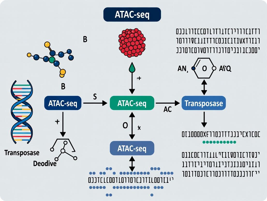
ATAC-seq Explained: A Complete Guide to Chromatin Accessibility for Researchers
This comprehensive guide provides researchers, scientists, and drug development professionals with a deep dive into Assay for Transposase-Accessible Chromatin using sequencing (ATAC-seq).
ATAC-seq Explained: A Complete Guide to Chromatin Accessibility for Researchers
Abstract
This comprehensive guide provides researchers, scientists, and drug development professionals with a deep dive into Assay for Transposase-Accessible Chromatin using sequencing (ATAC-seq). It covers foundational concepts linking chromatin architecture to gene regulation, details step-by-step experimental and bioinformatics workflows, addresses common pitfalls and optimization strategies, and guides the critical validation and interpretation of results within the broader genomics landscape. Learn how ATAC-seq can accelerate discoveries in disease mechanisms, biomarker identification, and therapeutic target discovery.
What is ATAC-seq? Unlocking the Genome's Regulatory Landscape
Chromatin accessibility refers to the degree of physical compaction of DNA and its associated histone proteins, which directly governs the ability of transcription factors and regulatory complexes to bind cis-regulatory elements. This in-depth guide frames chromatin accessibility within the foundational thesis of ATAC-seq (Assay for Transposase-Accessible Chromatin using sequencing) research, detailing its role as the primary determinant of cellular identity and function through the regulation of gene expression.
The Molecular Basis of Chromatin Accessibility
The eukaryotic genome is packaged into nucleosomes, the basic repeating units of chromatin. Each nucleosome consists of ~147 bp of DNA wrapped around an octamer of core histone proteins (H2A, H2B, H3, H4). The positioning, composition, and chemical modification of nucleosomes, along with the action of chromatin remodelers, dictate regional accessibility. Accessible chromatin regions, often depleted of nucleosomes, correspond to promoters, enhancers, silencers, and insulators—collectively known as regulatory elements.
Title: Hierarchy of Chromatin Compaction and Accessibility States
Key Methodologies for Profiling Accessibility
Several high-throughput sequencing methods probe chromatin accessibility. Their quantitative outputs form the basis for comparative analysis.
Table 1: Core Chromatin Accessibility Assays
| Method | Principle | Key Metric | Resolution | Primary Input |
|---|---|---|---|---|
| ATAC-seq | Hyperactive Tn5 transposase inserts adapters into accessible DNA. | Insertion site density. | Single-nucleotide (footprints possible). | 50k-100k viable nuclei. |
| DNase-seq | DNase I endonuclease cleaves accessible DNA. | Cleavage site density. | ~10-50 bp. | 1-50 million nuclei. |
| MNase-seq | Micrococcal Nuclease digests linker DNA between nucleosomes. | Protected DNA fragment length/signal. | Nucleosome (~147 bp). | 1-10 million cells. |
| FAIRE-seq | Phenol-chloroform extraction isolates nucleosome-depleted DNA. | Enrichment of DNA in aqueous phase. | 100-1000 bp. | 10-20 million cells. |
Detailed ATAC-seq Protocol
Title: Standard ATAC-seq Protocol for Cultured Cells. Principle: The hyperactive Tn5 transposase simultaneously fragments and tags accessible genomic DNA with sequencing adapters.
Reagents & Equipment:
- Cell lysis buffer (10 mM Tris-HCl pH 7.4, 10 mM NaCl, 3 mM MgCl2, 0.1% IGEPAL CA-630)
- Transposase reaction mix (commercial or homemade Tn5 loaded with adapters)
- Phosphate Buffered Saline (PBS) with 0.04% Bovine Serum Albumin (BSA)
- DNA purification beads (SPRI-based)
- Qubit fluorometer and PCR thermocycler
- Bioanalyzer/TapeStation for library QC
Procedure:
- Nuclei Preparation: Harvest ~50,000-100,000 cells. Wash with cold PBS. Lyse cells in ice-cold lysis buffer for 3-10 minutes on ice. Pellet nuclei at 500-800 rcf for 10 min at 4°C.
- Tagmentation: Resuspend nuclei pellet in transposase reaction mix (25 μL 2x TD Buffer, 2.5 μL Tn5 Transposase, 22.5 μL nuclease-free water). Incubate at 37°C for 30 minutes in a thermocycler with heated lid.
- DNA Purification: Immediately purify tagmented DNA using SPRI beads per manufacturer's protocol. Elute in 20-30 μL elution buffer (10 mM Tris-HCl, pH 8.0).
- Library Amplification: Amplify purified DNA using limited-cycle PCR (typically 5-12 cycles) with barcoded primers and a high-fidelity DNA polymerase. Determine optimal cycle number via qPCR side-reaction.
- Library Cleanup & QC: Perform a double-sided SPRI bead cleanup to remove primer dimers and large fragments. Assess library size distribution (~200-1000 bp modal size) and quantify.
Title: ATAC-seq Experimental Workflow
Data Interpretation and Key Findings
ATAC-seq data analysis yields peaks of signal corresponding to accessible chromatin regions. Comparative analysis reveals cell-type-specific patterns.
Table 2: Typical ATAC-seq Data Metrics by Sample Type
| Sample Type | Recommended Reads per Sample | Expected Peaks | % Reads in Peaks | FRiP Score Benchmark |
|---|---|---|---|---|
| Primary Human Cells (e.g., T-cells) | 50-100 million | 50,000 - 150,000 | 20-40% | >0.2 |
| Cell Line (e.g., HEK293, K562) | 50-80 million | 40,000 - 100,000 | 25-50% | >0.25 |
| Mouse Tissue (Homogeneous) | 60-100 million | 60,000 - 200,000 | 15-35% | >0.15 |
| Complex Tissue (e.g., Brain) | 100-200 million | 100,000 - 300,000 | 10-30% | >0.1 |
The Scientist's Toolkit: Essential Reagents & Materials
Table 3: Key Research Reagent Solutions for ATAC-seq
| Reagent/Material | Supplier Examples | Function |
|---|---|---|
| Hyperactive Tn5 Transposase | Illumina (Nextera), Diagenode, homemade | Enzyme that fragments and tags accessible DNA. Core of the assay. |
| Nuclei Extraction/Lysis Buffer | 10x Genomics, Sigma-Aldrich, homemade | Gently lyses plasma membrane while keeping nuclear membrane intact. |
| SPRI (Solid Phase Reversible Immobilization) Beads | Beckman Coulter, Sigma-Aldrich | Magnetic beads for size-selective DNA purification and cleanup. |
| High-Fidelity PCR Master Mix | NEB, Thermo Fisher, KAPA | For limited-cycle amplification of tagmented DNA with minimal bias. |
| Dual-Size Selection Beads | Beckman Coulter (SPRIselect) | Enables precise selection of library fragments (e.g., 100-600 bp). |
| Fluorescent DNA Quantification Assay | Thermo Fisher (Qubit), Promega (QuantiFluor) | Accurate dsDNA quantification for library normalization. |
| Bioanalyzer/TapeStation High Sensitivity DNA Kits | Agilent Technologies | Capillary electrophoresis for precise library fragment size analysis. |
| Cell Strainer (40 μm) | Falcon, PluriSelect | Removal of cell clumps to ensure single-nucleus suspensions. |
| Nuclease-Free Water and Buffers | Thermo Fisher, Sigma-Aldrich | Prevents degradation of nucleic acids during all reaction steps. |
Signaling Pathways Modulating Accessibility
Chromatin accessibility is dynamically regulated by signaling cascades that modify histones or recruit remodelers.
Title: Signaling to Chromatin Accessibility via Histone Modification
Chromatin accessibility, as the fundamental gatekeeper of gene expression, provides the mechanistic interface between the static genome and dynamic cellular responses. ATAC-seq has emerged as the preeminent tool for mapping this regulatory landscape due to its simplicity, low cell input, and high resolution. Understanding and manipulating chromatin accessibility is now a central thesis in basic research for developmental biology and immunology, as well as in applied drug discovery for oncology and neurological diseases, where epigenetic dysregulation is a key driver of pathology.
Within the broader study of chromatin accessibility basics, Assay for Transposase-Accessible Chromatin using sequencing (ATAC-seq) represents a paradigm shift. The core breakthrough was the utilization of a hyperactive mutant Tn5 transposase, preloaded with sequencing adapters, to simultaneously fragment and tag regions of open chromatin. This method streamlined the mapping of nucleosome positions and transcription factor footprints with unprecedented speed and sensitivity, using far fewer cells than previous techniques like DNase-seq and FAIRE-seq.
The Hyperactive Transposase: Tn5
The wild-type Tn5 transposase catalyzes the cut-and-paste transposition of transposon DNA. The hyperactive mutant (E54K, L372P) exhibits significantly increased enzymatic activity and stability. When pre-loaded in vitro with oligonucleotide adapters for next-generation sequencing, this engineered transposase inserts these adapters into accessible genomic regions in a single reaction step.
Table 1: Comparison of Chromatin Accessibility Assays
| Assay | Key Enzyme/Principle | Typical Cell Number | Resolution | Primary Output |
|---|---|---|---|---|
| ATAC-seq | Hyperactive Tn5 Transposase | 500 - 50,000 cells | Nucleosome (~200 bp) & TF footprint (<100 bp) | Open chromatin regions, nucleosome positioning |
| DNase-seq | DNase I Endonuclease | 1 - 50 million cells | ~100-200 bp | DNase I hypersensitive sites (DHSs) |
| FAIRE-seq | Phenol-Chloroform Extraction | 1 - 10 million cells | ~200-500 bp | Nucleosome-depleted regions |
| MNase-seq | Micrococcal Nuclease | 1 - 50 million cells | Nucleosome (~147 bp) | Protected DNA (nucleosome positions) |
Detailed ATAC-seq Experimental Protocol
Core Principle: Live nuclei are incubated with the pre-loaded Tn5 transposase, which inserts sequencing adapters into accessible DNA. The tagged DNA is then purified, amplified by PCR, and sequenced.
Key Steps:
- Cell Collection & Lysis: Harvest fresh cells (e.g., 50K-100K). Wash with cold PBS. Resuspend in cold lysis buffer (10 mM Tris-HCl pH 7.4, 10 mM NaCl, 3 mM MgCl2, 0.1% IGEPAL CA-630) to isolate intact nuclei. Centrifuge immediately.
- Transposition Reaction: Resuspend nuclei pellet in transposition mix containing the pre-loaded Tn5 enzyme (e.g., from Illumina Nextera or a homemade assembly) and buffer. Incubate at 37°C for 30 minutes with gentle mixing.
- DNA Purification: Use a standard column- or bead-based DNA clean-up kit to stop the reaction and purify the transposed DNA.
- PCR Amplification & Library Indexing: Amplify the purified DNA with limited-cycle PCR using primers compatible with the transposase-loaded adapters. Incorporate sample-specific barcodes (dual indexing is recommended).
- Library Clean-up & Quality Control: Purify the final library using SPRI beads. Assess library quality via Bioanalyzer/TapeStation (expect a periodicity of ~200 bp nucleosome ladder pattern). Quantify by qPCR.
- Sequencing: Sequence on an appropriate NGS platform (typically Illumina). A minimum of 25-50 million paired-end reads per sample is standard for mammalian genomes.
Table 2: Key Research Reagent Solutions for ATAC-seq
| Reagent/Material | Function & Critical Notes |
|---|---|
| Hyperactive Tn5 Transposase | Core enzyme, pre-loaded with sequencing adapters. Commercial kits (Illumina) or purified protein for custom assembly. |
| Cell Permeabilization Buffer | Gently lyses the plasma membrane while keeping nuclear membrane intact. Critical for enzyme access. |
| Nuclease-Free Water & Buffers | Essential to prevent degradation of nuclei, DNA, and enzyme activity. |
| SPRI (Solid Phase Reversible Immobilization) Beads | For size selection and clean-up of DNA fragments after transposition and PCR. |
| High-Fidelity PCR Master Mix | For limited-cycle amplification of transposed DNA fragments with high fidelity. |
| Dual Indexing PCR Primers | To multiplex samples, each gets a unique pair of barcodes added during PCR. |
| Qubit dsDNA HS Assay Kit | Accurate quantification of low-concentration DNA libraries. |
| Bioanalyzer High Sensitivity DNA Kit | Assesses library fragment size distribution and quality. |
Data Analysis & Biological Interpretation
Sequencing reads are aligned to a reference genome. The insert size distribution reveals sub-nucleosomal fragments (TF footprints), mononucleosomal (~200 bp), and dinucleosomal (~400 bp) fragments. Peak calling identifies regions of significant accessibility, which can be correlated with gene regulatory elements.
Diagram 1: ATAC-seq Experimental Workflow (79 chars)
Diagram 2: Fragment Sizes Map Chromatin Features (68 chars)
Advanced Applications & Impact on Drug Discovery
ATAC-seq's low cell requirement enabled its application to rare cell populations and clinical samples. Key derivatives include:
- Single-cell ATAC-seq (scATAC-seq): Profiles chromatin heterogeneity across cell populations.
- ATAC-seq with sequencing of extracted regulatory elements (ATAC-see): Visualizes open chromatin loci in situ.
- Multimodal assays: Paired with RNA-seq (multiome) or protein expression (CITE-seq).
In drug development, ATAC-seq is used to map the impact of chemical compounds or genetic perturbations on the global chromatin landscape, identifying mechanisms of action and off-target epigenetic effects.
Table 3: Quantitative Metrics for a Successful ATAC-seq Experiment
| Metric | Target Range / Expected Result | Purpose & Interpretation |
|---|---|---|
| Fraction of Reads in Peaks (FRiP) | >20-30% (cell lines); >10-15% (tissues) | Measures signal-to-noise. Low FRiP suggests poor transposition or over-digestion. |
| Transposition Fragment Size Distribution | Clear peaks at <100 bp, ~200 bp, ~400 bp | Confirms successful nucleosome patterning. Absence suggests technical failure. |
| Library Complexity (Non-Redundant Fraction) | >0.8 for bulk ATAC-seq | Measures library saturation. Low complexity indicates PCR over-amplification or low cell input. |
| Mitochondrial Read Percentage | <20-50% (varies by sample type) | High % indicates excessive nuclei lysis or poor cytoplasmic removal. |
| Total Sequencing Depth | 25-50 million aligned reads (mammalian) | Sufficient for peak calling and differential analysis. |
Assay for Transposase-Accessible Chromatin using sequencing (ATAC-seq) has become a cornerstone technique for probing chromatin architecture. The core analytical outputs—peaks, footprints, and nucleosome positioning—collectively translate raw sequencing data into a multi-scale map of regulatory genomics. This guide details the technical interpretation, generation, and integration of these outputs, forming a critical chapter in the thesis on ATAC-seq chromatin accessibility basics. Mastery of these elements is essential for researchers and drug development professionals aiming to identify functional regulatory elements, transcription factor (TF) occupancy, and epigenetic states linked to disease and treatment response.
Core Outputs: Definitions and Biological Significance
| Output | Genomic Feature Represented | Biological Interpretation | Key Analytical Challenge |
|---|---|---|---|
| Peaks | Broad regions of open chromatin. | Candidate cis-regulatory elements (cCREs) such as enhancers, promoters, and insulators. | Distinguishing true signal from background noise; peak-calling parameter sensitivity. |
| Footprints | Short (~6-12 bp) dips in ATAC-seq signal within a peak. | Putative transcription factor binding site (TFBS) where protein occupancy physically impedes Tn5 transposase cleavage. | Low signal-to-noise ratio; confounding effects of TF dynamics and chromatin structure. |
| Nucleosome Positioning | Periodic pattern of insert sizes from ATAC-seq fragments. | Positioning of nucleosomes along the DNA, inferred from protected fragments (~180-200 bp) and subnucleosomal particles. | Resolution limits; influence of data depth and computational deconvolution. |
Table 1: Comparative Summary of Core ATAC-seq Outputs.
Detailed Methodologies for Generating Core Outputs
Peak Calling Protocol
- Input: Aligned BAM files (paired-end reads), after removal of mitochondrial reads and duplicates.
- Tool: MACS2 (Model-based Analysis of ChIP-Seq 2) is standard.
Command Example:
-f BAMPE: Uses paired-end mode for superior fragment size estimation.--nomodel --shift -100 --extsize 200: Bypasses the internal shifting model, applying a fixed shift to center peaks on the transposition event.
- Output: BED file of peak locations (summits and intervals) and statistical scores.
Footprint Detection Protocol
- Input: ATAC-seq BAM file aligned to the reference genome, plus a BED file of peak regions.
- Tool: HINT-ATAC from the RGT suite (Recommended for current best practices).
Command Example:
Post-processing: Footprints are typically matched to known TF motifs (e.g., using JASPAR database) via tools like
rgt-motifanalysis matching.
Nucleosome Positioning Analysis Protocol
- Input: ATAC-seq BAM file.
- Method: Analysis of insert size distribution.
- Extract fragment (insert) sizes from the BAM file.
- Generate a genome-wide histogram of fragment lengths.
- Key Lengths: Peaks at ~<100 bp (nucleosome-free), ~180-200 bp (mononucleosome), ~360-400 bp (dinucleosome).
- Positioning Callers: Tools like NucleoATAC or DANPOS2 can be used to call nucleosome positions genome-wide by scanning for periodic patterns of protected fragments.
Visualizing Relationships and Workflows
Title: ATAC-seq Core Outputs Generation Workflow
Title: Multi-Scale Features at a Regulatory Locus
The Scientist's Toolkit: Essential Research Reagents & Materials
| Item | Function/Application in ATAC-seq | Key Consideration |
|---|---|---|
| Tn5 Transposase (Loaded) | Enzyme that simultaneously fragments and tags accessible genomic DNA with sequencing adapters. The core reagent. | Commercial kits (e.g., Illumina Nextera) ensure consistent activity and loading. |
| Cell Permeabilization Buffer | (For intact nuclei assays) Gently lyses the plasma membrane while keeping nuclear membrane intact for Tn5 entry. | Critical for optimizing signal-to-noise; often contains Digitonin. |
| Nuclei Isolation & Wash Buffers | Prepare clean nuclei from tissue/cells, removing cytoplasmic contaminants that inhibit transposition. | Must be ice-cold and often contain protease inhibitors. |
| Magnetic Beads (SPRI) | For post-PCR cleanup and size selection to remove primer dimers and select optimal fragment lengths. | Bead-to-sample ratio determines size cut-off. |
| PCR Amplification Mix | Amplifies the transposed library with indexed primers for multiplexing. | Use limited-cycle PCR to minimize amplification bias. |
| High-Sensitivity DNA Assay Kit | (e.g., Bioanalyzer, TapeStation, Qubit) Quantifies and assesses size distribution of final libraries before sequencing. | Essential for accurate sequencing pool normalization. |
| qPCR Primers for Accessible Loci | Validate ATAC-seq library quality by qPCR, comparing signal at open vs. closed genomic regions. | Quality control step before deep sequencing. |
Understanding the regulatory genome is foundational to modern molecular biology and therapeutic discovery. Within the broader thesis on ATAC-seq (Assay for Transposase-Accessible Chromatin using sequencing) chromatin accessibility basics, this whitepaper explores the subsequent critical step: interpreting open chromatin regions to predict transcription factor (TF) binding and consequential enhancer activity. ATAC-seq provides a genome-wide map of nucleosome-depleted, "open" regions, which are putative regulatory elements. However, not all accessible chromatin is functionally active. This guide details the computational and experimental frameworks used to move from a catalog of open regions to mechanistic, biological insight into gene regulation, with direct implications for understanding disease etiology and identifying novel drug targets.
From ATAC-seq Peaks to TF Motif Analysis
The primary output of an ATAC-seq experiment is a set of peaks representing regions of statistically significant chromatin accessibility. These peaks are candidates for enhancers, promoters, insulators, and other cis-regulatory elements.
De Novoand Known Motif Discovery
Purpose: To identify which transcription factors are likely binding within ATAC-seq peaks.
- Methodology (Known Motif Scanning): The sequence of each ATAC-seq peak is scanned against a database of position weight matrices (PWMs) representing known TF binding motifs. Tools like HOMER, MEME-ChIP, or FIMO are commonly used.
- Input: FASTA file of peak sequences.
- Process: Each PWM is scored against the sequence. A log-odds score or p-value threshold determines a "hit."
- Output: A list of TFs whose motifs are significantly enriched in the peak set compared to a background genomic sequence (e.g., shuffled peaks or genomic regions with matched GC content).
- Methodology (De Novo Motif Discovery): Used when novel or poorly characterized factors are involved. Tools like MEME or HOMER identify overrepresented sequence patterns within the peak set.
- Input: FASTA file of peak sequences.
- Process: Algorithm searches for ungapped, recurring patterns of a specified width.
- Output: One or more novel PWMs, which can then be compared to known databases for annotation.
Quantitative Data Summary: Table 1: Common Motif Discovery Tools and Their Key Parameters
| Tool | Primary Function | Key Statistical Output | Common Background Model |
|---|---|---|---|
| HOMER | Known scanning & de novo | p-value, % of targets with motif | Matched GC content, repeat-masked |
| MEME-ChIP | De novo & refinement | E-value (expectation) | Markov model from provided sequences |
| FIMO | Known motif scanning | q-value (FDR-adjusted p-value) | Specified nucleotide frequencies |
Footprinting for Precise TF Binding Inference
Purpose: To pinpoint the exact genomic location of a bound TF within an open chromatin region. Bound TFs protect their core binding site from transposase cleavage, creating a "footprint" of low ATAC-seq signal flanked by higher signal from accessible borders.
Experimental Protocol (Digital Genomic Footprinting from ATAC-seq Data):
- High-Depth Sequencing: Footprint detection requires high sequencing depth (>100 million reads) to resolve the subtle, localized depletion in cleavage events.
- Alignment & Processing: Align ATAC-seq reads to reference genome, filter for properly paired, non-mitochondrial, and high-quality reads.
- Tn5 Offset Adjustment: Account for the 9-bp stagger introduced by the Tn5 transposase and shift reads +/- 4-5 bp to represent the actual cleavage center.
- Footprint Calling: Use algorithms (e.g., TOBIAS, HINT-ATAC, Wellington) that calculate a per-nucleotide cleavage profile and identify statistically significant dips.
- Input: BAM file of shifted reads, peak regions (BED file).
- Process: Compare observed cleavage profile within a peak to a expected profile (often from a DNase I or Tn5 sequence bias model). Apply a statistical test (e.g., Wilcoxon rank-sum) to evaluate the significance of the footprint depression.
- Motif Integration: Overlap identified footprint sites with TF motifs to assign protein identity to the footprint.
Digital Genomic Footprinting Workflow
Predicting Enhancer Activity and Target Genes
Identifying a putative TF-bound region is insufficient; predicting its functional activity (enhancer vs. inactive open chromatin) and its target gene is the ultimate goal.
Chromatin State Integration
Purpose: Use complementary epigenomic marks to classify the functional state of an open chromatin region.
- Methodology (Chromatin Immunoprecipitation Sequencing - ChIP-seq): Perform ChIP-seq for histone modifications in the same cell type.
- H3K27ac: Marks active enhancers and promoters.
- H3K4me1: Marks poised and active enhancers (distal from promoters).
- H3K4me3: Marks active promoters.
- H3K27me3: Marks Polycomb-repressed regions.
Experimental Protocol (Integration with H3K27ac ChIP-seq):
- Generate Data: Perform standard ATAC-seq and H3K27ac ChIP-seq on biologically matched samples.
- Peak Calling: Call peaks for each dataset independently (e.g., using MACS2).
- Overlap Analysis: Intersect ATAC-seq peaks with H3K27ac peaks using tools like BEDTools.
- Classification: An ATAC-seq peak overlapping H3K27ac is a high-confidence active enhancer or promoter. An ATAC-seq peak lacking H3K27ac is a candidate inactive/poised regulatory element.
Chromatin Conformation Capture
Purpose: To empirically link distal enhancers to their target promoters through physical chromatin looping.
- Methodology (HiChIP or H3K27ac HiChIP): A method combining chromatin conformation capture with immunoprecipitation for a specific mark (e.g., H3K27ac). It provides high-resolution, mark-specific contact maps.
- Input: Cross-linked chromatin, digested and ligated, followed by immunoprecipitation with an antibody against H3K27ac.
- Output: Paired-end sequencing reads representing ligation junctions between spatially proximal DNA fragments, enriched for active regulatory elements.
Machine Learning Predictions
Purpose: To computationally predict enhancer activity and gene targets using integrated features.
- Methodology: Train supervised models (e.g., random forest, gradient boosting, deep neural networks) using known enhancer-promoter pairs (e.g., from eQTL studies or high-throughput reporter assays like STARR-seq).
- Features: Include sequence-based features (motif scores, conservation), chromatin features (ATAC-seq signal, histone marks), and 1D genomic distance.
- Tools: TargetFinder, PEP, and custom models are commonly used.
Quantitative Data Summary: Table 2: Methods for Linking Enhancers to Target Genes
| Method | Principle | Resolution | Key Advantage | Key Limitation |
|---|---|---|---|---|
| Nearest Gene | Genomic proximity | N/A | Simple, fast | Highly inaccurate, many false links |
| Chromatin Conformation (Hi-C/HiChIP) | Physical 3D contact | 1-10 kb | Empirical, genome-wide | Cost, complexity, moderate resolution |
| Machine Learning (TargetFinder) | Integrated feature prediction | N/A | Inexpensive, scalable | Depends on quality of training data |
| Enhancer Perturbation + RNA-seq | Functional causality | Single enhancer | Gold standard for function | Low-throughput, costly |
The Scientist's Toolkit: Research Reagent Solutions
Table 3: Essential Reagents and Kits for ATAC-seq and Downstream Analysis
| Item Name | Supplier Examples | Function in Workflow |
|---|---|---|
| Tn5 Transposase (Loaded) | Illumina (Nextera), Diagenode, custom | Enzyme that simultaneously fragments open chromatin and adds sequencing adapters. Core of ATAC-seq. |
| Nuclei Isolation Kit | Sigma-Aldrich, Thermo Fisher, 10x Genomics | Gentle lysis buffers and reagents to isolate intact nuclei from cells/tissues for ATAC-seq. |
| Magnetic Beads for Size Selection | SPRIselect (Beckman), AMPure XP (Beckman) | To purify and select appropriately sized DNA fragments post-tagmentation (e.g., remove large fragments >1000 bp). |
| High-Sensitivity DNA Assay Kits | Qubit (Thermo), Bioanalyzer/TapeStation (Agilent) | Accurate quantification and quality assessment of low-concentration ATAC-seq libraries prior to sequencing. |
| ChIP-Validated Antibodies | Cell Signaling, Abcam, Active Motif | For ChIP-seq of histone modifications (H3K27ac, H3K4me1) to validate enhancer activity. Critical for integration. |
| Chromatin Conformation Capture Kits | Arima HiC, Phase Genomics | Standardized reagents for Hi-C or HiChIP library preparation to map enhancer-promoter contacts. |
| TF Motif/PWM Databases | JASPAR, CIS-BP, HOCOMOCO | Curated collections of position weight matrices used for scanning ATAC-seq peaks to predict TF binding. |
Logical Framework for Predicting Enhancer Activity
Predicting transcription factor binding and enhancer activity from open chromatin data is a multi-layered inference problem. It requires moving beyond simple peak calling to integrate in silico motif analysis, digital footprinting, complementary epigenomic datasets, and 3D chromatin architecture. The experimental and computational protocols outlined here provide a rigorous pathway to transform ATAC-seq peak lists into testable hypotheses about transcriptional regulatory networks. For drug development professionals, this pipeline is essential for identifying disease-relevant non-coding regulatory elements that may serve as novel therapeutic targets or biomarkers, solidifying the critical role of chromatin accessibility basics in translational research.
Within the foundational research of ATAC-seq (Assay for Transposase-Accessible Chromatin using sequencing) for profiling chromatin accessibility, three technical advantages stand out as transformative: Speed, Sensitivity, and Low Cell Input Requirements. These characteristics have fundamentally accelerated epigenetic research and its application in drug discovery by enabling rapid, high-resolution mapping of regulatory landscapes from limited and precious clinical samples.
In-Depth Technical Analysis
Speed: Streamlined Workflow from Cells to Data
The primary driver of speed in ATAC-seq is the integration of tagmentation (transposition and fragmentation) into a single enzymatic step. Compared to traditional methods like DNase-seq or FAIRE-seq, which require multiple days, ATAC-seq can be completed from cells to sequencing libraries in approximately 3-4 hours.
Table 1: Protocol Duration Comparison
| Method | Cell Lysis & Tagmentation/Fragmentation | Library Preparation | Total Hands-On Time | Total Time to Library |
|---|---|---|---|---|
| ATAC-seq | 30 min | ~3 hours | ~4 hours | 1 day |
| DNase-seq | Several hours | 2-3 days | 1.5-2 days | 4-5 days |
| FAIRE-seq | Overnight | 2 days | 1-2 days | 4 days |
Detailed Protocol for Fast ATAC-seq Library Preparation:
- Cell Lysis & Tagmentation: Resuspend 50-100,000 cells in cold lysis buffer (10 mM Tris-HCl pH 7.4, 10 mM NaCl, 3 mM MgCl2, 0.1% IGEPAL CA-630). Incubate on ice for 3 min. Pellet nuclei and resuspend in transposition mix (25 μL 2x TD Buffer, 2.5 μL Tn5 Transposase, 22.5 μL nuclease-free water). Incubate at 37°C for 30 minutes in a thermomixer with shaking.
- DNA Purification: Immediately clean up tagmented DNA using a MinElute PCR Purification Kit or SPRI beads. Elute in 21 μL of Elution Buffer.
- Library Amplification: Amplify the purified DNA using 1x NPM, 1.25 μM of a universal i5 and a uniquely barcoded i7 primer, and 1x NEB Next High-Fidelity 2X PCR Master Mix. Use a cycle number determined by a qPCR side reaction or a pre-optimized number (typically 8-12 cycles for 50K cells).
- Library Clean-up: Perform a double-sided SPRI bead cleanup (e.g., 0.5x followed by 1.5x ratio) to remove primer dimers and select for appropriately sized fragments. Quantify and pool for sequencing.
Sensitivity: Capturing Rare Cell States and Faint Signals
ATAC-seq sensitivity stems from the highly efficient Tn5 transposase and the direct ligation of sequencing adapters during tagmentation. This efficiency allows for the detection of open chromatin regions even from small cell populations.
Table 2: Sensitivity Metrics in Low-Input ATAC-seq
| Cell Number Input | Recommended Sequencing Depth | Detectable Peaks | Key Application |
|---|---|---|---|
| 50,000 - 100,000 (Standard) | 50-100 million reads | ~80,000 - 120,000 | Bulk tissue analysis, cell lines |
| 5,000 - 10,000 (Low Input) | 50 million reads | ~50,000 - 80,000 | Fine needle aspirates, limited biopsies |
| 500 - 1,000 (Ultra-Low Input) | 100+ million reads | ~20,000 - 50,000 | Rare progenitor cells, sorted populations |
| Single Cell (scATAC-seq) | 10,000-50,000 reads/cell | 1,000 - 5,000/cell | Heterogeneity, cellular atlas construction |
Protocol for High-Sensitivity Low-Input ATAC-seq (5,000-10,000 cells):
- Cell Handling: Precisely count cells using a hemocytometer or automated counter. Wash cells gently in cold PBS.
- Modified Lysis: Perform lysis in a reduced volume (e.g., 50 μL) to minimize nucleus loss. Include a carrier (e.g., 0.1% BSA) in wash buffers to reduce adhesion.
- Tagmentation Optimization: Use a proportionally reduced but more concentrated transposition mix (e.g., 10 μL 2x TD Buffer, 1 μL Tn5, 9 μL water). Extend tagmentation time to 45-60 min.
- Library Amplification with qPCR: Perform library amplification alongside a 10 μL qPCR reaction using SYBR Green. Amplify the main reaction until the qPCR amplification curve reaches 1/3 of its plateau. This prevents over-cycling and preserves complexity.
- High-Depth Sequencing: Sequence the resulting library to a minimum depth of 50 million paired-end reads to ensure statistical power for peak calling.
Low Cell Input Requirements: Enabling Studies on Precious Samples
The low cell requirement is a direct consequence of high sensitivity. It allows researchers to profile chromatin accessibility from minute clinical samples (e.g., tumor biopsies, patient-derived xenografts, embryonic material) and rare immune cell subsets without the need for cell expansion.
Technical Foundations of Low-Input Compatibility:
- Efficient Tagmentation: A single Tn5 transposome can insert adapters into accessible DNA, requiring minimal starting material.
- Minimal Purification Steps: The streamlined protocol reduces sample loss.
- Optimized Buffers: Modern commercial buffers stabilize nuclei and maintain transposase activity in small volumes.
Table 3: Impact of Low Input Requirements on Research Applications
| Application Field | Traditional Method Challenge | ATAC-seq Advantage |
|---|---|---|
| Cancer Biology | Need for large tumor sections, obscuring heterogeneity. | Profiling of small, morphologically defined regions or circulating tumor cells. |
| Immunology | Difficulty in obtaining large numbers of rare immune subsets (e.g., antigen-specific T cells). | Epigenetic profiling of sorted populations from peripheral blood. |
| Neurobiology | Hard-to-acquire primary neuronal tissue. | Analysis of post-mortem brain regions or organoids. |
| Developmental Biology | Limited material from early embryos. | Mapping chromatin dynamics in embryonic stem cells or early lineages. |
The Scientist's Toolkit: Research Reagent Solutions
Table 4: Essential Reagents for Optimized ATAC-seq
| Item | Function & Importance |
|---|---|
| High-Activity Tn5 Transposase | Engineered hyperactive enzyme for efficient tagmentation in low-input and sensitive applications. Critical for success. |
| Nuclei Isolation & Lysis Buffer | Gently lyses cell membrane while keeping nuclei intact. Consistent formulation is key for batch-to-batch reproducibility. |
| Magnetic SPRI Beads | For size selection and clean-up. Enables removal of primers, dimers, and large fragments without column loss. |
| Unique Dual-Indexed PCR Primers | Allow multiplexing of hundreds of samples in a single sequencing run, reducing cost and handling time. |
| Nuclei Counting Dye (e.g., DAPI) | Accurate quantification of isolated nuclei before tagmentation is essential for optimizing enzyme-to-DNA ratio. |
| qPCR Master Mix with High-Fidelity Polymerase | For accurate determination of optimal PCR cycles during library amplification, preventing over-amplification. |
| High-Sensitivity DNA Assay Kit (e.g., Qubit, Bioanalyzer) | Accurate quantification and quality assessment of low-concentration final libraries. |
Visualizing the ATAC-seq Workflow and Advantages
Diagram 1: Integrated ATAC-seq workflow showcasing core advantages.
Diagram 2: Tn5 transposition mechanism enabling speed and sensitivity.
A Step-by-Step ATAC-seq Protocol: From Cell to Data
This technical guide details the critical pillars of robust experimental design for ATAC-seq (Assay for Transposase-Accessible Chromatin using sequencing) within the broader thesis of chromatin accessibility basics. ATAC-seq maps open chromatin regions genome-wide, identifying putative regulatory elements. The validity of these findings is fundamentally dependent on meticulous planning of cell type selection, biological replication, and appropriate controls to mitigate technical and biological variability.
Cell Type Considerations in ATAC-Seq
The choice of cell type is the primary determinant of the biological relevance of an ATAC-seq experiment. Chromatin accessibility is highly cell-type-specific.
Key Factors for Selection
- Biological Relevance: The cell type must accurately model the biological question (e.g., disease state, developmental lineage, treatment response).
- Heterogeneity: Primary cells reflect in vivo states but are heterogeneous. Cell sorting (FACS) using specific surface markers is often required.
- Proliferation State: ATAC-seq uses a transposase integration step. Actively dividing cells may yield different signal-to-noise ratios compared to quiescent cells due to variations in nuclear content and cell cycle stage.
- Input Cell Number: Standard protocols require 50,000-100,000 viable cells. Low-input and single-cell protocols exist but have distinct design implications.
Cell Type Comparison Table
Table 1: Common Cell Sources for ATAC-Seq Experiments
| Cell Type | Advantages | Disadvantages | Recommended Use Case |
|---|---|---|---|
| Primary Cells | Physiologically relevant, native chromatin state. | Limited availability, donor variability, hard to culture. | Disease profiling, population studies. |
| Cell Lines | Easily cultured, high yield, genetically uniform. | May have accumulated epigenetic artifacts from long-term culture. | Mechanistic studies, CRISPR screens, treatment time-courses. |
| Fresh/Frozen Tissue | Preserves native tissue context and heterogeneity. | Requires dissociation; nuclei isolation is critical and variable. | Translational research, tumor biology. |
| Sorted Populations (FACS) | High purity for specific cell types from a mixture. | Lower yield; sorting stress may affect chromatin. | Rare population analysis (e.g., stem cells, specific immune cells). |
| Cryopreserved Nuclei | Flexibility; batch experiments from same sample. | Potential for nuclear lysis or accessibility changes during freeze-thaw. | Large cohort studies, biobank resources. |
Replicates: Biological and Technical
Adequate replication is non-negotiable for statistical power and reproducibility.
Definitions and Purpose
- Biological Replicates: Cells or tissues harvested from independent biological sources (different animals, human donors, separate cultures). They capture biological variation and are essential for generalizing conclusions.
- Technical Replicates: The same biological sample processed through the experimental workflow multiple times (e.g., same nuclei split into multiple library preps). They assess technical noise from library preparation and sequencing.
Quantitative Guidelines for Replication
Recent community standards and statistical analyses provide concrete recommendations.
Table 2: Replication Guidelines for ATAC-Seq Experiments
| Parameter | Recommendation | Rationale |
|---|---|---|
| Minimum Biological Replicates | n=3 for each condition/cell type. n=2 is absolute minimum but severely limits statistical testing. | Enables assessment of variability and use of tools like DESeq2 for differential accessibility. |
| Technical Replicates | Typically not required for high-throughput sequencing if using unique molecular identifiers (UMIs). | Modern protocols are robust; sequencing depth is more critical. Use for troubleshooting. |
| Sequencing Depth per Rep | 20-50 million high-quality, non-mitochondrial, non-duplicate reads for bulk ATAC-seq. | Saturation of peak detection. Complex genomes or heterogeneous samples require higher depth. |
| Power Analysis | Use tools like ATACseqQC or ssize to determine replicates/depth based on expected effect size. |
For differential analysis, more replicates often outweigh deeper sequencing. |
Essential Controls in ATAC-Seq
Controls are required to distinguish biological signal from technical artifact.
Types of Controls
- Negative Control: A sample where accessible chromatin is expected to be absent or vastly different. Examples include:
- Cell-free Input Control: Tagmentation reaction performed on naked genomic DNA (without nuclei). Identifies sequence bias of the transposase.
- DNase I-treated DNA: Can be used as a control for nuclease accessibility patterns, though less common.
- Positive Control: A well-characterized cell line or sample (e.g., GM12878 lymphoblastoid cells) with a publicly available, high-quality ATAC-seq dataset. Used to benchmark experimental and bioinformatic pipelines.
- Process Control: Spike-in Nuclei. Adding a small number of nuclei from a different species (e.g., Drosophila melanogaster S2 cells to human samples) prior to tagmentation. Allows for normalization based on spike-in read counts, controlling for technical variation in tagmentation efficiency and PCR amplification.
Control Experiment Protocol:Spike-in Nuclei for Normalization
- Prepare Spike-in Nuclei: Culture D. melanogaster S2 cells. Harvest and isolate nuclei using the same protocol as your experimental cells. Count nuclei and aliquot for single-use. Determine the optimal spike-in ratio empirically (e.g., 1-10% of total nuclei).
- Spike-in Addition: Combine a precise volume of your experimental nuclei with the predetermined volume of S2 nuclei immediately before the tagmentation reaction. Mix gently but thoroughly.
- Proceed with ATAC-Seq: Continue with the standard ATAC-seq protocol (tagmentation, purification, PCR amplification).
- Bioinformatic Normalization: During analysis, align reads to a concatenated genome (e.g., hg38+dm6). Use the proportion of reads aligning to the spike-in genome to scale libraries for differential analysis.
The Scientist's Toolkit: ATAC-Seq Research Reagents
Table 3: Essential Reagents and Materials for ATAC-Seq
| Reagent / Material | Function | Key Consideration |
|---|---|---|
| Tn5 Transposase | Enzyme that simultaneously fragments and tags accessible DNA with sequencing adapters. | Commercial loaded enzymes (e.g., Illumina Tagmentase) ensure high efficiency and reproducibility. |
| Digitonin | Mild detergent used in lysis buffers to permeabilize nuclear membranes without destroying chromatin structure. | Concentration is critical; over-permeabilization leads to mitochondrial DNA contamination. |
| Sucrose Gradient | A cushion (e.g., 30% sucrose) used during nuclei isolation to purify nuclei from cellular debris. | Essential for reducing cytoplasmic contamination and improving signal-to-noise. |
| AMPure XP Beads | Magnetic beads used for size selection and cleanup of DNA libraries post-tagmentation and PCR. | Ratio of beads to sample determines size selection window (e.g., 0.5x to 1.8x for fragment selection). |
| PCR Indexed Primers | Primers that amplify the tagmented DNA and add unique dual indices for sample multiplexing. | Use unique dual indexing to minimize index hopping errors on patterned flow cells. |
| Cell Stains (DAPI, PI) | For assessing nuclei integrity and concentration via fluorescence microscopy or flow cytometry. | Viable, intact nuclei are critical. Avoid apoptotic cells. |
| ERCC Spike-in RNA | Optional: For single-nucleus ATAC-seq (snATAC-seq), these exogenous RNAs can be added to assess droplet encapsulation efficiency. | Not used in standard bulk ATAC-seq. |
| Nextera Index Kit | A common commercial source of indexed primers compatible with the Illumina Tn5 transposase. | Ensure primer indexes are compatible with your sequencer (iSeries adapters for NextSeq/Novaseq). |
Visualization of Experimental Workflow and Controls
ATAC-Seq Experimental Design and Control Workflow
How Replication Addresses Sources of Variation
This technical guide details the foundational sample preparation steps for the Assay for Transposase-Accessible Chromatin using sequencing (ATAC-seq), within the broader thesis of chromatin accessibility research. The quality of nuclei isolation and the efficiency of the transposition reaction are the most critical determinants of a successful ATAC-seq experiment, impacting data resolution, signal-to-noise ratio, and reproducibility for researchers and drug development professionals.
Table 1: Key Metrics for Nuclei Isolation Quality Control
| Metric | Optimal Range | Measurement Method | Impact of Deviation |
|---|---|---|---|
| Nuclei Count | 50,000 - 100,000 per reaction | Hemocytometer (Trypan Blue) | Low count: Poor library complexity. High count: Over-transposition. |
| Nuclei Integrity | >90% intact (smooth, round) | Microscopy (DIC or fluorescent stain) | Lysed nuclei: Release of genomic DNA & inhibitors. |
| Cellular Debris | Minimal to none | Flow cytometry (DAPI vs. SSC) | Debris: Non-specific transposition, high background. |
| Mitochondrial DNA Contamination | <20% of final reads | Post-sequencing bioinformatics | High mtDNA: Reduces usable reads for nuclear chromatin. |
| Nuclei Purity (Absence of intact cells) | No intact cells visible | Microscopy | Intact cells: Inaccessible chromatin, failed assay. |
Table 2: Transposition Reaction Optimization Parameters
| Parameter | Recommended Condition | Rationale | Typical Commercial Kit Value |
|---|---|---|---|
| Reaction Temperature | 37°C | Optimal activity for Tn5 transposase. | 37°C |
| Reaction Time | 30 min | Balance between completeness and over-fragmentation. | 30 min |
| Number of Nuclei per 50 µL rxn | 50,000 | Ensures sufficient template, avoids enzyme saturation. | 50,000 - 100,000 |
| Tn5 Transposase Concentration | As per kit (e.g., 2.5 µL) | Pre-optimized for insertion density & fragment size. | Fixed volume |
| Mg^{2+} Concentration (Final) | ~10 mM | Essential cofactor for transposase activity. | Provided in buffer |
Detailed Methodologies
Protocol: Nuclei Isolation from Cultured Mammalian Cells (Cold Lysis Method)
This protocol is designed for adherent or suspension cells, minimizing mechanical disruption.
Materials: Ice-cold PBS, Ice-cold Lysis Buffer (10 mM Tris-HCl pH 7.4, 10 mM NaCl, 3 mM MgCl2, 0.1% IGEPAL CA-630, 0.1% Tween-20, 0.01% Digitonin), Wash Buffer (10 mM Tris-HCl pH 7.4, 10 mM NaCl, 3 mM MgCl2, 0.1% Tween-20), Resuspension Buffer (10 mM Tris-HCl pH 7.4, 10 mM NaCl, 3 mM MgCl2, 1% BSA), DAPI solution.
Procedure:
- Harvest Cells: Collect ~50,000-100,000 viable cells. Wash once with ice-cold PBS.
- Cell Lysis: Resuspend cell pellet thoroughly in 50 µL of ice-cold Lysis Buffer. Incubate on ice for 3-5 minutes. Monitor lysis under a microscope (>90% lysed cells with released intact nuclei).
- Wash Nuclei: Immediately add 1 mL of ice-cold Wash Buffer. Pellet nuclei at 500 x g for 5 minutes at 4°C in a pre-chilled centrifuge.
- Remove Supernatant: Carefully aspirate supernatant. The pellet may be small.
- Wash Again: Repeat steps 3 and 4.
- Resuspend Nuclei: Gently resuspend the pellet in 50 µL of Resuspension Buffer. Filter through a 30-40 µm cell strainer if clumping is observed.
- Count and QC: Mix 10 µL of nuclei suspension with 10 µL of DAPI (1 µg/mL). Count intact, DAPI-positive nuclei using a hemocytometer. Adjust concentration to ~1,000 nuclei/µL. Proceed immediately to transposition or flash-freeze.
Protocol: The Transposition Reaction
Materials: Isolated nuclei, Tagmented DNA Buffer (Illumina), Tn5 Transposase (Illumina or equivalent), Nuclease-free water, DNA Cleanup Beads (SPRI).
Procedure:
- Assemble Reaction: Combine in a nuclease-free tube:
- 25 µL of 2x Tagmentation Buffer
- 5 µL of Tn5 Transposase (commercial preparation)
- Nuclease-free water (variable)
- 20 µL of nuclei suspension (~50,000 nuclei)
- Total Volume = 50 µL Mix gently by pipetting. Do not vortex.
- Incubate: Place the tube in a preheated thermal cycler at 37°C for 30 minutes.
- Cleanup: Immediately add 50 µL of DNA Cleanup Beads (1.0x ratio) to the 50 µL reaction. Mix thoroughly. Follow standard SPRI bead cleanup protocol, eluting in 20-30 µL of Elution Buffer or 10 mM Tris-HCl pH 8.0.
- QC: Analyze 1 µL of eluted DNA on a Bioanalyzer/TapeStation (HS DNA chip). A successful reaction shows a nucleosomal ladder pattern with a dominant peak < 1,000 bp.
Visualizations
Diagram 1: ATAC-seq Nuclei Isolation & Tagmentation Workflow
Diagram 2: Tn5 Transposase Mechanism in Chromatin Tagmentation
The Scientist's Toolkit: Research Reagent Solutions
| Reagent / Material | Function | Key Consideration |
|---|---|---|
| IGEPAL CA-630 (NP-40 Alternative) | Non-ionic detergent for cell membrane lysis. | Concentration is critical (typically 0.1%). Too high lyses nuclei. |
| Digitonin | Mild detergent targeting cholesterol-rich membranes. | Enhances nuclear membrane permeabilization for Tn5 entry at low concentrations (0.01%). |
| Tn5 Transposase (Loaded) | Enzyme that simultaneously fragments and tags accessible DNA with sequencing adapters. | Commercial pre-loaded kits (e.g., Illumina) ensure consistency. Home-loading is possible but requires optimization. |
| SPRI (Solid Phase Reversible Immobilization) Beads | Magnetic beads for DNA size selection and cleanup. | Bead-to-sample ratio (e.g., 1.0x) is used post-tagmentation to purify DNA and remove salts/enzymes. |
| BSA (Bovine Serum Albumin) | Additive in resuspension buffers. | Stabilizes nuclei and prevents adhesion to tube walls. |
| DAPI (4',6-diamidino-2-phenylindole) | Fluorescent DNA stain. | Used for nuclei counting and integrity assessment under a fluorescence microscope. |
Within the broader thesis on ATAC-seq (Assay for Transposase-Accessible Chromatin using sequencing) chromatin accessibility basics, the library preparation and sequencing steps are critical determinants of data quality and interpretability. ATAC-seq leverages a hyperactive Tn5 transposase to simultaneously fragment and tag accessible genomic regions with sequencing adapters. The subsequent decisions regarding sequencing depth and read configuration (single vs. paired-end) directly impact the ability to call peaks accurately, identify transcription factor binding sites, and discern nucleosome positioning patterns. This guide provides current, evidence-based guidelines to optimize these parameters for robust chromatin accessibility research and its applications in drug development.
Core Principles of ATAC-seq Library Preparation
The standard ATAC-seq protocol involves key steps where optimization is crucial.
Detailed Protocol:
- Cell Lysis and Transposition: Isolate nuclei from 50,000 to 100,000 viable cells. Resuspend nuclei in a transposition reaction mix containing the engineered Tn5 transposase preloaded with sequencing adapters (Nextera technology). Incubate at 37°C for 30 minutes.
- DNA Purification: Immediately clean up the transposed DNA using a SPRI bead-based purification system (e.g., AMPure XP beads) to remove enzymes and salts.
- PCR Amplification: Amplify the purified DNA using a limited-cycle PCR program (typically 5-12 cycles). Use a polymerase compatible with Nextera primers and incorporate dual-indexed primers to enable multiplexing.
- Library QC and Clean-up: Assess library fragment size distribution using a Bioanalyzer or TapeStation (expected nucleosome laddering pattern). Perform a second SPRI bead clean-up, often with a size selection ratio (e.g., 0.5x-1.5x) to remove large fragments and primer dimers.
- Quantification: Precisely quantify the final library using a fluorometric method (e.g., Qubit) before pooling for sequencing.
Guidelines for Paired-End Sequencing
Paired-end (PE) sequencing is the gold standard for ATAC-seq. In PE sequencing, both ends of each DNA fragment are read.
Advantages for ATAC-seq:
- Accurate Mapping: PE reads dramatically improve the mapping accuracy of short fragments, which is essential for defining precise boundaries of open chromatin regions.
- Nucleosome Positioning: The span of a paired-end read (the distance between R1 and R2) directly corresponds to the fragment length. This allows for the genome-wide profiling of fragment length distributions, enabling the inference of nucleosome positions (mono-, di-, tri-nucleosome fragments).
- Identification of Complex Events: PE data helps distinguish genuine open chromatin signals from technical artifacts like PCR duplicates.
Recommended Configuration: PE 50 bp x 2 (or PE 75 bp x 2) is typically sufficient. The read length should be long enough to map uniquely to the genome but need not exceed the insert size. For human or mouse genomes, 50-75 bp reads are standard. The paired-end nature is non-negotiable for high-quality analysis.
Diagram Title: Paired-End Sequencing Workflow for ATAC-seq
Guidelines for Sequencing Read Depth
Required read depth is a function of experimental goals and genome complexity. Saturation analysis is the best practice for determining optimal depth for a specific experimental system.
Key Considerations:
- Basic Peak Calling: For identifying broad open chromatin regions in a mammalian genome.
- Transcription Factor (TF) Analysis: For precise motif discovery within peaks, requiring finer resolution.
- Nucleosome Positioning: For profiling nucleosome spacing and occupancy, which requires high depth to capture long fragments.
- Differential Analysis: For comparing accessibility between conditions (e.g., drug-treated vs. control), which demands higher depth to achieve statistical power.
The following table summarizes current (2024) consensus guidelines based on recent literature and consortium recommendations (e.g., ENCODE4).
| Experimental Goal | Minimum Recommended Depth (Pass-Filter Reads) | Optimal Depth (Pass-Filter Reads) | Key Rationale |
|---|---|---|---|
| Genome-wide open chromatin map (Human/Mouse) | 25 million paired-end reads | 50-60 million paired-end reads | Ensures detection of major accessible regions; saturates peak discovery for broad patterns. |
| Transcription factor footprinting / Motif analysis | 50 million paired-end reads | 100+ million paired-end reads | High depth is needed to capture the subtle depletion of cleavage events at protein-bound sites within peaks. |
| Nucleosome positioning analysis | 50 million paired-end reads | 100+ million paired-end reads | Enables robust signal for long fragments (>300 bp) corresponding to mono/di-nucleosomes. |
| Differential ATAC-seq (between conditions) | 50 million per replicate | 100+ million per replicate | Provides statistical power to detect significant changes in accessibility, especially for subtle effects. |
Diagram Title: Read Depth vs. Experimental Goal in ATAC-seq
The Scientist's Toolkit: Essential Research Reagent Solutions
| Item | Function in ATAC-seq | Key Consideration |
|---|---|---|
| Hyperactive Tn5 Transposase | Engineered enzyme that simultaneously fragments accessible DNA and adds sequencing adapters. The core reagent. | Commercial pre-loaded complexes (e.g., Illumina Tagmentase) ensure batch-to-batch consistency. |
| Dual-Indexed PCR Primers | Amplify the transposed library and add unique sample indices for multiplexing. | Use unique dual indexes (UDIs) to minimize index hopping artifacts in NovaSeq workflows. |
| SPRI Magnetic Beads (e.g., AMPure XP) | Perform size-selective purification of DNA after transposition and PCR. Crucial for removing small artifacts and selecting optimal fragment sizes. | Bead-to-sample ratio controls size selection; a double-sided clean-up (e.g., 0.5x then 1.2x) effectively removes primer dimers. |
| High-Fidelity PCR Master Mix | Amplify libraries with minimal bias and error. | Use a polymerase specifically validated for amplifying Nextera-style libraries. |
| Cell Permeabilization/ Lysis Buffer | Gently lyse the cell membrane while keeping nuclei intact for transposition. | Must be optimized for specific cell types (e.g., primary cells, tissue samples). |
| Fluorometric DNA Quantification Kit (e.g., Qubit dsDNA HS) | Accurately measure low-concentration library DNA without interference from RNA or salts. | More accurate for library quantification than absorbance (Nanodrop). |
| High-Sensitivity DNA Bioanalyzer/TapeStation Kit | Assess library fragment size distribution and quality. Confirms the characteristic nucleosome ladder pattern. | Essential QC step before sequencing. |
This technical guide details a standard ATAC-seq (Assay for Transposase-Accessible Chromatin using sequencing) bioinformatics pipeline, framed within a broader thesis on chromatin accessibility basics. ATAC-seq is a foundational method for probing the regulatory genome, identifying regions of open chromatin that are typically associated with active regulatory elements such as enhancers and promoters. Understanding this landscape is critical for research in gene regulation, cellular differentiation, and disease mechanisms, providing essential insights for drug development professionals targeting epigenetic dysregulation.
The ATAC-seq Experimental Workflow
Detailed Experimental Protocol
Principle: The assay uses a hyperactive Tn5 transposase to simultaneously fragment and tag accessible genomic regions with sequencing adapters.
Reagents & Steps:
- Cell Lysis: Isolate nuclei from ~50,000-100,000 cells using a cold lysis buffer (e.g., 10 mM Tris-Cl pH 7.4, 10 mM NaCl, 3 mM MgCl2, 0.1% IGEPAL CA-630).
- Transposition: Incubate nuclei with the Tn5 transposase pre-loaded with adapters (Illumina Nextera chemistry) at 37°C for 30 minutes. The Tn5 inserts adapters into accessible DNA.
- DNA Purification: Use a standard column-based or SPRI bead DNA cleanup protocol.
- PCR Amplification: Amplify the tagged DNA fragments with limited-cycle PCR (typically 5-12 cycles) using indexed primers to introduce sample barcodes.
- Size Selection & QC: Use SPRI beads to selectively purify fragments primarily below ~700 bp (enriching for nucleosome-free regions). Assess library quality via Bioanalyzer/TapeStation (peak ~200-500 bp) and quantify via qPCR.
Computational Pipeline: From FASTQ to Peaks
Diagram Title: ATAC-seq Bioinformatics Pipeline Flow
Step-by-Step Methodology & Tools
Step 1: Quality Control (QC)
- Tool: FastQC (v0.12.1), MultiQC (v1.20)
- Protocol: Run
fastqc *.fastq.gzon raw FASTQ files. Aggregate reports withmultiqc .. Key metrics: per-base sequence quality, adapter contamination, sequence duplication levels.
Step 2: Adapter Trimming & Read Filtering
- Tool: Trimmomatic (v0.39), Cutadapt (v4.10), or fastp (v0.24.2).
- Protocol (fastp):
fastp -i read1.fastq -I read2.fastq -o clean1.fastq -O clean2.fastq --adapter_fasta adapters.fa --trim_poly_g --low_complexity_filter. Removes Nextera adapters and low-quality bases.
Step 3: Alignment to Reference Genome
- Tool: Bowtie2 (v2.5.3) or BWA-MEM2 (v2.2.1).
- Protocol (Bowtie2):
bowtie2 -x hg38 -1 clean1.fastq -2 clean2.fastq -X 2000 --local --very-sensitive | samtools sort -o aligned.bam. The-X 2000sets maximum insert size, crucial for ATAC-seq paired-end reads.
Step 4: Post-Alignment Processing & Filtering
- Tools: SAMtools (v1.20), picard-tools (v3.2.1).
- Protocol:
- Remove unmapped, low-quality, and non-primary alignments:
samtools view -b -h -f 2 -q 30 aligned.bam > filtered.bam. - Remove mitochondrial reads:
samtools idxstats aligned.bam | cut -f 1 | grep -v chrM | xargs samtools view -b aligned.bam > noMT.bam. - Mark duplicate reads using Picard:
java -jar picard.jar MarkDuplicates I=noMT.bam O=deduplicated.bam M=dup_metrics.txt. - Index the final BAM:
samtools index deduplicated.bam.
- Remove unmapped, low-quality, and non-primary alignments:
Step 5: Tn5 Shift Adjustment
- Concept: The Tn5 transposase binds as a dimer and inserts adapters offset by 9 bp. Reads aligning to the positive strand must be shifted +4 bp, and reads on the negative strand -5 bp.
- Tool: Custom script or
alignmentSievefrom deepTools (v3.5.6). - Protocol (deepTools):
alignmentSieve -b deduplicated.bam -o shifted.bam --ATACshift. This creates a BAM file with adjusted fragment ends representing the actual transposase cut site.
Step 6: Peak Calling
- Tools: MACS2 (v2.2.9.1) is the de facto standard.
- Protocol:
macs2 callpeak -t shifted.bam -f BAMPE -g hs -n output_prefix -B --call-summits --keep-dup all.-f BAMPEuses paired-end mode, critical for accurate fragment analysis. The--call-summitsoption identifies the precise point of signal enrichment within each broad peak.
Step 7: Peak Annotation & Downstream Analysis
- Tools: ChIPseeker (R/Bioconductor), HOMER (v4.12), deepTools.
- Protocol: Annotate peaks to genomic features (promoters, introns, intergenic) using
annotatePeaks.pl(HOMER). Generate coverage bigWig files for visualization (bamCoveragefrom deepTools). Perform differential accessibility analysis with tools like DESeq2 via DiffBind.
Key Metrics & Data Presentation
Table 1: Expected QC Metrics at Major Pipeline Stages
| Stage | Key Metric | Ideal Target/Threshold | Purpose |
|---|---|---|---|
| Raw Reads (FastQC) | % Bases ≥ Q30 | > 80% | Overall sequencing quality. |
| % Adapter Content | < 5% | Indicates level of adapter contamination. | |
| Post-Trimming | % Reads Retained | > 90% | Measures data loss from cleaning. |
| Alignment | Overall Alignment Rate | > 80% (for human) | Efficiency of mapping to genome. |
| Mitochondrial Read % | < 20% (can vary by tissue) | Quality of nuclear isolation. | |
| Post-Filtering | FRiP Score | > 20% (Cell type dependent) | Fraction of reads in peaks; signal-to-noise. |
| Peak Calling | Number of Peaks | 50,000 - 150,000 (for human) | Yield of accessible regions. |
| NSC / RSC (from MACS2) | NSC > 1.05, RSC > 0.8 | Normalized/Relative Strand Cross-correlation; measures peak quality. |
The Scientist's Toolkit: Key Research Reagent Solutions
Table 2: Essential Materials for ATAC-seq Experiment
| Item | Supplier/Example | Function in Protocol |
|---|---|---|
| Hyperactive Tn5 Transposase | Illumina (Nextera DNA Flex), Diagenode, or custom loaded | Core enzyme; simultaneously fragments and tags accessible DNA. |
| Cell Lysis Buffer | Homemade (Tris/NaCl/MgCl2/IGEPAL) or commercial kit (e.g., 10x Genomics) | Gently lyses cell membrane to isolate intact nuclei. |
| SPRI Beads | Beckman Coulter AMPure XP, or equivalents | Size selection and purification of DNA post-transposition and post-PCR. |
| Indexed PCR Primers | Illumina i5/i7 indexes or custom | Amplifies library and adds unique dual indexes for sample multiplexing. |
| High-Sensitivity DNA Assay | Agilent Bioanalyzer/TapeStation HS kit, Qubit dsDNA HS assay | Quantifies and assesses size distribution of final library. |
| PCR Enzyme Master Mix | NEB Next High-Fidelity 2X PCR Master Mix | High-fidelity amplification of library with minimal bias. |
| Reference Genome & Annotation | GENCODE, UCSC Genome Browser | Used for alignment (Bowtie2 index) and peak annotation. |
Downstream Analysis Pathways
Signaling & Regulatory Logic from Peaks
Diagram Title: From Chromatin Peaks to Biological Insight Pathway
This pipeline transforms raw sequencing data into a map of genomic regulatory potential. Within our thesis on chromatin accessibility basics, it provides the fundamental data layer upon which hypotheses about transcriptional regulation, cellular identity, and disease mechanisms are built, offering actionable targets for further mechanistic studies and therapeutic intervention.
Assay for Transposase-Accessible Chromatin with high-throughput sequencing (ATAC-seq) has become a cornerstone for probing the regulatory genome. Within the broader thesis of ATAC-seq chromatin accessibility basics, this guide details its advanced application in two critical areas: pinpointing non-coding genetic variants that dysregulate chromatin state in disease and reconstructing the dynamic trajectories of cell fate decisions. By mapping open chromatin regions, ATAC-seq provides a direct readout of active regulatory elements, serving as the functional canvas upon which genetic variation and cellular transitions are painted.
Identifying Disease-Associated Regulatory Variants
Regulatory variants, primarily single nucleotide polymorphisms (SNPs) and indels in non-coding regions, exert their pathogenic effects by altering transcription factor (TF) binding, chromatin accessibility, and ultimately gene expression. ATAC-seq is instrumental in their identification and functional characterization.
Core Workflow and Methodology
The standard pipeline integrates genotype data with ATAC-seq chromatin accessibility profiles.
Detailed Experimental Protocol:
- Cohort Selection & ATAC-seq Profiling: Perform ATAC-seq on primary cells, sorted cell populations, or nuclei from frozen tissue samples from a case-control cohort (e.g., 50 patients vs. 50 controls). Use the OMNI-ATAC protocol for high-quality signals from complex tissues.
- Cell Lysis: Use cold lysis buffer (10 mM Tris-HCl pH 7.4, 10 mM NaCl, 3 mM MgCl2, 0.1% IGEPAL CA-630).
- Tagmentation: Incubate nuclei with the Tn5 transposase (Illumina Tagment DNA TDE1 Enzyme) at 37°C for 30 minutes.
- Library Amplification: Amplify with indexed primers using 10-12 PCR cycles.
- Variant Calling & QTL Mapping: Perform whole-genome sequencing (WGS) on the same individuals. Align ATAC-seq reads (using Bowtie2/BWA) and call peaks (using MACS2). Test for statistical associations between genotype dosages and peak accessibility signals using a linear regression model (e.g., in QTLtools), generating chromatin accessibility quantitative trait loci (caQTLs).
- Variant Prioritization & Annotation: Integrate caQTLs with disease-associated SNPs from genome-wide association studies (GWAS). Use tools like HaploReg and RegulomeDB to annotate variant function. Overlap caQTL peaks with histone marks (H3K27ac, H3K4me1) and TF motifs (using HOMER) to predict impact on TF binding.
- Functional Validation:
- CRISPR-based Editing: Use CRISPR/Cas9 to introduce the risk allele into an isogenic cell line (e.g., iPSC-derived neurons).
- Post-edit ATAC-seq: Repeat ATAC-seq on edited vs. wild-type cells.
- Assay for Transposase-Accessible Chromatin (ATAC) & RNA-seq: Correlate accessibility changes with differential expression of putative target genes (e.g., via CRISPRi).
Key Data Outputs
Table 1: Example Summary of caQTL Analysis for Autoimmune Disease
| GWAS Locus | Lead SNP (rsID) | Associated caQTL Peak (Genomic Coordinates) | Nearest Gene | Effect Size (β) | P-value | Predicted Disrupted TF Motif |
|---|---|---|---|---|---|---|
| 6p21.32 | rs123456 | chr6:31,500,123-31,500,789 | HLA-DRB1 | 0.85 | 2.3e-12 | NF-κB |
| 1q23.3 | rs234567 | chr1:161,234,567-161,235,100 | FCGR2B | -0.42 | 4.1e-08 | STAT1 |
| 10p15.1 | rs345678 | chr10:6,789,012-6,789,450 | IL2RA | 0.61 | 7.8e-09 | FOXP3 |
ATAC-seq caQTL Mapping & Validation Pipeline
Reconstructing Cellular Trajectories
Single-cell ATAC-seq (scATAC-seq) enables the deconvolution of cellular heterogeneity and the inference of dynamic transitions, such as differentiation or disease progression, by modeling changes in chromatin accessibility over a pseudotemporal axis.
Core Workflow for Trajectory Inference
Detailed Computational Protocol:
- scATAC-seq Data Generation & Preprocessing: Generate data using the 10x Genomics Chromium platform or a droplet-based method. Process fragments files using Cell Ranger ATAC. Filter cells based on unique nuclear fragments (TSS enrichment >2, fragments in peaks >1000).
- Dimensionality Reduction & Clustering: Create a peak-by-cell matrix. Reduce dimensionality using Latent Semantic Indexing (LSI) (via Signac or ArchR). Perform graph-based clustering (Louvain/Leiden) on the LSI components in UMAP or t-SNE space to identify distinct cell states.
- Trajectory Inference: Construct a cellular manifold using a graph-based method (e.g., PAGA in Scanpy) or a principal graph method (e.g., Monocle3, Cicero). Calculate a diffusion map or learn a principal graph to order cells along a pseudotime trajectory. Root the trajectory using prior knowledge (e.g., most primitive cell cluster).
- Dynamic Accessibility Analysis: Identify pseudotime-dependent peaks using a generalized additive model (GAM) (tradeSeq in R) or kernel regression. Cluster these peaks into modules with similar accessibility dynamics. Link dynamic peaks to nearby genes and perform pathway enrichment analysis (GREAT, Enrichr).
Key Data Outputs
Table 2: Example Trajectory Analysis of Hematopoietic Differentiation (scATAC-seq)
| Pseudotime Interval | Inferred Cell State | # of Dynamic Peaks Gained | # of Dynamic Peaks Lost | Key TF Motifs Enriched (HOMER) | Associated Biological Pathway (GO Term) |
|---|---|---|---|---|---|
| 0.0 - 2.5 | Hematopoietic Stem Cell (HSC) | 120 | 15 | RUNX1, GATA2 | Stem Cell Maintenance |
| 2.5 - 5.0 | Multipotent Progenitor (MPP) | 345 | 110 | SPI1 (PU.1), CEBPA | Myeloid Differentiation |
| 5.0 - 8.0 | Granulocyte-Macrophage Progenitor (GMP) | 510 | 280 | CEBPE, KLF6 | Innate Immune Response |
| 8.0 - 10.0 | Mature Monocyte | 75 | 420 | MAFB, IRF8 | Phagocytosis |
scATAC-seq Trajectory of Myeloid Differentiation
The Scientist's Toolkit: Research Reagent Solutions
Table 3: Essential Reagents and Tools for Disease Variant & Trajectory Studies
| Item | Supplier/Example | Primary Function in Workflow |
|---|---|---|
| Nextera Tn5 Transposase | Illumina (Tagment DNA TDE1) | Enzymatic fragmentation of accessible DNA and simultaneous adapter ligation for library prep. |
| Chromium Next GEM Chip H | 10x Genomics | Generates single-cell gel beads in emulsion (GEMs) for high-throughput scATAC-seq. |
| Nuclei Isolation & Lysis Kit | MilliporeSigma (NUC201) | Prepares clean, intact nuclei from complex tissues for ATAC-seq. |
| AMPure XP Beads | Beckman Coulter | Size selection and purification of DNA libraries post-tagmentation/PCR. |
| CRISPR-Cas9 Ribonucleoprotein (RNP) | Synthego, IDT | For precise knock-in of risk alleles in isogenic cell lines for functional validation. |
| Cell-Permeable Histone Marker Antibodies | Cell Signaling Technology | For co-assay of chromatin accessibility and histone modifications (e.g., CUT&Tag). |
| MACS2 & HOMER Software | Open Source | Standardized peak calling and motif discovery/annotation. |
| ArchR / Signac Package | Bioconductor, Satija Lab | Comprehensive R toolkit for scATAC-seq data analysis, including trajectory inference. |
ATAC-seq Troubleshooting: Solving Common Pitfalls for Robust Data
Diagnosing and Fixing Low Library Complexity and High Mitochondrial Read Contamination
Within the broader thesis on ATAC-seq (Assay for Transposase-Accessible Chromatin using sequencing) chromatin accessibility research, two pervasive technical challenges critically impact data quality and biological interpretation: low library complexity and high mitochondrial read contamination. Low complexity, measured by metrics like non-redundant fraction (NRF) and PCR bottlenecking coefficient (PBC), indicates an insufficient diversity of unique genomic fragments, compromising statistical power. Concurrently, high mitochondrial DNA (mtDNA) reads, often constituting >20-50% of total sequencing output, consume sequencing depth and obscure nuclear chromatin accessibility signals. This whitepaper provides an in-depth technical guide for researchers, scientists, and drug development professionals to diagnose, troubleshoot, and resolve these issues, thereby ensuring robust, publication-quality ATAC-seq data.
Diagnostic Metrics and Quantitative Benchmarks
Effective diagnosis requires quantifying library complexity and mitochondrial contamination. The following tables summarize standard metrics and their interpretations.
Table 1: Library Complexity Metrics and Interpretation
| Metric | Calculation/Definition | Optimal Range | Suboptimal Range | Problematic Range |
|---|---|---|---|---|
| Non-Redundant Fraction (NRF) | (Non-redundant reads) / (Total reads) | NRF > 0.9 | 0.8 ≤ NRF ≤ 0.9 | NRF < 0.8 |
| PCR Bottlenecking Coefficient 1 (PBC1) | (Unique genomic locations) / (Distinct reads) | PBC1 > 0.9 | 0.5 ≤ PBC1 ≤ 0.9 | PBC1 < 0.5 |
| PCR Bottlenecking Coefficient 2 (PBC2) | (Non-redundant reads) / (Distinct reads) | PBC2 > 0.9 | 0.3 ≤ PBC2 ≤ 0.9 | PBC2 < 0.3 |
| Estimated Library Size | Estimated from saturation curve | > 10 million unique fragments | 1-10 million | < 1 million |
Table 2: Mitochondrial Read Contamination Benchmarks
| Sample Type | Expected mtDNA % (Optimal) | Tolerable mtDNA % (Acceptable) | High Contamination (Requires Action) |
|---|---|---|---|
| Cultured Cell Lines | < 5% | 5% - 20% | > 20% |
| Primary Cells / Tissues | < 10% | 10% - 30% | > 30% |
| Frozen or FFPE Samples | < 20% | 20% - 40% | > 40% |
Root Causes and Diagnostic Workflow
Low complexity and high mtDNA often share common etiologies but require distinct investigative paths.
Causes of Low Library Complexity:
- Insufficient Starting Material: Below 50,000 nuclei for standard protocols.
- Suboptimal Transposition: Incorrect reaction conditions (time, temperature, salt concentration).
- Excessive PCR Amplification: Too many PCR cycles leading to over-amplification of a subset of fragments.
- Sample Degradation: Poor nuclear integrity from apoptosis or improper handling.
Causes of High Mitochondrial Contamination:
- Cellular Stress/Apoptosis: Releases mtDNA due to outer membrane permeabilization.
- Inefficient Lysis: Failure to thoroughly remove cytoplasmic mitochondria prior to transposition.
- Transposase Bias: Tn5 transposase's ability to tag accessible mitochondrial DNA.
- Carryover of Cytoplasmic DNA: From incomplete washing steps.
The diagnostic relationship between sample quality, experimental steps, and outcomes is outlined below.
Diagram Title: Root Cause Analysis for ATAC-seq Quality Issues
Experimental Protocols for Mitigation and Resolution
Protocol 4.1: Optimized Nuclei Isolation for Low Complexity/High mtDNA
Objective: Obtain intact, clean nuclei free of cytoplasmic mitochondrial contamination. Reagents: See "The Scientist's Toolkit" (Section 7). Procedure:
- Harvest Cells: Pellet 50,000 - 100,000 cells. For tissues, perform fine dicing followed by gentle mechanical dissociation.
- Cold Lysis: Resuspend pellet in 1 mL of Ice-cold Lysis Buffer. Incubate on ice for 3-10 minutes (optimize per cell type). Monitor under trypan blue; nuclei should be released and free of cytoplasmic tags.
- Centrifuge: Spin at 500 rcf for 5 min at 4°C in a fixed-angle rotor to pellet nuclei.
- Wash: Carefully remove supernatant. Wash pellet gently with 1 mL of Nuclei Wash Buffer. Repeat centrifugation.
- Resuspend: Resuspend purified nuclei in 50 µL of Transposition Reaction Mix or storage buffer. Count using a hemocytometer; integrity should be >85%.
Protocol 4.2: Mitochondrial Depletion via Sucrose Gradient Centrifugation
Objective: Actively remove mitochondria from nuclear preparation. Procedure:
- Prepare a discontinuous sucrose gradient (e.g., 1.6 M / 2.0 M) in an ultracentrifuge tube.
- Layer the crude nuclear pellet (resuspended in 0.25 M sucrose buffer) on top.
- Centrifuge at 40,000 rcf for 60 minutes at 4°C.
- Collect the nuclei band at the interface. Dilute with wash buffer and pellet at 500 rcf for 5 min.
Protocol 4.3: qPCR-Based Pre-Sequencing QC
Objective: Quantify mitochondrial DNA burden before library amplification. Procedure:
- Extract a 5 µL aliquot of post-transposition DNA.
- Perform SYBR Green qPCR with two primer sets:
- Nuclear Target: e.g., a housekeeping gene locus (e.g., GAPDH).
- Mitochondrial Target: e.g., MT-ND1 or MT-COX1.
- Calculate ΔCq (CqmtDNA - Cqnuclear). A ΔCq < 5 indicates significant contamination (>10% mtDNA).
Protocol 4.4: Post-Sequencing Bioinformatics Mitigation
Objective: In silico removal of mitochondrial reads and complexity-aware downsampling. Procedure:
- Alignment: Align reads to a concatenated reference genome (e.g., hg38 + rCRS mitochondrial genome).
- Filter mtDNA Reads: Use
samtoolsto remove reads aligning primarily to the mitochondrial chromosome. - Assess Complexity: Use
preseqto estimate library complexity and saturation. - Downsampling: If complexity is low but uniform, use
samtoolsto randomly subsample the BAM file to a depth where complexity metrics are optimal for comparative analysis.
The Scientist's Toolkit: Essential Reagents and Materials
Table 3: Key Research Reagent Solutions for ATAC-seq Optimization
| Item | Function/Benefit | Example Product/Catalog |
|---|---|---|
| Digitonin-based Lysis Buffer | Selective permeabilization of plasma membrane while keeping nuclear membrane intact, reducing mtDNA contamination. | Cell Lysis Buffer (10x Genomics, 2000043) |
| PMSF (Phenylmethylsulfonyl fluoride) | Serine protease inhibitor to prevent nuclear protein degradation during isolation. | PMSF, 100mM in ethanol (Sigma, 93482) |
| Sucrose, Ultra Pure | For creating density gradients to separate nuclei from mitochondria via centrifugation. | Sucrose, RNase/DNase free (Invitrogen, AM9760) |
| Tagment DNA Buffer & Enzyme (Tn5) | Engineered hyperactive Tn5 transposase for simultaneous fragmentation and adapter tagging. | Illumina Tagment DNA TDE1 (20034197) |
| SPRIselect Beads | Size-selective purification of transposed DNA to remove small fragments (including some mtDNA). | Beckman Coulter, B23318 |
| KAPA HiFi HotStart ReadyMix | High-fidelity, low-bias PCR polymerase for limited-cycle amplification to preserve complexity. | KAPA Biosystems, KK2602 |
| DAPI Stain | Fluorescent dye for counting and assessing nuclei integrity via microscopy or flow cytometry. | DAPI, dilactate (Thermo, D3571) |
| Nuclear QC Standards | Pre-isolated nuclei for benchmarking sample preparation protocols. | Nuclei EZ Prep (Sigma, NUC101) |
Integrated Workflow for Prevention and Correction
A consolidated workflow integrating preventive best practices and corrective actions is essential.
Diagram Title: Integrated ATAC-seq Quality Control and Correction Workflow
Addressing low library complexity and high mitochondrial read contamination is not merely a technical exercise but a fundamental requirement for generating reliable ATAC-seq data within chromatin accessibility research. By implementing rigorous pre-sequencing QC (e.g., optimized nuclei isolation, qPCR checks), adhering to standardized complexity metrics, and applying strategic bioinformatic filtering, researchers can salvage valuable samples and ensure their findings reflect true biology rather than technical artifact. This systematic approach is indispensable for drug development professionals leveraging ATAC-seq to identify novel regulatory elements and therapeutic targets in disease models.
Optimizing Transposition Time and Input Cell/Nuclei Number
Assay for Transposase-Accessible Chromatin using sequencing (ATAC-seq) is a cornerstone method for profiling genome-wide chromatin accessibility. The core enzymatic step, transposition, integrates adapters into open genomic regions via a hyperactive Tn5 transposase preloaded with sequencing adapters. The efficiency of this reaction is governed by two critical, interdependent parameters: transposition time and input cell/nuclei number. Optimizing these factors is paramount for balancing data quality, signal-to-noise ratio, and cost-effectiveness in downstream drug discovery and basic research applications.
This technical guide synthesizes current methodologies to empirically determine the optimal transposition conditions, ensuring high-complexity libraries with minimal amplification bias and mitochondrial DNA contamination.
Table 1: Effect of Transposition Time on Library Metrics (Using 5,000 Nuclei)
| Transposition Time (min) | Median Fragment Size (bp) | Fraction of Reads in Peaks (FRiP) | Duplicate Rate (%) | Mitochondrial Read % | Key Observation |
|---|---|---|---|---|---|
| 5 | 180-200 | 0.15-0.25 | 25-35 | 40-60 | Under-transposition; high mito. DNA. |
| 30 (Standard) | 200-250 | 0.30-0.45 | 15-25 | 20-40 | Balanced profile. |
| 60 | 250-300 | 0.35-0.50 | 10-20 | 10-25 | Increased fragment length. |
| >120 | >300 | 0.20-0.35 | 8-15 | 5-15 | Over-transposition; reduced specificity. |
Table 2: Recommended Input Cell/Nuclei Numbers for scATAC-seq & Bulk ATAC-seq
| Application | Recommended Input (Cells/Nuclei) | Minimum Functional Input | Key Consideration for Optimization |
|---|---|---|---|
| Standard Bulk ATAC-seq | 50,000 | 500 | Lower input increases PCR duplicates. |
| High-Sensitivity Bulk | 5,000 - 10,000 | 100 | Requires increased PCR cycles; risk of bias. |
| Plate-based scATAC-seq | 1 (per well) | N/A | Transposition efficiency per cell is critical. |
| Droplet-based scATAC-seq | 5,000 - 100,000 (total load) | N/A | Aim for 10,000-20,000 recovered nuclei. |
Experimental Protocols for Optimization
Protocol 3.1: Titration of Transposition Time
Objective: To determine the optimal transposition incubation time for a fixed cell input. Materials: Pre-isolated nuclei, ATAC-seq Tagmentation Buffer, Loaded Tn5 Transposase (commercial or homemade), PBS, Qiagen MinElute PCR Purification Kit. Procedure:
- Isolate nuclei from 50,000 cells (in triplicate) using a lysis buffer (10 mM Tris-HCl pH 7.4, 10 mM NaCl, 3 mM MgCl2, 0.1% IGEPAL CA-630).
- Resuspend nuclei pellet in 50 µL of transposition reaction mix (25 µL 2x TD Buffer, 2.5 µL Loaded Tn5, 22.5 µL PBS + 0.1% Tween-20 + 0.01% Digitonin).
- Aliquot the reaction into 5 tubes (10 µL each). Incubate at 37°C for 5, 15, 30, 60, and 120 minutes.
- Immediately purify DNA using the MinElute Kit. Elute in 10 µL EB.
- Proceed with library amplification using ½ of the purified DNA with SYBR Green qPCR to determine additional cycles needed.
- Sequence libraries on a mid-output flow cell. Analyze metrics in Table 1.
Protocol 3.2: Titration of Input Cell Number
Objective: To determine the minimum functional input for a fixed transposition time (30 min). Materials: As in 3.1, but scale transposition reaction volume proportionally. Procedure:
- Isolate nuclei from 100,000, 10,000, 5,000, 1,000, and 500 cells (in triplicate). Centrifuge at 500 RCF to pellet, careful not to lose small pellets.
- Perform transposition in a scaled-down volume (e.g., 10 µL total for ≤5,000 cells: 5 µL 2x TD Buffer, 0.5 µL Tn5, 4.5 µL lysis buffer with digitonin). Incubate 30 min at 37°C.
- Purify directly with 2x volumes of SPRIselect beads (Beckman Coulter). Elute in 15 µL.
- Amplify entire library with qPCR monitoring. Note the cycle number where fluorescence deviates from baseline (Cq).
- Libraries from very low input (≤1,000 cells) will require 5-10 extra PCR cycles. Sequence and assess complexity via unique non-mitochondrial read count.
Visualizations
Diagram Title: Interplay of Input & Time on ATAC-seq Outcomes
Diagram Title: ATAC-seq Optimization Workflow
The Scientist's Toolkit: Key Research Reagent Solutions
Table 3: Essential Materials for Transposition Optimization
| Item & Vendor Example | Function in Optimization | Critical Specification |
|---|---|---|
| Hyperactive Tn5 Transposase (e.g., Illumina Tagmentase, DIY loaded) | Enzymatic insertion of sequencing adapters into open chromatin. | Lot-to-lot activity consistency; pre-loaded with adapters. |
| Cell Lysis/Nuclei Isolation Buffer (e.g., 10x Genomics Lysis Buffer, homemade) | Releases nuclei while preserving chromatin accessibility. | Concentration of IGEPAL/ Digitonin; must be empirically titrated. |
| ATAC-seq Tagmentation Buffer (2x) (Commercial or 100 mM TAPS, 50 mM MgCl2, 20% DMF) | Provides optimal chemical environment for Tn5 activity. | pH, Mg2+ concentration, and DMF % are critical for efficiency. |
| SPRIselect Beads (Beckman Coulter) or MinElute Columns (Qiagen) | Post-tagmentation DNA clean-up and size selection. | Bead-to-sample ratio determines size cut-off; crucial for removing small fragments. |
| SYBR Green qPCR Master Mix (e.g., NEB Next) | Determines required amplification cycles for low-input libraries. | Sensitivity and linear dynamic range for accurate Cq determination. |
| High-Sensitivity DNA Assay (e.g., Agilent Bioanalyzer/TapeStation) | Assesses final library fragment size distribution. | Accurate sizing in 100-1000 bp range to check over/under-transposition. |
Addressing Batch Effects and Technical Variability in Multi-Sample Studies
Within the broader thesis on ATAC-seq chromatin accessibility basics, a fundamental challenge emerges when scaling from single experiments to multi-sample studies. Batch effects—systematic technical variations introduced during different experimental runs—can confound biological signals, leading to false positives and irreproducible conclusions. This technical guide provides an in-depth analysis of the sources, detection, and correction of these artifacts, with a focus on ATAC-seq data for chromatin accessibility profiling.
Technical variability in ATAC-seq can arise at multiple stages:
- Sample Preparation: Variability in cell lysis, transposition reaction efficiency (Tn5 enzyme activity, concentration, reaction time), and DNA purification.
- Library Preparation: Differences in PCR amplification cycles, reagent lots (especially critical for the Tn5 transposase), and library quantification.
- Sequencing: Flow cell lot, sequencing lane, cluster density, and sequencing depth.
- Data Processing: Changes in software versions, reference genome builds, and alignment parameters.
Detection and Diagnosis
Effective correction requires robust detection. Key methods include:
3.1. Principal Component Analysis (PCA): The first principal components often correlate with technical batches rather than biological conditions. 3.2. Hierarchical Clustering: Samples may cluster by processing date rather than experimental group. 3.3. Quantitative Metrics:
- Inter-Metric Correlation: Correlation of quality control metrics (e.g., TSS enrichment, fragment length distribution) with batch identifiers.
- PVCA (Principal Variance Component Analysis): Quantifies the proportion of variance attributable to batch versus biological factors.
Table 1: Common Quantitative Metrics for Batch Effect Detection in ATAC-seq
| Metric | Description | Indicative of Batch Effect When... |
|---|---|---|
| Total Fragments | Number of sequenced read pairs. | Mean differs significantly between batches. |
| FRiP (Fraction of Reads in Peaks) | Proportion of fragments in called peaks. | Varies systematically by processing run. |
| TSS Enrichment Score | Signal-to-background ratio at transcription start sites. | Correlates with library preparation batch. |
| Fragment Size Distribution | Proportion of mono-, di-, and nucleosome-free fragments. | Profile shifts between sequencing lanes. |
| PCR Bottleneck Coefficient | Estimate of library complexity from pre- and post-PCR quantification. | Differs by PCR amplification batch. |
Experimental Protocols for Mitigation
Proactive experimental design is the most effective strategy.
4.1. Protocol for Randomized Block Design
- Objective: Distribute biological samples across technical batches to confound technical and biological variables.
- Methodology:
- Stratify Samples: Group samples by key biological factors (e.g., treatment, genotype).
- Randomize: Within each biological group, randomly assign samples to different library preparation batches and sequencing lanes.
- Replicate: Include at least one technical replicate (same biological sample processed in different batches) and one biological replicate per condition in each batch where possible.
- Key Reagent: Balanced sample allocation matrix.
4.2. Protocol for Reference Sample Integration
- Objective: Use a constant control to calibrate across batches.
- Methodology:
- Select Reference: Choose a stable, well-characterized cell line or pooled sample as a reference.
- Spike-in: Include aliquots of this reference sample in every experimental batch (library prep and sequencing).
- Normalization: Use the signal from the reference sample in each batch to derive a normalization factor (e.g., using methods like RUV or Remove Unwanted Variation).
- Key Reagent: Universally available reference cell line (e.g., K562 for human studies).
Computational Correction Methods
When batch effects persist, apply computational tools.
5.1. Protocol for Using sva/ComBat-seq (in R)
- Application: Corrects count-based data (like ATAC-seq peaks) post-alignment.
- Workflow:
- Generate a raw count matrix (rows=peaks, columns=samples).
- Create a model matrix for the biological condition of interest.
- Specify a batch vector (e.g., sequencing date).
- Run
ComBat_seqfrom thesvapackage to estimate and remove batch effects, preserving biological signal via the condition model.
- Input: Raw peak count matrix (
.txtor dataframe). - Output: Batch-corrected count matrix ready for differential analysis.
5.2. Protocol for Using Harmony (in R/Python)
- Application: Integrates embeddings or reduced dimensions (e.g., from PCA on peak counts).
- Workflow:
- Perform PCA on the normalized ATAC-seq peak-by-cell matrix (for single-cell) or sample-by-peak matrix (for bulk).
- Run
Harmonyon the PC embeddings, specifying the batch covariate. - Use the harmonized embeddings for downstream clustering and visualization.
- Input: PCA coordinates (e.g., from
RunPCAin Seurat). - Output: Integrated, batch-corrected low-dimensional embeddings.
Diagram Title: Workflow for Addressing Batch Effects in ATAC-seq
The Scientist's Toolkit: Research Reagent Solutions
Table 2: Essential Reagents and Materials for Batch-Robust ATAC-seq
| Item | Function in Mitigating Batch Effects |
|---|---|
| Commercially Pooled Tn5 Transposase | Ensures consistent transposase activity and integration efficiency across batches compared to in-house preparations. |
| Quant-iT PicoGreen dsDNA Assay Kit | Provides accurate, reproducible quantification of low-concentration DNA libraries, critical for balanced sequencing input. |
| Non-Indexed DNA Spike-In Control (e.g., S. cerevisiae) | Added in constant amount pre-library prep; allows normalization based on spike-in read counts to correct for technical variation. |
| Universal Human Reference RNA (or Genomic DNA) | Served as a reference sample processed with each batch to monitor and correct for inter-batch variability. |
| Dual-Index Barcode Adapters (i7 & i5) | Reduces index hopping and allows more samples to be multiplexed in a single lane, reducing lane effects. |
| Calibrated Fluorometric QC Instruments (e.g., Qubit) | Essential for reproducible quantification of DNA at key steps (post-Tn5, post-PCR) to standardize inputs. |
Validation and Reporting
After correction, validate that biological signal is retained.
- Negative Controls: Known negative control regions should not show spurious differential accessibility.
- Positive Controls: Expected biological differences between groups should remain significant.
- Variance Assessment: Report the proportion of variance explained by batch before and after correction (e.g., via PVCA).
Table 3: Example PVCA Results Pre- and Post-Correction
| Variance Component | Before Correction | After Correction |
|---|---|---|
| Biological Condition | 25% | 68% |
| Library Prep Batch | 55% | 8% |
| Sequencing Lane | 15% | 3% |
| Residual (Unexplained) | 5% | 21% |
Within ATAC-seq research, acknowledging and addressing batch effects is not ancillary but central to generating reliable, interpretable chromatin accessibility data. A combination of rigorous experimental design, continuous monitoring via QC metrics, and appropriate computational correction forms a mandatory pipeline. By implementing the strategies outlined, researchers can ensure that observed differences reflect biology, not technical artifact, thereby strengthening the foundation of downstream mechanistic and translational insights.
Best Practices for Sample Handling and Reagent Quality Control
The Assay for Transposase-Accessible Chromatin using sequencing (ATAC-seq) has become a cornerstone in epigenetic research, providing critical insights into gene regulation mechanisms in basic research and drug discovery. The technique's sensitivity to experimental variables makes rigorous sample handling and reagent quality control (QC) paramount. This guide details the standardized practices necessary to ensure robust, reproducible chromatin accessibility data, forming the foundational pillar of any thesis investigating chromatin dynamics.
Core Principles of Sample Handling for Nuclei Isolation
Pre-isolation: Tissue and Cell Collection
The integrity of ATAC-seq data is determined at the moment of sample collection. Key quantitative parameters are summarized below:
Table 1: Critical Time and Temperature Benchmarks for Sample Collection
| Sample Type | Max Delay to Processing (Fresh) | Optimal Storage Temp (Short-term) | Cryopreservation Medium |
|---|---|---|---|
| Primary Tissue (e.g., mouse liver) | 10 minutes | 4°C in cold PBS or media | Not recommended for ATAC; process fresh |
| Cultured Adherent Cells | Immediate trypsinization & quenching | 4°C in PBS + 0.04% BSA | N/A |
| Peripheral Blood Mononuclear Cells (PBMCs) | Process within 2 hours | Room Temp (in EDTA tubes) | Cryostor CS10 for long-term; assess viability post-thaw |
| Flash-Frozen Tissue | N/A | -80°C (for later nuclei prep) | N/A |
Nuclei Isolation Protocol
A standardized protocol for nuclei isolation from mammalian cells/tissues:
Method: Nuclei Isolation for ATAC-seq
- Lysis Buffer Preparation: Prepare cold lysis buffer (10 mM Tris-HCl pH 7.4, 10 mM NaCl, 3 mM MgCl2, 0.1% IGEPAL CA-630, 0.1% Tween-20, 0.01% Digitonin). Fresh digitonin is critical.
- Cell/Tissue Processing:
- For cells: Pellet 50,000-100,000 cells, resuspend in 50 µL cold lysis buffer.
- For tissue: Dounce homogenize in cold lysis buffer.
- Incubation: Incubate on ice for 3-10 minutes (optimize per cell type; monitor under microscope).
- Washing: Immediately add 1 mL of cold wash buffer (10 mM Tris-HCl pH 7.4, 10 mM NaCl, 3 mM MgCl2, 0.1% Tween-20), invert to mix.
- Pellet Nuclei: Centrifuge at 500 x g for 5 minutes at 4°C. Carefully discard supernatant.
- Resuspension: Gently resuspend nuclei pellet in 50 µL of cold ATAC-seq Resuspension Buffer (RSB) (10 mM Tris-HCl pH 7.4, 10 mM NaCl, 3 mM MgCl2). Filter through a 40 µm flow-through cell strainer.
- Counting and QC: Count using a hemocytometer with Trypan Blue. Target >95% intact nuclei and concentration of ~1,000-2,000 nuclei/µL. Proceed immediately to tagmentation.
Reagent Quality Control Framework
Critical Reagents and Their QC Parameters
The performance of the Tn5 transposase is the single most crucial variable.
Table 2: QC Specifications for Key ATAC-seq Reagents
| Reagent | Key QC Parameter | Acceptable Range | Test Method |
|---|---|---|---|
| Tn5 Transposase (Commercial or Homemade) | Enzyme Activity (Tagmentation Efficiency) | 20-50% DNA fragment in 100-600bp range post-PCR | Gel electrophoresis or Bioanalyzer of test reaction |
| Endotoxin Level | < 1 EU/µg | LAL assay | |
| PCR Master Mix | Amplification Efficiency | >90% | qPCR standard curve on control genomic DNA |
| Contamination (No-Template Control) | No detectable product | Gel electrophoresis post 30 cycles | |
| DNA Purification Beads (SPRI) | Size Selection Ratio (Sample to Bead) | 0.5x to 1.8x (dual-sided clean-up) | Fragment analyzer to assess size distribution |
| Nuclease-free Water | RNase/DNase Activity | Undetectable | Fluorescent assay incubation with substrate |
| Buffer Components (e.g., Digitonin) | Purity & Consistency | >95% purity (HPLC) | Vendor COA; in-house test lysis efficiency |
In-house Tn5 Activity Assay Protocol
Method: Functional QC of Tn5 Transposase Batch
- Reaction Setup: In a 20 µL volume, combine 100 ng of purified, high-quality genomic DNA (e.g., from HEK293T) with 1x Tagmentation Buffer and a fixed, limiting amount of the Tn5 enzyme batch (e.g., 0.5 µL).
- Tagmentation: Incubate at 37°C for exactly 30 minutes.
- Clean-up: Immediately add 20 µL of DNA Binding Buffer and purify using 1.8x SPRI beads. Elute in 20 µL TE buffer.
- Analysis: Run 2 µL of eluate on a 2% Agarose gel or Agilent Bioanalyzer High Sensitivity DNA chip.
- Interpretation: An effective batch will produce a smear centered between 100-600 bp. A high-molecular-weight band indicates under-tagmentation; a very low-molecular-weight smear indicates over-tagmentation or contaminating nuclease activity.
Integrated Workflow and Contamination Control
ATAC-seq Workflow with Critical QC Checkpoints
The Scientist's Toolkit: Essential Research Reagent Solutions
Table 3: Essential Materials for Robust ATAC-seq
| Item | Function & Rationale | Example Product/Type |
|---|---|---|
| Viable Nuclei Isolation Kit | Gentle lysis of plasma membrane while keeping nuclear envelope intact. Critical for accessible chromatin exposure. | EZ Nuclei Isolation Kit (Nuclei EZ Prep) or homemade buffer with digitonin. |
| QC'd Tn5 Transposase | Enzyme that simultaneously fragments and tags accessible genomic regions with sequencing adapters. Batch-to-batch consistency is key. | Illumina Tagment DNA TDE1 Enzyme or pre-loaded homemade Tn5. |
| SPRI (Solid Phase Reversible Immobilization) Beads | For size selection and clean-up post-tagmentation and PCR. Allows removal of primers, dimers, and large fragments. | AMPure XP or Sera-Mag SpeedBeads. |
| High-Fidelity PCR Master Mix | Amplifies tagmented DNA with minimal bias and high fidelity for accurate library representation. | KAPA HiFi HotStart ReadyMix or NEBNext Q5. |
| Fluorometric DNA Quantification Kit | Accurately measures low-concentration, dsDNA libraries without contamination from RNA or nucleotides. Critical for pooling. | Qubit dsDNA HS Assay or Picogreen. |
| Fragment Analyzer / Bioanalyzer | Provides precise size distribution of libraries pre-sequencing. Essential QC to confirm ideal fragment range (100-600 bp). | Agilent Bioanalyzer HS DNA chip or Fragment Analyzer. |
| Dual Indexed Sequencing Adapters | Allows multiplexing of samples while reducing index hopping errors. | Illumina IDT for Illumina UD Indexes or similar. |
| Nuclease-free, Low-binding Tubes & Tips | Minimizes sample loss and prevents enzymatic degradation throughout the workflow. | PCR tubes and tips certified nuclease-free. |
Implementing the stringent sample handling and reagent QC practices outlined here is non-negotiable for generating publication-grade ATAC-seq data. In the context of foundational chromatin accessibility research, these protocols ensure that observed differences reflect true biology, not technical artifact, thereby solidifying the validity of any subsequent thesis conclusions regarding gene regulation and therapeutic targeting.
This whitepaper serves as a technical deep-dive into the advanced frontiers of Assay for Transposase-Accessible Chromatin using sequencing (ATAC-seq), building upon the foundational thesis that established ATAC-seq as a pivotal method for mapping chromatin accessibility. As the field evolves, core challenges of cellular heterogeneity, throughput, and multimodal analysis are addressed through three interconnected pillars: sample multiplexing, single-cell resolution (scATAC-seq), and integration with complementary omics layers. This guide details the protocols, data analysis, and reagent solutions enabling these sophisticated modifications, which are critical for researchers and drug development professionals aiming to decipher gene regulatory networks in development and disease.
High-Throughput Multiplexing in ATAC-seq
Multiplexing allows pooling of multiple samples in a single sequencing lane, dramatically reducing per-sample cost and batch effects. Current methods primarily utilize lipid-based or antibody-tagged oligonucleotide barcodes added during nuclei preparation.
Key Quantitative Data: Multiplexing Platforms
Table 1: Comparison of Major ATAC-seq Multiplexing Methods
| Method | Barcoding Principle | Max Plexity (2024) | Key Advantage | Reported Efficiency (Cell Recovery) |
|---|---|---|---|---|
| CellPlex (10x Genomics) | Lipid-Oligo Nucleus Tag | 12-16 samples | Full compatibility with scATAC-seq | 85-90% |
| Multiplexed scATAC (mtscATAC) | Antibody-Tagged Oligos (Hashtags) | Up to 96 samples | Flexibility with frozen nuclei | 70-80% |
| SNARE-seq2 | Combinatorial Indexing (CI) | Up to 10^5 in silico | Extremely high cell throughput | 60-70% (doublet rate ~5%) |
| s3-ATAC | Split-and-Pool Combinatorial Indexing | Up to 10^6 nuclei | Lowest cost per nucleus | ~50% (highly scalable) |
Detailed Protocol: CellPlex-based Nucleus Multiplexing
Aim: To tag nuclei from up to 12 different samples with unique lipid-incorporated barcodes prior to droplet-based scATAC-seq. Reagents: Chromium Next GEM Chip K, CellPlex Kit (10x Genomics), Nuclei Buffer (10mM Tris-HCl pH 7.4, 10mM NaCl, 3mM MgCl2, 0.1% Tween-20, 0.1% Nonidet P40, 1% BSA, 1 U/µL RNase inhibitor). Procedure:
- Nuclei Isolation: Prepare fresh or frozen tissue/cells. Lyse in cold lysis buffer (10mM Tris-HCl pH 7.4, 10mM NaCl, 3mM MgCl2, 0.1% Tween-20, 0.1% NP-40) for 3-5 minutes on ice. Wash twice in Nuclei Buffer.
- Nucleus Tagging: Resuspend ~10,000 nuclei per sample in 50 µL Nuclei Buffer. Add 5 µL of unique CellPlex Tag to each sample. Incubate at room temperature for 5 minutes.
- Pooling: Combine all uniquely tagged nucleus suspensions into a single tube. Wash once with Nuclei Buffer to remove excess tags.
- Transposition: Centrifuge pooled nuclei. Resuspend pellet in transposition mix (25 µL 2x TD Buffer, 2.5 µL Tn5 Transposase, 22.5 µL nuclease-free water). Incubate at 37°C for 30 minutes.
- Clean-up: Purify DNA using SPRI beads (1.8x ratio). Proceed to library amplification or droplet encapsulation per 10x Genomics scATAC-seq protocol.
Diagram 1: CellPlex Nucleus Multiplexing and scATAC-seq Workflow
Single-Cell ATAC-seq (scATAC-seq) Methodologies
scATAC-seq deciphers chromatin accessibility landscapes at the resolution of individual cells, enabling the discovery of regulatory heterogeneity.
Key Quantitative Data: scATAC-seq Platform Performance
Table 2: Performance Metrics of Leading scATAC-seq Platforms
| Platform / Method | Read Depth per Cell (Recommended) | Cells per Run (Typical) | Key Output | Median TSS Enrichment |
|---|---|---|---|---|
| 10x Genomics Chromium | 20,000-50,000 fragments | 5,000-10,000 | Peak-cell matrix, barcoded fragments | 12-25 |
| sci-ATAC-seq (Combinatorial Indexing) | 5,000-15,000 fragments | 50,000-100,000 | Peak-cell matrix | 8-15 |
| DNBelab C4 (Nanoball) | 10,000-30,000 fragments | 20,000-50,000 | Peak-cell matrix | 10-20 |
| Fluidigm C1 (Microfluidics) | >100,000 fragments | 96-800 (plate-based) | High-quality individual libraries | 20-30 |
Detailed Protocol: 10x Genomics scATAC-seq v2
Aim: Generate chromatin accessibility profiles for thousands of single nuclei. Reagents: Chromium Next GEM Chip K, Chromium Next GEM ATAC Kit, SPRIselect Reagents, Dual Index Kit TT Set A. Procedure:
- Nuclei Preparation & Counting: As in 2.2, but aim for viability >90% and concentration ~1,000 nuclei/µL. Avoid clumps.
- Transposition: Mix 10,000 nuclei (10 µL) with 10 µL of ATAC Buffer and 2.5 µL of ATAC Enzyme. Incubate at 37°C for 60 min. Immediately proceed to cleanup with SPRIselect beads (0.6x ratio). Elute in 21 µL.
- GEM Generation & Barcoding: Load the transposed nuclei, Master Mix, and Partitioning Oil onto a Chromium Chip K. Run on a Chromium Controller to generate Gel Beads-in-emulsion (GEMs). Within GEMs, transposed fragments receive a cell-specific 10x Barcode and a unique molecular identifier (UMI).
- Post GEM-RT Cleanup & Amplification: Break GEMs, pool fractions, and perform SPRI clean-up. Amplify libraries via PCR (12 cycles).
- Library Construction: Perform size selection using SPRIselect beads (0.4x and 1.2x ratios to retain 200-600 bp fragments). Index with sample-specific i7 and i5 indexes.
- Sequencing: Sequence on Illumina platforms (NovaSeq recommended). Required read configuration: Read1: 50 bp (genomic DNA), i7 Index: 8 bp, i5 Index: 16 bp, Read2: 49 bp (genomic DNA).
Diagram 2: Droplet-Based Single-Cell ATAC-seq Experimental Pipeline
Integration with Other Omics Modalities
Multimodal omics profiling on the same single cell provides a unified view of the cellular state, linking chromatin accessibility to gene expression (RNA), surface proteins, or methylation.
Key Quantitative Data: Multiome Platforms
Table 3: Platforms for ATAC-seq Integration with Other Omics
| Integrated Modality | Leading Technology | Key Measured Features | Cell Throughput (Typical) | Paired Data Recovery Rate |
|---|---|---|---|---|
| Transcriptome (RNA) | 10x Genomics Multiome (ATAC + GEX) | Open chromatin + mRNA expression | 5,000-10,000 | >85% cells yield both modes |
| Surface Protein | CITE-seq / ASAP-seq | Open chromatin + ~100+ surface proteins | 5,000-8,000 | ~80% |
| DNA Methylation | scCOOL-seq / snmCAT-seq | Open chromatin + CpG methylation + copy number | 1,000-3,000 | ~70% |
| Histone Modification | scChIC-seq | Open chromatin + specific histone mark (H3K27ac) | 100-1,000 | >90% |
Detailed Protocol: 10x Genomics Multiome (ATAC + Gene Expression)
Aim: Simultaneously profile chromatin accessibility and whole-transcriptome mRNA from the same single nucleus. Reagents: Chromium Next GEM Chip K, Chromium Next GEM Single Cell Multiome ATAC + Gene Expression Kit, Dual Index Kit NT. Procedure:
- Nuclei Preparation: Isolate nuclei as before. It is critical to use a lysis buffer that preserves nuclear RNA (e.g., containing RNase inhibitor).
- Transposition & GEM Generation: Perform tagmentation as per scATAC-seq protocol (Step 2 in 3.2). Load transposed nuclei onto the Chromium controller alongside Multiome ATAC and GEX reagents. This generates GEMs containing a single nucleus, ATAC Reaction Mix, and GEX Reaction Mix.
- Co-Processing: In each GEM, two separate reactions occur: (a) ATAC: Amplification of transposed fragments with a cell-specific barcode. (b) GEX: Reverse transcription of poly-adenylated RNA with the same cell-specific barcode.
- Library Preparation: Post GEM cleanup, the material is split for separate ATAC and cDNA library construction following the manufacturer's protocol. Both libraries share a common 10x Barcode, enabling downstream pairing.
- Sequencing & Analysis: Sequence ATAC and GEX libraries separately (or pooled). Use Cell Ranger ARC pipeline for joint alignment, barcode processing, and generation of paired peak-cell and gene-cell matrices.
Diagram 3: Multiome ATAC + RNA Co-Assay in a Single Nucleus
The Scientist's Toolkit: Research Reagent Solutions
Table 4: Essential Reagents and Kits for Advanced ATAC-seq Modifications
| Item Name (Supplier) | Category | Primary Function in Experiment |
|---|---|---|
| Chromium Next GEM ATAC Kit (10x Genomics) | Platform Kit | Provides all specialized reagents for droplet-based scATAC-seq, including barcoded gel beads, partitioning oil, and enzymes. |
| CellPlex Kit (10x Genomics) | Multiplexing | Contains lipid-tagged oligonucleotides for sample multiplexing prior to scATAC-seq. |
| Chromium Next GEM Single Cell Multiome ATAC + GEX Kit (10x Genomics) | Multiome Kit | Enables simultaneous profiling of chromatin accessibility and gene expression from the same nucleus. |
| Tn5 Transposase (Illumina / Custom) | Enzyme | Engineered hyperactive transposase that simultaneously fragments and tags accessible chromatin with sequencing adapters. |
| SPRIselect Beads (Beckman Coulter) | Clean-up | Size-selective solid-phase reversible immobilization (SPRI) beads for DNA purification and size selection. |
| Nuclei Buffer (10x Genomics / Homemade) | Buffer | Isotonic buffer for nuclei isolation, washing, and resuspension; often contains BSA and RNase inhibitor. |
| Cell Staining Buffer (BioLegend) | Buffer | PBS-based buffer with BSA for antibody staining in surface protein multiomics (e.g., ASAP-seq). |
| TotalSeq-C Antibodies (BioLegend) | Protein Tagging | Antibodies conjugated to oligonucleotides for measuring surface protein abundance alongside ATAC (CITE-seq/ASAP-seq). |
| Dual Index Kit TT Set A (10x Genomics) | Sequencing | Provides unique dual indexes for library multiplexing on Illumina sequencers. |
| RNase Inhibitor, Murine (NEB) | Enzyme Inhibitor | Critical for preserving nuclear RNA in Multiome or other RNA-integration assays. |
Validating ATAC-seq Results: Ensuring Biological Relevance and Fidelity
This guide is framed within the foundational thesis that understanding chromatin accessibility via ATAC-seq is most powerful when integrated with complementary functional genomics datasets. The convergence of accessibility, gene expression, and transcription factor occupancy data enables the construction of causal regulatory models, critical for advancing fundamental biology and targeted drug development.
Table 1: Common Genomic Data Types for Integrative Analysis
| Data Type (Assay) | Primary Output | Key Quantitative Metrics | Temporal Resolution | Functional Insight |
|---|---|---|---|---|
| ATAC-seq | Peaks (Accessible regions) | Peak count, insert size distribution, TSS enrichment score | Snapshot | Chromatin accessibility landscape; putative regulatory elements (enhancers, promoters). |
| RNA-seq | Gene/Transcript counts | TPM/FPKM, Read Counts, Differential Expression (log2FC, p-value) | Snapshot/Dynamic | Steady-state gene expression levels; response to perturbation. |
| ChIP-seq | Peaks (Protein-binding sites) | Peak count, read density, fold-enrichment over control | Snapshot | In vivo transcription factor binding or histone modification marks. |
Table 2: Correlation Outcomes & Biological Interpretations
| Observed Correlation | Potential Biological Interpretation | Common Validation Approach |
|---|---|---|
| ATAC-seq peak + RNA-seq gene expression | The accessible region may be a functional enhancer/promoter for that gene. | CRISPRi/a of the peak; Reporter assay. |
| ATAC-seq peak + ChIP-seq peak (TF) | Accessibility may be facilitated by or facilitate TF binding. | Motif analysis within ATAC peak; TF perturbation followed by ATAC-seq. |
| ATAC-seq peak + RNA-seq + ChIP-seq peak (TF) | Strong evidence for a direct, functional TF-target gene regulatory interaction. | Integrated multi-omics (e.g., Triangulation). |
| ATAC-seq peak (no change) + RNA-seq (change) | Regulation may occur post-transcriptionally, or via a distal element not assayed. | Hi-C/3C data integration for chromatin looping. |
Detailed Experimental Protocols
Core Protocol: Multiomic Sample Preparation for Paired Analysis
For highest correlation accuracy, use biological replicates from the same cell population.
A. Paired ATAC-seq & RNA-seq from a Single Cell Population
- Cell Harvesting: Harvest ~50,000-100,000 viable cells. Wash with cold PBS.
- Aliquot for RNA-seq: Lyse a portion (e.g., 10-20k cells) in TRIzol or buffer RLT plus and store at -80°C for subsequent RNA extraction.
- ATAC-seq Reaction: Perform standard ATAC-seq on the remaining cells: cell lysis (NP-40/Tween-20), transposition with Illumina-loaded Tn5 transposase (37°C, 30 min).
- Library Prep & Sequencing: Purify transposed DNA, PCR amplify with indexed primers for ATAC-seq. In parallel, extract total RNA, perform poly-A selection/rRNA depletion, and prepare stranded RNA-seq library. Sequence ATAC-seq on HiSeq/NovaSeq (PE 50-150bp) and RNA-seq (PE 100-150bp).
B. Integrating with Existing ChIP-seq Data
- Data Acquisition: Use public (GEO, ENCODE) or in-house ChIP-seq datasets from a highly similar cellular context.
- Quality Control: Ensure ChIP-seq has high signal-to-noise (e.g., FRiP score > 1%, strong IDR for replicates).
- Re-analysis: Re-process raw reads through a unified pipeline (see below) to ensure consistent genomic alignment and peak calling.
Unified Computational Analysis Pipeline
A robust, version-controlled pipeline is essential.
Raw Read Processing:
- Adapter Trimming & QC: Use
Trim Galore!orfastpfor all datasets. - Alignment: Align ATAC-seq and ChIP-seq reads to reference genome (e.g., hg38) using
BWA-mem2orBowtie2. Align RNA-seq reads withSTARorHISAT2. - Post-Alignment Processing: Remove duplicates (
sambamba markdup), filter for mapping quality (MAPQ > 30 for ATAC/ChIP), and remove mitochondrial reads (ATAC-seq).
- Adapter Trimming & QC: Use
Peak/Gene Calling:
- ATAC-seq: Call peaks using
MACS2(--nomodel --shift -100 --extsize 200). - ChIP-seq: Call peaks using
MACS2with appropriate controls. - RNA-seq: Quantify gene expression with
featureCountsorHTSeq, then perform differential expression withDESeq2oredgeR.
- ATAC-seq: Call peaks using
Integrative Correlation Analysis:
- Peak-to-Gene Linkage: Link ATAC-seq peaks to gene promoters (e.g., ±2.5kb from TSS) or use tools like
GREATfor distal association. Corregate with DE genes. - Overlap Analysis: Use
bedtools intersectto find genomic overlap between ATAC-seq peaks and ChIP-seq peaks. Perform statistical enrichment withChIPpeakAnnoorHOMER. - Motif Analysis: Scan ATAC-seq peaks for known TF motifs (
HOMER findMotifsGenome.pl) and check for enrichment of motifs matching the integrated ChIP-seq TFs. - Visual Correlation: Generate aggregate plots of ATAC-seq signal around ChIP-seq peak summits (
deepTools plotProfile) or heatmaps of all three data types at loci of interest.
- Peak-to-Gene Linkage: Link ATAC-seq peaks to gene promoters (e.g., ±2.5kb from TSS) or use tools like
Visualizations
Diagram 1: Integrative Analysis Experimental Workflow
Diagram 2: Logical Regulatory Relationships Between Datasets
The Scientist's Toolkit: Research Reagent Solutions
Table 3: Essential Reagents & Kits for Integrated Assays
| Item | Function in Integrative Analysis | Example Product/Kit |
|---|---|---|
| Dual-indexed Tn5 Transposase | For preparing sequencing-ready ATAC-seq libraries. Essential for multiplexing samples destined for multi-modal correlation. | Illumina Tagment DNA TDE1, Nextera DNA Flex Library Prep. |
| Cell Permeabilization Buffer | Gently lyses cells to allow Tn5 access to chromatin while preserving RNA integrity for parallel RNA-seq. | 10x Genomics ATAC-seq Cell Lysis Buffer, homemade (IGEPAL/Tween-20 based). |
| RNA Stabilization Reagent | Immediately preserves RNA expression state in the aliquot split for RNA-seq, ensuring correlation fidelity. | RNAlater, TRIzol Reagent. |
| Magnetic Beads for Size Selection | Critical for ATAC-seq to isolate mononucleosomal fragments (~200-600 bp) and for RNA-seq library clean-up. | SPRIselect Beads (Beckman Coulter). |
| ChIP-validated Antibody | For generating new ChIP-seq data. Specificity is paramount for meaningful correlation with ATAC-seq peaks. | CST (Cell Signaling Technology) Antibodies with validated ChIP-seq protocols. |
| Universal qPCR Master Mix | Validating library quality (ATAC-seq, RNA-seq) and checking ChIP enrichments prior to sequencing. | SYBR Green-based master mixes. |
| Crosslinker (for ChIP-seq) | For in vivo fixation of protein-DNA interactions (ChIP-seq). Formaldehyde is standard. | Ultrapure Formaldehyde (e.g., Thermo Scientific 28906). |
Within the context of a broader thesis on ATAC-seq chromatin accessibility basics, the identification of putative regulatory elements is only the first step. ATAC-seq reveals regions of open chromatin, which are candidate enhancers or promoters. However, functional validation is required to confirm their ability to modulate gene expression. This technical guide details the integration of luciferase reporter assays and CRISPR-based genome editing as a definitive two-step workflow for validating the regulatory activity of elements discovered via ATAC-seq.
Luciferase Reporter Assays forCis-Regulatory Validation
Luciferase assays provide a quantitative, medium-throughput method to test the transcriptional activity of a candidate DNA sequence in a cellular context.
Core Experimental Protocol
Step 1: Cloning the Regulatory Element. The candidate regulatory sequence (typically 200-1000 bp, identified from an ATAC-seq peak) is PCR-amplified from genomic DNA and cloned into a reporter plasmid upstream of a minimal promoter (e.g., TK or SV40) driving the firefly luciferase gene. An empty vector (minimal promoter only) and a positive control (e.g., a known strong enhancer/promoter) are cloned in parallel.
Step 2: Cell Transfection. Transfect the reporter construct(s) into a relevant cell line. A Renilla luciferase plasmid under a constitutive promoter (e.g., CMV) is co-transfected as an internal control for transfection efficiency and cell viability. Use a standardized transfection reagent (e.g., Lipofectamine 3000) and plate cells to 70-80% confluency in a 96-well plate format.
Step 3: Luciferase Measurement. After 24-48 hours, lyse cells and measure firefly and Renilla luciferase activity using a dual-luciferase assay kit. Readings are taken on a luminometer.
Step 4: Data Analysis. Firefly luciferase activity is normalized to Renilla activity for each well. Fold-change is calculated relative to the empty vector control. Statistical significance is determined via a t-test (e.g., n=6 biological replicates).
Table 1: Example Luciferase Assay Results for Candidate Enhancers from an ATAC-seq Study
| Candidate Element (Location) | Normalized Luciferase Activity (Mean ± SEM) | Fold-Change vs. Empty Vector | p-value |
|---|---|---|---|
| Empty Vector (Control) | 1.00 ± 0.12 | 1.0 | - |
| Positive Control (SV40 Enhancer) | 15.30 ± 1.45 | 15.3 | <0.001 |
| Candidate Enhancer 1 (Chr5:55,234-55,789) | 5.67 ± 0.58 | 5.7 | <0.001 |
| Candidate Enhancer 2 (Chr12:102,456-102,900) | 1.45 ± 0.21 | 1.5 | 0.12 (ns) |
| Candidate Enhancer 3 (Chr8:876,123-876,600) | 3.22 ± 0.33 | 3.2 | <0.01 |
ns = not significant; SEM = Standard Error of the Mean
CRISPR-Based Functional Validation in the Genomic Context
While luciferase assays confirm inherent regulatory potential, CRISPR tools are required to validate function at the endogenous genomic locus, considering native chromatin architecture and long-range interactions.
Key CRISPR Methodologies
A. CRISPR Interference (CRISPRi) for Enhancer Knockdown. A catalytically dead Cas9 (dCas9) fused to a transcriptional repressor domain (KRAB) is targeted to the candidate enhancer via sgRNAs to disrupt its activity.
Protocol: Stably express dCas9-KRAB in the target cell line. Transfect with sgRNAs designed to tile across the ATAC-seq peak region. Measure expression changes of the putative target gene(s) via qRT-PCR 72-96 hours post-transfection.
B. CRISPR/Cas9 Deletion for Loss-of-Function. Wild-type Cas9 and two sgRNAs flanking the candidate element are used to create a precise deletion.
Protocol: Co-transfect Cas9 and a pair of sgRNAs. Single-cell clones are isolated, genotyped by PCR and sequencing to confirm homozygous deletion. The phenotype is assessed by measuring expression of associated genes and relevant cellular assays.
C. CRISPR Activation (CRISPRa) for Gain-of-Function. Targeting dCas9 fused to transcriptional activators (e.g., VPR) to a site can test if it can initiate gene expression.
Protocol: Useful for validating putative silenced or low-activity enhancers.
Table 2: Example CRISPR Validation Results for a Candidate Enhancer (Chr5:55,234-55,789)
| Validation Method | Target Gene Expression (vs. Wild-type) | Phenotypic Outcome | Key Measurement |
|---|---|---|---|
| CRISPRi (KRAB) | 60% reduction (p<0.001) | Reduced cell proliferation | EdU assay: 45% decrease in S-phase cells |
| CRISPR Deletion | 75% reduction (p<0.001) | Impaired differentiation | Flow cytometry: 70% reduction in marker+ cells |
| CRISPRa (VPR) | 5-fold increase (p<0.001) | - | Confirms element sufficiency |
Integrated Experimental Workflow
The logical progression from ATAC-seq discovery to functional validation is outlined below.
Workflow for Validating ATAC seq Regulatory Elements
Key Signaling Pathways Involving Enhancer-Promoter Interaction
The functional outcome of validated enhancers often involves specific signaling cascades that converge on transcription factor activation.
Signaling to Enhancer Activation and Gene Expression
The Scientist's Toolkit: Research Reagent Solutions
Table 3: Essential Reagents and Kits for Functional Validation of Regulatory Elements
| Reagent / Material | Supplier Examples | Function in Validation Workflow |
|---|---|---|
| Dual-Luciferase Reporter Assay System | Promega, Thermo Fisher | Quantifies firefly (experimental) and Renilla (control) luciferase activity from co-transfected cells. |
| Minimal Promoter Vectors (pGL4.23, pGL4.26) | Promega | Backbone plasmids for cloning candidate elements upstream of a minimal promoter driving firefly luciferase. |
| Lipofectamine 3000 Transfection Reagent | Thermo Fisher | High-efficiency reagent for plasmid delivery into a wide range of mammalian cell lines. |
| dCas9-KRAB & dCas9-VPR Expression Plasmids | Addgene (various labs) | For CRISPRi (repression) and CRISPRa (activation) at the endogenous genomic locus. |
| Wild-type SpCas9 Nuclease & sgRNA Cloning Vectors | Addgene, ToolGen | For generating precise deletions of candidate regulatory regions. |
| PCR Cloning Kit (Gibson Assembly or TA/Blunt) | NEB, Takara | For efficient cloning of amplified genomic regions into reporter vectors. |
| Genomic DNA Extraction Kit (for genotyping) | Qiagen, Thermo Fisher | Isolates high-quality DNA from CRISPR-edited cell clones for sequence verification. |
| Cell Culture Media & Reagents (for relevant cell line) | ATCC, Sigma | Maintains physiologically relevant cellular context for all experiments. |
Within the foundational research on ATAC-seq chromatin accessibility basics, it is imperative to understand its position relative to other canonical methodologies. Assay for Transposase-Accessible Chromatin with sequencing (ATAC-seq), DNase I hypersensitive sites sequencing (DNase-seq), and Micrococcal Nuclease sequencing (MNase-seq) are pivotal techniques for probing chromatin architecture, each with distinct mechanistic approaches and outputs. This guide provides a technical comparison for researchers and drug development professionals, framing ATAC-seq within the broader epigenomic toolkit.
Core Methodologies and Mechanistic Comparisons
ATAC-seq (Assay for Transposase-Accessible Chromatin with sequencing)
Protocol: Live or flash-frozen nuclei are isolated and incubated with a hyperactive Tn5 transposase pre-loaded with sequencing adapters. The transposase simultaneously fragments accessible DNA regions and tags them with adapters for PCR amplification and subsequent high-throughput sequencing. A critical step is the optimization of transposase concentration and reaction time to avoid over-digestion. Standard protocol involves cell lysis, transposition (37°C for 30 min), DNA purification, and library amplification (typically 10-12 PCR cycles).
DNase-seq (DNase I Hypersensitive Sites Sequencing)
Protocol: Isolated nuclei are treated with a titrated amount of DNase I enzyme, which cleaves nucleosome-depleted, accessible DNA. The reaction is stopped, and the cleaved DNA fragments are size-selected (typically 100-500 bp), ligated to adapters, and sequenced. Key is the careful titration of DNase I to achieve single-hit kinetics, where a fraction of accessible sites is cut exactly once per cell.
MNase-seq (Micrococcal Nuclease Sequencing)
Protocol: Nuclei are digested with Micrococcal Nuclease (MNase), which preferentially cleaves linker DNA between nucleosomes. After digestion, mononucleosomal DNA (~147 bp) is gel-purified and used for sequencing library construction. This protocol maps nucleosome positions and occupancy, indirectly revealing accessible regions as nucleosome-depleted valleys.
Diagram 1: Core Workflow Comparison of ATAC-seq, DNase-seq, and MNase-seq (Max 80 chars)
Quantitative Comparison of Strengths and Limitations
Table 1: Technical and Performance Comparison of Chromatin Accessibility Assays
| Parameter | ATAC-seq | DNase-seq | MNase-seq |
|---|---|---|---|
| Primary Output | Open chromatin regions, nucleosome positions | DNase I Hypersensitive Sites (DHS) | Nucleosome positions, occupancy, and phasing |
| Required Starting Material | 500 - 50,000 cells (standard); <500 (optimized) | 1 - 50 million cells | 1 - 10 million cells |
| Hands-on Time | ~4-5 hours | ~2-3 days | ~2 days |
| Sequencing Depth | 50-100 million reads (human) | 50-200 million reads (human) | 30-50 million reads (human) |
| Resolution | Single-base pair (insertion site) | ~10-50 bp (cut site cluster) | ~10-50 bp (nucleosome dyad) |
| Ability to Call Nucleosomes | Yes (from subnucleosomal fragments) | Indirect | Primary strength |
| Assay Complexity | Low (single enzyme step) | Moderate (titration, end-repair) | Moderate (titration, size selection) |
| Key Strength | Speed, low input, simultaneous fragmentation & tagging | Long-standing gold standard, extensive historical data | Direct nucleosome mapping, detects protected regions |
| Key Limitation | Mitochondrial read contamination, sequence bias of Tn5 | High cellular input, complex protocol | Underrepresents highly accessible regions |
Table 2: Application Suitability for Research and Drug Development
| Research Goal | Recommended Assay | Rationale |
|---|---|---|
| Mapping open chromatin from rare/primary cell types | ATAC-seq | Ultra-low input requirements, rapid protocol. |
| Defining regulatory elements for disease GWAS follow-up | DNase-seq or ATAC-seq | Both provide robust DHS/peak calls; choice depends on sample availability. |
| Detailed nucleosome positioning and phasing analysis | MNase-seq | Unmatched precision in mapping nucleosome boundaries and occupancy. |
| High-throughput epigenetic drug screening | ATAC-seq | Scalability and compatibility with automation in 96/384-well formats. |
| Creating reference epigenomes for large consortia | DNase-seq | Historical consistency and deeply validated protocols (e.g., ENCODE). |
| Mapping transcription factor footprints | DNase-seq (historically) or high-depth ATAC-seq | DNase I has less sequence bias at cut site; high-depth ATAC-seq is now competitive. |
The Scientist's Toolkit: Key Research Reagent Solutions
Table 3: Essential Materials for Chromatin Accessibility Experiments
| Item | Function & Role in Experiment |
|---|---|
| Hyperactive Tn5 Transposase | Core enzyme for ATAC-seq; simultaneously fragments and tags accessible chromatin with sequencing adapters. |
| DNase I, RNase-free | Endonuclease for DNase-seq; cleaves DNA in nucleosome-depleted regions. Requires careful titration. |
| Micrococcal Nuclease (MNase) | Endo-exonuclease for MNase-seq; digests linker DNA to isolate mononucleosomes for positioning studies. |
| Nuclei Isolation Buffer | (e.g., NP-40 or Igepal-based) For gentle cell lysis and release of intact nuclei, critical for all three protocols. |
| Size Selection Beads | (e.g., SPRI beads) For purifying and selecting DNA fragments of desired size range post-digestion/tagmentation. |
| Dual-Size DNA Marker | For gel verification of mononucleosomal (~147 bp) or subnucleosomal (<100 bp) fragments in MNase-seq and ATAC-seq. |
| PCR Library Amplification Kit | High-fidelity polymerase for limited-cycle amplification of tagged DNA fragments to create sequencing libraries. |
| Cell Permeabilization Reagents | (e.g., Digitonin) Used in ATAC-seq protocols for certain cell types to improve Tn5 access to chromatin. |
| Sequencing Control DNA | (e.g., E. coli DNA for DNase-seq titration) Provides a standard digestion curve for enzyme calibration. |
Advanced Considerations and Integrated Analysis
Diagram 2: Decision Logic for Assay Selection and Analysis Path (Max 78 chars)
The foundational thesis of ATAC-seq chromatin accessibility research positions it as a transformative method that balances speed, sensitivity, and information content. While DNase-seq remains a gold standard for certain applications like precise footprinting, and MNase-seq is unrivaled for nucleosome-centric questions, ATAC-seq's low input requirement and streamlined protocol have made large-scale, single-cell, and dynamic studies of chromatin accessibility broadly accessible. For drug development professionals, the choice of assay hinges on the specific biological question, sample constraints, and the need for integration with complementary genomic datasets to validate and prioritize regulatory targets.
Benchmarking Tools and Metrics for Peak Caller Performance
Within the broader thesis on ATAC-seq chromatin accessibility fundamentals, the accurate identification of open chromatin regions—peak calling—is a critical computational step. The performance of peak-calling algorithms directly influences downstream biological interpretations, including transcription factor binding site prediction and enhancer identification. This guide provides a technical framework for benchmarking these tools, essential for researchers and drug development professionals validating regulatory genomics data.
Core Metrics for Performance Evaluation
Benchmarking requires a set of quantitative metrics that compare algorithm outputs against a ground truth. The following table summarizes the core metrics used.
Table 1: Core Performance Metrics for Peak Callers
| Metric | Formula | Interpretation |
|---|---|---|
| Precision (Positive Predictive Value) | TP / (TP + FP) | Proportion of called peaks that are true positives. |
| Recall (Sensitivity) | TP / (TP + FN) | Proportion of true peaks successfully detected. |
| F1-Score | 2 * (Precision * Recall) / (Precision + Recall) | Harmonic mean of precision and recall. |
| Jaccard Index | TP / (TP + FP + FN) | Similarity between called and true peak sets. |
| False Discovery Rate (FDR) | FP / (TP + FP) or 1 - Precision | Expected proportion of false positives among called peaks. |
Standardized Benchmarking Workflow
A robust benchmark requires a controlled experimental setup with a known answer. Below is a detailed protocol for a in silico spike-in benchmarking experiment.
Protocol:In SilicoSpike-in Benchmarking Experiment
- Ground Truth Generation: Start with a real ATAC-seq dataset (e.g., from ENCODE). Define a high-confidence "gold standard" peak set using a consensus of multiple callers or orthogonal validation (e.g., ChIP-seq).
- Spike-in Simulation: Use a tool like
BEDToolsto randomly select a subset (e.g., 20%) of gold-standard peaks. Simulate synthetic ATAC-seq reads from these regions with a defined coverage and insert size distribution usingDWGSIMorART. - Background Data Creation: Take the remaining 80% of gold-standard peaks and add them to a "background" genome (e.g., S. cerevisiae) or shuffle their locations within the original genome to create false regions. Generate reads from this background.
- Mixed Dataset Assembly: Merge the simulated spike-in reads (true signal) with the background reads (noise/decoy) to create a final benchmarking dataset where the true positive regions are precisely known.
- Peak Calling: Run the target peak callers (e.g., MACS2, Genrich, HMMRATAC, SEACR) on the mixed dataset using their default or optimized parameters.
- Performance Calculation: Compare each caller's output against the known spike-in peaks using the metrics in Table 1. Tools like
BEDTools(for overlaps) and custom R/Python scripts are used for calculation.
Workflow Visualization
In Silico Benchmarking Workflow for ATAC-seq Peak Callers
The Scientist's Toolkit: Research Reagent Solutions
Table 2: Essential Computational Tools & Resources for Benchmarking
| Item | Function/Benefit | Example/Note |
|---|---|---|
| Reference Genome | Baseline for read alignment and coordinate definition. | GRCh38 (hg38), GRCm39 (mm39). Use a consistent version. |
| Simulation Tools | Generate synthetic sequencing reads with known origin for ground truth. | DWGSIM, ART, BEERS. |
| Peak Calling Software | The algorithms under evaluation. | MACS2, Genrich, HMMRATAC, SEACR, ZINBA. |
| Interval Comparison Tools | Perform overlap operations between peak sets. | BEDTools, bedops. Critical for calculating TP/FP/FN. |
| Metric Calculation Scripts | Compute precision, recall, F1-score, etc. | Custom R (rtracklayer, valr) or Python (pybedtools) scripts. |
| Visualization Packages | Generate reproducible plots of benchmarking results. | R: ggplot2, ComplexHeatmap. Python: matplotlib, seaborn. |
| Containerization | Ensures software version consistency and reproducibility. | Docker or Singularity containers for each peak caller. |
Advanced Metrics and Real-World Considerations
Beyond core metrics, advanced measures account for genomic context and peak quality.
Table 3: Advanced and Contextual Performance Metrics
| Metric | Description | Relevance to ATAC-seq |
|---|---|---|
| Peak Boundary Accuracy | Measures the nucleotide-level shift between called and true peak summits/edges. | Important for precise TF motif localization. |
| Signal-to-Noise Ratio (SNR) | Ratio of read density within called peaks vs. flanking regions. | Indicates peak sharpness and signal strength. |
| Runtime & Memory Use | Computational resource consumption on standardized data. | Practical for large-scale or high-throughput studies. |
| Reproducibility (IDR) | Measures consistency of peaks across replicates using Irreproducible Discovery Rate. | Adopted by ENCODE to define high-confidence sets. |
Protocol: Irreproducible Discovery Rate (IDR) Analysis
This protocol assesses the replicability of a peak caller, a key metric in consortium standards.
- Process Replicates: Run the chosen peak caller independently on two or more biological ATAC-seq replicates.
- Rank Peaks: For each replicate output, rank peaks by their statistical significance (e.g., -log10(p-value) or q-value).
- Pair and Sort: Use the
idrpackage to pair corresponding peaks across replicates based on spatial overlap and create a combined, sorted list. - Calculate IDR: The algorithm fits a copula mixture model to estimate the probability that a peak is irreproducible.
- Set Threshold: Apply a standard IDR cutoff (e.g., 1% or 5%) to obtain a conservative, reproducible set of peaks for downstream analysis.
IDR Analysis Workflow Visualization
IDR Workflow for Assessing Peak Caller Reproducibility
Effective benchmarking is multi-faceted. A comprehensive evaluation should integrate in silico spike-in experiments (for absolute accuracy), replicate concordance analysis via IDR (for real-world reliability), and assessment of computational efficiency. For most ATAC-seq studies focused on chromatin accessibility basics, it is recommended to prioritize callers that balance high F1-scores on synthetic benchmarks with robust IDR performance on biological replicates, ensuring both accuracy and reproducibility for downstream regulatory analysis.
Within the broader thesis on ATAC-seq chromatin accessibility basics, a critical step is the biological interpretation of identified peaks. This involves contextualizing results using public data repositories and motif analysis to distinguish technical artifacts from biologically significant regulatory elements, thereby linking accessibility to function.
Public Data Repositories: ENCODE and CistromeDB
Public repositories provide pre-processed, annotated data from thousands of experiments, serving as essential benchmarks.
The ENCODE Project
The Encyclopedia of DNA Elements (ENCODE) provides a comprehensive map of functional elements in the human and mouse genomes.
Key Data Types for ATAC-seq Contextualization:
- Histone modification ChIP-seq: Marks for enhancers (H3K27ac), promoters (H3K4me3), repressed regions (H3K27me3).
- Transcription Factor (TF) ChIP-seq: Binding sites for hundreds of TFs across cell lines and tissues.
- DNase-seq and ATAC-seq: Baseline chromatin accessibility maps.
- Chromatin state segmentation: Integrative annotations (e.g., active promoter, weak enhancer).
Protocol: Overlapping ATAC-seq Peaks with ENCODE Annotations
- Data Acquisition: Download relevant BED or narrowPeak files from the ENCODE portal (https://www.encodeproject.org).
- Genomic Interval Overlap: Use tools like
bedtools intersectto compute overlap between your ATAC-seq peaks and ENCODE features. - Statistical Enrichment: Calculate fold-enrichment and statistical significance (e.g., hypergeometric test) for overlap with specific annotation classes.
Cistrome DB
Cistrome DB (http://cistrome.org/) is a curated collection of chromatin profiling data, focusing on TF and histone mark ChIP-seq, with rigorous quality control and uniform processing.
Key Features for Contextualization:
- Quality-filtered data: All datasets have a quality score.
- Species-specific: Extensive data for human and mouse.
- Toolkit Integration: Provides a data browser and utilities for direct comparison.
Protocol: Using the Cistrome Data Browser for Comparison
- Upload Peaks: Input your ATAC-seq peak BED file into the Cistrome Data Browser.
- Select Reference Datasets: Choose relevant cell type or tissue-specific TF/ChIP-seq datasets.
- Run Overlap Analysis: The browser calculates overlaps and provides visualization.
Table 1: Representative Public Repository Metrics (Human Genome, hg38)
| Repository | Datasets (Approx.) | Primary Data Types | Key Metric for Contextualization |
|---|---|---|---|
| ENCODE (v4) | > 15,000 | TF ChIP-seq, Histone ChIP-seq, DNase-seq, ATAC-seq | > 80% of candidate cis-regulatory elements (cCREs) validated by functional assays |
| Cistrome DB (2024) | > 70,000 | TF ChIP-seq, Histone ChIP-seq | Datasets with quality threshold >1 have >95% IDR reproducibility |
Table 2: Example Contextualization Output for an ATAC-seq Peak Set
| Annotation Source (Cell Line: K562) | Overlapping Peaks | % of Total Peaks | Fold-Enrichment vs. Random | p-value |
|---|---|---|---|---|
| ENCODE H3K27ac (Enhancer) | 12,450 | 41.5% | 8.2 | < 1e-100 |
| ENCODE H3K4me3 (Promoter) | 5,880 | 19.6% | 5.6 | < 1e-75 |
| Cistrome: GATA1 ChIP-seq | 3,120 | 10.4% | 15.3 | < 1e-50 |
| Cistrome: CTCF ChIP-seq | 4,890 | 16.3% | 6.7 | < 1e-60 |
Analysis of Conserved Motifs
Identifying overrepresented DNA sequence motifs within ATAC-seq peaks reveals the TFs likely driving regulatory activity.
Core Methodology
Protocol: De Novo Motif Discovery with HOMER
- Input Preparation: Create a FASTA file of sequences from your ATAC-seq peak summits (±50-100 bp). Use
bedtools getfasta. - Background Selection: Generate a matched background file (e.g., genomic regions with similar GC content).
- Run HOMER:
- Interpretation: Review
knownResults.txtandhomerResults.html. Key outputs include:- Motif Logo: Visual representation of the binding preference.
- % of Targets: Percentage of input peaks containing the motif.
- p-value & q-value: Statistical significance of enrichment.
- Best Match/Annotation: Match to a known motif in databases (JASPAR, CIS-BP).
Advanced Context: Evolutionary Conservation
Incorporating phylogenetic conservation strengthens motif significance.
Protocol: Using AME (Analysis of Motif Enrichment) from MEME Suite with Conservation
- Prepare Conservation Scores: Obtain phyloP or phastCons scores for your peak regions from UCSC.
- Run AME with Conservation Filter:
- Filter: Prioritize motifs enriched in peaks with high conservation scores.
Diagram Title: ATAC-seq Peak Motif Discovery & Enrichment Analysis Workflow
Integrated Interpretation Framework
True insight emerges from synthesizing repository overlaps and motif data.
Logical Framework:
- Corroboration: An ATAC-seq peak overlapping a GATA1 ChIP-seq peak (Cistrome) and containing a GATA motif is strong evidence for a functional GATA1 binding site.
- Novelty: Peaks with a novel motif but overlapping accessible enhancer marks (H3K27ac from ENCODE) suggest binding by an uncharacterized or condition-specific TF.
- Functional Prioritization: Peaks with conserved motifs and overlapping TF binding sites in relevant cell types are high-priority candidates for experimental validation.
Diagram Title: Logic Flow for Interpreting ATAC-seq Peaks
The Scientist's Toolkit
Table 3: Essential Research Reagent Solutions for Validation
| Item/Reagent | Function in Follow-up Experiments | Example Vendor/Catalog |
|---|---|---|
| TF-specific Antibodies | For ChIP-qPCR validation of TF binding predicted by motif analysis. | Cell Signaling Technology, Abcam, Diagenode |
| CRISPRa/dCas9-VP64/gRNA Systems | Functional validation of enhancer activity by targeted activation. | Synthego, ToolGen |
| Dual-Luciferase Reporter Assay Systems | Measure transcriptional activity of cloned ATAC-seq peak sequences. | Promega (E1910) |
| siRNA or shRNA Libraries (TF-targeted) | Knockdown of TF to observe downstream gene expression changes (CRISPRi). | Horizon Discovery, Sigma-Aldrich |
| Next-Generation Sequencing Kits | For follow-up ChIP-seq, RNA-seq, or Capture-C to confirm mechanisms. | Illumina, Twist Bioscience |
Conclusion
ATAC-seq has firmly established itself as an indispensable tool for mapping the dynamic regulatory genome, offering unparalleled efficiency and resolution. By mastering its foundational principles, meticulous methodology, troubleshooting tactics, and rigorous validation frameworks, researchers can reliably translate chromatin accessibility maps into profound biological and clinical insights. Future directions point towards the routine integration of single-cell and multimodal assays, spatial epigenomics, and the application of machine learning to predict gene regulatory networks. This progression will further empower the identification of novel drug targets, the understanding of cellular differentiation in development and disease, and the ultimate realization of precision medicine.