ATAC-Seq Library Size Selection: Methods, Optimization, and Best Practices for Chromatin Accessibility Analysis
This comprehensive guide explores ATAC-seq library size selection, a critical step for high-quality chromatin accessibility data.
ATAC-Seq Library Size Selection: Methods, Optimization, and Best Practices for Chromatin Accessibility Analysis
Abstract
This comprehensive guide explores ATAC-seq library size selection, a critical step for high-quality chromatin accessibility data. Tailored for researchers and drug development professionals, we cover foundational principles of fragment distribution and selection goals, detail current methodological approaches including bead-based and gel-free techniques, provide troubleshooting strategies for common pitfalls, and offer comparative validation frameworks. Learn to optimize your protocol for robust, reproducible results in epigenetic and transcriptional regulation studies.
ATAC-Seq Size Selection Basics: Why Fragment Length Matters for Chromatin Profiling
FAQs & Troubleshooting Guides
Q1: Why do I see a fragment size periodicity of ~200 bp in my ATAC-seq library, and what does it signify? A: The ~200 bp periodicity in the fragment size distribution is a hallmark of nucleosome-protected DNA. It represents DNA wrapped around nucleosomes (mono-, di-, tri-nucleosome fragments). This pattern confirms successful Tn5 tagmentation and indicates the preservation of chromatin structure. A lack of this pattern may suggest over-digestion, excessive cell lysis, or poor chromatin integrity.
Q2: My library is predominantly composed of large (>1kb) fragments. What went wrong? A: A skew towards very large fragments often indicates insufficient Tn5 transposase activity or reaction inhibition. Common causes and solutions include:
- Insufficient cell lysis: Ensure effective lysis with a non-ionic detergent (e.g., NP-40, Digitonin) to expose chromatin.
- Inhibitors in the sample: Purify nuclei more thoroughly. Increase washes or use a density gradient.
- Suboptimal reaction conditions: Verify Mg²⁺ concentration (a critical cofactor for Tn5) and ensure the reaction is incubated at 37°C.
Q3: After size selection, my nucleosome-derived (~200-600 bp) fragments are depleted, leaving only short (<100 bp) fragments. How can I recover them? A: This is a common issue when using stringent size selection methods like double-sided SPRI bead purification. To retain nucleosome-bound fragments:
- Optimize bead-to-sample ratio: For selecting >200 bp fragments, use a lower bead ratio (e.g., 0.5x) to remove short fragments, then a second, higher ratio (e.g., 1.3x) to capture the larger fragments from the supernatant.
- Consider alternative methods: Use agarose gel extraction or PippinHT systems for more precise size cuts, especially crucial for nucleosome occupancy analyses.
Q4: How can I bioinformatically distinguish nucleosome-free from nucleosome-bound regions from my ATAC-seq data?
A: This is done by analyzing the insert size distribution. Standard computational pipelines (e.g., ATACseqQC) classify fragments:
- Nucleosome-Free Regions (NFRs): Fragments < 100 bp.
- Mononucleosome: Fragments ~ 180-247 bp.
- Dinucleosome: Fragments ~ 315-473 bp.
- Trinucleosome: Fragments ~ 550-615 bp.
Peak callers like
MACS2are typically run on the <100 bp fragments to identify open chromatin regions.
Key Quantitative Data in ATAC-Seq Fragment Analysis
Table 1: Characteristic Fragment Sizes in ATAC-Seq
| Fragment Class | Size Range (bp) | Biological Correlate | Primary Use in Analysis |
|---|---|---|---|
| Nucleosome-Free | < 100 | Transcription Factor footprints, accessible DNA | Primary peak calling for chromatin accessibility |
| Mononucleosome | 180 - 247 | DNA wrapped around one nucleosome | Nucleosome positioning & occupancy |
| Dinucleosome | 315 - 473 | DNA wrapped around two adjacent nucleosomes | Chromatin compaction studies |
| Trinucleosome+ | 550 - 615+ | DNA wrapped around three or more nucleosomes | Higher-order structure analysis |
Table 2: Impact of Size Selection Method on Fragment Recovery
| Size Selection Method | Target Range | Pros | Cons | Effect on Nuc-Free/Bound Ratio |
|---|---|---|---|---|
| Double-Sided SPRI Beads | e.g., 100-700 bp | Fast, scalable, automatable | Imprecise, can lose extremes | Can skew if ratios not optimized |
| Agarose Gel Extraction | Precise cut (e.g., 100-300 bp) | High precision, visual QC | Labor-intensive, low throughput | High purity for targeted ranges |
| PippinHT / SageELF | User-defined, precise | High precision, higher throughput | Equipment cost, protocol time | Excellent for defined populations |
Experimental Protocols
Protocol 1: Optimized ATAC-seq for Preserving Nucleosome-Bound Fragments
- Nuclei Isolation: Lyse 50,000-100,000 cells in cold lysis buffer (10 mM Tris-Cl pH 7.4, 10 mM NaCl, 3 mM MgCl2, 0.1% IGEPAL CA-630, 0.1% Tween-20, 0.01% Digitonin). Incubate on ice for 3 min. Immediately dilute with wash buffer (without detergent) and spin.
- Tagmentation: Resuspend purified nuclei in 25 µL transposition mix (1x TD Buffer, 0.01% Digitonin, 0.1% Tween-20, 2.5 µL Tn5 transposase). Incubate at 37°C for 30 min in a thermomixer with shaking (1000 rpm).
- DNA Purification: Immediately add DNA Cleanup Beads (e.g., SPRI) at a 2.0x ratio to bind all fragments. Elute in 21 µL EB buffer.
- Library Amplification: Amplify for ½ cycle number determined by a qPCR side reaction to avoid over-amplification. Use 1x NPM and custom index primers.
- Size Selection: Perform double-sided SPRI cleanup (e.g., 0.5x to remove shorts, then 1.3x to capture longs from supernatant) or run on a 2% agarose gel to extract fragments 100-700 bp.
Protocol 2: Bioinformatic Separation of Fragment Classes
- Alignment: Align paired-end reads to reference genome using
bowtie2orBWAwith parameters-X 2000to allow large fragments. - Filtering: Remove mitochondrial reads, duplicates, and low-quality alignments.
- Insert Size Calculation: Use
samtoolsto calculate insert sizes from properly paired reads. - Fragment Classification: Using
RpackageATACseqQC:
- Subset BAM Files: Generate separate BAM files for nucleosome-free (<100 bp) and mononucleosome (~180-247 bp) fragments for downstream analysis.
Visualizations
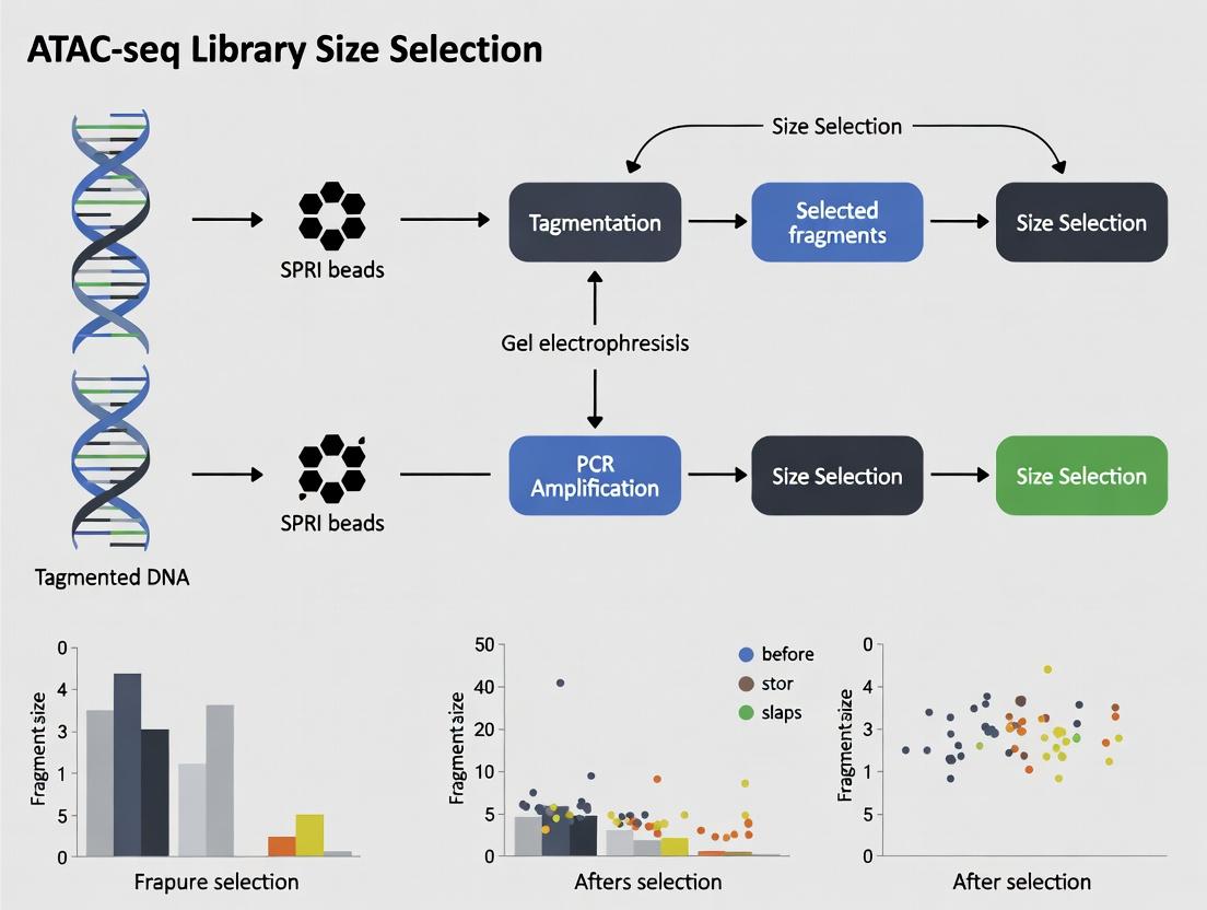
Title: ATAC-seq Experimental and Bioinformatics Workflow
Title: Tn5 Tagmentation on NFR vs. Nucleosome-Bound DNA
The Scientist's Toolkit: Research Reagent Solutions
Table 3: Essential Reagents for ATAC-seq Size Selection Research
| Item | Function | Key Consideration |
|---|---|---|
| Tn5 Transposase | Enzyme that simultaneously fragments and tags genomic DNA with adapters. | Commercial "loaded" enzymes (Nextera) ensure consistency. Activity lot-check is recommended. |
| Digitonin | A mild, cholesterol-dependent detergent used for cell membrane permeabilization. | Critical for nuclei integrity. Concentration must be titrated for each cell type. |
| SPRI (Solid Phase Reversible Immobilization) Beads | Magnetic beads for DNA clean-up and size selection via PEG/NaCl concentration. | Bead-to-sample ratio is the primary variable for controlling size selection windows. |
| PippinHT or SageELF Systems | Automated gel electrophoresis systems for high-precision DNA size selection. | Essential for generating highly defined fragment libraries (e.g., isolating pure mono-nucleosome fragments). |
| High-Sensitivity DNA Assay (e.g., Bioanalyzer, TapeStation, Fragment Analyzer) | For precise quantification and size profiling of libraries pre- and post-size selection. | Mandatory QC step to evaluate size distribution and calculate molarity for pooling. |
| Dual-Indexed PCR Primers | Amplify tagmented DNA and add unique sample indexes for multiplexing. | Using unique dual indexes (UDIs) reduces index hopping artifacts in sequencing. |
| AMPure XP or Similar Beads | The industry-standard SPRI bead for DNA purification and size selection. | Different lots can have subtle binding characteristics; maintain consistency within a study. |
Troubleshooting Guides and FAQs
Q1: After size selection, my ATAC-seq library yield is extremely low. What could be the cause and how can I fix it? A1: Low yield is common and often stems from over-tight size selection or excessive sample loss. First, verify your starting material quality (≥50,000 intact nuclei). If using SPRI beads, ensure the bead-to-sample ratio is calibrated precisely for your target size range (e.g., 0.5x to 0.8x for sub-nucleosomal fragments). Increase the elution buffer incubation time to 2 minutes at 37°C and elute in a minimal volume (15-20 µL). Consider using a low-retention tip for all bead handling steps. If using gel-based systems, stain the gel after cutting to confirm you excised the correct region.
Q2: My final library shows a high percentage of large (>1000 bp) fragments, indicating poor size selection. How can I improve resolution? A2: This indicates inefficient removal of long fragments, often from uncut or poorly digested chromatin. First, optimize the transposition step (ensure fresh DTT, correct Mg2+ concentration, and precise reaction timing). For bead-based cleanups, implement a double-sided size selection. Use a low SPRI ratio to remove large fragments (e.g., 0.55x), keep the supernatant, then add a higher ratio (e.g., 1.8x) to the supernatant to capture your target fragments (primarily <300 bp). See Table 1 for standard ratios.
Q3: My library complexity appears low (low PCR duplication rates). Could size selection be a contributing factor? A3: Yes. Overly stringent size selection that discards too much material forces excessive PCR amplification, leading to duplicate reads. To improve complexity, widen your size selection window (e.g., capture 100-700 bp instead of 150-300 bp). Use qPCR to quantify the library pre-amplification and aim to use the minimum number of PCR cycles (often 8-12). If complexity remains low, increase the number of input cells to ensure sufficient unique fragments are captured during selection.
Q4: I observe adapter dimer contamination (peak at ~120-150 bp) in my Bioanalyzer trace post-size selection. How did this happen and how do I remove it? A4: Adapter dimers form when transposed fragments are too short to have adapters ligated on both ends, allowing adapter-adapter ligation. Size selection should remove them, but if present, your lower size cut-off was too high. To resolve, perform an additional round of size selection with a slightly higher lower-bound SPRI ratio (e.g., 0.65x instead of 0.5x) to bind and remove the dimers. Always run a high-sensitivity assay (Bioanalyzer/TapeStation) after size selection and before pooling for sequencing.
Key Experimental Protocols
Protocol 1: Double-Sided SPRI Bead Size Selection for ATAC-seq
- Prepare Beads: Vortex SPRI beads thoroughly at room temperature.
- Remove Large Fragments: Add 0.55x volume of beads to purified post-ligation library. Mix thoroughly and incubate 5 min.
- Pellet on Magnet: Place on magnet until clear. Transfer supernatant (contains target fragments) to a new tube.
- Capture Target Fragments: To supernatant, add 0.95x volume of fresh beads (total ratio now ~1.5x). Mix and incubate 5 min.
- Wash: Place on magnet, discard supernatant. Wash pellet twice with 80% ethanol.
- Elute: Air dry 2 min, elute in 20 µL 10 mM Tris-HCl (pH 8.0). Incubate 2 min at 37°C, then place on magnet. Transfer eluate to new tube.
Protocol 2: PippinHT Gel-Based Size Selection for High-Precision Applications
- Prepare Cassette: Prime PippinHT 2% agarose gel cassette with SYBR Gold dye per manufacturer instructions.
- Load Sample: Mix 25-100 µL of library with internal standards. Load into a single well.
- Set Collection Windows: Program the instrument for automated size selection. Standard windows: "S1: 100-300 bp" for nucleosome-free fragments, "S2: 300-700 bp" for mono/di-nucleosome fragments.
- Run and Collect: Run at constant voltage until elution. Collect the eluted fractions into separate tubes.
- Clean Up: Concentrate and clean the eluted fractions using a 1x SPRI bead cleanup before PCR amplification.
Data Presentation
Table 1: Comparison of Size Selection Methods for ATAC-seq
| Method | Target Range | Approximate Yield Recovery | Adapter Dimer Removal | Hands-on Time | Best For |
|---|---|---|---|---|---|
| Single-Sided SPRI | >150 bp | 60-80% | Moderate | Low (30 min) | Routine, high-input projects |
| Double-Sided SPRI | 100-700 bp | 40-60% | Excellent | Medium (45 min) | High-purity applications |
| PippinHT Gel | User-defined (e.g., 100-300, 300-700 bp) | 30-50% | Excellent | High (2 hrs) | Precision studies, complex samples |
| Manual Gel Extraction | User-defined | 20-40% | Excellent | Very High (3+ hrs) | Low-throughput, when equipment is limited |
Table 2: Impact of Size Selection Window on Library Metrics
| Size Selection Window (bp) | % Reads in Peaks (Signal) | Mitochondrial Reads % (Noise) | PCR Duplication Rate | Estimated Library Complexity |
|---|---|---|---|---|
| No Selection | 15-25% | 40-60% | 50-80% | Low |
| 100-700 | 30-40% | 10-25% | 20-40% | High |
| 150-300 | 40-55% | 5-15% | 30-50% | Medium |
| 300-1000 | 20-30% | 30-40% | 40-60% | Low |
Diagrams
Title: The Core Goals of Library Size Selection Workflow
Title: Double-Sided SPRI Bead Size Selection Protocol
The Scientist's Toolkit: Research Reagent Solutions
| Item | Function in ATAC-seq Size Selection |
|---|---|
| SPRI (Solid Phase Reversible Immobilization) Beads | Magnetic beads that bind DNA fragments in a size-dependent manner in the presence of PEG and salt, enabling clean size separation and purification. |
| PippinHT Cassettes (2% Agarose) | Pre-cast, automated gel electrophoresis cassettes for high-precision, high-recovery excision of specific DNA size ranges. |
| High-Sensitivity DNA Assay Kits (e.g., Agilent Bioanalyzer HS DNA, Qubit dsDNA HS Assay) | Essential for accurate quantification and size distribution analysis of low-concentration libraries before and after size selection. |
| Low-Binding/Retention Microcentrifuge Tubes & Tips | Minimizes adsorption and loss of precious, low-yield ATAC-seq libraries during bead handling and transfer steps. |
| SYBR Gold Nucleic Acid Gel Stain | A highly sensitive stain for visualizing low-mass DNA fragments on gels during manual or automated size selection. |
| PCR Cleanup/Size Selection Beads (Different Sizes) | Some protocols use a combination of different bead sizes (e.g., SPRIselect) to fine-tune size selection boundaries more precisely. |
| Tris-EDTA (TE) Buffer or 10 mM Tris-HCl (pH 8.0) | A low-ionic-strength elution buffer that maximizes DNA recovery from SPRI beads and maintains fragment stability. |
Troubleshooting & FAQs
Q1: My final ATAC-seq library shows a broad or unexpected fragment size distribution after size selection. What are the primary causes? A1: Common causes include over-digestion by Tn5 transposase, insufficient or excessive PCR amplification, suboptimal size selection bead ratios, or sample degradation. Ensure fresh cells/nuclei, titrate Tn5 enzyme, use a minimal PCR cycle number, and accurately calculate bead-to-sample ratios for each selection step.
Q2: I aim to analyze nucleosome positioning, but my library is depleted of ~200 bp fragments (mononucleosome). How can I troubleshoot this? A2: This often indicates over-fixation, excessive lysis, or mechanical fragmentation during nuclei preparation. Use gentle lysis buffers without detergents like NP-40, avoid vortexing, and minimize centrifugation steps. Optimize the transposition time and temperature.
Q3: My library yield after double-sided size selection is too low for sequencing. What adjustments can I make? A3: Low yield can result from over-sizing. Slightly widen the fragment range selected (e.g., take 100-600 bp instead of 100-300 bp). Increase input cell number (50k-100k nuclei). Ensure AMPure XP or SPRI beads are thoroughly resuspended and at room temperature. Elute in a smaller volume (e.g., 15 µL of EB buffer).
Table 1: Optimal ATAC-seq Fragment Ranges and Their Biological Significance
| Fragment Size Range | Primary Chromatin Source | Key Application | Typical Selection Method |
|---|---|---|---|
| < 100 bp | Open chromatin, TF footprints | cis-regulatory element mapping | Double-sided SPRI beads |
| ~ 180-220 bp | Mononucleosome | Nucleosome positioning | Narrow-cut gel or beads |
| ~ 360-440 bp | Dinucleosome | Nucleosome phasing | Size selection beads |
| > 1000 bp | Large inaccessible regions | Often removed | Discarded in supernatant |
Table 2: Troubleshooting Common Size Selection Outcomes
| Observed Issue | Potential Cause | Recommended Action |
|---|---|---|
| High proportion of < 50 bp fragments | Over-transposition | Reduce Tn5 amount or incubation time |
| Lack of nucleosomal ladder | Poor nuclei isolation | Verify lysis microscopically; optimize protocol |
| Smear above 500 bp | Incomplete transposition, genomic DNA contamination | Add more Tn5; include QC step (Bioanalyzer) |
| Low library complexity | Excessive PCR cycles, low input | Use 1/4 reaction for qPCR to determine cycles; increase cell input |
Detailed Experimental Protocols
Protocol 1: Double-Sided SPRI Bead Size Selection for Open Chromatin (<100 bp)
- Post-PCR Cleanup: Bring final ATAC-seq PCR reaction to 50 µL with EB buffer. Add 50 µL (1.0x ratio) of room-temperature, resuspended AMPure XP beads. Mix and incubate 5 minutes.
- First Elution (Remove Large Fragments): Place on magnet. Transfer supernatant (containing fragments <~800 bp) to a new tube after beads clear.
- Second Bead Addition (Capture Small Fragments): To supernatant, add 20 µL (0.4x ratio) of fresh beads. Mix, incubate 5 min. Place on magnet and discard supernatant.
- Wash & Final Elution: With beads on magnet, wash 2x with 200 µL 80% ethanol. Air-dry 5 min. Elute in 20 µL EB buffer. This eluate contains fragments primarily <~800 bp.
- Third Bead Addition (Remove Very Small Fragments): To eluate, add 4 µL (0.2x ratio) of beads. Mix, incubate 5 min. Place on magnet. Transfer supernatant (now enriched for ~100-800 bp, with peak <100 bp) to a fresh tube. Quantify by Qubit.
Protocol 2: Gel-Based Extraction for Mononucleosome Enrichment (~200 bp)
- Gel Casting: Prepare a 2% low-melt agarose gel in 1x TAE.
- Loading & Run: Mix post-PCR library with loading dye. Load alongside a low-range DNA ladder (e.g., 25-500 bp). Run at 70-80V for 60-90 minutes.
- Visualization & Excision: Stain with SYBR Safe. Visualize on a blue light transilluminator to minimize DNA damage. Excise the gel slice corresponding to 150-250 bp using a clean scalpel.
- Purification: Use a gel extraction kit (e.g., QIAquick). Weigh gel slice, add 3 volumes of Buffer QG, incubate at 37°C until dissolved. Follow kit protocol, eluting in 15-20 µL EB buffer.
The Scientist's Toolkit: Essential Reagents & Materials
Table 3: Key Research Reagent Solutions for ATAC-seq Size Selection
| Reagent / Material | Function / Purpose | Example Product |
|---|---|---|
| AMPure XP/SPRI Beads | Magnetic bead-based size selection and cleanup; ratio determines cutoff. | Beckman Coulter AMPure XP |
| Low-Range DNA Ladder | Accurate sizing of fragments from 25-500 bp on gels. | NEB Low Molecular Weight DNA Ladder |
| Qubit dsDNA HS Assay | Accurate quantification of low-concentration libraries post-selection. | Thermo Fisher Qubit Assay |
| High-Sensitivity DNA Bioanalyzer Chip | Profiling fragment size distribution pre- and post-selection. | Agilent Bioanalyzer HS DNA Chip |
| Nuclei Isolation Buffer | Gently lyses plasma membrane without disrupting nucleosomes. | 10 mM Tris-HCl, 10 mM NaCl, 3 mM MgCl2, 0.1% IGEPAL CA-630 |
| Tn5 Transposase | Simultaneously fragments and tags accessible DNA with adapters. | Illumina Tagmentase, or homemade Tn5 |
Visualizations
Diagram 1: ATAC-seq Fragment Origin & Selection Workflow
Diagram 2: SPRI Bead Ratio-Based Size Selection Logic
Troubleshooting Guides & FAQs
Q1: After rigorous size selection, my ATAC-seq data shows a high proportion of reads in large, non-nucleosomal fragments (>1000 bp). What could be the cause and how can I fix it?
A: This indicates incomplete transposition or contamination with mitochondrial or genomic DNA. First, verify the integrity of your Tn5 transposase and freshness of reagents. Increase the number of AMPure XP bead clean-up cycles (e.g., double-size selection: 0.5x bead-to-sample ratio to remove large fragments, followed by 1.8x to retain nucleosomal fragments). Ensure tissue is thoroughly homogenized and nuclei are cleanly isolated. Include a DNase I treatment step post-nuclei isolation to remove contaminating DNA.
Q2: My peak calling results are inconsistent between replicates when using different size selection methods (gel vs. beads). Which method is more reliable?
A: Bead-based selection (e.g., with AMPure XP) generally provides higher reproducibility due to more precise size cut-offs and reduced manual handling. Gel extraction can introduce variability in the exact fragment range excised. Standardize on a double-sided bead selection protocol. Ensure your peak caller (e.g., MACS2) parameters are adjusted for your selected fragment range. Use --shift -75 --extsize 150 for sub-nucleosomal fragments and --nomodel for a broader range.
Q3: Following size selection for mononucleosomal fragments (~200-300 bp), motif discovery fails to identify expected transcription factor binding sites. Why?
A: Stringent selection for mononucleosomes may exclude important sub-nucleosomal fragments (<100 bp) containing transcription factor footprints. These shorter fragments are critical for precise motif localization. Re-analyze your data by including fragments down to 50 bp. Use a footprinting tool like HINT-ATAC or TOBIAS which are specifically designed to use the gradient of fragment lengths for footprint detection.
Q4: How does the ratio for SPRI bead size selection directly impact the TSS enrichment score and data interpretation?
A: The bead-to-sample ratio determines the lower size cut-off. A higher ratio (e.g., 1.8x) retains smaller fragments, increasing read density at Transcription Start Sites (TSS) due to inclusion of sub-nucleosomal fragments, thus raising TSS enrichment. A lower ratio (e.g., 0.5x) removes these fragments, decreasing TSS enrichment but potentially increasing nucleosome signal clarity.
Table 1: Impact of SPRI Bead Ratio on Key QC Metrics
| Bead Ratio | Approximate Size Retained (bp) | Effect on TSS Enrichment | Effect on Peak Number | Recommended For |
|---|---|---|---|---|
| 0.5x | >~800 | Greatly Reduces | Reduces | Removing large artifacts |
| 0.8x | ~300-800 | Moderate | Moderate | Isolating di/tri-nucleosomes |
| 1.2x | ~150-300 | Standard | Standard | Standard nucleosomal analysis |
| 1.8x | ~50-150 | Increases | Increases | Footprinting & open chromatin |
Experimental Protocols
Protocol: Two-Sided SPRI Bead Size Selection for ATAC-seq Objective: Isolate fragments primarily from 50-300 bp to enrich for open chromatin and nucleosomal fragments.
- Prepare AMPure XP Beads: Warm to room temperature for 30 min. Vortex thoroughly.
- First Selection (Remove Large Fragments): Add 0.5x volumes of beads to 1x volume of purified ATAC-seq library. Mix thoroughly by pipetting. Incubate at RT for 5 min.
- Capture Supernatant: Place on magnet. Wait until solution clears (~5 min). Transfer supernatant containing fragments <~800 bp to a new tube.
- Second Selection (Retain Target Fragments): Add 0.7x volumes of fresh beads to the supernatant (resulting in a net 1.2x ratio from original). Mix and incubate at RT for 5 min.
- Wash: Place on magnet. Wait for clearing. Discard supernatant. Keep tube on magnet, add 200 µl of 80% ethanol. Incubate 30 sec. Discard ethanol. Repeat wash. Air dry pellet for 5 min.
- Elute: Remove from magnet. Elute in 22 µl of 10 mM Tris-HCl (pH 8.0). Incubate at RT for 2 min. Place on magnet. Transfer 20 µl of eluate (containing ~50-300 bp fragments) to a new tube.
Protocol: Assessing Size Selection Efficacy via Bioanalyzer
- Use Agilent High Sensitivity DNA Kit.
- Load 1 µl of pre- and post-size selection library.
- Run on Agilent 2100 Bioanalyzer per manufacturer's instructions.
- Analyze electropherogram: Successful selection shows a strong peak ~200-300 bp (mononucleosome) and a smaller peak <100 bp (TF footprints). Large fragments (>800 bp) should be minimal.
Visualization
Title: Downstream Analysis Dependency on Size Selection
Title: Method Comparison for Fragment Isolation
The Scientist's Toolkit: Research Reagent Solutions
| Item | Function in ATAC-seq Size Selection |
|---|---|
| AMPure XP Beads | SPRI (Solid Phase Reversible Immobilization) beads for precise, bead-based size selection. Ratios determine fragment cut-offs. |
| Pippin HT or SageELF | Automated gel electrophoresis systems for high-throughput, reproducible size selection. |
| Agilent High Sensitivity DNA Kit | For precise quality control of library fragment size distribution pre- and post-selection. |
| NEB Next Ultra II DNA Library Prep Kit | Often used in conjunction with custom ATAC-seq protocols, includes buffers compatible with bead clean-up. |
| Tween-20 (0.1% in TE Buffer) | Low-EDTA TE buffer with Tween used in bead-based clean-up to improve elution efficiency and reduce fragment loss. |
| Qubit dsDNA HS Assay Kit | For accurate quantification of post-size selection libraries, as concentration changes significantly. |
Hands-On Protocols: Comparing Bead, Gel, and Automated ATAC-Seq Size Selection Methods
FAQs & Troubleshooting Guides
Q1: My final ATAC-seq library yield is consistently low after SPRI bead cleanup. What are the most common causes? A: Low yields typically result from incorrect bead-to-sample ratio, over- or under-drying of beads, or incomplete elution. Ensure you are using the correct ratio for your target size range (see Table 1). Do not over-dry the bead pellet, as this can reduce DNA elution efficiency; 2-5 minutes of air-drying is usually sufficient. Elute in a warm (37-55°C) elution buffer or nuclease-free water and ensure adequate mixing during resuspension.
Q2: How does the SPRI bead-to-sample ratio affect the size selection window in ATAC-seq? A: The ratio of SPRI bead volume to sample volume directly controls the size cutoff. A higher ratio binds smaller fragments, while a lower ratio is more selective for larger fragments. For typical ATAC-seq double-sided selection to isolate nucleosome-free fragments (e.g., < ~120 bp) and exclude large fragments (> ~800 bp), a sequential two-ratio protocol is used (see Experimental Protocol 1).
Q3: I am seeing excessive adapter dimer contamination (< 100 bp) in my final library. How can I remove this using SPRI beads? A: Adapter dimers (~50-80 bp) can be effectively removed by performing a left-side (or "high-cut") selection. Use a low bead ratio (e.g., 0.5x-0.6x) to bind and remove larger fragments, retaining the supernatant containing your target library and dimers. Then, perform a right-side selection on this supernatant with a high bead ratio (e.g., 1.8x-2.0x) to bind your target library while leaving dimers in the supernatant, which is discarded.
Q4: The size distribution of my selected library is inconsistent between replicates. What steps should I standardize? A: Key variables to standardize are: 1) Bead Mixing: Ensure beads are at room temperature and thoroughly vortexed before use. 2) Incubation Time: Adhere strictly to the 5-10 minute incubation time with beads. 3) Magnet Time: Leave samples on the magnet until the supernatant is completely clear. 4) Washing: Use freshly prepared 80% ethanol and do not disturb the bead pellet. 5) Elution Volume: Use precise, consistent elution volumes.
Q5: Can I perform SPRI bead-based size selection on a low-input ATAC-seq sample? A: Yes, but recovery may be lower. Use carrier RNA or linear acrylamide (e.g., 1 µL of 5 ng/µL) if sample input is below 10 ng. Reduce the number of washing steps to one quick wash with 80% ethanol. Elute in a smaller volume (e.g., 12-15 µL) to concentrate the sample.
Data Tables
Table 1: Common SPRI Bead Ratios for ATAC-seq Size Selection
| Target Fragment Size Range | Bead-to-Sample Ratio (v/v) | Primary Use in ATAC-seq |
|---|---|---|
| > 700 bp | 0.4x - 0.5x | "Left-side" selection to remove very large fragments & debris. |
| ~150 - 800 bp | 0.55x - 0.7x | Right-side selection after tagmentation to remove small adapter dimers. |
| < 200 bp | 1.6x - 2.0x | "Right-side" selection to concentrate nucleosome-free regions. |
| ~100 - 600 bp | 0.5x (supernatant) -> 1.8x (pellet) | Common double-sided selection protocol. |
Table 2: Troubleshooting Common SPRI Bead Selection Issues
| Problem | Potential Cause | Solution |
|---|---|---|
| Low library yield | Bead over-drying; Incorrect ratio | Limit air-dry time to <5 min. Verify ratio for input DNA mass. |
| Broad size distribution | Inconsistent incubation or mixing | Standardize incubation time to 5 min, mix by pipetting 10x. |
| High adapter dimer carryover | Inefficient left-side selection | Decrease the first bead ratio (e.g., to 0.45x) to bind more small fragments. |
| Loss of target fragments | Overly aggressive selection | Increase the second bead ratio (e.g., from 1.8x to 1.6x) to recover more material. |
| Beads carry over into eluate | Disturbed pellet during wash | Carefully remove supernatant; re-magnetize if needed. |
Experimental Protocols
Experimental Protocol 1: Double-Sided SPRI Selection for ATAC-seq
This protocol is designed to select nucleosome-free fragments (<~120 bp) while removing both adapter dimers and large genomic DNA.
Materials:
- SPRI beads (room temperature, vortexed)
- Freshly prepared 80% ethanol
- Nuclease-free water or TE buffer
- Magnetic stand suitable for tube/plate format
Method:
- First Selection (Remove Large Fragments): To the purified post-PCR ATAC-seq library, add SPRI beads at a 0.5x ratio (e.g., 25 µL beads to 50 µL sample). Mix thoroughly by pipetting at least 10 times.
- Incubate at room temperature for 5 minutes.
- Place on magnetic stand until the supernatant is clear (≥ 3 minutes). Retain the supernatant, which contains fragments smaller than the cutoff (~700 bp).
- Second Selection (Recover Target & Remove Dimers): Transfer the supernatant to a fresh tube. Add SPRI beads at a 1.8x ratio relative to the original sample volume (e.g., 90 µL beads for a 50 µL original sample). Mix thoroughly.
- Incubate at room temperature for 5 minutes.
- Place on magnetic stand until clear. Carefully discard the supernatant, which now contains adapter dimers and very small fragments.
- Wash: With the tube on the magnet, add 200 µL of 80% ethanol without disturbing the pellet. Incubate for 30 seconds, then remove and discard the ethanol. Repeat for a total of two washes.
- Dry: Briefly air-dry the bead pellet for 2-5 minutes until it shows a matte appearance with small cracks. Do not over-dry.
- Elute: Remove from the magnet. Resuspend the beads thoroughly in 22 µL of nuclease-free water or TE buffer. Incubate at 37°C for 5 minutes.
- Final Capture: Place on the magnet until clear (≥ 3 minutes). Transfer 20 µL of the clear supernatant (your size-selected library) to a fresh tube.
Diagrams
Title: Double-Sided SPRI Selection Workflow for ATAC-seq
Title: Fragment Partitioning in Double-Sided SPRI Selection
The Scientist's Toolkit: Key Research Reagent Solutions
| Item | Function in SPRI-Based ATAC-seq Size Selection |
|---|---|
| SPRI Beads (e.g., AMPure XP, SPRIselect) | Paramagnetic particles that bind DNA in a size-dependent manner in the presence of PEG and salt, enabling clean-up and selection. |
| Polyethylene Glycol (PEG) 8000 | The primary crowding agent in SPRI bead solutions; concentration dictates the effective size cutoff for binding. |
| High-Salt Binding Buffer | Provides the ionic conditions necessary for DNA to adsorb to the beads. Often included in the bead suspension. |
| 80% Ethanol (Freshly Prepared) | Used to wash the bead-bound DNA without eluting it, removing salts and other contaminants. |
| Nuclease-Free Water or TE Buffer | Low-ionic-strength solution used to elute purified DNA from the beads after washing. |
| Carrier RNA / Linear Acrylamide | Inert molecules added to stabilize and improve recovery of very low-input samples during SPRI cleanups. |
| Magnetic Stand (96-well or 1.5 mL tube) | Holds tubes/plates to immobilize SPRI beads for supernatant removal during washing and elution steps. |
This technical support center is framed within a research thesis on ATAC-seq library size selection optimization. It addresses common experimental hurdles for researchers and development professionals.
Troubleshooting Guides & FAQs
Q1: In manual gel excision for ATAC-seq, my final library yield is consistently low. What are the primary causes? A: Low yield typically stems from three areas: 1) UV Damage: Excessive UV exposure during band visualization can nick DNA. Limit exposure to <30 seconds using a hand-held UV lamp at 365 nm preferentially. 2) Incomplete Elution: For a 200 bp ATAC-seq fragment, ensure gel dissolution at 55-60°C and use adequate elution buffer volume (e.g., 3x gel volume). 3) Inaccurate Cutting: Cutting too close to the band removes buffer space; include 2-3 mm margin on each side to capture the full size distribution.
Q2: My Pippin Prep system is not recovering my target ATAC-seq fragment range (e.g., 100-200 bp). What should I check? A: First, verify the cassette specifications. For a 100-200 bp target, use a 2% agarose cassette with appropriate internal markers (e.g., 75 bp and 300 bp). Second, confirm sample loading volume does not exceed 40 µL to prevent overflow. Third, ensure the elution buffer is fresh and the collection tube is properly seated. Run a system check with a known DNA ladder.
Q3: How does contamination from primer dimers or adapter artifacts differ between the two methods? A: Manual cutting offers visual discretion; you can avoid cutting very low molecular weight regions of the gel. Pippin Prep, while automated, relies on pre-set markers. If primer dimers (<100 bp) co-migrate near your target lower bound, they may be collected. Using a Pippin "tight" collection window or a post-cleanup with AMPure beads at a higher ratio (e.g., 1.8x) can mitigate this.
Q4: Which method provides better reproducibility for high-throughput ATAC-seq studies? A: The Pippin Prep provides superior reproducibility (see Table 1) due to automated, software-defined size gates, minimizing inter-user variability. Manual cutting's reproducibility is highly dependent on technician skill.
Data Presentation
Table 1: Comparison of Manual Gel Cutting vs. Pippin Prep for ATAC-seq Size Selection
| Parameter | Manual Gel Cutting | Pippin Prep System | Notes |
|---|---|---|---|
| Average Yield Recovery | 40-60% | 60-80% | Yield highly dependent on fragment size and user skill for manual method. |
| Size Accuracy (SD) | ± 20-40 bp | ± 5-15 bp | Pippin Prep uses internal markers for precise calibration. |
| Hands-on Time | 30-45 minutes | 5-10 minutes | Manual time includes gel casting, cutting, and extraction steps. |
| Reproducibility (CV) | 15-25% | <5% | Coefficient of Variation (CV) for fragment size distribution. |
| Typical Cost per Sample | Low (agarose, buffers) | High (proprietary cassettes) | |
| Optimal Fragment Range | Broad (>50 bp) | Defined windows (e.g., 100-200 bp) | Pippin excels at tight library size selection. |
Experimental Protocols
Protocol 1: Manual Gel Extraction for ATAC-seq Libraries
- Prepare Gel: Cast a 2-3% high-resolution agarose gel (e.g., MetaPhor) in 1x TAE with a well-suited DNA ladder (e.g., 25 bp increment ladder).
- Load & Run: Mix purified ATAC-seq library with 6x loading dye. Load alongside ladder. Run at 5-6 V/cm until sufficient separation (∼45 min).
- Visualize & Excise: Stain with SYBR Gold or GelGreen. Using a clean scalpel, quickly excise the gel slice containing your target nucleosomal fragment band (e.g., ∼190 bp mononucleosome). Minimize gel mass.
- Extract DNA: Use a commercially available gel extraction kit. Dissolve gel slice at 55°C, bind DNA to silica membrane, wash, and elute in 15-30 µL nuclease-free water or TE buffer.
Protocol 2: Size Selection with Pippin Prep for ATAC-seq
- Instrument Setup: Power on Pippin Prep. Select protocol "2% Agarose Cassette, 100-200 bp" (or appropriate range).
- Sample Preparation: Dilute your ATAC-seq library in elution buffer to a total volume of ≤ 40 µL. Load into the designated sample well on the Pippin cassette.
- Run: Insert the cassette into the instrument and start the run. Duration is approximately 2 hours.
- Recovery: Post-run, retrieve the collection tube containing your size-selected library. Quantify via qPCR or fluorometry.
The Scientist's Toolkit
Key Research Reagent Solutions for ATAC-seq Size Selection
| Item | Function in Experiment |
|---|---|
| High-Sensitivity DNA Assay (e.g., Qubit dsDNA HS) | Accurately quantifies low-concentration DNA post-extraction. |
| SYBR Gold Nucleic Acid Gel Stain | A highly sensitive, low-background stain for visualizing faint DNA bands. |
| Certified Low-Range Ultra Agarose | Provides superior resolution for fragments in the 50-500 bp range. |
| Pippin Prep Cassette (2%, 100-200 bp) | Pre-cast agarose cassette with internal fluorescent markers for automated collection. |
| SPRIselect Beads | Used for post-gel cleanup and buffer exchange, with size selection capabilities via ratio adjustment. |
| DNA LoBind Tubes | Minimizes adsorption of low-input ATAC-seq libraries to tube walls. |
Visualizations
Troubleshooting Guides & FAQs
Magnetic Rack Method FAQs
Q1: My bead slurry appears heterogeneous or has formed a precipitate. What should I do? A1: Vortex the bead stock thoroughly for at least 30 seconds, or until the mixture is completely homogeneous with no visible aggregates. Always prepare and use beads at room temperature to prevent PEG and salt precipitation. If a precipitate persists, briefly spin the tube and transfer the homogeneous supernatant to a new tube.
Q2: I am observing low recovery of my target fragments after SPRI selection. What are the primary causes? A2: The most common causes are:
- Incorrect bead-to-sample ratio: Ensure the ratio is calibrated for your target size range. Standard ratios are: 0.6X-0.8X for large fragment removal (>~500 bp), 1.0X-1.2X for standard selection (~200-500 bp), and 1.5X-1.8X for small fragment selection (<~200 bp).
- Incomplete mixing: Mix beads and sample thoroughly by pipetting or vigorous vortexing to ensure uniform binding.
- Ethanol contamination in the elution buffer: Ensure the 80% ethanol wash is thoroughly removed. Let the bead pellet air-dry for 2-5 minutes with the tube lid open before elution.
- Elution buffer volume too high: Use the minimum recommended elution volume (e.g., 15-25 µL for a 50 µL starting sample) to maximize concentration.
Q3: I see carryover of small fragments when trying to select for larger ATAC-seq libraries. How can I improve size selectivity? A3: Perform a double-sided (or double-spin) size selection. First, use a low bead ratio (e.g., 0.55X) to bind and discard very large fragments. Recover the supernatant, then add more beads to achieve a higher final ratio (e.g., 1.2X) to bind your target fragments. This narrows the size distribution. See the workflow diagram below.
Experimental Protocols
Detailed Protocol: Double-Sided SPRI Size Selection for ATAC-seq Libraries
This protocol is optimized for selecting nucleosome-free (< 200 bp) and mononucleosome (~200-500 bp) fragments in ATAC-seq.
- Prepare Beads: Vortex SPRI beads (e.g., AMPure XP, SPRIselect) thoroughly until no pellet is visible. Ensure beads are at room temperature.
- First Binding (Remove Large Fragments): To your purified ATAC-seq library in a low-EDTA TE buffer or water, add SPRI beads at a 0.55X ratio (e.g., 55 µL beads to 100 µL sample). Mix thoroughly by pipetting 10-15 times.
- Incubate: Incubate at room temperature for 5 minutes.
- Separate: Place the tube on a magnetic rack. Allow the solution to clear completely (≈5 minutes). Do not discard the supernatant.
- Transfer Supernatant: Carefully transfer the cleared supernatant (containing fragments smaller than the cut-off) to a new tube.
- Second Binding (Recover Target Fragments): To the supernatant, add fresh SPRI beads at a volume calculated to achieve a final combined ratio of 1.2X. Since you already added a 0.55X volume, calculate the addition: (1.2X total - 0.55X already added) = 0.65X (e.g., add 65 µL beads to the supernatant from step 5). Mix thoroughly.
- Incubate & Separate: Incubate for 5 minutes. Place on the magnetic rack until clear.
- Wash: With the tube on the magnet, remove and discard the supernatant. While on the magnet, add 200 µL of freshly prepared 80% ethanol. Incubate for 30 seconds, then remove and discard the ethanol. Repeat for a total of two washes.
- Dry: Let the bead pellet air-dry for 2-5 minutes with the tube lid open. Do not over-dry.
- Elute: Remove the tube from the magnet. Add your desired volume of elution buffer (e.g., 10mM Tris-HCl, pH 8.0-8.5) to the bead pellet. Mix thoroughly by pipetting. Incubate at room temperature for 2 minutes.
- Recover Eluate: Place the tube back on the magnetic rack. Once clear, transfer the eluate (containing size-selected DNA) to a new tube. Proceed to quantification.
Table 1: Comparison of Gel-Free Size Selection Methods for ATAC-seq
| Parameter | Single-Ratio SPRI | Double-Sided SPRI | Magnetic Rack with Size-Specific Beads |
|---|---|---|---|
| Typical Size Range | Broad (e.g., 150-700 bp) | Narrow (e.g., 200-500 bp) | Tunable, based on bead chemistry |
| Average Yield | 60-80% of input | 40-60% of input | 50-70% of input |
| Hands-On Time | ~15 minutes | ~25 minutes | ~20 minutes |
| Cost per Sample | Low | Medium | Medium-High |
| Best for ATAC-seq | Quick cleanup, broad profiling | High-resolution mapping (nucleosome positions) | Specialized applications (e.g., selecting very small fragments) |
| Primary Limitation | Broad size distribution | Lower yield | Proprietary bead kits, cost |
Table 2: Recommended SPRI Bead Ratios for ATAC-seq Fragment Selection
| Target Fragment Pool | Bead Ratio (Sample Volume:X) | Approximate Size Cut-off (bp) | Purpose in ATAC-seq |
|---|---|---|---|
| Large Fragment Removal | 0.5X - 0.7X | Removes > 500-700 bp | Eliminate di-/tri-nucleosomes & large artifacts |
| Nucleosome-Free + Mono-nucleosome | 1.0X - 1.3X | Retains ~150-600 bp | Standard library for open chromatin & footprinting |
| Nucleosome-Free Only | 1.6X - 2.0X | Retains ~< 200 bp | Isolate regions of very high accessibility |
| Stringent Mono-nucleosome | Double-sided: 0.55X + 0.65X | Selects ~200-500 bp | Clean nucleosome positioning data |
Visualizations
Title: Double-Sided SPRI Size Selection Workflow
Title: Troubleshooting Low Yield in SPRI Workflow
The Scientist's Toolkit: Research Reagent Solutions
Table 3: Essential Materials for SPRI-based ATAC-seq Size Selection
| Item | Function | Key Considerations |
|---|---|---|
| SPRI Magnetic Beads (e.g., AMPure XP, SPRIselect) | Bind DNA in high PEG/salt buffer; size-dependent binding efficiency. | Critical: Must be room temp & homogeneous. Lot-to-lot variability may require ratio re-calibration. |
| Magnetic Rack | Separates bead-bound DNA from solution. | Use a rack designed for your tube size (e.g., PCR strips, 1.5 mL tubes). Ensure strong, even magnetic field. |
| Molecular Biology Grade Ethanol (80%) | Washes away salts and contaminants while keeping DNA bound to beads. | Must be freshly prepared from pure ethanol and nuclease-free water to prevent dilution errors. |
| Low-EDTA TE Buffer or Nuclease-Free Water | Elution buffer. Releases purified DNA from beads. | TE buffer stabilizes DNA. EDTA can inhibit downstream enzymatic steps if concentration is too high. |
| PEG/NaCl Buffer | Provided with beads. Creates binding conditions. | Ensure it is mixed thoroughly with the bead stock. |
| PCR Tubes/Plates & Non-Binding Tips | Sample handling. | Prevents loss of material and sample adhesion to tube walls. |
Technical Support Center
Troubleshooting Guides & FAQs
Q1: During magnetic bead-based size selection on the liquid handler, my final ATAC-seq library yields are consistently low. What could be the cause?
A: Low yields often stem from incomplete bead mixing or inaccurate supernatant removal. Ensure the method includes a "bead resuspension" step with vigorous pipette mixing (e.g., 10 cycles of 200 µL aspirate/dispense at medium speed) after the initial binding and before each wash. Verify that the liquid handler's magnetic module engagement time is sufficient for full bead capture (typically 2-5 minutes) before supernatant removal. Calibrate the Z-height for supernatant aspiration to be close to the bead pellet without disturbing it.
Q2: I observe high size variability between replicates in my high-throughput ATAC-seq run. How can I improve reproducibility?
A: This typically points to inconsistent bead-to-sample ratios or temperature fluctuations. First, calibrate all pipetting channels for the viscous bead solutions. Implement a protocol where beads are temperature-equilibrated to room temperature (RT) for 30 minutes before use. Use fresh, high-quality 80% ethanol for washes. Most critically, use a dual-size selection "bead ratio" method. See the standardized protocol below.
Q3: My liquid handler is producing errors when transferring SPRISelect/AMPure XP beads. What specific adjustments should I make?
A: Bead solutions are viscous and can foam. Program the liquid handler to:
- Use wider-bore tips if available.
- Reduce aspirate and dispense speeds to 50-75% of normal.
- Include a 5-10 second post-aspiration delay to allow beads to settle in the tip.
- Include a tip touch-off on the vial wall after dispensing to ensure full delivery.
- Pre-wet tips by aspirating and dispensing the bead solution once before the transfer step.
Q4: After automated size selection, my ATAC-seq libraries have excessive adapter dimer contamination (peak ~100 bp). How do I resolve this?
A: Adapter dimers indicate insufficient size selection stringency. You must optimize the bead volume ratios for your target fragment range. For ATAC-seq targeting nucleosomal fragments (~150-500 bp), a common dual-ratio is:
- First selection (remove large fragments): Sample Volume : Bead Volume = 1 : 0.5 (e.g., 50 µL sample + 25 µL beads). Keep supernatant.
- Second selection (remove small fragments): Supernatant Volume : Bead Volume = 1 : 1.2 (e.g., 75 µL supernatant + 90 µL beads). Discard supernatant. Always validate new ratios with a Bioanalyzer/TapeStation before full runs.
Experimental Protocol: Automated Dual-Bead Ratio Size Selection for ATAC-seq
Objective: Reproducible isolation of nucleosome-protected DNA fragments (primarily mono-, di-, tri-nucleosome) using a liquid handler.
Materials:
- Purified, adapter-ligated ATAC-seq library in 50 µL EB buffer.
- SPRISelect or AMPure XP beads.
- Freshly prepared 80% Ethanol.
- Nuclease-free water or EB buffer for elution.
- Liquid Handler (e.g., Beckman Coulter Biomek i7, Hamilton STAR, Tecan Fluent) with magnetic module and Peltier-cooled deck (set to 4°C for bead storage).
Method:
- Equilibration: Place beads at RT for 30 min. Vortex thoroughly for >1 min until homogenous.
- First Selection (Remove Large Fragments & Cleanup):
- Program the handler to combine 50 µL library with 25 µL beads (0.5x ratio) in a deep-well plate. Mix thoroughly for 5 min.
- Engage magnets. Wait 5 min for clear separation.
- Transfer 75 µL of supernatant to a new well.
- Second Selection (Remove Small Fragments & Adapter Dimers):
- To the 75 µL supernatant, add 90 µL of beads (1.2x ratio). Mix thoroughly for 5 min.
- Engage magnets. Wait 5 min.
- Aspirate and discard supernatant completely.
- Ethanol Washes (2x):
- With magnets engaged, add 200 µL of 80% ethanol. Incubate 30 sec. Aspirate and discard fully. Repeat.
- Air-dry beads for 5-7 min (programmable). Do not over-dry.
- Elution:
- Disengage magnets. Add 22 µL of EB buffer or nuclease-free water. Mix thoroughly for 2 min.
- Engage magnets for 2 min.
- Transfer 20 µL of clear eluate to a fresh output plate. Store at -20°C.
- QC: Analyze 1 µL on a High Sensitivity Bioanalyzer or TapeStation.
Table 1: Comparison of Manual vs. Automated Size Selection Performance (Hypothetical Data from Aggregated Studies)
| Metric | Manual Protocol (n=12) | Automated Liquid Handler (n=12) | Improvement |
|---|---|---|---|
| Average Yield (nM) | 8.5 ± 3.2 | 9.1 ± 0.8 | +7%, 68% lower CV |
| Fragment Size Peak (bp) | 285 ± 45 | 290 ± 12 | 73% lower CV |
| Adapter Dimer Contamination (% of total) | 4.1 ± 2.8% | 1.5 ± 0.5% | Reduced by 64% |
| Hands-on Time per 96 Samples | ~6 hours | ~1 hour | 83% reduction |
| Inter-replicate CV of Library Complexity | 18% | 7% | 61% reduction |
Table 2: Recommended Bead Ratios for Different ATAC-seq Target Ranges
| Target Fragment Range | Primary Goal | First Bead Ratio (Sample:Beads) | Second Bead Ratio (Supernatant:Beads) | Expected Elution Volume |
|---|---|---|---|---|
| Nucleosomal (150-500 bp) | Remove long fragments, then short dimers | 1 : 0.5 | 1 : 1.2 | 20-22 µL |
| Short Fragments (<300 bp) | Enrich for accessible regions | 1 : 0.8 | 1 : 0.9 | 17-20 µL |
| Long Fragments (>500 bp) | Enrich for bound/closed regions | 1 : 0.3 | 1 : 1.5 | 22-25 µL |
Visualizations
Title: Automated Dual-Bead Ratio Size Selection Workflow
Title: Thesis Framework: ATAC-seq Size Selection Methods Research
The Scientist's Toolkit: Research Reagent Solutions
Table 3: Essential Materials for Automated ATAC-seq Size Selection
| Item | Function | Critical Note for Automation |
|---|---|---|
| SPRISelect or AMPure XP Beads | Size-selective binding of DNA fragments based on PEG/NaCl concentration. | Most critical. Must be vortexed thoroughly and temp-equilibrated. Viscosity affects pipetting accuracy. |
| Fresh 80% Ethanol | Washing away salts and contaminants while DNA is bound to beads. | Prepare fresh daily. Old ethanol can carry over water and reduce yield. |
| Nuclease-free EB Buffer (10mM Tris-Cl, pH 8.5) | Eluting purified DNA from beads. | Lower salt content vs. TE buffer improves downstream sequencing. |
| Magnetic Ring-Stand or Plate | Holding samples during bead separation on the liquid handler deck. | Must be compatible with the deck layout and magnet module height. |
| Low-Binding, V-Bottom 96-Well Plates | Reaction vessels for binding, washing, and elution. | V-bottom improves bead pellet consistency and supernatant removal. |
| Wide-Bore or Filtered Liquid Handler Tips | Accurate transfer of viscous bead solutions and prevention of aerosol contamination. | Reduces bead clogging and foam formation during transfers. |
| Bioanalyzer HS DNA Chips or TapeStation HSD1000 Screens | Quality control of final library size distribution and quantification. | Essential for validating and tuning automated method parameters. |
Troubleshooting Guides & FAQs
Q1: My Bioanalyzer/TapeStation trace after size selection shows a significant fraction of adapter dimers (~120-130 bp). What went wrong and how can I salvage the library? A: This indicates incomplete purification during size selection with SPRI beads. The ratio of beads to sample was likely too high, pulling small fragments like dimers. To salvage: 1) Repeat size selection using a more stringent bead ratio. For a target range of 200-600 bp, use a double-sided selection (e.g., 0.5x beads to remove large fragments, keep supernatant; then add beads to supernatant at 1.8x ratio to capture target fragments). 2) Re-run the QC. If dimers persist, treat the library with a heat-labile duplex-specific nuclease (DSN) to selectively digest double-stranded adapter dimers before a final SPRI clean-up.
Q2: The library yield measured by Qubit is acceptable (>10 nM), but qPCR for library quantification shows very low concentration, and subsequent sequencing fails. What is the cause? A: This discrepancy suggests a high proportion of your library molecules are not amplifiable, often due to: 1) Over-digestion with Tn5 transposase, leading to very short fragments with damaged ends. 2) Carryover of contaminants from the ATAC-seq reaction (e.g., detergents, salts) that inhibit polymerase during qPCR. Solution: Re-purify the library using a column-based clean-up kit designed to remove small fragments and salts. Perform a serial dilution of your library in the qPCR reaction to check for inhibition. Ensure the Tn5 reaction was not incubated for an excessively long time.
Q3: My fragment distribution is correct, but the Bioanalyzer peak is broad and "fuzzy," not sharp. Does this affect sequencing, and what does it indicate? A: A broad, fuzzy peak indicates high heterogeneity in fragment sizes within your selected range. While it may not preclude sequencing, it can lead to uneven coverage. It often results from: 1) Over-fragmented chromatin due to excessive transposase activity or over-sonication (if used). 2) Inefficient size selection where the cutoff slopes are not sharp. Solution: For future preps, titrate the transposase amount or digestion time. For the current library, if yield allows, you can perform a tighter size selection using a Pippin Prep or BluePippin system with more precise cassette-based gel cutting.
Q4: The qPCR amplification curve has a late Ct (e.g., >25 cycles when using 1:10,000 dilution of a 1 nM library standard), but the final yield is normal. Is this a problem? A: A late Ct by itself is not problematic if the final yield after PCR amplification is sufficient. It likely indicates that the starting concentration of amplifiable fragments post-selection is low, but the library has good PCR efficiency. Monitor the melting curve for a single peak to ensure specificity. The key metric is the calculated concentration from the standard curve. If the final yield (Qubit) matches the qPCR-predicted yield post-amplification, the library is valid.
Table 1: Expected QC Metrics for ATAC-seq Libraries Post Size-Selection
| QC Method | Target Metric | Suboptimal Result | Implied Issue |
|---|---|---|---|
| Bioanalyzer 2100 (High Sensitivity DNA) | Sharp peak in 200-600 bp range. DV200 > 70%. | Peak < 150 bp; broad smear. | Adapter dimer contamination; over-fragmentation. |
| TapeStation (High Sensitivity D1000) | Peak(s) in target range. Smear value low. | High smear value; secondary peaks. | Incomplete size selection; gDNA contamination. |
| Qubit dsDNA HS Assay | Yield > 1.5 ng/µL in 15 µL elution. | Yield < 0.5 ng/µL. | Loss during SPRI steps; low cell input. |
| qPCR (Library Quant) | Ct difference from standard < 2 cycles. | Ct greatly delayed or undetected. | Inhibitor carryover; low adapter ligation efficiency. |
Table 2: Troubleshooting SPRI Bead Ratios for Double-Sided Size Selection
| Target Insert Size | First Bead Ratio (Remove Large Fragments) | Keep Supernatant | Second Bead Ratio (Capture Target) | Discard Supernatant |
|---|---|---|---|---|
| 150-300 bp | 0.3x - 0.4x | Yes | 1.3x - 1.5x | Yes |
| 200-600 bp | 0.5x - 0.6x | Yes | 1.6x - 1.8x | Yes |
| 300-1000 bp | 0.7x - 0.8x | Yes | 1.1x - 1.3x | Yes |
Detailed Experimental Protocols
Protocol 1: Post-Selection QC via Bioanalyzer High Sensitivity DNA Assay
- Prepare Gel-Dye Mix: Vortex the High Sensitivity DNA dye concentrate for 10 sec. Pipette 25 µL of filtered gel matrix into a spin filter. Add 1 µL of dye. Centrifuge at 2240 x g for 10 min. Protect from light.
- Prime Chip: Load 9 µL of gel-dye mix into the well marked "G". Close chip priming station. Press plunger until held by clip. Wait 30 sec. Release clip. Wait 5 sec. Slowly pull plunger back.
- Load Samples: Pipette 5 µL of marker into each sample and ladder well. Load 1 µL of High Sensitivity DNA ladder into the ladder well. Load 1 µL of each purified library (diluted 1:5 in nuclease-free water) into separate sample wells.
- Run Chip: Vortex chip for 1 min. Place in Agilent 2100 Bioanalyzer. Run within 5 minutes.
Protocol 2: Library Quantification via SYBR Green qPCR
- Prepare Standards: Dilute a commercial library standard (e.g., Kapa Biosystems) to 10 pM in 10 mM Tris-HCl, pH 8.0. Perform a 1:10 serial dilution to create a 6-point standard curve from 10 pM to 0.001 pM.
- Prepare Samples: Dilute the test library 1:10,000 and 1:100,000 in the same buffer.
- Set Up Reactions: For each reaction, mix: 10 µL 2X SYBR Green qPCR Master Mix, 2 µL primer mix (10 µM each forward/reverse), 4 µL diluted standard or sample, 4 µL nuclease-free water.
- Run qPCR Program: 95°C for 5 min; 40 cycles of: 95°C for 30 sec, 60°C for 45 sec (acquire fluorescence); followed by a melting curve analysis.
Visualization: Workflow & Relationships
Title: ATAC-seq Post-Selection QC Workflow & Decision Points
Title: Post-Selection QC Problem Diagnosis & Resolution Pathways
The Scientist's Toolkit: Essential Research Reagent Solutions
Table 3: Key Reagents & Materials for Post-Selection QC
| Item Name | Supplier Examples | Function in QC |
|---|---|---|
| SPRIselect Beads | Beckman Coulter, Kapa Biosystems | Magnetic beads for precise double-sided size selection and clean-up. |
| Agilent High Sensitivity DNA Kit | Agilent Technologies | Provides gel-dye matrix and chips for fragment analysis on Bioanalyzer. |
| D1000/High Sensitivity D1000 ScreenTapes | Agilent Technologies | Pre-cast gels for fragment analysis on TapeStation systems. |
| Kapa Library Quantification Kit (SYBR) | Roche | Optimized qPCR master mix and primers for accurate quant of Illumina libraries. |
| Qubit dsDNA HS Assay Kit | Thermo Fisher Scientific | Fluorometric dye for specific, sensitive quantification of double-stranded DNA. |
| Nuclease-free Water | Various | Critical for all dilutions to prevent degradation or enzymatic interference. |
| Pippin Prep System & Cassettes | Sage Science | Automated gel electrophoresis for high-precision, reproducible size selection. |
| Heat-Labile DSN Enzyme | e.g., MCB | Selective digestion of double-stranded adapter dimers to clean up libraries. |
Solving Common ATAC-Seq Size Selection Problems: A Troubleshooting Guide for Researchers
Diagnosing and Fixing Low Library Yield After Size Selection
Troubleshooting Guide & FAQs
Q1: What are the primary causes of low yield after ATAC-seq size selection? A: Low yield typically stems from four main categories: 1) Insufficient input material, 2) Excessive DNA loss during bead-based size selection, 3) Inefficient library amplification, and 4) Suboptimal fragment distribution prior to selection. The table below quantifies common failure points and their impact.
Table 1: Common Causes and Impact on Post-Selection Yield
| Cause | Typical Yield Loss | Diagnostic Check |
|---|---|---|
| Insufficient Cell Input | 60-80% | Quantify nuclei count post-lysis. Target >50,000 nuclei. |
| Overly Stringent Bead Ratios | 40-70% | Re-run bioanalyzer on pre- and post-selection material. |
| PCR Cycle Over-/Under-Amplification | 30-60% | Run qPCR side-reaction to determine optimal cycles. |
| Tn5 Transposition Efficiency | 50-90% | Assess fragment size pre-amplification. Smear should center <1kb. |
| Post-PCR Cleanup Loss | 20-40% | Use lower bead-to-sample ratio (e.g., 0.8x) for cleanup. |
Q2: How can I optimize bead-based double-sided size selection to maximize yield? A: The standard protocol using SPRI beads often uses a 0.5x left-side (to remove large fragments) and a 0.8x right-side (to recover the target fragment) selection. Yield loss occurs at both steps. An optimized protocol is detailed below.
Experimental Protocol: Optimized Double-Sided SPRI Selection for ATAC-seq
- Pre-amplified Library Preparation: Generate the ATAC-seq library following standard transposition and a limited-cycle (e.g., 5-cycle) PCR amplification.
- First Size Selection (Remove Large Fragments):
- Bring a 50 µL PCR reaction to room temperature. Vortex SPRI beads thoroughly.
- Add 0.45x sample volume of SPRI beads (22.5 µL) instead of 0.5x. Mix thoroughly by pipetting.
- Incubate at RT for 5 min. Place on magnet until supernatant is clear.
- Transfer all supernatant (target ~72.5 µL) to a new tube. Do not discard.
- Second Size Selection (Recover Target Fragments & Remove Primer Dimer):
- To the supernatant, add 0.10x original sample volume of SPRI beads (5 µL, for a total ratio of 0.55x). Mix thoroughly.
- Incubate at RT for 5 min. Place on magnet until clear.
- Discard this supernatant.
- With tube on magnet, wash beads twice with 200 µL of 80% ethanol.
- Air-dry beads for 2-3 min. Elute in 22 µL of 10 mM Tris-HCl (pH 8.0).
- This recovers fragments primarily between ~150-800 bp.
- Final Amplification: Perform 3-5 additional qPCR cycles on the size-selected material to generate sufficient sequencing library. Clean up with a 0.8x bead ratio.
Q3: My post-selection bioanalyzer trace shows the correct fragment range but very low concentration. What should I check? A: This indicates successful selection but massive material loss. First, verify pre-selection concentration and profile. Then, systematically check:
- Bead shelf-life and storage: Use fresh beads, stored at 4°C.
- Ethanol concentration: Use fresh 80% ethanol.
- Elution buffer: Use Tris-HCl, pH 8.0-8.5, not water. Ensure it's pre-heated to 55°C for elution.
- Elution time: Allow elution to proceed for a full 2 minutes off the magnet before pelleting beads.
Diagram 1: Diagnostic workflow for low ATAC-seq library yield.
The Scientist's Toolkit: Key Research Reagent Solutions
Table 2: Essential Materials for ATAC-seq Size Selection Optimization
| Item | Function & Role in Yield Recovery |
|---|---|
| SPRIselect Beads | Magnetic beads for size-selective binding of DNA. Critical for reproducible ratio-based selection. |
| Fresh 80% Ethanol | Essential for effective bead washing without overdrying. Old or incorrectly diluted ethanol causes loss. |
| High-Sensitivity DNA Assay (e.g., Qubit dsDNA HS) | Accurate quantification of low-yield libraries post-selection. Avoids overestimates from spectrophotometry. |
| High-Sensitivity Bioanalyzer/ TapeStation | Provides fragment distribution profile pre- and post-selection to diagnose size-selection efficacy. |
| Nuclease-Free Water & Tris-EDTA | Elution buffers. Slightly basic (pH 8.0-8.5) Tris improves DNA recovery from beads vs. water. |
| Real-Time PCR (qPCR) Master Mix | Enables precise determination of optimal library amplification cycles to prevent over-/under-amplification. |
| TD Buffer and Tn5 Transposase | The core transposition enzyme mix. Batch variability directly impacts initial fragment distribution and yield. |
Diagram 2: ATAC-seq workflow with critical yield loss points highlighted.
This technical support center is framed within a broader thesis research on ATAC-seq library size selection methods. It addresses the common issue of broad or bimodal fragment distributions in next-generation sequencing (NGS) libraries, which can severely impact data quality and experimental outcomes.
FAQs & Troubleshooting Guides
Q1: What are the primary causes of a broad fragment size distribution after AMPure or SPRI bead cleanup? A1: The main causes are:
- Incorrect bead-to-sample ratio: Deviating from the recommended ratio (e.g., 0.8x for size selection) drastically alters the size cutoff.
- Improper bead mixing or incubation: Incomplete homogenization of the bead slurry or inconsistent incubation times lead to non-specific binding.
- Ethanol carryover during wash steps: Residual ethanol inhibits elution and can degrade DNA, causing smearing.
- Over-drying or under-drying beads: Over-drying makes DNA difficult to elute, reducing yield and skewing distribution; under-drying leaves ethanol.
- Variable sample composition: High salt, EDTA, or other contaminants in the sample buffer can interfere with bead binding kinetics.
Q2: Why does my Bioanalyzer/TapeStation trace show a pronounced bimodal distribution (e.g., peaks at ~150bp and ~300bp) for my ATAC-seq library? A2: In ATAC-seq, a bimodal distribution often indicates:
- Incomplete tagmentation: The Tn5 transposase may have partially reacted, leading to a population of under-tagmented fragments (larger peak) and properly tagmented fragments (smaller peak). This is frequently due to suboptimal cell input, reaction time, or transposase concentration.
- Over-digestion during tagmentation: Excessive Tn5 can lead to over-fragmentation and very short fragments that may be lost or appear as a separate, very low peak.
- Inefficient size selection: The chosen double-sided SPRI bead cleanup (e.g., 0.5x/1.5x ratios) failed to adequately remove the large or small fragment populations.
Q3: How can I correct a broad size distribution before sequencing? A3: Corrective actions include:
- Re-perform size selection: Precisely re-optimize double-sided SPRI bead cleanups. Use the table below for guidance.
- Gel extraction: For critical or stubborn samples, excise the desired size range from an agarose or PippinHT gel for maximum purity.
- Re-assess input DNA quality: Check the integrity of your starting material (e.g., nuclei prep for ATAC-seq) on a gel. Degraded input causes broad outputs.
- Re-optimize enzymatic steps: For ATAC-seq, titrate the Tn5 enzyme amount and tagmentation time.
Data Presentation: SPRI Bead Ratios for Size Selection
Table 1: Common SPRI Bead Ratios for Fragment Size Selection
| Bead Ratio (Sample Volume) | Primary Target Size Range | Removes | Typical Use Case |
|---|---|---|---|
| 0.5x | >~700 bp | Small fragments (<~100-150 bp) | "Right-side" selection, post-PCR cleanup. |
| 0.6x | >~500 bp | Small fragments (<~100 bp) | Enrich for larger fragments. |
| 0.8x | >~300 bp | Small fragments (<~50 bp) | Standard post-ligation cleanup. |
| 1.0x | >~150 bp | Very small fragments | Standard post-PCR cleanup. |
| 1.5x | >~50 bp | Primers, adaptor dimers | "Left-side" selection, dimer removal. |
| Double-Sided (0.5x/1.5x) | ~150-700 bp | <150 bp and >700 bp | ATAC-seq library isolation. |
| Double-Sided (0.6x/1.2x) | ~200-500 bp | <200 bp and >500 bp | Focused selection for exome/panel sequencing. |
Table 2: Troubleshooting Broad/Bimodal Distributions
| Observed Issue | Likely Cause | Recommended Corrective Action |
|---|---|---|
| Very broad smear (>500bp range) | Bead ratio far from optimal, ethanol carryover, degraded input. | Re-cleanup with precise bead ratio. Ensure fresh 80% ethanol, proper aspiration, and adequate air-dry time (5-7 mins). |
| Bimodal peak (ATAC-seq: ~150bp & ~300bp) | Incomplete tagmentation or inefficient size selection. | Optimize Tn5 concentration and time. Perform a stringent double-sided SPRI selection (e.g., 0.4x/1.6x). |
| Low yield after size selection | Over-drying beads, selecting too narrow a range, low input. | Do not over-dry beads (>10 mins). Elute in warmer buffer (e.g., 30-37°C) and re-assess input quantity/quality. |
| Persistent adapter dimer peak (~128bp) | Inadequate removal during left-side (high-ratio) selection. | Increase bead ratio for the left-side selection (e.g., from 1.5x to 1.8x). Use fresh beads. |
Experimental Protocols
Protocol 1: Optimized Double-Sided SPRI Bead Cleanup for ATAC-seq Libraries This protocol aims to isolate the nucleosome-free (<200bp) and mononucleosome (~200-600bp) fragments.
- First Cleanup (Remove Large Fragments): Bring post-PCR ATAC-seq library to 50µL in nuclease-free water. Add 0.5x volume (25µL) of well-resuspended SPRI beads. Mix thoroughly by pipetting. Incubate at room temperature (RT) for 5 minutes.
- First Separation: Place on a magnet. Wait 5 minutes until the supernatant is clear. Transfer the supernatant (containing fragments <~700 bp) to a new tube. Discard the beads (with bound large fragments).
- Second Cleanup (Remove Small Fragments/Dimers): To the supernatant, add 1.5x volume of the original supernatant volume (75µL) of fresh SPRI beads. Mix thoroughly. Incubate at RT for 5 minutes.
- Second Separation: Place on magnet. Wait 5 minutes. Keep the beads. While on the magnet, wash twice with 200µL of freshly prepared 80% ethanol. Wait 30 seconds per wash, then aspirate carefully.
- Elution: Air-dry beads for 5-7 minutes (no longer). Remove from magnet. Elute in 22µL of 10mM Tris-HCl (pH 8.0-8.5). Mix well. Incubate at RT for 2 minutes. Place on magnet for 2 minutes. Transfer 20µL of eluate to a new tube. Quantify.
Protocol 2: Troubleshooting Incomplete Tagmentation in ATAC-seq To address the bimodal distribution caused by under-tagmentation.
- Titrate Tn5: Set up a series of 50,000 nuclei reactions with varying Tn5 enzyme amounts (e.g., 2.5µL, 5µL, 7.5µL of commercial enzyme) while keeping time constant (30 mins, 37°C).
- Purify DNA: Stop reaction with SDS and Proteinase K. Purify DNA via a standard MinElute column cleanup.
- Analyze Distribution: Run 1µL of purified DNA on a High Sensitivity DNA Bioanalyzer chip or TapeStation. The optimal condition will show a strong nucleosomal ladder with a dominant sub-200bp peak and diminished high-molecular-weight genomic DNA smear.
Visualizations
Title: Troubleshooting Workflow for Fragment Distribution Issues
Title: ATAC-seq Library Prep with Key Size Selection Step
The Scientist's Toolkit: Research Reagent Solutions
Table 3: Essential Materials for Size Selection & Troubleshooting
| Item | Function in Protocol | Key Consideration |
|---|---|---|
| SPRI (Ampure/SPRIselect) Beads | Magnetic beads for size-based DNA purification and selection. | Lot-to-lot consistency is critical. Always resuspend thoroughly before use. |
| Fresh 80% Ethanol | Wash buffer to remove salts and contaminants without eluting DNA. | Must be prepared fresh weekly from pure ethanol to prevent acetic acid formation. |
| Low TE Buffer or Nuclease-Free Water | Elution buffer for purified DNA. | TE (pH 8.0-8.5) stabilizes DNA; water is compatible with downstream enzymatic steps. |
| High Sensitivity DNA Assay (Bioanalyzer/TapeStation/Qubit) | Accurate quantification and size profiling of libraries. | Essential for diagnosing distribution issues before sequencing. |
| Tn5 Transposase (Commercial Kit or Home-made) | Enzyme for simultaneous fragmentation and tagging in ATAC-seq. | Concentration and activity are the primary determinants of fragment size distribution. |
| PippinHT or BluePippin System | Automated, high-resolution gel-based size selection. | Gold standard for obtaining tight, specific fragment distributions when beads fail. |
| PCR Cleanup Kit (MinElute, NucleoSpin) | For DNA purification after tagmentation or to clean up re-amplification. | Useful for intermediate cleanup steps to remove enzymes and buffers. |
Optimizing Bead-to-Sample Ratios for Specific Target Ranges and Sample Types
Troubleshooting Guides & FAQs
Q1: My post-bead purification ATAC-seq libraries show excessive adapter dimer contamination (~128 bp peak). What is the likely cause and solution? A: This typically indicates an insufficient bead-to-sample ratio during the cleanup, failing to remove small fragments. For standard SPRI beads, the recommended ratio to remove fragments <~100 bp is 0.8x. If dimers persist, increase the ratio incrementally to 0.9x or 1.0x. Ensure beads are at room temperature and thoroughly mixed with the sample for 5 minutes.
Q2: I am losing my large (>1000 bp) nucleosome-associated fragments after bead selection. How can I optimize recovery? A: Loss of large fragments suggests the bead ratio is too high, pulling down all fragments and not allowing the large ones to remain in the supernatant. For target enrichment of large fragments (e.g., mono-/di-nucleosome regions), use a dual-sided selection. First, use a low ratio (e.g., 0.5x) to remove small fragments; transfer supernatant to new tube. Then, add beads to the supernatant to achieve a high combined ratio (e.g., 1.8x) to capture the desired large fragments. See Table 1.
Q3: How should I adjust bead ratios for low-input or degraded samples (e.g., from FFPE tissue)? A: Degraded/fragmented samples have a shifted size distribution towards smaller fragments. Use a slightly higher initial bead ratio (e.g., 0.9x) for the first cleanup to stringently remove short artifacts. For the final library cleanup, a standard 1.0x or 1.1x ratio is often sufficient. Always perform a Bioanalyzer/TapeStation check post-cleanup.
Q4: My bead cleanup efficiency seems variable between experiments. What critical steps am I missing? A: Key troubleshooting steps:
- Bead Settling: Do not let beads settle for more than 2-3 minutes before discarding supernatant. Over-settling increases non-specific binding.
- Ethanol Wash: Use fresh 80% ethanol. Do not disturb the bead pellet during washes.
- Elution Buffer: Elute in a low-salt, low-EDTA buffer (e.g., 10 mM Tris-HCl, pH 8.0). Ensure it is pre-warmed to 55°C and incubated with beads for 5 min.
- Bead Lot Variability: Different SPRI bead lots may have varying binding kinetics. Re-calibrate ratios with a test sample when using a new lot.
Q5: For targeting a specific library insert size range (e.g., 150-500 bp), what bead strategy is best? A: A sequential selection with two different bead ratios is most effective. See the detailed protocol below and Table 1.
Data Presentation
Table 1: Recommended SPRI Bead Ratios for ATAC-seq Size Selection
| Target Insert Size Range | Sample Type / Goal | Initial Bead Ratio (Remove Small Fragments) | Second Bead Ratio (Capture Target) | Combined Ratio | Expected Yield Impact |
|---|---|---|---|---|---|
| 100-300 bp | Standard Cytoplasmic/Nuclear Extract | 0.45x (discard beads) | 1.35x (keep beads) | 1.8x | Moderate (30-50%) |
| 150-500 bp | Enrich for Nucleosome-Associated Fragments | 0.5x (discard beads) | 1.3x (keep beads) | 1.8x | Low-Moderate (20-40%) |
| >500 bp | Long Fragment Recovery | 0.4x (discard beads) | 1.6x (keep beads) | 2.0x | Low (10-25%) |
| Remove Adapter Dimers | All, Post-PCR Cleanup | 0.8x (keep beads) | N/A | 0.8x | Minimal (<5% loss of >200 bp) |
| High Recovery | Precious/Low-Input | 0.6x (discard beads) | 1.2x (keep beads) | 1.8x | High (60-80%) |
Ratios are volumetric (µL of beads per µL of sample). Based on standard AMPure/SPRI bead binding kinetics at room temperature in PEG/NaCl buffer.
Experimental Protocols
Protocol: Dual-Sided SPRI Bead Selection for Target Range 150-500 bp
This protocol is framed within thesis research on optimizing ATAC-seq library selection to improve signal-to-noise in chromatin accessibility profiles.
Materials: Purified post-amplification ATAC-seq library, SPRI beads (e.g., AMPure XP), fresh 80% ethanol, TE buffer, magnetic rack, pipettes.
Method:
- Vortex SPRI beads thoroughly to ensure an even suspension.
- First Selection (Remove Small Fragments):
- Transfer 50 µL of library to a clean tube.
- Add 0.5x volume (25 µL) of room-temperature SPRI beads. Mix thoroughly by pipetting 10 times.
- Incubate at room temperature for 5 minutes.
- Place on magnetic rack for 2 minutes or until supernatant clears.
- Transfer 72.5 µL of the supernatant (contains fragments >~150 bp) to a new tube. Discard the tube with beads.
- Second Selection (Capture Target Range):
- To the 72.5 µL supernatant, add 1.3x volume (94.3 µL) of SPRI beads. Mix thoroughly.
- Incubate at room temperature for 5 minutes.
- Place on magnetic rack for 2 minutes. Carefully remove and discard supernatant.
- Wash:
- With beads on the magnet, add 200 µL of fresh 80% ethanol. Incubate for 30 seconds, then remove supernatant.
- Repeat wash once. Ensure all ethanol is removed.
- Air-dry bead pellet for 2-3 minutes (do not over-dry).
- Elute:
- Remove from magnet. Add 22 µL of pre-warmed (55°C) TE or 10 mM Tris-HCl (pH 8.0).
- Mix well and incubate at room temperature for 2 minutes.
- Place on magnet for 2 minutes.
- Transfer 20 µL of clear supernatant containing size-selected library to a new tube.
- Quantity using Qubit and analyze fragment distribution on Bioanalyzer.
Mandatory Visualization
Dual-Sided SPRI Bead Selection Workflow
Fragment Size Selection by Sequential Bead Ratios
The Scientist's Toolkit: Research Reagent Solutions
| Item | Function in Bead-Based Size Selection |
|---|---|
| SPRI Beads (e.g., AMPure XP, SPRIselect) | Paramagnetic carboxyl-coated particles that bind DNA in PEG/NaCl buffer. The binding capacity and size cutoff are controlled by the bead-to-sample ratio. |
| Polyethylene Glycol (PEG) 8000 / NaCl Buffer | The chemical environment provided by the beads. High concentrations promote binding of smaller DNA fragments. Critical for reproducible ratio effects. |
| Fresh 80% Ethanol (in nuclease-free water) | Wash solution to remove salts and PEG without eluting the bound DNA from the beads. Must be fresh to prevent dilution by absorbed water. |
| Low-EDTA TE Buffer or 10 mM Tris-HCl (pH 8.0) | Elution buffer. Low EDTA is crucial for downstream enzymatic steps (e.g., re-amplification, sequencing). Pre-warming to 55°C increases elution efficiency. |
| Magnetic Separation Rack | Holds tubes to separate bead-bound DNA from supernatant. A rack with strong, even magnets ensures clean supernatant removal. |
| High-Sensitivity DNA Assay (e.g., Qubit dsDNA HS) | Accurate quantification of low-concentration, size-selected libraries. Fluorometric assays are essential as spectrophotometers cannot detect adapter dimers. |
| High-Sensitivity DNA Bioanalyzer/TapeStation | Capillary electrophoresis to visually assess library fragment size distribution before and after bead selection, enabling ratio optimization. |
| PCR Tubes/Low-Bind Microcentrifuge Tubes | Minimizes DNA loss due to adsorption to tube walls, which is significant for low-input samples post-selection. |
Mitigating Adapter Dimer Contamination and Small Fragment Carryover
Troubleshooting Guides & FAQs
Q1: What are the primary consequences of adapter dimer contamination in my ATAC-seq library? A1: Adapter dimers (~120-150 bp) consume sequencing cycles and reagents, drastically reducing the fraction of usable reads from true nucleosome-derived fragments, compromising library complexity, sequencing depth, and peak-calling sensitivity.
Q2: How does small fragment carryover from pre-size-selection steps affect downstream data? A2: Carryover of sub-nucleosomal fragments (<100 bp) leads to:
- Increased background noise by mapping to open chromatin regions without nucleosome periodicity.
- Inflated duplication rates due to limited diversity of these small fragments.
- Skewed insert size distribution, interfering with nucleosome positioning analyses.
Q3: What are the most effective strategies to prevent adapter dimer formation during library preparation? A3:
- Use of Purified, Bead-Linked Transposase: Ensures precise tagmentation with minimal free adapter.
- Optimized Adapter Concentration: Titrate adapter input to the minimum required for efficient tagmentation.
- Post-Tagmentation Cleanup: Implement a double-sided SPRI bead cleanup (e.g., 0.5x followed by 1.0x ratio) to remove free adapters and very small fragments before PCR.
- Qubit/QC Diligence: Quantify libraries before and after PCR. A large amplification jump may indicate adapter dimer amplification.
Q4: My post-size-selected library still shows a small peak at ~120-150 bp on the Bioanalyzer. What should I do? A4: Perform a second, stringent size selection. For a target insert size of 150-800 bp, use a dual-SPRI bead protocol (e.g., 0.55x to discard small fragments, then 0.8x to retain the supernatant containing large fragments). Re-run the QC.
Q5: Are there post-sequencing bioinformatic tools to salvage data from libraries with adapter dimer contamination?
A5: Yes, but prevention is superior. Tools like cutadapt or fastp can aggressively trim adapter sequences. However, this leads to significant data loss and cannot recover the library complexity lost to dimer sequencing.
Experimental Protocol: Dual-SPRI Bead Size Selection for ATAC-seq This protocol is optimized to minimize dimer (<150 bp) and small fragment (<100 bp) carryover.
- Prepare Libraries: Complete tagmentation and PCR amplification of your ATAC-seq library.
- First Bead Addition (Remove Small Fragments): Vortex SPRI beads thoroughly. Add a 0.55x volume of beads to the PCR product (e.g., 55 μL beads to 100 μL sample). Mix thoroughly by pipetting.
- Incubate & Separate: Incubate at room temperature for 5 minutes. Place on a magnet stand until the supernatant is clear (~5 minutes).
- Retain Supernatant: Carefully transfer the supernatant (containing desired fragments) to a new tube. Discard the beads-bound small fragments and dimers.
- Second Bead Addition (Recover Target Fragments): Add a 0.8x volume of fresh SPRI beads to the supernatant (e.g., 80 μL beads to 100 μL supernatant). Mix thoroughly.
- Incubate & Wash: Incubate 5 minutes. Place on magnet. Discard supernatant. With tube on magnet, wash beads twice with 200 μL of 80% ethanol.
- Elute: Air-dry beads for ~5 minutes, then elute DNA in nuclease-free water or TE buffer (e.g., 22 μL). Transfer to a clean tube.
- Quality Control: Assess library profile using a High Sensitivity Bioanalyzer or TapeStation.
Summary of Size Selection Method Efficacy Table 1: Comparison of common size selection methods for ATAC-seq.
| Method | Target Range | Adapter Dimer Removal | Small Fragment Removal | Yield Impact | Hands-on Time |
|---|---|---|---|---|---|
| Single-Sided SPRI Beads (e.g., 1.0x) | >~200 bp | Poor | Moderate | High Loss | Low |
| Dual-Sided SPRI Beads (e.g., 0.55x/0.8x) | ~150-800 bp | Excellent | Excellent | Moderate Loss | Moderate |
| Gel Extraction | Precise (user-defined) | Excellent | Excellent | High Loss | High |
| Pippin HT/SageELF | Very Precise | Excellent | Excellent | Moderate Loss | Low |
Title: Workflow for Dual-Sided SPRI Bead Size Selection
The Scientist's Toolkit: Key Research Reagent Solutions
Table 2: Essential materials for mitigating contamination in ATAC-seq.
| Item | Function & Relevance |
|---|---|
| High-Quality, Bead-Linked Transposase (e.g., Tn5) | Minimizes free adapter in reaction, the primary source of adapter dimers. |
| SPRI (Solid Phase Reversible Immobilization) Beads | Enable rapid, buffer-based size selection. The core reagent for dual-sided cleanups. |
| High-Sensitivity DNA QC Kit (Bioanalyzer/TapeStation) | Essential for visualizing library profile and detecting low-level adapter dimer peaks. |
| Magnetic Stand | For efficient separation of SPRI beads from solution during cleanups. |
| qPCR Kit with SYBR Green | For accurate quantification of amplifiable library fragments, less influenced by dimer contamination than Qubit. |
| Low-Binding Microcentrifuge Tubes | Prevents DNA loss during critical cleanup and size selection steps. |
| Precision Pipettes & Filter Tips | Ensure accurate bead-to-sample ratios, which are critical for reproducible size selection. |
Technical Support Center
Troubleshooting Guides & FAQs
Q1: My ATAC-seq library yield from low-input cells (e.g., < 10,000 cells) is extremely low or undetectable after PCR amplification. What could be the cause and how can I fix it? A: This is often due to excessive loss of material during bead-based cleanups or insufficient transposition. Ensure you are using a Th5 transposase optimized for low input. Omit all intermediate cleanups; perform a single-sided SPRI bead cleanup after PCR, using a lower bead-to-sample ratio (e.g., 0.5x to 0.8x) to retain smaller fragments. Increase the number of PCR cycles judiciously (e.g., up to 15-18 cycles), but include a qPCR side-reaction to avoid over-amplification. Using carrier DNA or RNA during precipitation steps is not recommended as it interferes with library quantification.
Q2: When using frozen tissue sections, I observe high mitochondrial DNA contamination in my ATAC-seq libraries. How can I mitigate this?
A: High mitochondrial signal is common in samples where nuclear integrity is compromised. Optimize your nuclei isolation protocol: use a gentle but effective homogenization method (e.g., Dounce homogenizer) in a chilled, isotonic lysis buffer with non-ionic detergents (e.g., NP-40, Triton X-100). Filter the homogenate through a 40-μm cell strainer and perform a low-speed centrifugation (500 rcf) to pellet nuclei without pelleting intact cells. A sucrose gradient centrifugation can further purify nuclei. During data analysis, tools like ArchR or MACS2 can help filter out mitochondrial reads bioinformatically.
Q3: For clinical specimens like FFPE tissue or fine-needle aspirates, the DNA after transposition appears highly fragmented (< 50 bp). Is this usable? A: Extensive fragmentation is a challenge with degraded clinical samples. While standard ATAC-seq size selection (aiming for the nucleosomal ladder) may fail, you can shift your strategy. Target the "sub-nucleosomal" fraction (e.g., < 120 bp) which may represent transcription factor footprints. Use a double-sided SPRI bead cleanup (e.g., 0.5x left-side to remove very small fragments, then 1.2x right-side to capture the footprint-sized DNA). Note that library complexity will be lower; sequence deeply and use analysis pipelines designed for open chromatin data from degraded samples.
Q4: After size selection with SPRI beads, my library size distribution is incorrect (missing the nucleosomal periodicity). What went wrong? A: Inaccurate bead calibration is the most common issue. SPRI bead behavior is sensitive to PEG concentration, salt concentration, temperature, and fragment size distribution. Precisely control the sample-to-bead ratio and temperature (use a thermal cycler set to 20°C). For challenging samples, consider replacing a single bead-based size selection with a combination of a Pippin HT or BluePippin system (for precise excision of the nucleosomal ladder, e.g., 150-300 bp) followed by a bead cleanup. Always run a high-sensitivity Bioanalyzer or TapeStation trace before and after size selection.
Q5: I am comparing ATAC-seq data from fresh vs. frozen PBMCs, and the cluster separation in my analysis appears driven by sample type, not biology. A: This indicates a strong batch effect from protocol adaptation. Key variables are nuclei isolation efficiency and transposition time. For frozen cells, extend the lysis time slightly to ensure complete cell membrane disruption while keeping nuclei intact. Standardize the transposition reaction time and temperature rigorously across all sample types (e.g., 30 min at 37°C). Include a pilot experiment where you process an aliquot of fresh cells both immediately and after freezing to identify the step introducing bias. Use batch correction tools like Harmony or Seurat's integration methods in downstream analysis.
Table 1: Performance Metrics of ATAC-seq Protocol Adaptations
| Sample Type | Recommended Cell/Nuclei Input | Typical Library Yield (after PCR) | Optimal Size Selection Range | Key Quality Metric (Bioanalyzer) | Success Rate |
|---|---|---|---|---|---|
| Low Input (<10k cells) | 500 - 10,000 cells | 2 - 15 nM | 150 - 500 bp | Clear peak ~200 bp (mononucleosome) | 60-75% |
| Frozen Tissue (e.g., mouse cortex) | 50 mg tissue | 10 - 30 nM | 150 - 700 bp | Visible nucleosomal laddering | 70-85% |
| Clinical FFPE | 5-10 slides (5μm) | 1 - 10 nM | < 120 bp (footprint) or wide (100-300 bp) | Smear, no ladder | 40-60% |
| Fresh PBMCs (Standard) | 50,000 cells | > 50 nM | 150 - 700 bp | Strong nucleosomal ladder | >90% |
Table 2: SPRI Bead Ratio Effects on Fragment Retention
| Bead-to-Sample Ratio (v/v) | Approximate Fragment Size Retained (bp) | Primary Use Case in ATAC-seq |
|---|---|---|
| 0.5x | > 300 bp | Remove large fragments/gDNA |
| 0.8x | > 150 bp | Standard cleanup post-PCR |
| 1.0x | > 100 bp | Post-transposition cleanup |
| 1.5x | > 50 bp | Concentrate all fragments |
| 0.5x + 0.8x (Double-sided) | ~150-300 bp | Precise nucleosome selection |
Experimental Protocols
Detailed Protocol: ATAC-seq from Low-Input Cryopreserved Cells
- Thawing & Washing: Rapidly thaw cryopreserved cells in a 37°C water bath. Immediately transfer to 10 mL pre-warmed culture medium. Centrifuge at 300 rcf for 5 min at 4°C. Resuspend gently in 1 mL cold PBS.
- Nuclei Isolation & Counting: Lyse cells in 50 μL of cold ATAC-seq Lysis Buffer (10 mM Tris-HCl pH 7.4, 10 mM NaCl, 3 mM MgCl2, 0.1% IGEPAL CA-630). Incubate on ice for 3 min. Immediately add 1 mL of Wash Buffer (10 mM Tris-HCl pH 7.4, 10 mM NaCl, 3 mM MgCl2) and invert. Pellet nuclei at 500 rcf for 10 min at 4°C. Resuspend pellet in 50 μL of Transposase Mix (from commercial kit, e.g., Illumina Tagment DNA TDE1 Kit). Count nuclei using a hemocytometer under trypan blue stain if input is critical.
- Tagmentation: For inputs below 10,000 nuclei, scale the transposase reaction volume proportionally down to 20 μL. Incubate at 37°C for 30 min in a thermal mixer with agitation (1000 rpm). Immediately purify using MinElute PCR Purification Kit (do not use beads).
- Library Amplification: Amplify the purified tagmented DNA in a 25 μL PCR reaction using NEBNext High-Fidelity 2X PCR Master Mix and 1.25 μM of custom Ad1 and Ad2 primers. Run a 5 μL qPCR side reaction to determine additional cycles needed:
C(t) = -1/log2(1+E) * log2(Input) + C(t)(1). Typically, for <10k cells, add 5-7 cycles to the standard 5-cycle pre-qPCR count. - Size Selection & Cleanup: Combine PCR reactions. Perform a double-sided SPRI bead cleanup: Add 0.5x bead volume, incubate 5 min, separate. Transfer supernatant to a new tube. Add 0.8x bead volume to the supernatant (final ratio 1.3x), incubate, and wash. Elute in 22 μL EB buffer. Quantify by Qubit HS dsDNA assay and analyze fragment distribution on Bioanalyzer HS DNA chip.
Diagrams
ATAC-seq Workflow for Challenging Samples
Size Selection Strategy Decision Tree
The Scientist's Toolkit: Research Reagent Solutions
Table 3: Essential Materials for ATAC-seq with Challenging Samples
| Item | Function | Example Product/Catalog Number |
|---|---|---|
| Tn5 Transposase, Low-Input Optimized | Enzyme for simultaneous fragmentation and adapter tagging; low-input versions reduce loss. | Illumina Tagment DNA TDE1 Kit (20034197) |
| Nuclei Isolation Buffer (with detergent) | Lyse cell membrane while keeping nuclear membrane intact; critical for tissue. | 10x Genomics Nuclei Isolation Kit (1000494) or homemade (Tris-NaCl-MgCl2-IGEPAL) |
| Magnetic SPRI Beads | For DNA cleanup and size selection; require precise calibration. | Beckman Coulter AMPure XP (A63880) |
| High-Sensitivity DNA Assay Kit | Accurate quantification of low-yield libraries. | Thermo Fisher Qubit dsDNA HS Assay (Q32851) |
| High-Sensitivity DNA Bioanalyzer Kit | Assess library fragment size distribution and quality. | Agilent High Sensitivity DNA Kit (5067-4626) |
| PCR Purification Columns | Alternative to beads for minimal loss post-tagmentation. | Qiagen MinElute PCR Purification Kit (28004) |
| qPCR Master Mix with High GC | For accurate determination of additional PCR cycles needed. | KAPA SYBR Fast qPCR Kit (KK4601) |
| Programmable Gel Electrophoresis System | For highly precise size selection when beads fail. | Sage Science Pippin HT (PPI-1001) |
Benchmarking Size Selection Techniques: Data Quality, Reproducibility, and Cost Analysis
Technical Support Center
Troubleshooting Guides
Issue 1: Low Yield After Bead-Based Size Selection
- Problem: Final library concentration is too low for sequencing.
- Root Cause: Inaccurate bead-to-sample ratio or over-washing.
- Solution: Precisely calibrate bead volume using a standard curve of your target fragment size. Use fresh 80% ethanol for washes and ensure beads are fully dried before elution. Validate with a high-sensitivity assay (e.g., Qubit, Bioanalyzer).
Issue 2: Smear or Broad Size Distribution on Bioanalyzer TapeStation
- Problem: Library does not show a tight, defined peak.
- Root Cause (Gel): Over-exposure to UV light during excision or improper gel melting.
- Solution: Minimize UV exposure time (<30 sec). Ensure complete dissolution of gel slice at the correct temperature and with sufficient agitation.
- Root Cause (Beads): Incorrect bead ratio leading to inefficient size cutoff.
- Solution: Re-optimize the double-sided SPRI (Solid Phase Reversible Immobilization) bead ratio. For ATAC-seq, a typical sequential selection might use 0.5x beads (remove large fragments) followed by 1.8x beads (capture target fragments).
Issue 3: High Duplicate Rate in Sequencing Data
- Problem: High PCR duplicate levels indicating low library complexity.
- Root Cause: Over-amplification due to insufficient starting material post-size selection.
- Solution: Increase input cells for ATAC-seq reaction. Use the minimum number of PCR cycles. If using beads, re-check the recovery efficiency of your target fragment range (e.g., nucleosomal fragments for ATAC-seq).
Issue 4: Inconsistent Fragment Size Between Technical Replicates
- Problem: High variability in the median insert size.
- Root Cause (Gel): Manual excision variability.
- Solution: Use a pre-cast gel with precision markers and a sharp, clean scalpel. Have the same operator perform all excisions.
- Root Cause (Beads): Inconsistent bead mixing or temperature fluctuations.
- Solution: Use a calibrated multichannel pipette for bead dispensing. Perform all incubations at a controlled room temperature on a consistent rotator/mixer.
Frequently Asked Questions (FAQs)
Q1: For ATAC-seq, which method provides better reproducibility for nucleosomal ladder selection? A: Bead-based methods generally offer higher technical reproducibility due to automation potential and less manual intervention. Gel extraction can be more variable but allows visual verification of the nucleosomal ladder excision.
Q2: Can I use a single bead ratio for all my ATAC-seq libraries? A: It requires optimization. While a standard double-sided selection (e.g., 0.5x/1.8x) works for many, the optimal ratio can vary with initial transposition efficiency and cell type. Always run a quality control check (e.g., Bioanalyzer) on the final library.
Q3: How does the choice of size selection method impact the detection of transcription factor binding sites (TFBS) in ATAC-seq? A: Inefficient selection of mononucleosomal fragments (<~150 bp) can lead to under-sampling of open chromatin regions, reducing sensitivity for TFBS detection. Bead-based methods with optimized ratios can more consistently capture these small fragments compared to gel extraction, which may have lower recovery for small DNA fragments.
Q4: My bead-cleaned libraries show primer dimer contamination. What should I do? A: Perform a more stringent clean-up. If you used a 1.0x bead ratio to capture your library, try a 1.1x or 1.2x ratio to more effectively exclude primer dimers (<100 bp). Alternatively, run the product on a gel and excise the correct size band.
Data Quality Metrics Comparison
Table 1: Quantitative Comparison of Beads vs. Gel Extraction for ATAC-seq Size Selection
| Metric | Bead-Based Selection | Gel Extraction & Purification |
|---|---|---|
| Median Insert Size Consistency (CV%) | 3-5% (High) | 8-15% (Moderate) |
| Library Yield Recovery (100-300 bp) | 60-75% | 40-60% |
| Hands-on Time (per 8 samples) | ~30 minutes | ~90 minutes |
| Technical Replicate Correlation (R²) | 0.98 - 0.99 | 0.92 - 0.97 |
| PCR Duplicate Rate (Typical) | 15-30%* | 20-40%* |
| Cost per Sample | Low | Moderate |
| Automation Potential | High (Liquid handlers) | Low |
*Dependent on sequencing depth and starting cell number.
Experimental Protocols
Protocol A: Double-Sided SPRI Bead Selection for ATAC-seq Libraries
- Prepare Libraries: Complete transposition and PCR amplification of ATAC-seq libraries.
- First Bead Addition (Remove Large Fragments): Add SPRI beads at a 0.5x ratio (bead volume = 0.5 x sample volume) to the pooled PCR reactions. Mix thoroughly.
- Incubate & Separate: Incubate at RT for 5 min. Place on magnet until clear. Retain supernatant, which contains fragments ≤ ~1000 bp.
- Second Bead Addition (Capture Target Fragments): Transfer supernatant to a new tube. Add beads at a 1.8x ratio to the supernatant volume. Mix.
- Wash: Incubate 5 min, place on magnet, discard supernatant. Wash beads twice with 80% ethanol.
- Elute: Air-dry beads 5 min. Elute DNA in nuclease-free water or TE buffer. Quantify.
Protocol B: Manual Agarose Gel Extraction for ATAC-seq Size Selection
- Prepare Gel: Load entire ATAC-seq library on a 2-3% high-resolution agarose gel (e.g., MetaPhor). Include a low-mass DNA ladder.
- Electrophoresis: Run gel at low voltage (5 V/cm) until sufficient separation of the nucleosomal ladder (mono-, di-, tri-nucleosome bands).
- Visualize & Excise: Stain with SYBR Gold or GelGreen. Visualize under low-energy blue light (minimize UV). Precisely excise the region containing mononucleosomal fragments (~100-250 bp).
- Purify: Use a silica-membrane spin column kit designed for gel extraction. Follow manufacturer's instructions, ensuring the gel slice is fully dissolved.
- Elute & Quantify: Elute in a minimal volume (e.g., 15 µL). Measure concentration with a fluorometric assay.
Visualizations
Title: Comparative Workflow for ATAC-seq Size Selection Methods
Title: Decision Tree for Selecting Beads vs. Gel Extraction
The Scientist's Toolkit
Table 2: Essential Research Reagent Solutions for ATAC-seq Size Selection
| Item | Function | Example Brand/Type |
|---|---|---|
| SPRI Magnetic Beads | Bind DNA based on size in PEG/NaCl buffer; enable double-sided clean-up. | AMPure XP, SPRIselect, KAPA Pure Beads |
| High-Sensitivity DNA Assay | Accurately quantify low-concentration libraries post-selection. | Qubit dsDNA HS Assay, Picogreen |
| Fragment Analyzer | Assess library size distribution and purity pre-sequencing. | Agilent Bioanalyzer (HS DNA chip), Fragment Analyzer systems |
| Low-Range DNA Ladder | Accurate sizing of nucleosomal fragments on gels. | 25 bp or 50 bp DNA ladders |
| High-Resolution Agarose | Provide superior separation of small DNA fragments (50-500 bp). | MetaPhor, NuSieve GTG Agarose |
| Gel Extraction Kit | Purify DNA from excised agarose gel slices. | QIAquick Gel Extraction, Monarch Gel Extraction |
| Nuclease-Free Water | Elution buffer that prevents degradation of libraries. | Various molecular biology grade suppliers |
| Ethanol (80%) | Wash buffer for SPRI beads; must be freshly prepared. | Molecular biology grade ethanol diluted with nuclease-free water |
Technical Support Center: Troubleshooting ATAC-seq Library Size Selection
This support center addresses common issues arising from ATAC-seq library size selection methods, a critical variable in our broader research thesis. Proper size selection is paramount for enriching nucleosome-free fragments (open chromatin) and directly impacts downstream sequencing metrics.
FAQs and Troubleshooting Guides
Q1: After bead-based size selection, my library yield is extremely low. What could be the cause? A: Low yield often stems from over-zealous size selection. The target range for ATAC-seq (e.g., < 100 bp for mononucleosome-free fragments) is small.
- Troubleshoot: Verify your bead-to-sample ratio. A higher ratio removes more small fragments. Precisely calibrate using a bioanalyzer/tapestation trace of your post-Tn5 reaction pool. Consider using a more gradual double-sided size selection (e.g., removing both large >700bp and very small <50bp fragments) to retain the target peak.
- Protocol (Double-Sided SPRI Bead Cleanup):
- Bring sample to 50 µL with nuclease-free water.
- Add 30 µL (0.6X ratio) of room-temperature SPRI beads to bind and remove large fragments (>~700bp). Incubate 5 min, separate on magnet, and keep supernatant.
- Transfer supernatant to a new tube. Add 20 µL (0.4X ratio) of fresh SPRI beads to the supernatant (total effective ratio = 1.0X). This will bind the desired fragments and remove salts and primers.
- Incubate 5 min, wash twice with 80% ethanol, elute in suitable buffer.
Q2: My sequencing data shows unexpectedly high duplication rates (>50%). Is this related to size selection? A: Yes, excessively narrow size selection can drastically limit library complexity, leading to high PCR duplication. An incomplete removal of very small adapter-dimer fragments can also consume sequencing reads without biological information.
- Troubleshoot: Run a high-sensitivity assay (Bioanalyzer, TapeStation, or gel) before and after size selection. Ensure adapter-dimer peaks (~80-100bp) are absent. If the post-selection profile is very tight, consider slightly widening the selected size range to recover more unique fragments.
Q3: The fragment length distribution from sequencing shows a weak or absent nucleosomal periodicity. Could size selection be the issue? A: Absolutely. Overly aggressive selection of short fragments (<100bp) will enrich only the nucleosome-free region but discard all nucleosome-associated fragments, obliterating periodicity. Conversely, poor removal of large fragments (>1000bp) can create a noisy background.
- Troubleshoot: Optimize selection windows. A common successful range is to capture fragments from ~50-800 bp. This retains the open chromatin peak and several nucleosome ladders. Re-evaluate the concentration of nuclei used in the assay; over-digestion can also fragment nucleosomal DNA.
Q4: My mapping rates to the reference genome are lower than expected. How might library preparation influence this? A: While mapping rates primarily depend on sequencing quality and reference completeness, library artifacts from suboptimal size selection can contribute. Excessive primer or adapter dimers (very short fragments) will generate reads that fail to map.
- Troubleshoot: Implement a strict cleanup post-PCR to remove primers and dimers. Use a size selection method that reliably excludes fragments below 50 bp. Verify the quality of the transposase complex and ensure it is not overloaded.
Table 1: Comparison of Sequencing Metrics Across Different Size Selection Methods in ATAC-seq.
| Size Selection Method (Cutoff Range) | Average Mapping Rate (%) | Duplication Rate (%) | Fraction of Reads in Peaks (FRiP) | Nucleosomal Periodicity Visibility |
|---|---|---|---|---|
| SPRI Beads, Single 0.5X (Removes >~300bp) | 88.5 | 45.2 | 0.22 | Weak |
| SPRI Beads, Double 0.6X/1.0X (~50-700bp) | 92.7 | 28.1 | 0.35 | Strong |
| Pippin Prep Gel, Tight (100-300bp) | 90.1 | 65.8 | 0.40 | Absent (Open chromatin only) |
| Pippin Prep Gel, Wide (100-700bp) | 93.5 | 30.5 | 0.38 | Strong |
| No Size Selection | 85.3 | 52.4 | 0.18 | Very Weak/Noisy |
The Scientist's Toolkit: Essential Reagents for ATAC-seq Size Selection
Table 2: Key Research Reagent Solutions for Library Size Selection.
| Item | Function in ATAC-seq Size Selection |
|---|---|
| SPRI (Solid Phase Reversible Immobilization) Beads | Polyethylene glycol (PEG)/salt solution that binds DNA by size; the cornerstone of bead-based clean-up and selection. |
| Pippin Prep or BluePippin System (Sage Science) | Automated gel electrophoresis system for high-precision, reproducible DNA size selection. |
| High-Sensitivity DNA Assay Kits (Agilent/PerkinElmer) | For accurate quantification and sizing of libraries pre- and post-selection using capillary electrophoresis. |
| Low EDTA TE Buffer | Optimal elution and storage buffer for size-selected libraries, preserving integrity for sequencing. |
| NucleoMag NGS Clean-up and Size Select Beads | Magnetic beads with optimized buffers for standardized NGS library clean-up and size selection. |
Experimental Workflows and Diagrams
Diagram 1: Workflow from Size Selection to Key Metrics
Diagram 2: Decision Path for High Duplication Issues
Technical Support & Troubleshooting Center
Frequently Asked Questions (FAQs)
Q1: After size selection, my ATAC-seq libraries yield fewer peaks than expected. What could be the cause? A: This is commonly due to over-selection for shorter fragments, which removes nucleosomal DNA and skews data toward transcription factor footprints. Verify your size selection method (e.g., SPRI bead ratio, gel cut range). For nucleosome occupancy studies, aim to retain fragments up to ~1000 bp. Use a bioanalyzer or tape station to visualize your post-selection library size distribution.
Q2: How does library size selection directly impact the Signal-to-Noise Ratio (SNR) in my data? A: Improper size selection increases background noise. Adapter dimers and very short (<50 bp) non-informative fragments sequence reads, lowering the fraction of reads in peaks (FRiP), a key SNR metric. Aggressive cleanup using double-sided SPRI bead selection (e.g., 0.5x left-side, 1.5x right-side) is critical to remove these contaminants and improve SNR.
Q3: My nucleosome positioning signal is weak. Could my size selection be at fault? A: Yes. If the size selection window is too narrow (e.g., only selecting ~150-200 bp for "mononucleosome" fragments), you may be excluding longer, phased nucleosome arrays that are key for positioning analysis. Consider using a wider selection range (e.g., 150-500+ bp) or performing sequential elutions from SPRI beads to collect multiple fractions.
Q4: I see a peak in my bioanalyzer trace at ~150 bp, but also a large smear of longer fragments. Should I be concerned? A: A predominant ~150 bp peak (open chromatin/no nucleosome) with a ladder of higher fragments (mono-, di-, tri-nucleosome) is expected in successful ATAC-seq. Your concern should be the relative proportions. A large, diffuse long-fragment smear may indicate over-digestion or genomic DNA contamination. Ensure your lysis and transposition steps are optimized and timed.
Q5: Does the choice of size selection method (e.g., SPRI beads vs. Pippin Prep gel) affect peak calling consistency? A: Yes. SPRI beads offer a smoother size cut-off, while gel-based systems provide a sharper, more defined window. Gel-based selection can lead to more consistent fragment distributions between replicates, potentially improving peak caller performance. However, SPRI beads are faster and higher throughput. The method should be consistent within a study.
Troubleshooting Guides
Issue: Low Library Complexity / High Duplicate Rate
- Check: Post-size selection library concentration and profile.
- Action: Overly stringent size selection can lead to loss of unique molecules. Widen the selection window or reduce the SPRI bead ratio for the right-side (large fragment) selection. Re-amplify with minimal PCR cycles.
- Protocol Reference: For double-sided SPRI cleanup, standard ratios are 0.5x to remove short fragments, followed by 1.1-1.5x to retain the target size range. Adjust the second ratio based on your desired upper limit.
Issue: Inconsistent Nucleosomal Ladder Between Replicates
- Check: Transposition reaction efficiency and size selection reproducibility.
- Action: Standardize cell counting and lysis. For size selection, switch to a gel-based system (e.g., Sage Pippin, 2% agarose, 150-500 bp cut) for higher precision if bead-based methods yield variable results.
- Protocol: For manual SPRI bead size selection, calibrate using a bioanalyzer: test 1.2x, 1.3x, 1.5x bead-to-sample ratios to map the resulting fragment distribution.
Issue: High Background Noise (Low FRiP Score)
- Check: Bioanalyzer trace for a peak below 50 bp (adapter dimers).
- Action: Perform a more stringent left-side size selection. Use 0.6x or 0.7x SPRI beads instead of 0.5x to remove more small fragments. Ensure the transposase (Tn5) is properly titrated and not overloaded.
- Protocol: Adapter Dimer Removal Protocol: Perform a 0.6x SPRI bead cleanup. Retain the supernatant (contains adapter dimers). Pellet the beads, which now contain your library fragments. Wash beads, and elute in buffer. Then, perform a standard 1.2x selection on this eluate to capture the target fragments.
Table 1: Impact of Size Selection Window on ATAC-seq Metrics
| Size Selection Method (bp) | Mean Peaks Called | FRiP Score (SNR proxy) | % Reads in Nucleosomal (>150 bp) Fraction | Key Application |
|---|---|---|---|---|
| Strict (150-250) | 25,000 | 0.35 | 15% | TF Footprinting |
| Moderate (100-400) | 45,000 | 0.28 | 40% | Mixed Analysis |
| Broad (100-700) | 48,000 | 0.25 | 55% | Nucleosome Positioning |
| No Selection | 40,000 | 0.18 | 30% | Not Recommended |
Table 2: Comparison of Common Size Selection Methodologies
| Method | Precision | Recovery Yield | Throughput | Cost | Best For |
|---|---|---|---|---|---|
| Double-Sided SPRI Beads | Low-Moderate | High | High | $ | Routine profiling, high-throughput |
| Agarose Gel Extraction | High | Low | Low | $$ | Critical applications, nucleosome studies |
| Automated Gel (Pippin) | Very High | Moderate | Medium | $$$ | Reproducible nucleosome mapping, multisample studies |
| Magnetic Bead Rack | Moderate | Moderate | Medium | $$ | Standardized mid-throughput workflows |
Experimental Protocols
Protocol 1: Double-Sided SPRI Bead Size Selection for ATAC-seq Libraries
- First Cleanup (Remove Short Fragments): Bring final post-PCR library volume to 50 µL with water. Add 30 µL of well-resuspended SPRI beads (0.6x ratio). Mix thoroughly and incubate for 5 min at RT.
- Place on magnet for 5 min until supernatant is clear. Transfer supernatant (containing fragments >~100 bp) to a new tube.
- Second Cleanup (Retain Target Fragments): Add 30 µL of fresh SPRI beads to the supernatant (1.2x ratio relative to original 50 µL). Mix and incubate for 5 min at RT.
- Place on magnet for 5 min. Discard supernatant.
- Wash beads twice with 200 µL of 80% ethanol while on the magnet.
- Air dry for 5 min. Elute DNA in 22 µL of 10 mM Tris-HCl (pH 8.0).
- Quantify via qPCR or fluorometry.
Protocol 2: Gel-Based Size Selection Using Pippin Prep System
- Prepare ATAC-seq library as usual, with minimal PCR cycles (e.g., 8-11).
- Load 100-200 ng of library into a single well of a 2% agarose cassette with internal SYBR stain (Sage Science).
- On the Pippin Prep instrument, set the collection window based on desired fragments (e.g., 150-500 bp for nucleosome positioning).
- Run the cassette. The instrument will automatically collect the eluate in a recovery tube.
- Purify the eluted fraction with a standard PCR cleanup kit to remove agarose and salts.
- Quantify and validate distribution on a bioanalyzer.
Diagrams
Title: ATAC-seq Library Prep & Size Selection Workflow
Title: How Size Selection Affects Key Data Metrics
The Scientist's Toolkit: Research Reagent Solutions
| Item | Function in ATAC-seq Size Selection Context |
|---|---|
| SPRI (Solid Phase Reversible Immobilization) Beads | Magnetic beads that bind DNA in a size-dependent manner in PEG/NaCl buffer. The cornerstone of bead-based size selection via ratio adjustment. |
| 2% Agarose Pippin Prep Cassettes (Sage Science) | Pre-cast gel cassettes for automated, highly reproducible gel extraction of specific DNA size ranges. Critical for nucleosome studies. |
| High-Sensitivity DNA Assay Kits (e.g., Qubit, Bioanalyzer) | Accurate quantification and size profiling of libraries before and after selection to guide ratio adjustments and assess success. |
| Next-Generation Sequencing Library Quantification Kit (qPCR-based) | Precisely measures the concentration of amplifiable, adapter-ligated fragments, crucial for pooling after size selection. |
| Purified, Loaded Tn5 Transposase | Generates the initial fragmented library. Over- or under-activity affects the starting fragment distribution, impacting size selection efficacy. |
| PCR Amplification Kit with Low Bias Polymerase | Used for limited-cycle amplification post-tagmentation. Minimizes amplification bias which can distort the post-selection representation of fragments. |
| Ethanol (80%, nuclease-free) | Used in SPRI bead washing steps to remove salts and contaminants without eluting bound DNA. |
Technical Support Center
Troubleshooting Guide & FAQs
Q1: During manual bead-based size selection for my ATAC-seq library, I observe poor size discrimination and low yield. What are the likely causes and solutions? A: This is typically due to imprecise bead-to-sample ratio (BSR) handling or inconsistent incubation timing.
- Cause: Deviations from the optimal BSR (e.g., 0.5x for large fragment removal, 1.5x for target fragment selection) dramatically alter the effective size cutoff.
- Solution: Implement a calibrated digital pipette for PEG/NaCl buffer and bead volume addition. Use a dedicated thermal mixer for all incubations to ensure consistent temperature (20°C) and mixing (1000 rpm). Protocol: For a target range of 200-600 bp: 1) Add 0.5x beads, incubate 5 min, separate. Keep supernatant. 2) Add 0.5x beads to supernatant (total 1.0x), incubate 5 min, separate. Discard supernatant. 3) Wash beads twice with 80% ethanol. 4) Elute in 22 µL of 10 mM Tris-HCl, pH 8.0.
Q2: My automated liquid handler (e.g., Beckman Coulter Biomek) is consistently aspirating magnetic beads during supernatant removal, leading to fragment loss. How do I troubleshoot this? A: This is a common issue related to the liquid class and deck magnet configuration.
- Cause: An overly aggressive aspiration flow rate or improper positioning of the tip relative to the bead pellet.
- Solution: 1) Calibrate Liquid Class: Create a custom liquid class for the PEG/bead mixture, reducing the aspiration speed to 50% of the default for aqueous solutions. 2) Optimize Deck Position: Ensure the sample plate is positioned correctly on the deck-mounted magnet. Validate that the magnet engages fully for at least 2 minutes before aspiration. 3) Tip Positioning: Program a post-magnet engagement Z-offset lift of 0.5-1.0 mm to ensure tips avoid the bead pellet. A visual validation step using dye is recommended.
Q3: When scaling from 8 to 96 samples, my library complexity (as measured by PCR duplication rate) drops significantly in automated preparations. What should I check? A: This points to suboptimal bead mixing or evaporation during long run times.
- Cause: Inefficient bead resuspension on the platform leads to uneven fragment capture. Uncovered plates cause variable reagent concentration.
- Solution: 1) Mixing Program: Replace orbital mixing with a vigorous pipette-mixing step (e.g., 5 cycles of aspirating/dispensing 50 µL) within the protocol whenever beads are added. 2) Evaporation Control: Use a plate seal during long incubation steps (e.g., on-magnet separation) and employ an in-line seal piercer if the protocol allows. For open steps, ensure laboratory humidity is controlled (>40% RH).
Q4: How do I validate the performance of a new automated ATAC-seq size selection protocol against my manual gold standard? A: Perform a direct comparative QC run using a standardized, pre-fragmented DNA sample (e.g., a validated human genomic DNA digest).
- Protocol: 1) Split a single prepped ATAC-seq library (pre-size selection) into 12 aliquots. 2) Process 6 manually and 6 on the automated platform using identical buffers and target size ranges. 3) Quantify yield (Qubit), profile size distribution (TapeStation/Bioanalyzer), and sequence 2 libraries from each group. 4) Key Metrics: Compare the median insert size distribution, the ratio of fragments in the nucleosomal ladder (>300 bp) to subnucleosomal fragments (<300 bp), and library complexity (unique non-duplicate reads). See Table 1.
Data Presentation
Table 1: Comparative Analysis of Manual vs. Automated Size Selection for 96 Samples
| Metric | Manual (Gold Standard) | Automated (Liquid Handler) | Notes |
|---|---|---|---|
| Total Hands-on Time (hrs) | 8.0 | 1.5 | Automated offers ~80% reduction. |
| Total Process Time (hrs) | 4.5 | 5.5 | Automated run time includes deck movement. |
| Average Yield (ng, target band) | 85 ± 12 | 78 ± 8 | Yields are comparable (p=0.15, t-test). |
| Size Selectivity (% in 200-600 bp) | 88% ± 3% | 91% ± 2% | Automated offers superior consistency. |
| Inter-sample CV (Yield) | 14.2% | 7.5% | Automation significantly reduces variability. |
| Estimated Cost per Sample | $8.50 | $12.00 | Automated includes reagent + consumable premium. |
| Library Complexity (PCR dup rate) | 12% ± 3% | 14% ± 4% | No statistically significant difference. |
Experimental Protocols
Protocol A: Manual Double-Sided SPRI Bead Size Selection
- Prepare Beads: Vortex SPRIselect beads thoroughly. For each 50 µL reaction, prepare two separate bead aliquots in strip tubes: one of 0.5x volume (25 µL) and one of 0.5x volume (25 µL).
- First Cleanup (Remove Large Fragments): Add 0.5x beads (25 µL) to 50 µL sample. Mix thoroughly by pipetting 15 times. Incubate at RT for 5 min.
- Place on magnetic stand for 5 min until clear. Transfer 75 µL of supernatant to a new tube.
- Second Cleanup (Recover Target Fragments): Add 0.5x beads (25 µL) to the 75 µL supernatant. Mix thoroughly. Incubate at RT for 5 min.
- Place on magnetic stand for 5 min. Discard supernatant.
- Wash: With beads on magnet, add 200 µL of 80% ethanol. Incubate 30 sec. Discard. Repeat once.
- Air dry bead pellet for 3-5 min. Remove from magnet.
- Elute: Add 22 µL of 10 mM Tris-HCl (pH 8.0). Mix well. Incubate at RT for 2 min. Place on magnet for 2 min. Transfer 20 µL of eluate to a new tube.
Protocol B: Automated Method Validation QC
- Input Standardization: Dilute a pre-fragmented control DNA (e.g., 100-1000 bp ladder) to 2 ng/µL in 10 mM Tris-HCl, pH 8.0.
- Plate Setup: Dispense 50 µL of standardized DNA into Columns 1-6 (Manual) and 7-12 (Automated) of a 96-well PCR plate.
- Parallel Processing: Run Manual Protocol A on Columns 1-6. Simultaneously, execute the automated script (which mirrors Manual Protocol A's BSR and timings) on Columns 7-12.
- QC Analysis: Pool eluates column-wise. Analyze 1 µL from each pool on a High Sensitivity D1000 TapeStation. Quantify with Qubit dsDNA HS Assay.
- Data Analysis: Calculate yield recovery and size profile consistency using TapeStation software. Perform statistical comparison (t-test) between the two groups.
Mandatory Visualizations
Diagram Title: Manual Double-Sided SPRI Bead Size Selection Workflow
Diagram Title: Decision Logic for Method Selection Based on Scale & Resources
The Scientist's Toolkit: Key Research Reagent Solutions
| Item | Function in ATAC-seq Size Selection |
|---|---|
| SPRIselect Beads | Paramagnetic carboxyl-coated particles used for size-selective binding of DNA fragments in PEG/NaCl buffer. The precise bead-to-sample ratio determines the size cutoff. |
| Buffer EB (10 mM Tris-HCl, pH 8.0) | Low-EDTA elution buffer. Provides a stable, slightly alkaline environment for eluting DNA from beads and is compatible with downstream enzymatic steps (e.g., PCR). |
| 80% Ethanol (Molecular Grade) | Used for washing bead-bound DNA to remove salts, PEG, and other impurities without eluting the target fragments. |
| High-Sensitivity DNA Assay Kits | Fluorometric assays (e.g., Qubit dsDNA HS) for accurate quantification of low-concentration, size-selected libraries without interference from RNA or contaminants. |
| High-Sensitivity DNA Bioanalyzer/TapeStation Kits | Microfluidics-based capillary electrophoresis for precise size distribution analysis of the final library, critical for validating selection efficiency. |
| Pre-Fragmented Control DNA | A standardized DNA ladder used for method development and validation, allowing isolation of size selection performance from upstream ATAC-seq variability. |
Troubleshooting Guides & FAQs
Q1: After using our size selection method, libraries prepared with Illumina's Tagment DNA TDE1 Enzyme and Buffer Kits show low yields. What is the cause and solution? A: This is often due to excessive size selection stringency removing the optimally tagged fragments. The TDE1 enzyme produces a characteristic fragment distribution centered ~200 bp. Our method's default double-sided SPRI bead ratio (0.5x left-side, 0.8x right-side) may be too aggressive.
- Troubleshooting Protocol: Repeat the library prep, splitting the post-tagmentation reaction into three aliquots after stop buffer addition. Perform size selection with three different right-side (upper) SPRI bead ratios: 0.6x, 0.8x (standard), and 1.0x. Quantify with Qubit and analyze fragment distribution on a Bioanalyzer. The 0.6x ratio often recovers more of the nucleosomal ladder fragments compatible with Illumina kits.
- Expected Data:
Q2: When integrating with the Nextera DNA Flex Library Prep kit, we observe high adapter-dimer formation. How can this be mitigated? A: The Nextera DNA Flex kit uses a bead-linked transposome, and adapter-dimers arise from unincorporated, free adapters. Our size selection method must be placed correctly in the workflow.
- Revised Protocol: Follow the manufacturer's protocol through post-PCR amplification. Do not perform a post-PCR cleanup with the kit's beads. Instead, purify the entire reaction with a single 0.7x SPRI bead cleanup to remove enzymes and salts. Then, apply our optimized double-sided size selection (0.42x left, 0.8x right) to the eluate. This two-stage bead cleanup dramatically reduces adapter-dimer carryover into final libraries.
Q3: Are there compatibility issues with Diagenode's ATAC-Seq kit? A: Yes, a key consideration is compatibility with their proprietary transposase buffer system. Their kit chemistry can produce a slightly different fragment profile, requiring adjustment of the left-side (lower) size selection cutoff to retain mononucleosomal fragments.
- Optimization Protocol: After tagmentation and post-tagmentation cleanup per Diagenode's protocol, split the sample. Perform our size selection method with varying left-side SPRI bead ratios: 0.3x, 0.4x, and 0.5x, keeping the right-side ratio at 0.8x. Analyze on a Bioanalyzer. For this kit, a 0.4x left-side cutoff typically optimizes the signal-to-noise ratio by excluding small mitochondrial DNA fragments more effectively.
Research Reagent Solutions
| Item | Function in ATAC-Seq Size Selection Optimization |
|---|---|
| SPRI Magnetic Beads | The core reagent for size-selective binding of DNA fragments based on polyethylene glycol (PEG) concentration. Different bead-to-sample ratios select different fragment size ranges. |
| High-Sensitivity DNA Assay (e.g., Qubit, Bioanalyzer) | Essential for accurate quantification and size distribution analysis of low-concentration, pre-sequencing libraries post-size selection. |
| TD Buffer (Illumina) / Tagmentation Buffer (Diagenode) | Proprietary buffer systems that affect transposase efficiency and the resulting fragment length distribution, influencing optimal size selection parameters. |
| PCR Amplification Mix (with unique dual index primers) | Used after size selection to amplify the selected fragment pool. The choice of polymerase can affect GC-bias in the final library. |
| Nuclease-Free Water | Used for eluting DNA from beads; low-EDTA TE buffer can also be used but may interfere with downstream enzymatic steps if carryover occurs. |
ATAC-Seq Library Prep & Size Selection Workflow
Troubleshooting Logic for Kit Compatibility
Conclusion
Effective ATAC-seq library size selection is not a mere technical step but a fundamental determinant of data quality and biological insight. A strategic approach, informed by the foundational understanding of chromatin fragment origins, is essential. Researchers must select a method—bead-based for robustness and scalability, gel-based for precision, or automated for throughput—that aligns with their experimental goals, sample type, and resource constraints. Rigorous troubleshooting and validation against key sequencing and biological metrics are non-negotiable for ensuring reproducibility. As ATAC-seq moves further into clinical and drug discovery pipelines, where sample material is often limited and data consistency paramount, optimized and validated size selection protocols will be crucial for unlocking reliable epigenetic biomarkers and therapeutic targets. Future advancements in single-cell and ultra-low-input methods will continue to push the boundaries of size selection technology, demanding ongoing optimization from the research community.