ATAC-seq Nuclei Isolation: A Complete Troubleshooting Guide for Researchers
This comprehensive guide addresses the critical, yet often challenging, step of nuclei isolation for ATAC-seq.
ATAC-seq Nuclei Isolation: A Complete Troubleshooting Guide for Researchers
Abstract
This comprehensive guide addresses the critical, yet often challenging, step of nuclei isolation for ATAC-seq. We first establish the foundational principles of why high-quality nuclei are non-negotiable for robust chromatin accessibility data. We then walk through current, optimized methodological workflows for diverse sample types, from fresh tissues to challenging frozen or FFPE specimens. The core of the article provides a detailed, symptom-based troubleshooting framework for common pitfalls like low yield, clumping, and poor chromatin integrity. Finally, we discuss validation strategies and comparative analysis of alternative protocols to ensure data reliability. This resource empowers researchers and drug development scientists to diagnose, solve, and prevent nuclei isolation issues, thereby unlocking consistent, publication-quality ATAC-seq results.
The Nucleus in ATAC-seq: Why Isolation Quality Dictates Data Destiny
Troubleshooting Guides & FAQs
Q1: Our ATAC-seq library has low complexity (high duplication rate). Could this stem from initial nuclei isolation? A: Yes, low library complexity frequently originates from compromised nuclei integrity. Damaged nuclei release genomic DNA and nucleases, leading to excessive DNA fragmentation and the loss of accessible regions. This results in a limited number of unique, mappable fragments.
- Primary Cause: Physical or osmotic lysis of nuclei during isolation.
- Solution: Implement a gentle, optimized nuclei isolation buffer (e.g., with spermidine and NP-40 substitute) and minimize mechanical shear. Always count and assess nuclei integrity with Trypan Blue or DAPI staining before proceeding.
Q2: We observe a poor signal-to-noise ratio, with high background in sequencing data. How is this linked to nuclei quality? A: High background (reads in closed chromatin or mitochondrial regions) is directly linked to nuclei purity and integrity. Contamination with cytosolic nucleases or mitochondrial debris leads to non-specific DNA cleavage.
- Primary Cause: Incomplete cell lysis leaving cytoplasmic debris, or isolation of damaged nuclei from stressed cells.
- Solution: Optimize cell lysis time and detergent concentration. Include a sucrose gradient or gentle centrifugation wash step to purify nuclei. Use a mitochondrial inhibitor (e.g., oligomycin) during cell processing if apoptosis is suspected.
Q3: What are the critical quantitative metrics to assess nuclei quality pre-ATAC-seq? A: The following metrics, summarized in the table below, are predictive of final library quality.
Table 1: Nuclei Quality Assessment Metrics and Target Ranges
| Metric | Assessment Method | Target Range | Impact on Library |
|---|---|---|---|
| Nuclei Integrity | Microscopy (DAPI), Flow cytometry (FSC/SSC) | >85% intact, smooth morphology | Low Integrity: Increases duplication rate, reduces complexity. |
| Nuclei Concentration | Hemocytometer (Trypan Blue), Fluorometer | 50K-100K nuclei in 10µL for transposition | Too High: Over-transposition, short fragments. Too Low: Under-transposition, low yield. |
| Cytoplasmic Contamination | Microscopy (phase contrast, LysoTracker) | Minimal visible cytoplasmic debris | High Contamination: Increases mitochondrial reads & background. |
| Nuclei Clumping | Microscopy | Single, dispersed nuclei | Clumping: Inconsistent tagmentation, lower complexity. |
Detailed Methodologies
Protocol 1: Gentle Nuclei Isolation from Cultured Cells for ATAC-seq
- Harvest: Collect ~50,000-100,000 viable cells. Wash 1x with cold PBS.
- Lysis: Resuspend cell pellet in 50 µL of cold Nuclei Isolation Buffer (10 mM Tris-HCl pH 7.5, 10 mM NaCl, 3 mM MgCl2, 0.1% IGEPAL CA-630, 0.1% Tween-20, 0.01% Digitonin, 1 mM DTT, 1x protease inhibitor). Vortex immediately for 10 sec.
- Quench: Add 150 µL of cold Wash Buffer (10 mM Tris-HCl pH 7.5, 10 mM NaCl, 3 mM MgCl2, 0.1% Tween-20, 1 mM DTT) to quench lysis.
- Purify: Centrifuge at 500 x g for 5 min at 4°C. Carefully remove supernatant.
- Wash: Resuspend pellet in 50 µL Wash Buffer. Count using a hemocytometer with Trypan Blue.
- Tagmentation: Immediately use 10 µL containing ~50,000 nuclei in the transposition reaction.
Protocol 2: Nuclei Integrity Assessment via DAPI Staining & Microscopy
- Stain: Mix 10 µL of isolated nuclei suspension with 10 µL of DAPI solution (1 µg/mL).
- Load: Pipette 10 µL onto a hemocytometer.
- Image: Using a fluorescence microscope with a DAPI filter, capture images at 20x magnification.
- Score: Count intact nuclei (smooth, round, bright DAPI staining) versus damaged nuclei (diffuse, irregular, faint staining). Calculate percentage integrity.
Visualizations
ATAC-seq Workflow from Cells to Data
The Scientist's Toolkit: Research Reagent Solutions
Table 2: Essential Reagents for Robust ATAC-seq Nuclei Isolation
| Reagent | Function & Rationale | Example Product/Catalog # |
|---|---|---|
| Digitonin | A mild, cholesterol-dependent detergent. Critical for permeabilizing the plasma membrane while leaving the nuclear membrane intact during initial lysis. | Millipore Sigma #D141-100MG |
| IGEPAL CA-630 (NP-40 Substitute) | Non-ionic detergent used in wash buffers or at low concentration with digitonin for complete cell lysis. | Sigma-Aldrich #I8896-50ML |
| Spermidine | A polycation that stabilizes chromatin and nuclei structure, reducing clumping and damage during isolation. | Sigma-Aldrich #S0266-1G |
| Sucrose (OptiPrep) | Used to create density gradients for high-purity nuclei purification, removing cytoplasmic and mitochondrial contaminants. | Sigma-Aldrich #D1556-250ML |
| Tn5 Transposase | Engineered hyperactive transposase that simultaneously fragments and tags accessible genomic DNA. | Illumina Tagment DNA TDE1 (20034197) |
| DAPI (4',6-diamidino-2-phenylindole) | Fluorescent DNA stain for microscopic assessment of nuclei count, integrity, and morphology. | Thermo Fisher Scientific #D1306 |
| Protease Inhibitor Cocktail | Prevents proteolytic degradation of nuclear proteins (especially histones) during isolation. | Roche #11873580001 |
| Dithiothreitol (DTT) | Reducing agent that maintains protein activity (e.g., Tn5) and prevents oxidative damage during the protocol. | Thermo Fisher Scientific #R0861 |
Troubleshooting Guides & FAQs
Q1: My nuclei yield is consistently low after isolation. What are the primary causes? A: Low yield typically stems from tissue quality or mechanical disruption issues. For frozen tissue, ensure optimal freezing/thawing; slow thawing on ice is critical. For mechanical disruption, use a loose Dounce homogenizer (e.g., 15-20 strokes with pestle A) and confirm homogenization under a microscope. Over-homogenization can lyse nuclei, reducing yield. Use a nuclei counting method with Trypan Blue or DAPI on a hemocytometer. Expected yields vary: 50,000-100,000 nuclei per mg of mammalian tissue (e.g., mouse liver) is a good benchmark.
Q2: How do I assess nuclei purity, and what are common contaminants? A: Purity is assessed by visual inspection (microscopy) and flow cytometry. Common contaminants are cytoplasmic debris and unlysed cells. Stain with DAPI (nuclear DNA) and a cytoplasmic marker (e.g., Phalloidin for actin or an antibody for a cytoplasmic protein). A "pure" preparation should have >95% of events being DAPI+/cytoplasmic marker- via flow cytometry. Debris appears as small, irregular particles under a microscope. The presence of intact cells indicates inadequate lysis.
Q3: What defines good nuclear integrity, and how is it measured? A: Intact nuclei are round, smooth, and non-clumped with clearly defined edges. Integrity is measured visually and via the Nuclei Integrity Number (NIN) assay. The NING assay involves staining with an intercalating dye (e.g., SYTOX Green) that only enters nuclei with compromised membranes. Calculate NIN = (1 - (SYTOX Green+ nuclei count / Total DAPI+ nuclei count)) x 100%. A NIN >80% is generally acceptable for ATAC-seq.
Q4: My nuclei are clumping aggressively. How can I prevent this? A: Clumping is often due to chromatin stickiness released from lysed cells. Key solutions: 1) Include 0.1–0.5% BSA or 1–2% FBS in your wash and resuspension buffers to block non-specific sticking. 2) Use a wide-bore pipette tip for handling. 3) Add an appropriate concentration of an RNase inhibitor, as RNA can mediate clumping. 4) Filter nuclei through a 40-µm cell strainer after isolation. 5) Ensure buffers contain Mg2+ (e.g., 5 mM MgCl2) to help maintain nuclear envelope stability.
Q5: I see a lot of debris in my sample. How can I clean it up? A: Perform a density gradient centrifugation. After initial homogenization and filtration, layer the nuclei suspension over a cushion of 1–2 M sucrose in nuclei preparation buffer or commercial solutions like OptiPrep. Centrifuge at 1,300 x g for 10-20 min at 4°C. Intact nuclei will pellet while much of the debris remains in the supernatant. Carefully aspirate the supernatant and resuspend the pellet.
Q6: How do I accurately quantify nuclei for the ATAC-seq reaction? A: Rely on fluorescence-based counting rather than light microscopy. Stain an aliquot of nuclei with a DNA dye like DAPI (1-5 µg/mL) or Hoechst. Use a hemocytometer with fluorescence capability or an automated cell counter. Count multiple squares to ensure accuracy. The target for a standard ATAC-seq reaction is 50,000 nuclei, but protocols can be scaled down.
Table 1: Key Metrics for "Good" ATAC-seq Nuclei
| Metric | Definition | Measurement Method | "Good" Threshold | Impact on ATAC-seq |
|---|---|---|---|---|
| Yield | Number of intact nuclei recovered. | Fluorescence counting (DAPI/Hoechst). | Tissue-dependent; >50k per mg (liver). | Ensures sufficient material for library prep. |
| Purity | Proportion of nuclei vs. cellular debris. | Microscopy; Flow cytometry (DAPI+/Cyto-). | >95% pure nuclei population. | Reduces background signal from open chromatin in debris. |
| Integrity (NIN) | % of nuclei with intact membranes. | SYTOX Green exclusion assay. | >80% Nuclei Integrity Number. | Prevents Tn5 transposase access to internal DNA, reducing background. |
| Morphology | Physical appearance (round, smooth). | Bright-field/fluorescence microscopy. | Round, non-clumped, distinct edges. | Indicates healthy, uncontaminated preparation. |
Table 2: Common Problems & Solutions in Nuclei Isolation
| Problem | Potential Causes | Troubleshooting Solutions |
|---|---|---|
| Low Yield | Excessive mechanical force, outdated tissue, improper buffer osmolarity. | Optimize homogenization strokes; use fresh/fresh-frozen tissue; verify buffer salt concentrations. |
| High Debris | Incomplete filtration, tissue fat content, cell lysis. | Use sequential filtration (70µm then 40µm); for fatty tissue, include a wash step; avoid over-homogenization. |
| Nuclear Clumping | Release of chromatin/DNA, high nuclei concentration, ionic conditions. | Add BSA (0.1-0.5%), use wide-bore tips, optimize divalent cation (Mg2+) concentration, filter before use. |
| Poor ATAC-seq Signal | Nuclear integrity loss, contaminating nucleases, low purity. | Verify NIN >80%; include nuclease inhibitors (e.g., PMSF, EDTA); perform sucrose gradient purification. |
Experimental Protocols
Protocol 1: Nuclei Isolation from Frozen Mouse Tissue for ATAC-seq
Materials: Frozen tissue (~10-50 mg), Liquid N2, Mortar & Pestle, Homogenization Buffer (10 mM Tris-HCl pH 7.4, 10 mM NaCl, 3 mM MgCl2, 0.1% NP-40, 0.1% Tween-20, 1% BSA, 1x Protease Inhibitor), Dounce homogenizer, 40-µm strainer.
- Grind Tissue: Under liquid N2, pulverize frozen tissue to a fine powder.
- Suspend: Transfer powder to 1 mL cold Homogenization Buffer on ice.
- Homogenize: Transfer to Dounce. Perform 15-20 slow strokes with the loose pestle (A). Keep on ice.
- Filter & Wash: Pass homogenate through a 40-µm strainer into a tube. Centrifuge at 500 x g for 5 min at 4°C.
- Resuspend & Count: Gently resuspend pellet in 500 µL Wash Buffer (Homogenization Buffer without detergents). Count with DAPI stain.
Protocol 2: Nuclei Integrity Number (NIN) Assay
Materials: Isolated nuclei, 1x PBS, 5 mg/mL DAPI stock, 50 µM SYTOX Green stock, Flow cytometer/hemocytometer.
- Prepare Aliquots: Create two 50 µL aliquots of nuclei suspension (~10,000 nuclei).
- Stain:
- Tube A (Total): Add DAPI to 5 µg/mL final.
- Tube B (Compromised): Add DAPI (5 µg/mL) AND SYTOX Green (50 nM final).
- Incubate: Incubate for 5 min on ice, protected from light.
- Analyze: Analyze via flow cytometry (DAPI channel vs. FITC/SYTOX channel) or count under a fluorescence microscope.
- Calculate: NIN = (1 - [SYTOX Green+ count from Tube B / DAPI+ count from Tube A]) x 100%.
Visualizations
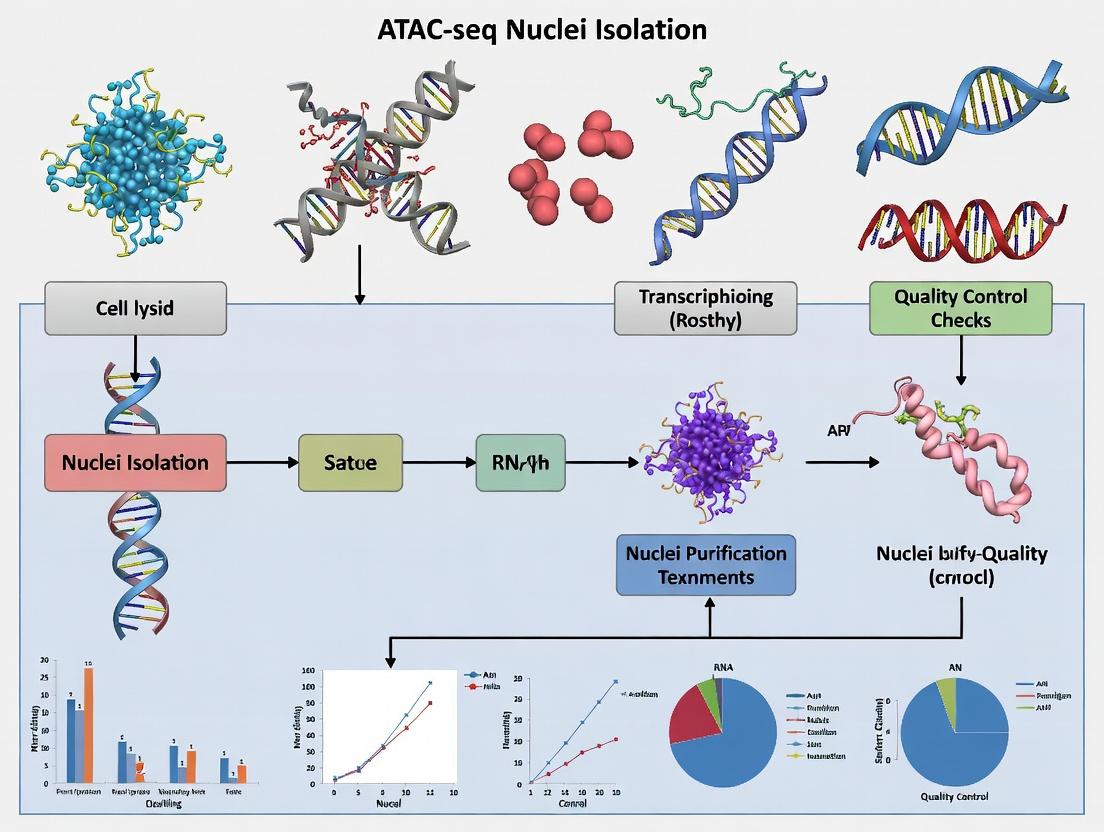
Diagram 1: ATAC-seq Nuclei Isolation & QC Workflow
Title: Workflow for Isolating and Checking ATAC-seq Nuclei Quality
Diagram 2: Key Metrics for Good Nuclei & Impact on Data
Title: How Nuclei Quality Metrics Affect Final ATAC-seq Data
The Scientist's Toolkit: Research Reagent Solutions
| Item | Function in ATAC-seq Nuclei Isolation |
|---|---|
| Dounce Homogenizer (Loose Pestle A) | Provides controlled mechanical shearing to lyse cell membranes while preserving nuclear integrity. |
| Non-ionic Detergents (NP-40, Tween-20) | Gently solubilize the plasma and organelle membranes without disrupting the nuclear envelope. |
| BSA (Bovine Serum Albumin) | Acts as a blocking agent to reduce nuclear clumping and non-specific adhesion to tubes. |
| MgCl₂ | Divalent cation crucial for maintaining nuclear structure and stabilizing the nuclear envelope. |
| Sucrose or OptiPrep | Forms a density cushion for gradient purification, pelleting nuclei away from lighter debris. |
| DAPI / Hoechst Stain | Fluorescent DNA intercalating dyes used for accurate counting and visualization of nuclei. |
| SYTOX Green / Blue | Membrane-impermeant DNA dyes that only stain nuclei with compromised membranes, assessing integrity. |
| 40-µm Cell Strainer | Removes large debris, tissue clumps, and potential nuclei aggregates to prevent clogging in downstream steps. |
| Protease/Nuclease Inhibitors (e.g., PMSF, EDTA) | Preserve nuclear proteins and prevent degradation of chromatin during isolation. |
Technical Support Center: ATAC-seq Nuclei Isolation Troubleshooting
FAQs
Q1: Why is my nuclei yield from frozen tissue significantly lower than from fresh tissue in ATAC-seq? A: Frozen tissue is prone to ice crystal formation during freezing/thawing, which physically shears nuclear membranes. For optimal yield: 1) Snap-freeze in liquid nitrogen or dry ice-cooled isopentane. 2) Use a optimized lysis buffer with added RNase inhibitor and spermidine to stabilize nuclei. 3) Minimize thawing time before homogenization.
Q2: I get high background reads from my FFPE samples. What's the cause? A: FFPE cross-linking causes DNA-protein and DNA-DNA crosslinks, leading to open chromatin artifacts and transposase insertion into non-nucleosomal DNA. Key steps: 1) Optimize de-crosslinking time and temperature (e.g., 65°C for 2 hours). 2) Include a post-isolation wash with 0.5% SDS to inactivate excess transposase. 3) Use a higher concentration of detergent in the lysis buffer (e.g., 0.5% NP-40).
Q3: My fresh tissue nuclei appear clumped and sticky. How can I improve dispersion? A: This is often due to cytoplasmic contamination or inadequate lysis. Solution: 1) Increase homogenization rigor (use a Dounce homogenizer with more strokes). 2) Add a filtration step through a 40µm cell strainer. 3) Incorporate a BSA (0.1%) or sucrose (0.2 M) cushion during centrifugation to reduce mechanical stress and clumping.
Q4: Why does ATAC-seq data from FFPE tissue show a bias against open chromatin regions? A: Formalin fixation preferentially crosslinks lysine and arginine residues, which are abundant in nucleosome cores, making them less accessible to the Tn5 transposase. Mitigation strategy: 1) Use a higher Tn5 transposase concentration and longer incubation time. 2) Employ a "tagmentation buffer booster" like PEG 8000. 3) Consider performing a limited proteinase K treatment after deparaffinization but before nuclei isolation.
Troubleshooting Guides
Issue: Low Tagmentation Efficiency (Low Library Complexity)
- Fresh Tissue: Check cell viability prior to lysis (>90%). Ensure nuclei are not over-lysed; confirm intact nuclei under a microscope using DAPI.
- Frozen Tissue: Assess RNA contamination (a common issue); treat nuclei with RNase A for 5 min on ice post-isolation.
- FFPE Tissue: Quantify crosslink reversal by measuring DNA fragment size post-decrosslinking (should be a smear >300 bp). Re-optimize heating time.
Issue: High Mitochondrial Read Alignment
- All Types: This indicates cytoplasmic contamination or damaged nuclei.
- Solution for Fresh/Frozen: Use a milder detergent (e.g., digitonin) or reduce its concentration in the lysis buffer. Perform a low-speed centrifugation (300 x g) to pellet intact nuclei while leaving mitochondria in suspension.
- Solution for FFPE: This is harder to mitigate due to inherent damage. Use computational tools (e.g.,
ATAC-seqQC) to filter mitochondrial reads post-sequencing.
Issue: Inconsistent Replicates with Frozen Tissue
- Root Cause: Inconsistent freezing rates or storage times.
- Protocol: Standardize freezing: Submerge tissue piece (< 0.5 cm³) in OCT compound directly in a dry ice-ethanol bath. Store at -80°C for < 6 months for best results. Document storage time.
Table 1: Nuclei Yield and Quality Metrics by Sample Type
| Metric | Fresh Tissue (Ideal) | Frozen Tissue (Optimal Protocol) | FFPE Tissue (Optimized) |
|---|---|---|---|
| Average Nuclei Yield/mg | 5,000 - 15,000 | 3,000 - 8,000 | 500 - 3,000 |
| Viability (DAPI+/PI-) | >95% | 70-90% | 50-75% |
| Median Fragment Size | ~200 bp | ~250 bp | ~300-500 bp |
| % Mitochondrial Reads | 10-30% | 20-40% | 30-60% |
| TNF5 Integration Sensitivity | High | Moderate | Reduced |
| Key Isolation Challenge | Apoptosis, RNase activity | Ice crystal damage, RNase release | Crosslinks, protein aggregates |
Table 2: Recommended Protocol Modifications by Tissue Type
| Step | Fresh Tissue | Frozen Tissue | FFPE Tissue |
|---|---|---|---|
| Homogenization | Dounce (15-20 strokes) | CryoMill grinding or mortar/pestle | Deparaffinization, rehydration |
| Lysis Buffer Detergent | 0.1% NP-40 | 0.25% NP-40, 0.1% Digitonin | 0.5% NP-40, 0.1% SDS |
| Critical Additives | None | 1 mM Spermidine, RNase Inhibitor | Proteinase K (limited), 10 mM EDTA |
| Incubation Time/Temp | 30 min on ice | 30 min on ice | 2 hrs at 65°C (decrosslink) |
| Post-Lysis Cleanup | 40µm filter | 40µm filter, BSA cushion | Centrifugal filter (100kDa MWCO) |
Experimental Protocols
Protocol 1: Nuclei Isolation from Frozen Tissue for ATAC-seq (Optimized for Yield)
- Pre-chill all buffers and equipment on ice.
- Grinding: In a liquid N2-chilled mortar, pulverize 10-25 mg frozen tissue to a fine powder. Transfer powder to a tube with 1 mL cold Lysis Buffer (10 mM Tris-HCl pH 7.5, 10 mM NaCl, 3 mM MgCl2, 0.25% NP-40, 0.1% Digitonin, 1 mM Spermidine, 1x Roche cOmplete protease inhibitor).
- Homogenize: Gently pipette mix. Incubate on ice for 30 min, inverting tube every 5 min.
- Filter & Pellet: Pass lysate through a pre-wet 40µm cell strainer. Centrifuge at 500 x g for 5 min at 4°C.
- Wash: Gently resuspend pellet in 1 mL Wash Buffer (Lysis Buffer without detergents). Centrifuge at 500 x g for 5 min at 4°C.
- Resuspend: Resuspend nuclei in 50 µL of Tagmentation Buffer or 1x PBS with 0.1% BSA. Count using a hemocytometer with DAPI stain.
Protocol 2: Nuclei Preparation from FFPE Tissue for ATAC-seq (Decrosslinking-Focused)
- Deparaffinize & Rehydrate: Cut 2-3 x 10 µm sections. Incubate in xylene (2 x 5 min), then in an ethanol series (100%, 95%, 70%, 50% - 2 min each). Rinse in distilled water.
- Decrosslink: Incubate tissue in 1 mL of Decrosslinking Buffer (50 mM Tris pH 8.0, 1 mM EDTA, 0.5% Tween-20) with 0.1 mg/mL Proteinase K at 65°C for 2 hours with gentle agitation.
- Quench: Place on ice, add 10 µL of 100 mM PMSF.
- Homogenize: Transfer tissue to a Dounce homogenizer with 1 mL Lysis Buffer (10 mM Tris-HCl pH 7.5, 10 mM NaCl, 3 mM MgCl2, 0.5% NP-40, 0.1% SDS). Dounce 30-40 strokes.
- Filter & Concentrate: Filter through a 40µm strainer. Concentrate nuclei using a 100 kDa molecular weight cutoff centrifugal filter at 1000 x g for 10 min.
- Resuspend: Recover nuclei in 30-50 µL of 1x PBS with 0.1% BSA. Quantify.
Visualizations
Title: Tissue-Specific Challenges and Solutions for Nuclei Isolation
Title: ATAC-seq Nuclei Isolation Optimization Workflow
The Scientist's Toolkit: Research Reagent Solutions
Table 3: Essential Materials for ATAC-seq Nuclei Isolation Across Sample Types
| Item | Function | Sample Type Specificity |
|---|---|---|
| Digitonin | Mild detergent that selectively permeabilizes plasma membranes over nuclear membranes, reducing cytoplasmic contamination. | Critical for frozen tissue; beneficial for fresh. |
| Spermidine (Triamine) | Stabilizes chromatin structure and nuclei during isolation, reducing aggregation and loss. | Essential for frozen tissue; recommended for all. |
| RNase Inhibitor | Protects RNA and, indirectly, nuclear integrity by preventing degradation-induced lysis. | Critical for frozen tissue (high RNase release). |
| PEG 8000 | Macromolecular crowding agent that boosts Tn5 transposase activity on suboptimal chromatin. | Most beneficial for FFPE and some frozen samples. |
| Proteinase K | Digests crosslinked proteins. Used in limited, controlled doses for FFPE tissue reversal. | Exclusive to FFPE tissue protocols. |
| BSA (Fraction V) | Added to wash and resuspension buffers to coat nuclei and prevent sticking to tubes. | Beneficial for all, especially sticky fresh/frozen preps. |
| Sucrose Cushion | A dense solution (e.g., 1.2 M sucrose) over which lysate is centrifuged to pellet pure nuclei through debris. | Alternative for difficult fatty or fibrous tissues (all types). |
| 40µm Cell Strainer | Removes large cellular aggregates and connective tissue to obtain a single-nuclei suspension. | Mandatory for all tissue types. |
Technical Support Center: ATAC-seq Nuclei Isolation Troubleshooting
Troubleshooting Guides & FAQs
Q1: My nuclei yield after lysis is too low. What could be wrong? A: Low nuclei yield often stems from overly harsh lysis. Key troubleshooting steps include:
- Verify Detergent Concentration & Type: Nonidet P-40 Substitute (NP-40) or Igepal CA-630 are standard. Concentrations between 0.1% and 0.5% are typical for ATAC-seq. Excessive detergent (>0.5%) can lyse nuclei. Titrate within this range.
- Optimize Incubation Time: Lysis on ice for 3-10 minutes is standard. Extending beyond 10 minutes can decrease yield.
- Check Cell Viability: Start with >90% viable cells. Apoptotic or dead cells lyse more easily.
- Validate Buffer Osmolarity: The lysis buffer must be isotonic to preserve nuclei. Ensure sucrose concentration is correct (typically 10-25 mM).
Q2: I observe excessive cytoplasmic contamination (actin or organelle debris) around my nuclei. How can I improve purity? A: Cytoplasmic debris indicates incomplete lysis or insufficient washing.
- Increase Detergent Concentration Slightly: If using 0.1% NP-40, try 0.2%.
- Add a Wash Step: After lysis, pellet nuclei (500g, 5-10 min, 4°C) and gently resuspend in a wash buffer (e.g., Nuclei Wash Buffer: 10 mM Tris-HCl, 10 mM NaCl, 3 mM MgCl2, 0.1% Tween-20).
- Include Bovine Serum Albumin (BSA): Adding 1% BSA to the lysis buffer can reduce non-specific sticking of debris.
Q3: My nuclei are clumping, which affects downstream tagmentation. How do I prevent this? A: Clumping is frequently caused by nuclei damage and release of DNA.
- Add EDTA/EGTA: Include 0.1-1 mM EDTA in your lysis buffer to chelate divalent cations and inhibit nucleases.
- Ensure Fresh Protease Inhibitors: Always use fresh protease inhibitor cocktails (PIC) to prevent protease activity that can degrade nuclear envelope proteins.
- Gentle Handling: Avoid vortexing or pipetting harshly after lysis. Use wide-bore pipette tips for resuspension.
- Filter Nuclei: Pass the nuclei suspension through a 40-μm cell strainer or a 5-mL round-bottom tube with a cell strainer cap.
Q4: My read distribution from ATAC-seq shows high mitochondrial contamination. What is the source? A: Mitochondrial reads originate from damaged nuclei or free mitochondrial DNA released during lysis.
- Optimize Centrifugation Force: Pellet nuclei at 300-500g. Higher speeds can pellet intact mitochondria.
- Re-evaluate Lysis Buffer: Ensure the buffer contains MgCl2 (typically 3-5 mM) or CaCl2 to stabilize nuclear membranes without pelleting organelles.
- Use a Sucrose Cushion: Layer the lysate over a 1.2 M sucrose solution and centrifuge (1200g, 20 min). Nuclei pellet through, while lighter debris remains at the interface.
Q5: My nuclei appear intact but show poor tagmentation efficiency. Could reagents be interfering? A: Yes, carryover of critical reagents can inhibit the Tn5 transposase.
- Remove All Detergents: Tn5 is highly sensitive to ionic (SDS) and non-ionic (NP-40, Triton) detergents. Ensure adequate washing post-lysis. Use a wash buffer with 0.1% Tween-20, which is less inhibitory to Tn5 than NP-40.
- Dilute Inhibitors: Ensure PIC is diluted out. Standard protocols use ≥1000x dilution in the tagmentation reaction.
- Quantify Nuclei Accurately: Use a hemocytometer or automated counter. Overloading nuclei (>50,000 per reaction) can deplete Tn5 activity.
Key Reagent Data & Protocols
Table 1: Common Detergents in ATAC-seq Lysis Buffers
| Detergent | Type | Typical Conc. Range | Primary Function in Lysis | Notes for ATAC-seq |
|---|---|---|---|---|
| NP-40 / Igepal CA-630 | Non-ionic | 0.1% - 0.5% | Disrupts plasma & organelle membranes | Most common; concentration is critical. |
| Triton X-100 | Non-ionic | 0.1% - 0.5% | Membrane solubilization | Slightly harsher than NP-40. |
| Tween-20 | Non-ionic | 0.1% - 0.5% | Mild membrane permeabilization | Often used in wash buffers; less inhibitory to Tn5. |
| Digitonin | Weak non-ionic | 0.01% - 0.05% | Selective cholesterol binding | Can provide cleaner nuclei; more expensive. |
Table 2: Common Protease Inhibitor Cocktail (PIC) Components
| Inhibitor | Target Protease Class | Typical Working Conc. | Stability in Solution |
|---|---|---|---|
| PMSF | Serine proteases | 0.1 - 1 mM | Unstable (~30 min in aqueous). Add fresh. |
| Aprotinin | Serine proteases | 0.3 - 3 µM | Stable for hours. |
| Leupeptin | Serine & Cysteine proteases | 0.5 - 2 µM | Stable for hours. |
| Pepstatin A | Aspartic proteases | 1 - 2 µM | Stable for hours. |
| EDTA / EGTA | Metalloproteases | 0.5 - 2 mM | Stable. Also chelates nucleases. |
Detailed Protocol: Nuclei Isolation for ATAC-seq from Cultured Cells (Adapted from Corces et al., 2017)
- Harvest Cells: Collect ~50,000-100,000 viable cells. Pellet at 500g for 5 min at 4°C. Discard supernatant.
- Cold Lysis: Resuspend cell pellet thoroughly in 50 µL of Cold Lysis Buffer (10 mM Tris-HCl pH 7.4, 10 mM NaCl, 3 mM MgCl2, 0.1% Igepal CA-630, 0.1% Tween-20, 0.01% Digitonin, 1x PIC). Piper gently 5-10 times.
- Incubate: Incubate on ice for 3-10 minutes. Monitor lysis under a microscope.
- Quench & Wash: Add 1 mL of Cold Wash Buffer (10 mM Tris-HCl pH 7.4, 10 mM NaCl, 3 mM MgCl2, 0.1% Tween-20, 1x PIC). Invert tube to mix gently.
- Pellet Nuclei: Centrifuge at 500g for 5 min at 4°C. Carefully aspirate supernatant.
- Resuspend: Gently resuspend nuclei in 50 µL of Resuspension Buffer (10 mM Tris-HCl pH 7.4, 10 mM NaCl, 3 mM MgCl2, 0.1% Tween-20, 1x PIC). Do not vortex.
- Filter (Optional): Pass through a pre-wetted 40-μm cell strainer.
- Count: Quantify nuclei using trypan blue on a hemocytometer. Proceed immediately to tagmentation.
The Scientist's Toolkit: Research Reagent Solutions
| Item | Function in ATAC-seq Nuclei Isolation |
|---|---|
| Igepal CA-630 / NP-40 | Non-ionic detergent for controlled plasma membrane lysis. |
| Digitonin | Cholesterol-binding detergent for selective membrane permeabilization. |
| Tween-20 | Mild non-ionic detergent for washing nuclei without inhibiting Tn5. |
| Protease Inhibitor Cocktail (PIC) | Prevents degradation of nuclear proteins and histones. |
| EDTA / EGTA | Chelates Mg2+/Ca2+; inhibits metalloproteases and nucleases. |
| Sucrose | Provides osmotic support to protect nuclei from swelling/rupture. |
| MgCl2 / CaCl2 | Divalent cations that help maintain nuclear envelope integrity. |
| BSA (Fraction V) | Reduces non-specific adsorption of nuclei to tubes and debris clumping. |
| RNase A | Optional addition to wash buffer to degrade cytoplasmic RNA. |
Visualizations
ATAC-seq Nuclei Isolation & Troubleshooting Flow
Reagent Action on Cellular Structures
Step-by-Step Protocols: Optimized Nuclei Isolation for Diverse Samples
Technical Support Center: ATAC-seq Nuclei Isolation Troubleshooting
FAQs & Troubleshooting Guides
Q1: My final nuclei preparation has excessive clumping. What are the primary causes and solutions? A: Clumping is often due to incomplete tissue dissociation, genomic DNA release from lysed nuclei, or insufficient homogenization/buffering. Follow this troubleshooting table.
| Potential Cause | Diagnostic Check | Corrective Action |
|---|---|---|
| Incomplete Tissue Dissociation | Visible tissue chunks prior to lysis. | Optimize mechanical mincing. For tough tissues, use a gentleMACS Octo Dissociator or a Dounce homogenizer (10-15 strokes with loose pestle). |
| Cellular Debris & DNA Release | Viscous lysate, poor flow through strainer. | 1. Increase BSA concentration in lysis buffer to 0.5-1%. 2. Add CaCl₂ (0.5-1mM) to stabilize nuclear membrane. 3. Use wide-bore pipette tips for all nuclei handling. 4. Add RNase A (0.1 mg/mL) to digest RNA scaffolds. |
| Insufficient NP-40/Detergent | High intact cell count under microscope. | Titrate NP-40 or Igepal CA-630 (0.1% to 0.5%). Validate with a viability dye (DAPI/PI) and adjust to minimize intact cells while maximizing nuclei integrity. |
| Centrifugation Speed Too High | Pellet is very tight and difficult to resuspend. | Reduce centrifugation to 300-500 x g for 5-10 minutes at 4°C. Always resuspend pellet gently with wide-bore tips. |
Q2: I observe a low yield of nuclei from my starting material. How can I improve efficiency? A: Low yield stems from nuclei loss during processing or inadequate initial tissue input. Key metrics from recent optimization studies are summarized below.
| Tissue Type | Typical Input (mg) | Expected Nuclei Yield (Range) | Critical Step for Yield Recovery |
|---|---|---|---|
| Mouse Cortex | 10-20 mg | 50,000 - 150,000 nuclei | Gentle Dounce homogenization; avoid over-homogenization. |
| Human PBMCs | 1x10⁶ cells | 400,000 - 600,000 nuclei | Precise lysis time (5-7 min on ice); immediate dilution with wash buffer. |
| Mouse Heart | 20-30 mg | 20,000 - 60,000 nuclei | Thorough mincing; optional 0.2-0.4 U/mL collagenase pre-digestion (5 min, 37°C). |
| Tumor (dissociated) | 5x10⁵ cells | 200,000 - 350,000 nuclei | Use a 40µm cell strainer followed by a 20µm strainer to remove debris but retain nuclei. |
Q3: My nuclei show poor tagmentation efficiency (low/over-fragmented library). What nuclei quality parameters are critical? A: Tagmentation efficiency is highly sensitive to nuclei purity, integrity, and buffer composition. Follow this detailed QC protocol.
Detailed Protocol: Nuclei Quality Assessment for ATAC-seq
- Count & Concentration: Use a hemocytometer or automated counter. Dilute nuclei in PBS + 1% BSA. Target concentration: 2,000-5,000 nuclei/µL.
- Viability/Integrity Staining: Mix 10 µL nuclei suspension with 10 µL of staining solution (1X PBS, 2 µg/mL DAPI, 0.2% Triton X-100). Incubate 5 min on ice.
- Microscopy QC: Visualize under fluorescence microscope (DAPI channel). Healthy nuclei are intact, round/oval, with smooth edges and uniform DAPI staining. Calculate percentage of intact nuclei (target >90%).
- Flow Cytometry QC (Optional but recommended): Analyze nuclei suspension on a flow cytometer measuring FSC/SSC and DAPI area vs. width to gate on single, intact nuclei.
Q4: How do I adapt this protocol for difficult, fibrotic tissues (e.g., liver, lung, tumor)? A: Fibrotic tissues require additional mechanical and/or enzymatic dissociation. Detailed Protocol for Fibrotic Tissue Pre-processing:
- Mince: Finely mince 20-30 mg tissue in 1 mL cold Nuclei EZ Lysis Buffer (or similar) on a petri dish placed on ice.
- Dounce Homogenize: Transfer to a Dounce homogenizer. Use loose pestle (A) for 15-20 strokes. Avoid generating foam.
- Filter: Pass homogenate through a 70µm strainer into a new tube.
- Optional Enzymatic Step: For persistent clumps, incubate filtered homogenate with 0.5 U/mL dispase II + 10 U/mL DNase I for 5 minutes at room temperature to digest extracellular matrix without damaging nuclei.
- Proceed with Standard Lysis: Add 1-2 volumes of cold Lysis Buffer (with detergent) to the filtered homogenate. Incubate on ice for 5-8 minutes with gentle inversion.
- Final Filtration: Pass lysate through a 40µm flow-through strainer followed by a 20µm pluriStrainer to collect clean nuclei. Centrifuge at 500 x g for 5 min at 4°C.
The Scientist's Toolkit: Key Research Reagent Solutions
| Item | Function & Rationale |
|---|---|
| Dounce Homogenizer (loose pestle) | Provides controlled mechanical shear to break tissue architecture without destroying nuclei. Superior to vortexing or pipetting. |
| Nuclei EZ Lysis Buffer (or homemade equivalent) | Isotonic buffer with non-ionic detergent (e.g., NP-40) and MgCl₂. Lyzes plasma membranes while keeping nuclear membrane intact. |
| Wide-Bore/Low-Binding Pipette Tips | Prevents physical shearing of genomic DNA and reduces nuclei adhesion to tip walls, minimizing loss and clumping. |
| pluriStrainer (20µm, 30µm) | Specialized cell strainers designed for optimal nuclei recovery and debris removal. Crucial step after standard 40µm filtration. |
| BSA (Molecular Biology Grade, 5% Solution) | Added to all buffers (0.1-1%) to reduce non-specific sticking of nuclei to tubes and tips. Stabilizes nuclei suspension. |
| DAPI (4',6-diamidino-2-phenylindole) Staining Solution | Fluorescent DNA dye used for rapid microscopic and flow cytometric assessment of nuclei count, integrity, and singlet status. |
| CaCl₂ Stock Solution (100mM) | Divalent cations can stabilize the nuclear envelope. Adding 0.5-1mM final concentration to lysis buffer can reduce nuclear lysis and DNA release. |
| RNase A (DNase-free) | Degrades RNA that can form sticky networks between nuclei, reducing clumping. Use after nuclei isolation is complete. |
Diagram 1: ATAC-seq Nuclei Isolation Workflow
Diagram 2: Troubleshooting Logic for Common Issues
Troubleshooting Guides & FAQs
Q1: My frozen tissue yields low nuclei count after pulverization and homogenization for ATAC-seq. What are the main culprits? A: Low nuclei yield often stems from inefficient tissue disintegration or excessive mechanical force damaging nuclei. Ensure tissue is kept fully frozen during pulverization (use liquid nitrogen). Over-homogenization with a rotor-stator can shear nuclei; use short, gentle bursts. Inadequate lysis buffer composition or incubation time can also prevent nuclei release. Verify buffer freshness and include appropriate detergent (e.g., IGEPAL CA-630) concentration.
Q2: How can I reduce unwanted background (non-nuclear) signal in my ATAC-seq libraries from frozen samples? A: Background signal typically arises from damaged nuclei or mitochondrial DNA release. Key steps include:
- Effective Debris Removal: Use a low-speed centrifugation step (e.g., 200-500 rcf for 5 min at 4°C) after homogenization to pellet debris before filtering the supernatant through a cell strainer.
- Optimized Nuclei Wash: Pellet nuclei through a sucrose or BSA cushion buffer (e.g., 1% BSA in PBS) at 500-700 rcf for 10 min at 4°C to remove cytoplasmic contaminants.
- Mitochondrial Depletion: Consider adding a brief, low-concentration digitonin wash (e.g., 0.01% for 5 min on ice) after isolation, followed by careful pelleting and washing. Caution: Over-digitonin will lyse nuclei.
Q3: My pulverized tissue forms a sticky, hard-to-process pellet. How do I mitigate this? A: Stickiness indicates residual water or cellular release of DNA/protein. Solutions:
- Ensure Complete Freezing: Submerge tissue in liquid nitrogen for >30 seconds before pulverization.
- Use a Cryogenic Mill: More effective than mortar/pestle for uniform powder.
- Immediate Buffer Addition: Transfer powder directly to cold lysis buffer with gentle vortexing to disperse clumps.
- Add RNase A: Contaminating RNA can cause viscosity. Adding RNase A (e.g., 20 µg/mL) during lysis can reduce stickiness.
Q4: What is the critical factor for balancing nuclei integrity vs. accessibility during lysis from frozen tissue? A: The detergent type, concentration, and incubation time are critical. A non-ionic detergent like IGEPAL CA-630 (NP-40) is standard. For frozen tissue, which often has compromised membranes, a lower concentration (e.g., 0.1% instead of 0.5%) and shorter incubation on ice (3-5 minutes) may preserve integrity while allowing access. Always check nuclei under a microscope after lysis.
Data Presentation
Table 1: Comparison of Pulverization Methods for Frozen Tissue ATAC-seq
| Method | Equipment | Typical Yield* (Nuclei/mg tissue) | Integrity (Microscopy) | Risk of Thawing | Cost & Speed |
|---|---|---|---|---|---|
| Mortar & Pestle | Porcelain/Pre-chilled steel | Low-Moderate (1-3k) | Variable, often clumped | High | Low cost, Slow |
| Cryogenic Mill | Specially designed ball mill (e.g., Retsch) | High (5-10k) | High, single nuclei | Very Low | High cost, Fast |
| Biomasher II / Homogenizer | Handheld pestle in microtube | Moderate (2-5k) | Moderate, some clusters | Medium | Low cost, Medium speed |
*Yield is tissue-type dependent; values are illustrative for murine liver/spleen.
Table 2: Common Lysis Buffer Components and Their Functions
| Component | Typical Concentration | Function | Consideration for Frozen Tissue |
|---|---|---|---|
| Tris-HCl (pH 7.4-7.8) | 10 mM | Maintains physiological pH | Critical for nuclease activity control. |
| NaCl | 10 mM | Maintains ionic strength | Helps stabilize nuclei. |
| MgCl₂ | 3-5 mM | Stabilizes nuclear membrane | Higher concentration may protect fragile nuclei. |
| IGEPAL CA-630 (NP-40) | 0.1% - 0.5% | Non-ionic detergent, lyses plasma membrane | Use lower end (0.1-0.2%) for frozen tissue. |
| Digitonin | 0.01% (optional wash) | Cholesterol-binding detergent, permeabilizes nuclear membrane | Can be used post-isolation for cleaner access. |
| Sucrose or BSA | 0.5-1% | Cushion to protect nuclei during pelleting | Highly recommended to prevent damage. |
Experimental Protocols
Protocol: Nuclei Isolation from Frozen Tissue for ATAC-seq (Adapted from Corces et al., 2017) Materials: Pre-cooled mortar/pestle or cryomill, Liquid N₂, Lysis Buffer (10 mM Tris-HCl pH 7.4, 10 mM NaCl, 3 mM MgCl₂, 0.1% IGEPAL CA-630, 0.5% BSA, 1x Protease Inhibitor), Wash Buffer (10 mM Tris-HCl pH 7.4, 10 mM NaCl, 3 mM MgCl₂, 0.5% BSA), 40µm cell strainer, 1.5 mL LoBind tubes.
Pulverization:
- Submerge 10-50 mg frozen tissue block in liquid N₂ in mortar. Pulverize vigorously until a fine powder forms. Alternatively, use a cryomill following manufacturer's instructions.
- Using a pre-cooled spatula, quickly transfer powder to a tube containing 1 mL of ice-cold Lysis Buffer.
Homogenization & Lysis:
- Gently vortex the tube for 5-10 seconds to disperse the powder.
- Incubate on ice for 5 minutes. Gently invert tube every minute. Monitor lysis: Take 5 µL, mix with DAPI/ Trypan Blue, and check under a microscope. Nuclei should be released and intact (round, smooth membrane).
Debris Removal & Washing:
- Filter the lysate through a pre-wet 40µm cell strainer into a new tube.
- Centrifuge at 500 rcf for 5 minutes at 4°C to pellet nuclei.
- Carefully discard supernatant. Resuspend pellet in 1 mL ice-cold Wash Buffer by gentle pipetting (avoid foaming).
- Centrifuge at 500 rcf for 5 minutes at 4°C. Discard supernatant.
Nuclei QC & Counting:
- Resuspend nuclei in 50-100 µL of Wash Buffer or ATAC-seq Resuspension Buffer.
- Count using a hemocytometer or automated cell counter. Stain with DAPI (for count) or Trypan Blue (for integrity). Proceed to transposition.
Mandatory Visualization
ATAC-seq Nuclei Isolation from Frozen Tissue Workflow
Troubleshooting Logic for Common Frozen Tissue Issues
The Scientist's Toolkit: Research Reagent Solutions
Table 3: Essential Materials for Frozen Tissue ATAC-seq
| Item | Function | Example/Note |
|---|---|---|
| Liquid Nitrogen | Cryogenic cooling for pulverization; prevents thawing and RNA degradation. | Essential for manual methods. |
| Cryogenic Mill (e.g., Retsch MM 400) | Provides consistent, efficient mechanical disruption while keeping tissue frozen. | Ideal for hard or fibrous tissues. |
| IGEPAL CA-630 (Octylphenoxy) | Non-ionic detergent for plasma membrane lysis. Critical for nuclei release. | Preferred over NP-40 for consistency. |
| Digitonin | Cholesterol-binding detergent for precise nuclear membrane permeabilization. | Use in post-isolation wash to reduce mitochondrial contamination. |
| Molecular Grade BSA | Acts as a protective agent, reducing nuclei loss to tube walls and damage during pelleting. | Use in lysis and wash buffers. |
| 40µm Nylon Cell Strainer | Removes large tissue debris and clumps after lysis. | Pre-wet with buffer to improve flow-through. |
| DAPI Stain (or Trypan Blue) | Dyes for counting and assessing nuclei integrity under a microscope. | QC is mandatory before transposition. |
| Sucrose Cushion Buffer | Provides a dense medium for gentle pelleting of nuclei, minimizing shear forces. | Alternative to BSA in wash buffer. |
Technical Support Center
Troubleshooting Guides & FAQs
Q1: Why is my nuclei yield from FFPE tissue sections extremely low after de-crosslinking and digestion? A: Low nuclei yield often stems from incomplete reversal of formaldehyde crosslinks or insufficient proteinase K digestion. Ensure de-crosslinking is performed at 65°C for at least 2 hours. Optimize proteinase K concentration; a common starting point is 0.2 mg/mL for 30 minutes at 50°C. Over-digestion can lyse nuclei, while under-digestion traps nuclei in the matrix. Include a post-digestion wash with a mild detergent like 0.1% Triton X-100 in Nuclei Buffer.
Q2: I observe high-molecular-weight DNA smearing on my Bioanalyzer trace after tagmentation from a crosslinked sample. What does this indicate? A: High-molecular-weight smearing suggests incomplete tagmentation, usually due to residual crosslinks or chromatin proteins blocking Tn5 enzyme access. Key steps: 1) Ensure thorough de-crosslinking. 2) Increase the number of nuclei used for tagmentation (2-5x more than for fresh/frozen samples). 3) Consider increasing the duration of the tagmentation reaction by 25-50% and validate the optimal input nuclei number in a pilot experiment.
Q3: My ATAC-seq library from an FFPE sample has very low complexity and high duplicate rates. How can I improve this? A: Low complexity arises from low accessible chromatin yield. Critical fixes:
- Increase input material: Start with 5-10 tissue sections (10 µm thick) instead of 1-2.
- Optimize de-crosslinking: Test extended incubation times (up to 4 hours) and the use of heat-assisted methods.
- Purify nuclei aggressively: Use density gradient centrifugation (e.g., with iodixanol) to isolate intact nuclei from debris after digestion.
- Post-lysis QC: Use a hemocytometer and DAPI staining to count intact nuclei before tagmentation. Target >50,000 nuclei per reaction.
Q4: After nuclei isolation from FFPE tissue, I see excessive cytoplasmic debris. How do I clean the nuclei preparation? A: Use a sucrose cushion or gradient centrifugation. Layer the crude nuclei suspension over a 1.2 M sucrose solution in Nuclei Buffer and centrifuge at 13,000g for 10 min at 4°C. Intact nuclei will pellet while debris remains at the interface. A gentle wash with 0.1% BSA in PBS can also help reduce stickiness and aggregation.
Table 1: Comparative Performance of ATAC-seq on Fixed vs. Fresh Frozen Tissues
| Parameter | Fresh/Frozen Tissue | FFPE Tissue (Optimized) | Formaldehyde Crosslinked (Optimized) |
|---|---|---|---|
| Recommended Input | 50 mg tissue / 50,000 cells | 5-10 x 10µm sections | 1x10^6 cells fixed for <10 min |
| Typical Nuclei Yield | 70-90% | 20-40% | 40-60% |
| De-crosslinking Required | No | 65°C, 2-4 hrs + Proteinase K | 65°C, 30 min - 2 hrs |
| Tagmentation Input (Nuclei) | 25,000 - 50,000 | 50,000 - 100,000 | 50,000 - 75,000 |
| *Library Complexity (NRF) | > 0.8 | 0.4 - 0.7 | 0.6 - 0.8 |
| Key Optimization Step | Gentle lysis | Aggressive de-crosslinking | Crosslinking duration control |
*NRF: Non-Redundant Fraction, a measure of library complexity.
Experimental Protocols
Protocol 1: Nuclei Isolation from FFPE Tissue Sections for ATAC-seq This protocol is framed within thesis research on isolating intact nuclei from challenging samples.
- Deparaffinization: Place 5-10 curls (10 µm) in a 1.5 mL tube. Add 1 mL xylene, vortex, incubate 10 min at RT. Centrifuge at max speed for 5 min. Discard supernatant. Repeat with fresh xylene.
- Rehydration: Wash sequentially with 1 mL of: 100% ethanol (2x), 95% ethanol, 80% ethanol, 70% ethanol, 50% ethanol. Centrifuge 5 min at max speed between each wash. Perform a final wash with PBS.
- De-crosslinking: Resuspend pellet in 200 µL Digestion Buffer (10 mM Tris-HCl pH 8.0, 100 mM NaCl, 1 mM EDTA, 0.1% SDS) with 0.2 mg/mL Proteinase K. Incubate at 50°C for 30 min with shaking (1000 rpm), then at 65°C for 2 hours.
- Nuclei Release & Purification: Cool on ice. Add 200 µL of 2x Nuclei Buffer (NB: 0.6 M sucrose, 10 mM MgCl2, 20 mM Tris-HCl pH 8.0, 2% Triton X-100, protease inhibitors). Homogenize with 20 strokes of a Dounce pestle. Filter through a 40 µm cell strainer.
- Density Gradient: Layer filtrate over 500 µL of 1.2 M sucrose cushion in NB. Centrifuge at 13,000g for 10 min at 4°C. Discard supernatant.
- Wash & Resuspend: Gently resuspend nuclei pellet in 100 µL of 1x NB + 0.1% BSA. Count using hemocytometer with DAPI stain. Proceed to tagmentation with 50,000-100,000 nuclei.
Protocol 2: ATAC-seq on Reversibly Formaldehyde-Crosslinked Cell Cultures
- Fixation: Harvest 1x10^6 cells. Resuspend in 1 mL PBS with 1% formaldehyde. Incubate at RT for 5 min with gentle rotation.
- Quenching: Add 100 µL of 1.25 M glycine (final ~0.125 M). Incubate 5 min at RT. Pellet cells at 500g for 5 min at 4°C. Wash 2x with cold PBS.
- Nuclei Isolation & De-crosslinking: Lyse cells in 50 µL cold ATAC-seq Lysis Buffer (10 mM Tris-HCl pH 7.4, 10 mM NaCl, 3 mM MgCl2, 0.1% IGEPAL CA-630). Pellet nuclei at 500g for 10 min at 4°C. Resuspend pellet in 50 µL of Digestion Buffer (see Protocol 1, step 3) without SDS or Proteinase K. Incubate at 65°C for 1 hour.
- Wash: Add 1 mL of ATAC-seq Wash Buffer (10 mM Tris-HCl pH 7.4, 10 mM NaCl, 3 mM MgCl2, 0.1% Tween-20). Pellet nuclei at 500g for 10 min at 4°C. Resuspend in 50 µL of Tagmentation Buffer.
- Tagmentation: Proceed with standard ATAC-seq tagmentation (Illumina or equivalent Tn5) using 50,000-75,000 nuclei. Increase tagmentation time by 50% (e.g., 15 min to 22.5 min) if pilot results show low fragmentation.
Diagrams
Diagram Title: FFPE Tissue ATAC-seq Experimental Workflow
Diagram Title: ATAC-seq on Fixed Tissue Troubleshooting Decision Tree
The Scientist's Toolkit: Research Reagent Solutions
Table 2: Essential Materials for ATAC-seq on Fixed Tissues
| Reagent/Material | Function/Principle | Key Consideration |
|---|---|---|
| Proteinase K | Serine protease that digests proteins, critical for reversing formaldehyde crosslinks in FFPE tissues. | Quality and activity vary. Titrate for each tissue type; over-digestion damages nuclei. |
| Tn5 Transposase | Enzyme that simultaneously fragments and tags accessible chromatin with sequencing adapters. | Commercial "loaded" Tn5 (e.g., Illumina) is standard. Increased input or time may be needed for fixed samples. |
| Sucrose (OptiPrep/Iodixanol) | Forms density gradient for purifying intact nuclei from cellular debris and unlysed cells. | Crucial for clean nuclei prep from heterogeneous FFPE digests. Reduces background in sequencing. |
| Igepal CA-630 / Triton X-100 | Non-ionic detergents for permeabilizing cell and nuclear membranes during isolation and wash steps. | Concentration is critical (typically 0.1%-0.5%). Triton X-100 is harsher; use Igepal for delicate nuclei. |
| Glycine | Quenches formaldehyde fixation by reacting with residual formaldeyde, stopping crosslinking. | Essential for reversible crosslinking protocols. Ensures consistency between samples. |
| DAPI Stain (4',6-diamidino-2-phenylindole) | Fluorescent DNA dye used to visually count and assess the integrity of isolated nuclei under a microscope. | Distinguishes intact nuclei from anucleate debris. Critical for accurate input normalization before tagmentation. |
High-Throughput and Automated Approaches for Drug Screening & Large Cohorts
Technical Support Center: Troubleshooting Guides & FAQs
This support center addresses common issues encountered during high-throughput ATAC-seq nuclei isolation, a critical preprocessing step for drug screening on large patient cohorts.
Frequently Asked Questions (FAQs)
Q1: During automated nuclei isolation from frozen tissue for a 500-sample cohort, we observe a >60% reduction in final nuclei yield compared to manual protocol. What are the primary causes? A: The most common causes are (1) Incomplete tissue dissociation due to fixed time sonication or homogenization settings not accounting for tissue heterogeneity. (2) Carryover of inhibitory contaminants (e.g., detergents, salts) between samples on an automated liquid handler due to insufficient wash steps. (3) Aggregation and loss of nuclei on filter membranes or plate wells. First, verify tissue dissociation visually using a fluorescent nuclear stain in a pilot batch. Increase wash volumes between samples on the handler by 30%. Pre-treat plates with a 1% BSA solution to prevent adhesion.
Q2: Our ATAC-seq data from a drug-treated cohort shows high background ("open" signal noise) and low signal-to-noise ratio in key regulatory regions. Could this stem from nuclei isolation? A: Yes. This often indicates cytoplasmic contamination or nuclear lysis during isolation, releasing ambient DNA and nucleases. Ensure the lysis buffer formulation is consistent and contains sufficient non-ionic detergent (e.g., IGEPAL CA-630) and a nuclease inhibitor. On automated platforms, check that mechanical agitation speed (e.g., orbital shaking) is optimized to prevent shear stress. Validate nuclei integrity and purity by flow cytometry (DAPI vs. cytoplasmic stain) for 5-10 random samples per 96-well plate.
Q3: When processing whole blood samples in a 384-well format, we see high well-to-well variability in sequencing library complexity. What is the troubleshooting path? A: Focus on consistent erythrocyte lysis and white blood cell (WBC) counting normalization. Automated systems may unevenly aspirate the WBC pellet after centrifugation. Implement a pre-isolation step using an automated cell counter or a fluorescence-based plate reader for DNA quantification to normalize input across wells. Ensure temperature control for lysis buffers is active on the deck.
Q4: After implementing a new magnetic bead-based nuclei isolation kit on our automated platform, we get frequent clogging of tips. How can we modify the protocol? A: Bead aggregation is typical. (1) Introduce a brief, low-frequency sonication or vortex pulse step immediately before aspiration. (2) Increase tip bore size if possible. (3) Modify the protocol to include a 1:1 dilution of the bead solution with a low-EDTA TE buffer to reduce viscosity. Always perform bead calibration (incubation time vs. yield) for any new lot.
Table 1: Comparison of Nuclei Isolation Methods for High-Throughput ATAC-seq
| Method | Throughput (Samples/Day) | Avg. Nuclei Yield (% of Theoretical Max) | Median Library Complexity (Unique Fragments per 10k Nuclei) | Common Failure Mode |
|---|---|---|---|---|
| Manual (Dounce) | 24-48 | 65-80% | 8,542 | Operator variability, low throughput |
| Automated Liquid Handler (Filter-based) | 960 | 45-60% | 7,115 | Filter clogging, variable lysis time |
| Automated (Magnetic Bead-based) | 576 | 70-75% | 9,230 | Bead aggregation, higher cost |
| Semi-Automated (Centrifugation-assisted) | 288 | 75-85% | 8,950 | Centrifuge downtime, batch effects |
Table 2: Impact of Common Troubleshooting Interventions on Key Metrics
| Intervention | Target Issue | Effect on Nuclei Yield (Mean ∆) | Effect on Data Quality (TSS Enrichment ∆) |
|---|---|---|---|
| BSA Plate Pre-treatment | Nuclei adhesion | +22% | +1.5 |
| Wash Volume Increase (30%) | Contaminant carryover | +15% | +2.1 |
| Input Normalization by DNA Quant | Cell count variability | +5% (Consistency) | +3.4 |
| Lysis Time Optimization (Tissue-specific) | Incomplete dissociation | +40% | +4.0 |
Experimental Protocols
Protocol 1: Validation of Automated Nuclei Integrity for ATAC-seq Purpose: To quality-check nuclei post-isolation from an automated platform before proceeding to tagmentation. Steps:
- Sampling: From each 96-well plate, select wells A1, H1, A12, H12 for validation.
- Staining: Transfer a 10 µL aliquot from each selected well to a 96-well V-bottom plate. Add 10 µL of staining solution (1x PBS, 2 µg/mL DAPI, 0.2 µg/mL Phalloidin-AF568).
- Incubation: Incubate for 15 minutes at 4°C protected from light.
- Analysis: Load onto a flow cytometer or automated counter. Gate on DAPI+ events to determine nuclei count. The Phalloidin (cytoplasmic F-actin) signal should be low in the DAPI+ population (<10% co-staining indicates clean nuclei).
- Decision Point: If nuclei yield is <50% of expected or cytoplasmic contamination >15%, flag the entire plate for re-isolation or data annotation.
Protocol 2: Calibration of Bead-Based Automated Isolation Purpose: To establish optimal incubation time for a new lot of magnetic beads. Steps:
- Setup: Use a control cell line (e.g., K562) aliquoted into 8 wells of a 96-well plate (10,000 cells/well).
- Lysis: Perform standard cell lysis on the automated deck.
- Bead Incubation Gradient: Program the handler to add beads to all wells simultaneously but to incubate for different times (1, 2, 3, 4, 5, 7, 10, 15 min) before magnetic separation.
- Elution & Quantification: Elute nuclei into a fixed volume. Quantify nuclear DNA using a fluorescent plate reader (e.g., with Hoechst 33342).
- Analysis: Plot DNA yield vs. time. Select the minimum time that reaches the yield plateau for the high-throughput protocol.
Visualizations
The Scientist's Toolkit: Research Reagent Solutions
Table 3: Essential Materials for High-Throughput ATAC-seq Nuclei Isolation
| Item | Function | Key Consideration for HTS/Automation |
|---|---|---|
| Non-ionic Detergent (e.g., IGEPAL CA-630) | Lyses plasma membrane without disrupting nuclear envelope. | Use liquid handler-compatible stabilized formulations; prepare single-use aliquots to prevent degradation. |
| Nuclease Inhibitor (e.g., RNase A inhibitor, Spermidine) | Prevents degradation of accessible chromatin. | Critical for extended automated runs; add fresh to lysis buffer just before run. |
| Magnetic Beads (Chromatin Capture) | High-purity nuclei separation. | Test for lot-to-lot aggregation; requires precise magnetic separation timing on deck. |
| BSA (Molecular Biology Grade) | Blocks adhesion to plastic tips and plates. | Use at 0.1-1% for pre-treating assay plates to minimize nuclei loss. |
| DAPI (4',6-diamidino-2-phenylindole) | Fluorescent DNA stain for nuclei counting/QC. | Compatible with automated imagers and flow cytometers on deck. |
| Hoechst 33342 | Cell-permeable DNA stain for live-cell normalization. | Used in plate-reader DNA quant for input normalization across samples. |
| Low-Binding 96/384-Well Plates | Sample processing and storage. | Minimizes nuclei and DNA adhesion; essential for consistent yields. |
| Cryopreservation Medium (DMSO-based) | Long-term storage of isolated nuclei. | Enables batch processing of isolation and decoupling from downstream library prep. |
Diagnose & Fix: A Symptom-Based Troubleshooting Matrix for Common Pitfalls
Troubleshooting Guides & FAQs
Q1: What are the primary causes of low nuclei yield during ATAC-seq sample preparation? A: The two most common technical causes are Incomplete Tissue/Cell Dissociation and Over-Lysis of nuclei. Incomplete dissociation fails to release all nuclei from the tissue matrix, while over-lysis damages nuclear membranes, causing chromatin leakage and loss during centrifugation.
Q2: How can I diagnose incomplete dissociation versus over-lysis? A: Monitor your preparation under a microscope at each step.
- Incomplete Dissociation: Visible tissue chunks or clumps remain after the lysis step. The supernatant after centrifugation contains few nuclei.
- Over-Lysis: After lysis, nuclei appear swollen, "ghost-like," or fragmented. The pellet after centrifugation is very small or sticky.
Q3: What are the critical optimization points for mechanical dissociation? A: Optimize the method and duration based on your tissue type.
- Soft Tissues (e.g., spleen, liver): Use gentle douncing (10-15 strokes with loose pestle). Over-douncing causes over-lysis.
- Hard Tissues (e.g., heart, tumor): May require gentle mincing with scalpels followed by enzymatic digestion (e.g., Collagenase/Dispase) for 5-15 minutes at 37°C before douncing.
Q4: How do I optimize lysis buffer composition and incubation time? A: The goal is to lyse the plasma membrane while keeping the nuclear membrane intact. Key variables are detergent concentration and lysis time.
Table 1: Optimization Parameters for Nuclei Lysis
| Parameter | Typical Range | Effect of Too Low | Effect of Too High | Recommendation |
|---|---|---|---|---|
| NP-40 or IGEPAL CA-630 Concentration | 0.1% - 0.5% (v/v) | Incomplete cell lysis, low yield | Nuclear membrane damage, over-lysis | Start at 0.1% for sensitive cells (e.g., primary neurons), 0.25% for standard cell lines. |
| Digitonin Concentration | 0.01% - 0.1% (w/v) | Incomplete lysis | Increased background, nuclear damage | Use 0.01-0.02% for permeabilizing nuclei for tagmentation in situ protocols. |
| Lysis Incubation Time | 2 - 10 minutes (on ice) | Low yield from incomplete lysis | Nuclear clumping/aggregation, degradation | Do not exceed 10 minutes on ice. Monitor microscopically after 3, 5, and 7 minutes. |
| Centrifugation Force | 300 - 500 RCF for 5 min | Incomplete pelleting of nuclei | Nuclear deformation/rupture | Use 500 RCF at 4°C for most cell types. Reduce to 300 RCF for fragile nuclei. |
Q5: Are there tissue-specific considerations for nuclei isolation? A: Yes. Different tissues require protocol adjustments.
Table 2: Tissue-Specific Recommendations
| Tissue Type | Major Challenge | Key Adjustment | Expected Yield (Nuclei/mg tissue)* |
|---|---|---|---|
| Mouse Cortex (Neuronal) | High fragility, over-lysis | Use low-detergent (0.1% NP-40), omit vortexing, use sucrose cushion. | 5,000 - 20,000 |
| Mouse Spleen | Easy dissociation, RBC contamination | Gentle douncing only. Add RBC lysis step if needed. | 100,000 - 500,000 |
| Solid Tumors | Fibrotic matrix, heterogeneity | Include 15-30 min collagenase digestion at 37°C before lysis. Filter through 70μm strainer. | 10,000 - 100,000 |
| Cultured Adherent Cells | Over-confluence, apoptosis | Harvest at 80-90% confluence. Use trypsinization, not scraping, for single-cell start. | 50,000 - 100,000 per well (6-well) |
*Yields are highly variable and depend on exact protocol and tissue condition.
Experimental Protocol: Optimized Nuclei Isolation for ATAC-seq
Method for Murine Spleen or Liver (Adapted from Corces et al., 2017, Nature Methods):
- Homogenization: Place 1-2 mg of fresh tissue in 1 mL of cold Nuclei EZ Lysis Buffer (or homemade buffer: 10 mM Tris-HCl, pH 7.5, 10 mM NaCl, 3 mM MgCl2, 0.1% NP-40, 0.1% Tween-20, 0.01% Digitonin). Gently dounce with loose pestle (10 strokes).
- Incubate: Place on ice for 5 minutes.
- Filter: Pass the lysate through a 40 μm cell strainer into a 2 mL Eppendorf tube.
- Wash: Add 1 mL of Wash Buffer (10 mM Tris-HCl, pH 7.5, 10 mM NaCl, 3 mM MgCl2, 0.1% Tween-20). Invert to mix.
- Pellet Nuclei: Centrifuge at 500 RCF for 5 minutes at 4°C. Carefully decant supernatant.
- Resuspend: Gently resuspend the pellet in 50 μL of Resuspension Buffer (10 mM Tris-HCl, pH 7.5, 10 mM NaCl, 3 mM MgCl2). Avoid pipette mixing; use wide-bore tips.
- Count & Quality Control: Mix 10 μL of nuclei with 10 μL of Trypan Blue. Count using a hemocytometer. Assess integrity under a microscope (40x). Intact nuclei are round and refractile.
Diagrams
ATAC-seq Nuclei Isolation Workflow
Troubleshooting Decision Tree
The Scientist's Toolkit: Key Research Reagent Solutions
| Reagent / Material | Function in Nuclei Isolation | Key Consideration |
|---|---|---|
| NP-40 Alternative (IGEPAL CA-630) | Non-ionic detergent for plasma membrane lysis. | Standard concentration is 0.1-0.5%. Less harsh than SDS. |
| Digitonin | Cholesterol-binding detergent for precise membrane permeabilization. | Used at low conc. (0.01-0.1%) for in situ tagmentation or fragile nuclei. |
| Sucrose Cushion (e.g., 1.8M Sucrose) | A dense solution layer at the bottom of the centrifuge tube. | Protects nuclei from mechanical shear during pelleting; improves purity. |
| Dounce Homogenizer | Glass homogenizer with tight/loose pestles for mechanical tissue disruption. | Number of strokes must be optimized; over-douncing causes over-lysis. |
| Wide-Bore or Filtered Pipette Tips | Tips with a larger opening at the end. | Prevents shearing and physical damage to isolated nuclei during pipetting. |
| Nuclei EZ Lysis Buffer (Sigma) | Commercial, ready-to-use lysis buffer optimized for nuclei isolation. | Provides consistency but may require optimization for specific tissues. |
| Protease/RNase Inhibitors | Cocktails added to lysis/wash buffers. | Prevent degradation of nuclear proteins and RNA, maintaining nuclear integrity. |
| 40μm Cell Strainer | Nylon mesh filter. | Removes large tissue aggregates and clumps to obtain a single-nuclei suspension. |
Technical Support Center: ATAC-seq Nuclei Isolation Troubleshooting
Frequently Asked Questions (FAQs)
Q1: Why are my nuclei forming clumps or aggregates during ATAC-seq isolation, and how does this affect my data? A1: Nuclei clumping is a common issue caused by insufficient membrane lysis, residual cytoskeletal components, or the presence of divalent cations. Aggregates lead to uneven tagmentation, causing severe data biases such as low library complexity, high PCR duplication rates, and poor signal-to-noise ratios in peak calling. It directly compromises the assessment of chromatin accessibility.
Q2: How can I adjust detergent concentration to minimize aggregation? A2: The concentration of non-ionic detergent (e.g., NP-40, Igepal CA-630) is critical. Too little leads to incomplete lysis and sticky nuclei; too much can damage nuclear integrity. A titration approach is recommended.
Q3: What is the role of divalent cations (Mg²⁺, Ca²⁺) in nuclei aggregation? A3: Divalent cations can act as bridges between negatively charged molecules on nuclear surfaces, promoting aggregation. However, Mg²⁺ is often a component of nuclei resuspension buffers. The key is to use a chelating agent (like EDTA/EGTA) in the lysis buffer to sequester cations released from the cytosol, while providing optimal, buffered Mg²⁺ later for tagmentation enzyme activity.
Q4: When and how should I use filtration to resolve clumping? A4: Gentle filtration is a mechanical solution to break apart loose aggregates. It is used as a final step after lysis and washing, just prior to counting. It is a corrective measure; optimal lysis and buffer conditions should be established first to prevent clumping.
Troubleshooting Guides
Problem: Severe nuclei aggregation observed after centrifugation wash steps.
Diagnosis & Action Plan:
- Immediate Fix: Pass the nuclei suspension through a pre-wet, cell-strainer capped flow cytometry tube (e.g., 35 µm nylon mesh). Do not force the suspension. Re-count.
- Systematic Investigation: Follow the workflow below to identify and correct the root cause.
Diagram 1: Nuclei Clumping Troubleshooting Decision Tree
Detailed Protocol: Systematic Optimization of Isolation Conditions
Objective: To identify the optimal combination of detergent concentration and chelator presence for obtaining a single-nuclei suspension from your specific cell type.
Reagents:
- Cell lysis buffer base (10 mM Tris-Cl pH 7.4, 10 mM NaCl, 3 mM MgCl₂, nuclease-free water).
- 10% Igepal CA-630 (or NP-40) stock.
- 0.5 M EDTA, pH 8.0.
- Nuclei wash & resuspension buffer (NRB: 1x PBS, 1% BSA, 0.2 U/µl RNase Inhibitor, 1x protease inhibitor).
- Cell strainer (35 µm), pre-wet with NRB.
Method:
- Prepare six variations of lysis buffer in 1.5 mL tubes as per Table 1.
- Harvest 1x10⁵ cells per condition. Pellet and fully aspirate supernatant.
- Resuspend each pellet in 50 µL of the assigned pre-chilled lysis buffer. Vortex gently for 3 seconds.
- Incubate on ice for 5 minutes.
- Immediately add 1 mL of ice-cold NRB to each tube to stop lysis.
- Centrifuge at 500 rcf for 5 minutes at 4°C. Carefully aspirate supernatant.
- Resuspend the pellet in 50 µL NRB. Pass through a pre-wet 35 µm strainer.
- Count using a hemocytometer with Trypan Blue. Assess clumping visually (score 0-3) and record yield.
Table 1: Optimization Matrix and Expected Outcomes
| Condition | Igepal CA-630 (%) | EDTA (mM) | Expected Nuclei Integrity | Expected Clumping Score (0-3) | Primary Use Case |
|---|---|---|---|---|---|
| 1 | 0.1 | 0 | Poor lysis, low yield | High (2-3) | Not recommended |
| 2 | 0.1 | 1 | Moderate, may be sticky | Moderate (1-2) | Tough-to-lyse cells |
| 3 | 0.25 | 0 | Good, but aggregation likely | Moderate (1-2) | Standard, low-chelator protocol |
| 4 | 0.25 | 1 | Optimal for most cell types | Low (0-1) | Recommended starting point |
| 5 | 0.5 | 1 | Good, risk of damage | Low (0-1) | Fibrous or adherent cells |
| 6 | 0.5 | 5 | Excellent, but may inhibit tagmentation | Very Low (0) | Problematic, sticky samples |
Note: Score 0=no clumps, 1=minor, 2=moderate, 3=severe. Yield is cell-type dependent.
Protocol: Integrated Filtration for Aggregate Removal
Objective: To implement a standardized, gentle filtration step within the nuclei isolation workflow.
Method:
- After the final wash and resuspension in NRB (or tagmentation buffer if proceeding directly), gently pipette the nuclei suspension up and down 5-10 times.
- Place a sterile 35 µm cell strainer cap on a 5 mL polystyrene round-bottom FACS tube.
- Pre-wet the strainer with 100 µL of NRB.
- Slowly pipette your nuclei suspension onto the center of the mesh. Allow it to flow through by gravity. Do not press or grind the sample.
- Rinse the original tube with 100 µL of buffer and pass it through the same strainer.
- Proceed to counting and tagmentation. The filtered sample should be used immediately.
The Scientist's Toolkit: Key Reagent Solutions
Table 2: Essential Reagents for ATAC-seq Nuclei Isolation
| Reagent | Function / Role in Preventing Aggregation | Example Product / Specification |
|---|---|---|
| Non-ionic Detergent | Disrupts the plasma and cytoplasmic membranes without dissolving the nuclear envelope. Critical concentration prevents incomplete lysis (sticky nuclei). | Igepal CA-630 (0.25-0.5%), NP-40 Alternative |
| EDTA / EGTA | Chelates divalent cations (Mg²⁺, Ca²⁺) released during lysis, preventing them from forming ionic bridges between nuclei. | 0.1-1 mM in lysis buffer; Omni-ATAC uses 0.1 mM EGTA. |
| Nuclei Wash & Resuspension Buffer (NRB) | Provides a protective, nuclease-free environment. BSA acts as a blocking agent to reduce non-specific sticking. | 1x PBS, 1% BSA, 0.2 U/µl RNase Inhibitor |
| Cell Strainer | Mechanically breaks apart loose nuclei aggregates post-lysis, ensuring a single-cell suspension for counting and tagmentation. | 35 µm nylon mesh, low-protein binding. |
| Sucrose Gradient | Alternative physical method. Nuclei pass through a dense sucrose layer, purifying them from cytosolic debris that can cause sticking. | 24% sucrose cushion in some protocols. |
| Digitonin | A mild, cholesterol-specific detergent. Can be used for very delicate nuclei or in sequential lysis with Igepal for difficult samples. | Use at low concentration (0.01-0.05%). |
Technical Support Center
Troubleshooting Guides & FAQs
Q1: My ATAC-seq libraries show extremely low fragment diversity and high mononucleosomal peaks. Is this indicative of nuclease contamination?
A: Yes. Excessive mononucleosomal fragments and a lack of larger fragments are classic signs of endogenous nuclease activity (e.g., from DNase I or RNase) or contaminated reagents. This degrades nuclei before the assay transposase can act.
Diagnostic Protocol:
- Run a nuclear integrity QC: After isolation, stain nuclei with DAPI (1 µg/mL) and check under a fluorescence microscope. Contaminated preps will show diffuse, "fuzzy" DAPI staining and nuclear debris.
- Perform a "No-Tn5" control: Process a sample through library prep without adding the Tn5 transposase. Run the final product on a Bioanalyzer. Any visible library smear or peak indicates contaminating nucleases have created spurious ends that were subsequently amplified.
- Quantitative Data from Typical Outcomes:
| Condition | Bioanalyzer Profile | Estimated Library Yield | Primary Cause |
|---|---|---|---|
| Healthy Nuclei | Broad smear (100-1000bp) | 5-20 nM | Proper Tn5 insertion. |
| Nuclease Contamination | Sharp peak ~200bp (mono-nucleosome) | <1 nM or very high (>50nM) of wrong size | Pre-assay DNA cleavage. |
| No-Tn5 Control (Clean) | No peak, flat line | 0 nM | No contaminating nucleases. |
| No-Tn5 Control (Contaminated) | Low smear/peak | 0.1-2 nM | Reagents or buffers contain nucleases. |
Mitigation Protocol:
- Use fresh, certified nuclease-free water and buffers.
- Add broad-spectrum nuclease inhibitors (e.g., 0.1 U/µL RNase inhibitor, 0.1 mM PMSF) to all lysis and wash buffers.
- Clean surfaces and equipment with DNA/RNA degradation solutions (e.g., DNA-Zap, RNaseZap).
- Use dedicated, purified reagents and aliquot upon arrival.
Q2: How can I distinguish between over-fixation and nuclease contamination? Both seem to cause low library complexity.
A: While both reduce yield, their mechanistic fingerprints differ. Over-fixation (excessive formaldehyde crosslinking) physically blocks Tn5 access, whereas nuclease contamination pre-cuts the DNA.
Diagnostic Quadrant Protocol:
- Split your cell sample into four treatment conditions:
- Condition A: Standard, non-fixed nuclei isolation.
- Condition B: Isolate nuclei, then treat with a known, controlled amount of DNase I (e.g., 0.01 U/µg DNA, 5 min, 37°C) before Tn5.
- Condition C: Lightly fix cells (0.1% formaldehyde, 5 min, room temp) before nuclei isolation.
- Condition D: Over-fix cells (1% formaldehyde, 20 min, room temp) before nuclei isolation.
- Process all four in parallel through ATAC-seq.
- Compare Bioanalyzer traces and sequencing library metrics.
| Condition | Expected Fragment Profile | Library Complexity (Unique Reads) | Inference |
|---|---|---|---|
| A: Standard | Normal nucleosomal periodicity | High (Benchmark) | Baseline. |
| B: +Controlled DNase | Strong ~200bp peak, loss of >300bp fragments | Very Low | Model for nuclease contamination. |
| C: Light Fixation | Slightly reduced >600bp fragments, otherwise normal | Moderately Reduced | Acceptable crosslinking. |
| D: Over-Fixation | Severe reduction of all fragments, especially >300bp | Extremely Low | Chromatin is inaccessible. |
Q3: I suspect over-fixation in my samples. Is there a protocol to salvage them?
A: Partial reversal of over-fixation is possible, but success is variable. The optimal approach is prevention via titration.
Reversal Protocol (Attempt):
- Quench thoroughly: After fixation, use 2.5M glycine (final concentration 0.125M) for 5 min at room temperature. Wash cells 2x with cold PBS.
- Increased Nuclear Permeabilization: Isolate nuclei as usual. Add a stronger detergent step: resuspend nuclear pellet in 50 µL of cold ATAC-seq lysis buffer containing 0.2% SDS. Incubate ON ICE for 5 minutes.
- Detergent Quench: Immediately add 50 µL of cold ATAC-seq lysis buffer containing 2% Triton X-100 to quench the SDS. Incubate on ice for 5 min.
- Wash: Pellet nuclei (500g, 5 min, 4°C). Carefully remove supernatant and resuspend in 50 µL of 1X Tagmentase Buffer. Proceed with Tn5 tagmentation.
- Critical Control: Always run a non-fixed control in parallel.
Prevention Protocol (Essential):
- Fixation Titration: Perform a pilot experiment fixing cells with a range of formaldehyde concentrations (e.g., 0.1%, 0.25%, 0.5%) for 10 minutes at room temperature.
- Always quench with glycine.
- QC nuclei post-isolation: Over-fixed nuclei will appear more refractive and clumpy under a microscope.
The Scientist's Toolkit: Research Reagent Solutions
| Reagent/Material | Function & Importance | Example Product/Catalog |
|---|---|---|
| High-Purity, Nuclease-Free Water | Solvent for all buffers; common source of contamination. | Invitrogen UltraPure DNase/RNase-Free Water |
| Broad-Spectrum Nuclease Inhibitor | Inactivates contaminating nucleases during nuclei prep. | Protector RNase Inhibitor |
| Digitonin | Critical. Detergent for gentle nuclear membrane permeabilization. Concentration must be titrated for each cell type. | Millipore Sigma Digitonin |
| Tn5 Transposase (Loaded) | Engineered enzyme that simultaneously fragments and tags accessible DNA. | Illumina Tagmentase TDE1 / DIY Tn5 |
| Formaldehyde, Methanol-Free | For crosslinking studies. Must be fresh and titrated. | Thermo Scientific Pierce 16% Formaldehyde |
| SPRI Beads | For post-tagmentation clean-up and size selection. | Beckman Coulter AMPure XP |
| DAPI Stain | Fluorescent DNA dye for nuclear integrity QC via microscopy. | Thermo Scientific DAPI |
| Nuclear Isolation Buffer (NIB) | Sucrose-based buffer to maintain nuclear stability. Typical: 10mM Tris-HCl, 3mM CaCl2, 2mM MgAc, 0.32M Sucrose, 1mM DTT, 0.1% Triton X-100. | Often prepared in-lab. |
Experimental Workflow & Decision Pathways
Title: ATAC-seq Poor Accessibility Diagnostic Workflow
Title: Mechanisms of Poor Accessibility: Nuclease vs Over-Fixation
Cytoplasmic Contamination & DNAse/RNAse Activity - Optimizing Wash Steps and Inhibitors
Technical Support Center: Troubleshooting Guides & FAQs
Q1: How can I visually assess cytoplasmic contamination in my isolated nuclei for ATAC-seq? A1: Use fluorescence microscopy with DAPI (nuclear stain) and a cytoplasmic dye (e.g., Trypan Blue or an antibody for a cytoplasmic marker like β-tubulin). A clean nucleus will show a crisp, round DAPI stain without a diffuse cytoplasmic halo. High levels of co-staining indicate contamination. Quantitative assessment via flow cytometry is more precise.
Q2: Which RNase inhibitor is most effective during ATAC-seq nuclei isolation, and at what concentration? A2: Recombinant RNasin Ribonuclease Inhibitors or SUPERase•In are widely recommended. Use a concentration of 0.5-1 U/µL in all lysis and wash buffers. SUPERase•In is particularly effective as it is active in a broader range of conditions, including in the presence of divalent cations often required in subsequent tagmentation steps.
Q3: How many wash steps are optimal, and what buffer composition minimizes contamination without losing nuclei? A3: Two rigorous but gentle wash steps are typically optimal. Over-washing (>3 times) leads to significant nuclear loss and damage. The buffer should contain a non-ionic detergent (e.g., 0.1% NP-40 or Igepal CA-630), salts (e.g., 10mM Tris-Cl, pH 7.4, 10mM NaCl, 3mM MgCl2), and inhibitors. The first wash should contain detergent; the second should be detergent-free to remove residual detergent before tagmentation.
Q4: How do I troubleshoot high background and low signal in my ATAC-seq library? A4: This is often a direct symptom of cytoplasmic contamination (carryover of mitochondria, RNases/DNases) or residual detergent. Ensure:
- Tissue is freshly dissociated or freshly frozen.
- Use the recommended inhibitor cocktail (see table below).
- Centrifuge steps are performed at 500g (not higher) at 4°C.
- Carefully remove supernatant after each wash, leaving a small volume (10-20 µL) to avoid pellet disturbance.
Q5: Are there specific inhibitors for DNase activity during isolation? A5: While specific DNase inhibitors exist (e.g., Actinomycin D, G-actin), they are often toxic or interfere with downstream steps. The most practical approach is to use EDTA or EGTA (2-5mM) in your wash buffers. These chelate Mg²⁺ and Ca²⁺ ions, which are essential cofactors for most DNase and RNase activity, thereby inhibiting them.
Table 1: Efficacy of Common RNase Inhibitors in Nuclei Isolation Buffers
| Inhibitor | Recommended Concentration | Key Advantage | Drawback for ATAC-seq |
|---|---|---|---|
| Recombinant RNasin | 0.5-1 U/µL | High specificity against RNase A-family enzymes. | Can be inactivated by oxidation; less effective in some salt conditions. |
| SUPERase•In | 0.5-1 U/µL | Broad-spectrum, resistant to oxidation, active in >5mM Mn²⁺ or Mg²⁺. | Higher cost. |
| RNAsin Plus | 0.5-1 U/µL | Broader specificity than standard RNasin. | May not fully inhibit all RNase types. |
| DEPC-treated water/buffers | 0.1% v/v (pre-treatment) | Inactivates RNases in solutions. | CANNOT be used with nuclei – toxic, destroys nucleic acids. For pre-treating tubes/equipment only. |
Table 2: Impact of Wash Step Protocol on Nuclear Purity and Yield
| Protocol Variation | Cytoplasmic Contamination (Relative Fluorescence) | Nuclear Yield (%) | Final ATAC-seq Library Complexity (Mapped Reads %) |
|---|---|---|---|
| Single wash, 0.1% NP-40 | High (100 ± 12) | 95 ± 5 | Low (45 ± 10) |
| Two washes (1st: 0.1% NP-40, 2nd: none) | Low (18 ± 5) | 85 ± 7 | High (78 ± 6) |
| Three washes (all 0.1% NP-40) | Very Low (10 ± 3) | 60 ± 10 | Medium-High (70 ± 8) * |
| Two washes + Inhibitor Cocktail | Lowest (8 ± 2) | 82 ± 6 | Highest (82 ± 5) |
*Note: Excessive washing reduces yield and can damage nuclei, impacting accessibility.
Experimental Protocols
Protocol: Optimized Nuclei Isolation with Wash and Inhibitor Steps for ATAC-seq (from Frozen Tissue)
Homogenization: Dounce homogenize 20-50 mg frozen tissue in 2 mL of Cold Lysis Buffer (10mM Tris-Cl pH 7.4, 10mM NaCl, 3mM MgCl2, 0.1% NP-40, 0.1% Tween-20, 1% BSA, 0.5 U/µL SUPERase•In, 2mM EDTA). Use 15-20 strokes with the "loose" pestle (A), then 10-15 with the "tight" pestle (B).
First Wash (Detergent-Containing): Filter homogenate through a 40µm cell strainer into a 15mL tube. Pellet nuclei at 500g for 5 min at 4°C. Carefully aspirate supernatant, leaving ~50 µL.
Resuspension & Second Wash (Detergent-Free): Gently resuspend pellet in 1 mL of Cold Wash Buffer (10mM Tris-Cl pH 7.4, 10mM NaCl, 3mM MgCl2, 1% BSA, 0.5 U/µL SUPERase•In, 2mM EDTA) by pipetting slowly 5-10 times. Do not vortex. Pellet again at 500g for 5 min at 4°C.
Final Resuspension: Aspirate supernatant completely, leaving ~10 µL. Resuspend nuclei in 50-100 µL of Resuspension Buffer (10mM Tris-Cl pH 7.4, 10mM NaCl, 3mM MgCl2, 0.5 U/µL SUPERase•In). Count nuclei using a hemocytometer and DAPI staining.
Quality Control: Assess a 10 µL aliquot by fluorescence microscopy for contamination and integrity before proceeding to tagmentation.
Visualizations
Diagram 1: ATAC-seq Nuclei Isolation & Contamination Troubleshooting Workflow
Diagram 2: Mechanism of Cytoplasmic Contaminant Interference
The Scientist's Toolkit: Key Research Reagent Solutions
Table 3: Essential Materials for Contamination-Free Nuclei Isolation
| Item | Function in Experiment | Key Consideration for ATAC-seq |
|---|---|---|
| SUPERase•In RNase Inhibitor | Broad-spectrum inhibition of RNases in harsh conditions (e.g., with Mg²⁺). | Critical for protecting the transposase mRNA during nuclei prep and tagmentation. |
| EDTA (0.5-5mM) | Chelates divalent cations (Mg²⁺, Ca²⁺), inhibiting metal-dependent DNases/RNases. | Do not exceed 5mM as it may chelate Mg²⁺ needed for Tn5 transposase later. |
| Igepal CA-630 / NP-40 (0.1-0.5%) | Non-ionic detergent for cell membrane lysis. | Use high-purity grade. Concentration is tissue/cell-type dependent; optimize. |
| BSA (0.1-1%) | Stabilizes nuclei, reduces stickiness and clumping. | Use nuclease-free, fatty-acid free BSA. |
| Dounce Homogenizer (loose & tight pestles) | Provides controlled, mechanical cell disruption. | Pre-chill. Number of strokes must be optimized for each tissue type. |
| 40µm Nylon Cell Strainer | Removes large debris and tissue clumps. | Pre-wet with lysis buffer to improve yield. |
| Nuclease-Free Water & Tubes | Prevents introduction of external nucleases. | Essential for all buffer preparation and sample handling. |
Technical Support Center: ATAC-Seq Nuclei Isolation Troubleshooting
Troubleshooting Guides & FAQs
FAQ 1: My final sequencing library shows high background/ low complexity. What buffer-related issues could be the cause?
- Answer: This is frequently linked to suboptimal transposase activity due to degraded or improperly prepared lysis buffers. The Tris-HCl in your lysis buffer is critical for maintaining correct pH. If old or made with impure water, pH can drift, reducing lysis efficiency and increasing background.
- Solution: Prepare fresh lysis buffer weekly from high-quality, nuclease-free water and molecular biology-grade reagents. Aliquot and store at -20°C. Always check pH before use if stored >1 week.
FAQ 2: I observe excessive nuclei clumping during isolation. How can temperature control mitigate this?
- Answer: Clumping is often caused by nuclear membrane stickiness from endogenous nucleases and proteases released during tissue/cell disruption. These enzymes become more active at higher temperatures, degrading proteins and creating sticky debris.
- Solution: Perform all centrifugation and wash steps in a pre-cooled (4°C) centrifuge. Keep all buffers (especially wash and resuspension buffers) on ice at all times. Process samples quickly and in batches to minimize time outside the cold.
FAQ 3: At what QC checkpoints should I assess my nuclei, and what are the quantitative thresholds for proceeding?
- Answer: Two critical checkpoints are required. See Table 1 for thresholds.
- Solution (Checkpoint 1): After isolation and final resuspension, stain nuclei with a viability dye (e.g., Trypan Blue) and count. Assess integrity and clumping via microscopy.
- Solution (Checkpoint 2): After the transposition reaction, purify the DNA and run a small aliquot on a high-sensitivity Bioanalyzer/TapeStation to assess the fragment size distribution before PCR amplification.
FAQ 4: My transposition efficiency seems low. How do I systematically check my reagent and temperature setup?
- Answer: Low efficiency stems from (a) inactive transposase due to temperature fluctuation, (b) inhibitors in the nuclei prep, or (c) incorrect buffer composition.
- Solution: Ensure the transposase is always stored at -80°C and thawed on ice immediately before use. Verify that your thermal mixer for the transposition reaction is accurately calibrated to 37°C. Include a positive control (e.g., purified genomic DNA from a known cell line) in your transposition reaction batch to isolate the issue to the nuclei prep versus the enzyme/buffer.
Table 1: Critical QC Checkpoints & Metrics for ATAC-Seq Nuclei Isolation
| QC Checkpoint | Assessment Method | Optimal Metric (Quantitative) | Threshold to Proceed | Action if Failed |
|---|---|---|---|---|
| Isolated Nuclei | Automated Cell Counter / Hemocytometer | Nuclei Count & Viability | >50,000 viable nuclei per reaction; >90% unstained (intact) | Optimize lysis time; add more starting material; use gentler pipetting. |
| Isolated Nuclei | Fluorescence Microscopy (DAPI stain) | Morphology & Clumping | Singlet nuclei, smooth edges, minimal debris/clumps (<10% clumped). | Increase detergent concentration slightly; add more thorough washing steps; filter through a 40µm flowmi. |
| Post-Transposition DNA | Bioanalyzer (High-Sensitivity DNA) | Fragment Size Distribution | Primary peak between 100 - 600 bp. | Optimize transposase input; check for inhibitors; ensure correct reaction temperature/duration. |
Experimental Protocols
Protocol 1: Rapid QC for Nuclei Count and Integrity
- Gently mix 10 µL of isolated nuclei suspension with 10 µL of Trypan Blue stain.
- Load 10 µL onto a hemocytometer.
- Image under a phase-contrast microscope at 20x magnification.
- Count all unstained (bright, refractive) nuclei in the four corner grids. Calculate concentration: (Total Count / 4) * Dilution Factor (2) * 10^4 = nuclei/mL.
- Assess clumping and debris in the central grid. >10% clumping indicates suboptimal isolation.
Protocol 2: Assessing Post-Transposition DNA Fragment Profile
- Purify DNA from 50% of the transposition reaction using a SPRI bead-based clean-up (1.8x ratio).
- Elute in 15 µL of nuclease-free water or low-EDTA TE buffer.
- Follow manufacturer instructions for Agilent High-Sensitivity DNA Kit (or similar).
- Load 1 µL of purified DNA onto the Bioanalyzer.
- Analyze the electrophoretogram for the expected nucleosomal ladder pattern with a dominant sub-nucleosomal (<300 bp) peak.
The Scientist's Toolkit: Research Reagent Solutions
Table 2: Essential Reagents for Robust ATAC-Seq Nuclei Isolation
| Reagent/Material | Function | Critical Consideration |
|---|---|---|
| Digitonin | Mild detergent for cell membrane permeabilization while keeping nuclear membrane intact. | Batch variability is high. Titrate for each new lot. Concentration typically 0.01%-0.1% in lysis buffer. |
| Sucrose Gradient Solution | Provides osmotic and density cushion to protect nuclei during centrifugation. | Prevents nuclei from pelleting too hard, which causes clumping and rupture. |
| Nuclease-Free Bovine Serum Albumin (BSA) | Stabilizes nuclei, reduces stickiness, and blocks non-specific binding. | Must be nuclease-free. A 0.1-1% solution in wash buffers significantly improves yield. |
| Protease Inhibitor Cocktail (PIC) | Inhibits endogenous proteases released during cell lysis. | Essential for fresh or difficult tissues (e.g., spleen, liver). Add fresh to cold lysis buffer. |
| Tn5 Transposase (Loaded) | Enzyme that simultaneously fragments and tags accessible chromatin DNA. | Sensitive to freeze-thaw cycles and temperature. Aliquot upon arrival. Use high-activity commercial preparations. |
| DAPI (4',6-diamidino-2-phenylindole) | Fluorescent DNA stain for microscopic nuclei visualization and counting. | Use at a low concentration (e.g., 1 µg/mL) for quick viability/ integrity checks. |
Visualizations
Beyond Isolation: Validating Nuclei Quality and Comparing Protocol Efficacy
FAQs & Troubleshooting Guide
Q1: During ATAC-seq nuclei isolation, my Trypan Blue viability count is high (>90%), but my flow cytometry data shows a large debris peak and low event rate. What is wrong? A: This discrepancy often indicates intact cells, not clean nuclei. Trypan Blue stains only cells with compromised plasma membranes, but does not differentiate between intact cells and nuclei. The debris in flow cytometry suggests cellular debris or incomplete lysis. Verify your lysis protocol: ensure correct detergent concentration (e.g., 0.1% IGEPAL CA-630) and incubation time on ice (typically 3-5 minutes, not longer). Vortexing or pipetting too harshly after lysis can also rupture nuclei, creating debris.
Q2: My microscopy shows apparently intact nuclei, but the ATAC-seq library has low complexity. Could my QC methods be missing something? A: Yes. Microscopy and Trypan Blue assess gross morphology and membrane integrity but not biochemical state. Nuclei may be intact but metabolically inactive or have degraded chromatin, leading to poor tagmentation. Incorporate DAPI staining in microscopy to check for bright, uniform chromatin fluorescence. Faint, patchy DAPI signal suggests degradation. For flow cytometry, always include DAPI or similar DNA dye to gate specifically on DNA-containing events (nuclei) and exclude debris.
Q3: When using flow cytometry for nuclei QC, what should my target event rate be, and what are common causes of low rates? A: Target event rates vary by cytometer and nozzle size, but a stable rate of 1,000-3,000 events/sec for a 70µm nozzle is typical. Common causes of low rates are:
- Clogging: Always filter your nuclei suspension through a 30-40µm cell strainer before analysis.
- Nuclei Aggregation: Overly concentrated samples or residual divalent cations can cause clumping. Ensure lysis and wash buffers contain EDTA (e.g., 1mM).
- Nuclei Loss: Pellet nuclei at low RCF (300-500 x g) for 5-10 minutes to avoid pelleting through resuspension.
Q4: My flow cytometry forward scatter (FSC) vs. side scatter (SSC) plot for nuclei looks very different from my cell plots. Is this normal? A: Yes. Nuclei are smaller and have less internal complexity than whole cells. Expect your nuclei population to appear with lower FSC (size) and lower SSC (granularity/complexity) compared to cells. The population should be tight and homogeneous. A broad scatter indicates size heterogeneity, possibly from incomplete lysis or nuclear damage.
Troubleshooting Table: Common QC Discrepancies in ATAC-seq Nuclei Isolation
| Observation (Method) | Potential Cause | Solution | Expected QC Metric After Fix |
|---|---|---|---|
| Low viability via Trypan Blue (Microscopy) | Mechanical damage during tissue dissociation or pipetting. Over-lysed nuclei. | Gentle pipetting with wide-bore tips. Optimize lysis time; stop before nuclei become fragile. | Viability >80% via Trypan Blue. |
| High debris, low DNA+ events (Flow Cytometry) | Incomplete tissue dissociation or lysis. Cellular debris. | Optimize digestion/lysis protocol. Filter through 30µm strainer. Use DNA dye (DAPI) to gate. | >70% of events in DAPI+ region. |
| High event rate, but broad FSC/SSC (Flow Cytometry) | Nuclear clumps/aggregates. Contamination with intact cells. | Add 1mM EDTA to buffers. Vortex gently post-lysis. Check with microscopy for clumps. | Tight, homogeneous population in FSC vs. SSC. |
| Good QC metrics but poor tagmentation | Chromatin degradation during isolation. Nuclei in non-native buffer. | Work quickly on ice. Use nucleus-specific buffers (e.g., sucrose-containing). Check buffers are ice-cold. | Bright, uniform DAPI signal under microscope. |
Detailed Protocols
Protocol 1: Integrated Nuclei QC Workflow for ATAC-seq
- Isolate nuclei per your tissue/cell-specific ATAC-seq protocol.
- Immediate Microscopy & Trypan Blue:
- Dilute 10µl of nuclei suspension with 10µl of 0.4% Trypan Blue.
- Load 10µl onto a hemocytometer.
- Under a phase-contrast microscope at 20x magnification, count clear (viable) and blue (non-viable) nuclei in the four corner grids.
- Calculate concentration and viability. Note: Viability refers to intact nuclear membrane.
- In a separate aliquot, stain with DAPI (1µg/mL final) and visualize under a fluorescence microscope for chromatin integrity.
- Flow Cytometry Preparation:
- Pass the nuclei suspension through a pre-wet 30µm cell strainer.
- Add DAPI to a final concentration of 1µg/mL.
- Keep samples on ice, protected from light, and analyze within 30 minutes.
- Flow Cytometry Analysis:
- Use a 70µm nozzle and low sample pressure.
- Trigger on a DAPI fluorescence channel (e.g., 450/50 nm filter).
- Collect ~10,000 DAPI-positive events.
- Create plots: FSC-A vs. SSC-A, FSC-A vs. FSC-H (for doublet discrimination), and DAPI vs. SSC.
Protocol 2: Rapid Lysis Optimization Titration If QC suggests incomplete lysis or over-lysis, perform this quick test:
- Aliquot 50µl of your cell suspension into 5 separate tubes.
- Add lysis buffer containing IGEPAL CA-630 to each tube for final concentrations of 0.05%, 0.1%, 0.2%, 0.3%, and 0.5%.
- Incubate on ice for 5 minutes.
- Immediately add 150µl of wash buffer with 1% BSA to stop lysis.
- Perform Trypan Blue and DAPI microscopy on each aliquot.
- Select the lowest detergent concentration that yields >90% free nuclei (by DAPI) with intact, bright morphology.
The Scientist's Toolkit: Essential Reagents for Nuclei QC
| Item | Function in Nuclei QC |
|---|---|
| IGEPAL CA-630 (Nonidet P-40) | Non-ionic detergent for gentle cell membrane lysis while leaving nuclear membrane intact. Critical concentration must be optimized. |
| DAPI (4',6-diamidino-2-phenylindole) | Fluorescent DNA dye. Stains nuclei for integrity checks in microscopy and is essential for gating DNA-containing events in flow cytometry. |
| Trypan Blue (0.4%) | Vital dye excluded by intact nuclear membranes. Used in hemocytometer counting for rapid assessment of "viability" (membrane integrity) and concentration. |
| EDTA (0.1-1mM) | Chelating agent added to all buffers to sequester divalent cations (Mg2+, Ca2+), inhibiting nucleases and preventing nuclear aggregation. |
| BSA (1%) | Added to wash/resuspension buffers to reduce nuclei loss from non-specific adhesion to tube walls. |
| 30-40µm Cell Strainer | Essential for removing large debris and aggregates prior to flow cytometry to prevent instrument clogs and ensure clean data. |
| Sucrose (in buffers) | Provides osmotic support to maintain nuclear integrity and prevent swelling or rupture during isolation. |
Visualizations
Title: Integrated Nuclei QC Workflow for ATAC-seq
Title: Diagnosing Poor ATAC-seq Results from QC Data
Technical Support Center
Troubleshooting Guides & FAQs
Q1: My final ATAC-seq data shows low FRiP (<0.2) and low TSS enrichment (<10). Which nuclei isolation metrics are most likely to blame? A: Low FRiP and TSS enrichment are strongly correlated with two primary nuclei isolation issues:
- Excessive Cytoplasmic Contamination: This leads to high background from mitochondrial and other non-nuclear reads. Check your Nuclei Purity (DAPI vs. Cytoplasmic Stain Ratio) and % Intact Nuclei metrics.
- Nuclear Lysis or Over-digestion: This fragments chromatin excessively before tagmentation. Review your Nuclei Integrity Score from bioanalyzer traces and the Digestion Time/Concentration used.
Table 1: Correlation of Nuclei Metrics with Final ATAC-seq QC Scores
| Nuclei Isolation Metric | Optimal Range | Impact on FRiP | Impact on TSS Enrichment | Corrective Action |
|---|---|---|---|---|
| % Viable Nuclei (by DAPI) | >85% | High (>0.3) | High (>15) | Optimize homogenization buffer; use fresh protease inhibitors. |
| Nuclei Purity (Nuclear/Cytoplasmic stain ratio) | >8 | High | High | Increase wash steps; consider gradient centrifugation. |
| Nuclei Concentration (per µL) | 1000-5000 | Moderate | Moderate | Adjust starting tissue/cell input; avoid over-dilution. |
| Median Nuclei Diameter (µm) | Tissue-specific (±10% of expected) | Moderate | Moderate | Titrate detergent (e.g., NP-40, IGEPAL) concentration. |
| % Clumps/Aggregates | <5% | Low | Low | Increase filtration steps; use gentle pipetting. |
Q2: I have good nuclei counts and viability, but my TSS enrichment is still poor. What hidden factor should I investigate? A: This often points to chromatin accessibility being compromised during isolation. Even intact nuclei can have epigenetically degraded chromatin if:
- Nuclease activity was not fully inhibited. Ensure your lysis buffer contains sufficient EDTA/EGTA and protease inhibitors.
- Isolation was performed at room temperature. Always keep samples and buffers on ice after cell lysis.
- Nuclei were stored too long before tagmentation. Proceed to tagmentation within 1-2 hours of isolation for best results.
Q3: How can I systematically test the effect of homogenization intensity on final data quality? A: Perform a Homogenization Titration Experiment.
Protocol: Homogenization Titration for Tissue Samples
- Sample Preparation: Aliquot identical tissue pieces (e.g., 20mg liver) into 5 tubes.
- Variable Application: Homogenize each aliquot with a Dounce homogenizer using a graded series of strokes (e.g., 5, 10, 15, 20, 25 strokes with the tight pestle).
- Constant Processing: Immediately after homogenization, filter through a 40µm strainer and centrifuge all samples under identical conditions.
- Dual QC Analysis:
- Nuclei-level QC: Stain an aliquot from each sample with DAPI and a cytoplasmic dye (e.g., Trypan Blue). Calculate % viable nuclei and purity ratio using a hemocytometer or automated counter.
- ATAC-seq QC: Process the rest of each sample through your standard ATAC-seq library prep.
- Correlation Analysis: Plot homogenization strokes vs. nuclei metrics, and those same metrics vs. the final FRiP and TSS scores from sequencing.
Q4: My nuclei yield is consistently low from a difficult primary tissue. How can I improve yield without sacrificing purity? A: This is a common thesis research problem. Implement a Differential Centrifugation Protocol to separate nuclei from debris.
Protocol: Differential Centrifugation for Low-Yield Tissues
- Homogenize tissue in 2 mL of ice-cold Hypotonic Lysis Buffer (10mM Tris-HCl pH7.5, 10mM NaCl, 3mM MgCl2, 0.1% IGEPAL CA-630, 1% BSA, 1x Protease Inhibitors) with 15 Dounce strokes.
- Filter the homogenate through a 70µm pre-wet filter.
- First Spin (Debris Removal): Centrifuge the filtrate at 200 x g for 5 min at 4°C. Carefully collect the supernatant, which contains nuclei, into a new tube. Pellet contains large debris and unlysed cells.
- Second Spin (Nuclei Pellet): Centrifuge the supernatant at 500 x g for 10 min at 4°C. Discard the supernatant (contains small cytoplasmic debris).
- Gently resuspend the pellet (nuclei) in 1 mL of Wash Buffer (PBS + 1% BSA + 1x Protease Inhibitors).
- Filter through a 40µm strainer. Count and assess purity.
The Scientist's Toolkit: Research Reagent Solutions
Table 2: Essential Materials for ATAC-seq Nuclei Isolation & QC
| Item | Function | Example/Catalog Note |
|---|---|---|
| Dounce Homogenizer (tight pestle) | Mechanical tissue disruption to release intact nuclei. | Kimble Chase 885300-0002 or similar. Glass is preferred. |
| IGEPAL CA-630 (or NP-40) | Non-ionic detergent for cell membrane lysis. Critical concentration must be titrated. | Sigma-Aldrich I8896. Use at 0.1%-0.5% typically. |
| Digitonin | Detergent used in tagmentation buffer to permeabilize nuclear membranes. | Thermo Fisher BN2006. Prepare fresh stock. |
| DAPI (4',6-diamidino-2-phenylindole) | Fluorescent DNA dye for staining and counting nuclei. Assesses viability (intact nuclei). | Thermo Fisher D1306. Use at final concentration of 1-5 µg/mL. |
| RNase A | Removes RNA that can interfere with nuclei staining and downstream molecular steps. | Qiagen 19101. Add during wash steps. |
| 40µm Cell Strainer | Removes nuclei clumps and large aggregates to ensure single-nuclei suspensions. | Falcon 352340 or PluriSelect 43-50040-50. |
| BSA (Bovine Serum Albumin) | Reduces non-specific adhesion of nuclei to tubes and tips during isolation. | New England Biolabs B9000S. Use at 0.1%-1% in buffers. |
| Protease Inhibitor Cocktail (EDTA-free) | Prevents degradation of nuclear proteins and chromatin by endogenous proteases. | Roche 04693159001. Must be added fresh to all buffers. |
| Automated Cell Counter (with fluorescence) | Provides accurate count, viability (% DAPI-positive), and size distribution of nuclei. | Bio-Rad TC20 or Countess II FL. Superior to manual hemocytometer. |
| High-Sensitivity DNA Assay | For quantifying tagmented DNA libraries prior to PCR amplification. Critical for avoiding over-cycling. | Agilent Bioanalyzer HS DNA chip or Qubit dsDNA HS Assay. |
Experimental Workflow & Relationship Diagrams
Title: ATAC-seq Nuclei Isolation QC & Troubleshooting Workflow
Title: How Nuclei Metrics Influence Final ATAC-seq QC Scores
Technical Support Center: ATAC-seq Nuclei Isolation Troubleshooting
Frequently Asked Questions (FAQs)
Q1: My isolated nuclei from the manual sucrose gradient protocol appear clumpy and aggregated. What could be the cause and how can I fix this? A: This is often due to cellular debris contamination or insufficient homogenization. Ensure fresh protease inhibitors are used and the tissue is thoroughly minced before douncing. Filter nuclei through a 40µm cell strainer after isolation. If using frozen tissue, ensure it was flash-frozen and not thawed prior to homogenization.
Q2: I am using a commercial kit, but my nuclei yield is consistently lower than expected from my cell culture samples. What steps should I take? A: First, verify cell count and viability prior to lysis. Low yield often stems from over-lysis. Precisely follow the kit's incubation time for the lysis buffer. Do not exceed 5-10 minutes on ice. For adherent cells, ensure complete detachment. Consider adding a quick centrifugation (500 rcf, 5 min) after douncing to pellet nuclei if the kit protocol allows, to improve recovery.
Q3: After isolation on an automated platform, my nuclei show poor chromatin accessibility in downstream ATAC-seq. The platform log indicated no errors. What should I troubleshoot? A: Automated systems are sensitive to input consistency. First, confirm your input cell suspension was single-cell and free of clusters before loading. Check that the system's wash buffers were freshly prepared and at the correct pH. The most common issue is excessive shearing force. Consult the platform manual to verify and potentially reduce the speed or pressure of the isolation steps.
Q4: I observe high levels of mitochondrial DNA contamination in my ATAC-seq libraries regardless of isolation method. How can I mitigate this during nuclei preparation? A: Mitochondrial contamination suggests cytoplasmic carryover. For manual protocols, increase the number of washes with the nuclei wash buffer (containing low detergent). For kits, ensure the wash steps are performed with full volume and without disturbing the pellet. For all methods, consider adding a low-concentration digitonin (e.g., 0.01%) wash step post-isolation to strip remaining membranes.
Q5: My nuclei isolated via commercial kit fail the viability stain (DAPI/Propidium Iodide), but the manual method works. Is the kit damaging nuclei? A: Commercial kit lysis buffers can be more stringent. Check the expiration date of the kit. Also, ensure you are not centrifuging nuclei at high speeds; most kits specify a gentle spin at 500 rcf. The stain failure might indicate permeabilized nuclear membranes. Compare the buffer compositions—the manual method's sucrose gradient may be more protective. Try reducing the kit's lysis time by 50%.
Troubleshooting Guides
Issue: Low Nuclei Purity (High Cytoplasmic Contaminant)
- Check: Homogenization efficiency (manual), lysis time (kit), or pressure settings (automated).
- Action: Add a 30-40µm filtration step post-isolation. Perform a visual check under a microscope with trypan blue.
- Protocol Adjustment (Manual): Increase sucrose concentration in the gradient from 32% to 40% for tougher tissues.
Issue: Inconsistent Yield Between Replicates (Automated Platform)
- Check: Input sample viscosity and temperature. Automated pipetting can be affected by these.
- Action: Standardize the resuspension buffer for your input cells/tissue. Allow all reagents and samples to equilibrate to the same temperature (typically 4°C) before run.
- Platform Calibration: Run a system purge and clean cycle. Calibrate the liquid handling arms if the platform allows it.
Issue: Nuclei Lysis During Isolation (All Methods)
- Check: Osmolarity of all buffers. Use a osmometer to verify.
- Action: Prepare fresh stocks of sucrose and MgCl2. Avoid vortexing; always pipette gently. For kits, do not vortex the nucleus storage buffer.
- Critical Step: After isolation, resuspend the final nuclei pellet in a salt-free, sucrose-based storage buffer (e.g., 10mM Tris-HCl, pH 8.0, 250mM sucrose, 5mM MgCl2, 0.1% NP-40) to stabilize until tagmentation.
Table 1: Performance Metrics Comparison of Isolation Methods
| Metric | Manual (Sucrose Gradient) | Commercial Kit (Column-Based) | Automated Platform (Liquid Handler) |
|---|---|---|---|
| Avg. Yield (%) | 65-75% | 70-80% | 60-70% |
| Purity (A260/A280) | 1.8-2.0 | 1.7-1.9 | 1.8-2.0 |
| Process Time (Hands-on) | 2.5-3.5 hours | 1-1.5 hours | 0.5 hours |
| Cost per Sample (Reagents) | Low | High | Medium-High |
| Inter-User Variability | High | Medium | Low |
| Recommended Input | 50mg-100mg tissue | 1x10^6 - 5x10^6 cells | 5x10^6 - 1x10^7 cells |
Table 2: Common Failure Modes and Primary Solutions
| Failure Mode | Likely Cause in Manual | Likely Cause in Kit | Likely Cause in Automated |
|---|---|---|---|
| Low Yield | Incomplete homogenization | Over-lysis | Clogged tips/lines |
| Clumping | Insufficient filtration | Buffer carryover | Inconsistent mixing |
| Poor ATAC Signal | Nuclease contamination | Inhibitor in storage buffer | Excessive shear force |
| High Mitochondrial DNA | Insufficient washing | Inefficient column binding | Incorrect wash volumes |
Experimental Protocols
Protocol 1: Manual Nuclei Isolation via Sucrose Gradient for ATAC-seq (Adapted from Buenrostro et al.)
- Homogenize: Mince 50mg fresh tissue in 1mL of cold Lysis Buffer (10mM Tris-HCl pH 7.4, 10mM NaCl, 3mM MgCl2, 0.1% NP-40, 0.1% Tween-20, 1% BSA, 1x Protease Inhibitor). Dounce 15-20 times with a loose pestle.
- Filter: Pass homogenate through a 40µm cell strainer into a 15mL tube.
- Layer: Carefully layer the filtrate over 1mL of ice-cold Sucrose Cushion Buffer (10mM Tris-HCl pH 7.4, 10mM NaCl, 3mM MgCl2, 32% sucrose) in a 2mL tube.
- Centrifuge: Spin at 850 rcf for 30 minutes at 4°C.
- Wash: Discard supernatant. Gently resuspend pellet in 1mL of Wash Buffer (10mM Tris-HCl pH 7.4, 10mM NaCl, 3mM MgCl2, 1% BSA). Centrifuge at 500 rcf for 5 min at 4°C.
- Resuspend: Discard supernatant. Resuspend nuclei in 50-100µL of Nuclei Storage Buffer (10mM Tris-HCl pH 8.0, 250mM sucrose, 5mM MgCl2, 0.1% NP-40). Count.
Protocol 2: Quality Control Assay for Isolated Nuclei (DAPI Staining & Count)
- Dilute: Mix 10µL of isolated nuclei suspension with 10µL of 0.4% Trypan Blue.
- Stain: Add 0.5µL of 1mg/mL DAPI stock solution (final ~5µg/mL).
- Incubate: Incubate for 3 minutes at 4°C in the dark.
- Count: Load 10µL onto a hemocytometer. Image under a fluorescence microscope (DAPI filter). Count intact, round, DAPI-positive nuclei. Calculate concentration.
Visualizations
The Scientist's Toolkit: Research Reagent Solutions
| Item | Function in ATAC-seq Nuclei Isolation | Example/Note |
|---|---|---|
| Dounce Homogenizer | Mechanical disruption of tissue/cells to release nuclei while minimizing shear. | Use loose pestle (A) first, then tight (B) for manual protocol. |
| Sucrose (Ultra Pure) | Forms density gradient for purification; cushions nuclei during centrifugation. | Prepare fresh 32-40% solution in nucleus-friendly buffer. |
| Nonidet P-40 (NP-40) | Non-ionic detergent for cell membrane lysis. Critical for nuclear envelope integrity balance. | Typically used at 0.1-0.5%. Varies by protocol. |
| Protease Inhibitor Cocktail | Prevents degradation of nuclear proteins and chromatin by proteases. | Must be added fresh to all ice-cold buffers before use. |
| MgCl₂ (Molecular Grade) | Divalent cation essential for maintaining nuclear structure and chromatin integrity. | Contributes to buffer osmolarity. Critical component. |
| BSA (Nuclease-Free) | Reduces non-specific binding and loss of nuclei to tube walls during processing. | Use at 1-5% in wash buffers. Improves yield. |
| 40µm Cell Strainer | Removes large cellular debris and aggregates post-homogenization. | Pre-wet with lysis buffer to improve recovery. |
| DAPI Stain (1mg/mL) | Fluorescent DNA dye for quantifying and assessing integrity of isolated nuclei. | Use at ~5µg/mL final concentration for microscopy. |
Technical Support Center: ATAC-seq Nuclei Isolation Troubleshooting
Thesis Context: This support center is part of a broader research thesis on optimizing and troubleshooting nuclei isolation for ATAC-seq across challenging sample types to ensure high-quality, open chromatin data for downstream analysis in basic and translational research.
Troubleshooting Guides & FAQs
Q1: My nuclei yield from a rare cell population (e.g., circulating tumor cells) is extremely low for ATAC-seq. What can I optimize? A: Low yield from rare cells often stems from cell loss during sorting or lysis. Key steps:
- Carrier Strategy: Use a fixed, inert carrier cell (e.g., Drosophila melanogaster S2 cells or mouse fibroblasts) during sorting and initial processing. The carrier provides necessary bulk for pelleting and nuclei integrity without contributing significantly to the final human/mouse sequencing library if properly bioinformatically filtered.
- Gentle Lysis: Use a low-percentage detergent (e.g., 0.1% IGEPAL CA-630) in a hypotonic lysis buffer. Incubate on ice for only 3-5 minutes, with gentle flicking. Over-lysing will destroy your scarce nuclei.
- Concentration: Avoid post-lysis centrifugation if possible. Use a low-binding filter plate or concentrate nuclei using a slow-speed, refrigerated centrifuge (300-500 g for 5 min at 4°C) with a cushion buffer.
Q2: My frozen clinical biopsy sample (e.g., tumor biopsy) produces very high debris and low nuclei integrity after ATAC-seq processing. How do I improve this? A: Frozen tissues, especially fibrous or necrotic biopsies, are challenging due to release of genomic DNA and cellular debris.
- Wash Thoroughly: Perform multiple gentle washes of the nuclei pellet with cold PBS + 0.1% BSA after homogenization to remove soluble DNA and debris.
- Debris Removal: Implement a density-based purification step. Layer the crude nuclei suspension over a chilled 30% iodixanol solution and centrifuge at 1000 g for 10 min at 4°C. Intact nuclei will pellet, while much of the debris remains in the iodixanol layer.
- Nuclei Count & QC: Always use a fluorescent nuclear dye (e.g., DAPI) and a hemocytometer or automated cell counter for an accurate count of intact nuclei. Do not rely on bright-field microscopy alone.
Q3: Nuclei isolated from post-mortem brain or neuronal cultures show poor Tagmentation efficiency and high mitochondrial reads. What is the cause and solution? A: Neurons are large, fragile, and rich in mitochondria. The primary issues are nuclear envelope damage and co-pelleting of intact mitochondria.
- Gentle Homogenization: Use a Dounce homogenizer with a loose pestle (Clearance ~0.09-0.12mm). Perform 7-12 gentle strokes—not the 15-25 used for other tissues. Check nuclei integrity after every 3-4 strokes.
- Mitochondrial Read Depletion: Add a sucrose cushion step. After lysis, layer the nuclei suspension over 1.2M sucrose solution and centrifuge at 13,000 g for 10 min at 4°C. Nuclei pellet, while mitochondria and myelin float or remain suspended. This can reduce mitochondrial reads from >50% to <20%.
- Buffer Additives: Include spermidine (0.1-0.5 mM) in your lysis and wash buffers. This polycation helps stabilize the nuclear membrane and chromatin structure.
Q4: How do I determine if my nuclei isolation was successful prior to tagmentation? A: Perform the following QC check:
- Procedure: Take a 2-5 µL aliquot of your final nuclei suspension. Mix 1:1 with a Trypan Blue or DAPI solution. Load onto a hemocytometer.
- Success Criteria:
- Viability Stain (Trypan Blue): Intact nuclei should exclude the dye (appear bright/clear). Lysed nuclei will stain blue.
- Fluorescence (DAPI): Intact nuclei show bright, uniform, round DAPI staining under a fluorescence microscope. Clumpy, irregular, or dim staining indicates poor quality.
- Yield: Compare expected vs. actual count. A >70% recovery rate from the expected cell number is generally good for challenging samples.
Table 1: Protocol Selection & Expected Nuclei Yield by Sample Type
| Sample Type | Recommended Isolation Kit/Protocol | Critical Modification | Expected Nuclei Yield (from 10,000 cells) | Key QC Metric |
|---|---|---|---|---|
| Rare Cells (Sorted) | Commercial ATAC-seq Kit (e.g., 10x Genomics) | Use of inert carrier cells (1:10 - 1:100 ratio) | 2,000 - 6,000 nuclei | Post-sort viability >90%, DAPI+ count. |
| Frozen Clinical Biopsy | Omni-ATAC or ONTAC Protocol | Density purification (Iodixanol cushion) | 1,000 - 50,000 nuclei* | Debris field <30% in DAPI image. |
| Post-Mortem Brain / Neurons | Neuronal ATAC-seq Protocol | Sucrose cushion centrifugation | 5,000 - 20,000 nuclei per 50mg tissue | Mitochondrial read fraction <25%. |
*Yield highly variable based on tissue cellularity and necrosis.
Table 2: Troubleshooting Common ATAC-seq Library Metrics Post-Sequencing
| Symptom | Possible Cause During Nuclei Isolation | Suggested Isolation Fix | Target Post-Fix Metric (from Sequencing) |
|---|---|---|---|
| High Mitochondrial Reads (>30%) | Co-isolation of intact mitochondria; Over-homogenization. | Implement sucrose cushion; Reduce homogenization strokes. | <20% mtDNA reads. |
| Low Fraction of Reads in Peaks (FRiP < 15%) | Nuclei lysis or damage, leading to poor tagmentation accessibility. | Optimize detergent concentration & lysis time; Add spermidine. | FRiP > 20%. |
| Low Library Complexity (High Duplication Rate) | Very low starting nuclei count; Nuclei clumping. | Increase input using carrier strategy; Add 0.1% BSA to buffers. | PCR bottleneck coefficient < 0.8. |
| Broad Fragment Size Distribution | Contamination with cytoplasmic nucleases or DNA. | Additional wash steps; Use RNase A treatment (on nuclei). | Clear nucleosomal ladder on Bioanalyzer. |
Experimental Protocols
Protocol: Density Purification for Debris-Rich Clinical Biopsies (Iodixanol Cushion)
- Following tissue homogenization and initial filtration through a 40µm strainer, centrifuge the crude nuclei suspension at 500 g for 5 min at 4°C.
- Gently resuspend the pellet in 1 mL of cold Nuclei Wash Buffer (10 mM Tris-HCl pH 7.5, 10 mM NaCl, 3 mM MgCl2, 0.1% BSA, 0.1% IGEPAL CA-630).
- In a new tube, prepare 1 mL of a 30% iodixanol solution (diluted in Wash Buffer).
- Carefully layer the 1 mL resuspended nuclei onto the top of the iodixanol cushion.
- Centrifuge at 1,000 g for 10 min at 4°C with the brake OFF.
- Discard the supernatant, including the iodixanol layer. The pellet contains purified nuclei.
- Resuspend gently in Tagmentation Buffer or PBS + 0.1% BSA for counting.
Protocol: Sucrose Cushion for Neuronal Samples to Deplete Mitochondria
- After Dounce homogenization of brain tissue in a sucrose-based homogenization buffer, filter through a 70µm strainer.
- Prepare a cushion of 1.2M sucrose in a thin-walled ultracentrifuge tube.
- Layer the homogenate carefully on top of the sucrose cushion.
- Centrifuge at 13,000 g for 10 min at 4°C.
- The nuclei will form a tight pellet. Mitochondria and myelin will remain at the interface.
- Carefully aspirate and discard the entire supernatant and interface.
- Gently resuspend the pellet in a small volume of Wash Buffer for counting and tagmentation.
Visualizations
Title: Decision Flow for ATAC-seq Nuclei Isolation from Challenging Samples
Title: Iodixanol Density Purification Workflow for Debris Removal
The Scientist's Toolkit: Key Reagent Solutions
| Reagent / Material | Function in Troubleshooting |
|---|---|
| Inert Carrier Cells (e.g., Drosophila S2) | Provides bulk for pelleting during rare cell sorts, reduces wall adhesion loss, and does not cross-map in analysis. |
| DAPI (4',6-diamidino-2-phenylindole) | Fluorescent DNA dye for accurate counting and visual assessment of nuclei integrity and debris. |
| Iodixanol (OptiPrep) | Forms a density gradient cushion to separate intact nuclei from lighter cellular debris and soluble DNA. |
| UltraPure Sucrose | Component of high-molarity cushions to separate dense nuclei from lighter mitochondria, critical for neuronal samples. |
| Spermidine (Trihydrochloride) | Polycation added to lysis buffers to stabilize nuclei and chromatin, preventing aggregation and damage. |
| BSA (Bovine Serum Albumin), Nuclease-Free | Added to wash buffers (0.1%) to reduce nuclei sticking to tubes and tips, improving recovery. |
| LoBind DNA/RNA Tubes | Minimizes adsorption of scarce nuclei and chromatin fragments to tube walls. |
| Dounce Homogenizer (Loose Pestle) | Allows controlled, mechanical disruption of tough tissues (e.g., brain, tumor) with minimal shear force. |
Conclusion
Mastering nuclei isolation is the foundational gatekeeper to successful ATAC-seq experiments. As outlined, this requires a deep understanding of the underlying principles (Intent 1), meticulous execution of tailored protocols (Intent 2), systematic diagnosis of problems (Intent 3), and rigorous validation of outcomes (Intent 4). Moving forward, the field is trending towards more robust, standardized, and high-throughput isolation methods, particularly for precious clinical and biobank samples. Implementing the troubleshooting and optimization strategies discussed here will not only save time and resources but will also enhance the reproducibility and biological relevance of chromatin accessibility studies. This advancement is crucial for accelerating discoveries in functional genomics, identifying novel drug targets, and developing epigenetic biomarkers for precision medicine.