ATAC-seq Nucleosome Positioning Protocol: A Complete Guide for Researchers and Drug Discovery Scientists
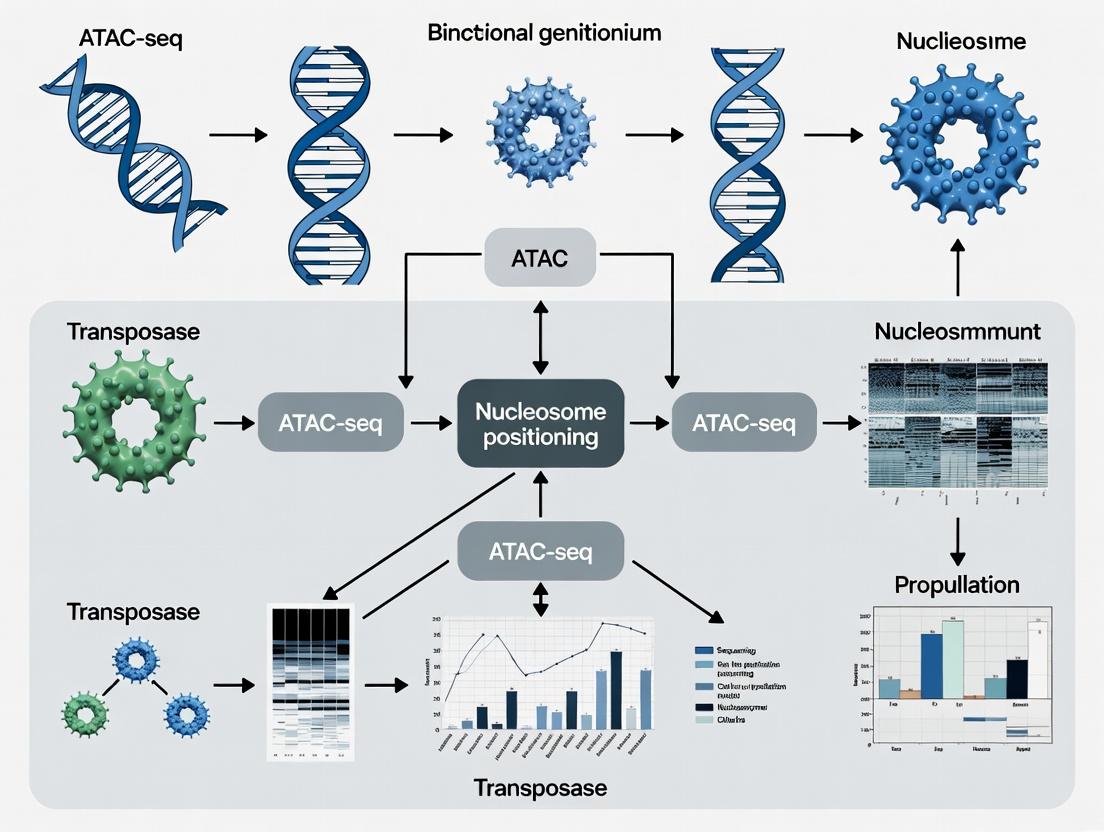
This comprehensive guide details the ATAC-seq (Assay for Transposase-Accessible Chromatin with high-throughput sequencing) protocol for mapping nucleosome positions, a critical determinant of chromatin architecture and gene regulation.
ATAC-seq Nucleosome Positioning Protocol: A Complete Guide for Researchers and Drug Discovery Scientists
Abstract
This comprehensive guide details the ATAC-seq (Assay for Transposase-Accessible Chromatin with high-throughput sequencing) protocol for mapping nucleosome positions, a critical determinant of chromatin architecture and gene regulation. We provide foundational knowledge on nucleosome biology and the assay's principle, followed by a step-by-step methodological workflow from cell preparation to data generation. Practical sections address common troubleshooting, optimization for challenging samples, and strategies for data validation and cross-method comparison. Designed for researchers, scientists, and drug development professionals, this article bridges technical execution with biological interpretation to empower epigenomic studies in development, disease, and therapeutic discovery.
Understanding the Nucleosome Landscape: The Why and How of ATAC-seq for Chromatin Accessibility
Within the nucleus of eukaryotic cells, genomic DNA is intricately packaged into a dynamic polymer called chromatin. This architecture is not merely for compaction; it is a fundamental regulator of all DNA-templated processes, including gene expression, DNA replication, and repair. The primary repeating unit of chromatin is the nucleosome, comprising approximately 147 base pairs of DNA wrapped around a histone octamer core. The positioning and chemical modification of nucleosomes, along with higher-order folding, create a landscape that determines the accessibility of regulatory elements. This article, framed within a broader thesis on ATAC-seq nucleosome positioning protocol research, details the core principles of chromatin architecture and provides application-focused protocols for its analysis, directly relevant to researchers in drug development targeting epigenetic mechanisms.
Core Components and Quantitative Data
Table 1: Core Components of Chromatin Architecture
| Component | Molecular Composition | Primary Function | Key Quantitative Metrics |
|---|---|---|---|
| DNA | Double-stranded helix of nucleotides. | Carries genetic information. | ~3.2 billion bp (human genome). |
| Histone Octamer | (H2A, H2B, H3, H4) x 2. | Protein core for DNA wrapping. | 8 histone molecules per nucleosome. |
| Nucleosome Core Particle | Histone octamer + 147 bp DNA. | Primary packaging unit. | ~1.65 left-handed superhelical turns. |
| Linker DNA | DNA between nucleosomes. | Connects core particles; binding site for linker histone H1. | Varies from ~10 to 80 bp (species/cell-type dependent). |
| Chromatosome | Nucleosome + linker histone H1. | Stabilizes nucleosome and promotes higher-order folding. | Binds ~20 additional bp of DNA (total ~167 bp). |
| 30-nm Fiber | Array of nucleosomes folded into a solenoid. | Secondary level of chromatin compaction. | Theoretical model; in vivo existence debated. |
Table 2: Common Histone Modifications and Functional Impact
| Modification Type | Common Sites (Human Histone H3) | General Regulatory Consequence | Enzymes (Examples) |
|---|---|---|---|
| Acetylation | K9, K14, K27, K56. | Neutralizes charge, reduces histone-DNA affinity, promotes open chromatin (euchromatin). | Histone Acetyltransferases (HATs) like p300/CBP. |
| Mono-/Di-/Tri-Methylation | K4me3, K36me3, K79me3. | Associated with active transcription. | SET1/COMPASS, SETD2, DOT1L. |
| Tri-Methylation | K9me3, K27me3. | Associated with transcriptionally silent heterochromatin. | SUV39H1/2, EZH2 (PRC2 complex). |
| Phosphorylation | S10, S28 (H3); S139 (H2AX, γH2AX). | Chromatin condensation during mitosis; DNA damage response. | Aurora B kinase, ATM/ATR. |
Detailed Protocol: Nucleosome Positioning via ATAC-seq
Title: Protocol for Nucleosome Positioning Analysis Using ATAC-seq
I. Principle: The Assay for Transposase-Accessible Chromatin with high-throughput sequencing (ATAC-seq) uses a hyperactive Tn5 transposase to simultaneously fragment and tag accessible genomic regions with sequencing adapters. Regions protected by nucleosomes are less accessible, leading to a pattern of insert sizes with a ~200-bp periodicity corresponding to nucleosome-bound DNA.
II. Reagent Solutions & Materials (The Scientist's Toolkit)
| Reagent/Material | Function/Description | Example Vendor/Catalog |
|---|---|---|
| Hyperactive Tn5 Transposase | Engineered enzyme for simultaneous DNA cutting and adapter tagging. | Illumina (Tagment DNA TDE1 Enzyme). |
| Nuclei Isolation Buffer | (10 mM Tris-Cl pH 7.5, 10 mM NaCl, 3 mM MgCl2, 0.1% IGEPAL CA-630). Lyses plasma membrane while keeping nuclear membrane intact. | Prepare fresh or use commercial kits. |
| Magnetic Beads (SPRI) | For post-tagmentation DNA clean-up and size selection. | Beckman Coulter (AMPure XP). |
| qPCR Kit for Library QC | Quantify library yield and check for adapter-dimer contamination. | Kapa Biosystems (Kapa Library Quant Kit). |
| High-Sensitivity DNA Bioanalyzer/PCR Assay | Assess fragment size distribution. | Agilent (Bioanalyzer High Sensitivity DNA chip). |
| Indexed Sequencing Adapters | For multiplexing samples during sequencing. | Illumina (Nextera Index Kit). |
III. Step-by-Step Methodology
- Cell Harvesting & Lysis: Harvest 50,000 - 100,000 viable cells. Wash with cold PBS. Resuspend pellet in 50 µL of cold Nuclei Isolation Buffer. Incubate on ice for 3-10 minutes (monitor under microscope for released nuclei). Immediately add 1 mL of cold Wash Buffer (10 mM Tris-Cl pH 7.5, 10 mM NaCl, 3 mM MgCl2, in nuclease-free water) and centrifuge at 500 rcf for 10 min at 4°C.
- Tagmentation: Resuspend the nuclei pellet in 25 µL of Tagmentation Mix (12.5 µL 2x Tagmentation Buffer, 2.5 µL Tn5 Transposase, 10 µL nuclease-free water). Mix gently and incubate at 37°C for 30 minutes in a thermomixer.
- DNA Purification: Immediately add 250 µL of DNA Binding Buffer to the tagmentation reaction. Purify using SPRI magnetic beads per manufacturer's protocol. Elute DNA in 21 µL of Elution Buffer (10 mM Tris-Cl, pH 8.0).
- PCR Amplification: Amplify the tagmented DNA using 25 µL of 2x PCR Master Mix, 2.5 µL of forward and 2.5 µL of reverse indexed primers (1.25 µM final), and the 21 µL eluted DNA. PCR cycle: 72°C for 5 min (gap filling); 98°C for 30 sec; then 5-12 cycles of [98°C for 10 sec, 63°C for 30 sec, 72°C for 1 min].
- Library Clean-up & Size Selection: Purify the PCR product with SPRI beads using a 0.5x-1.2x double-sided size selection to remove primer dimers (<100 bp) and large fragments (>1kb). Elute in 20 µL Elution Buffer.
- Quality Control & Sequencing: Quantify library concentration by qPCR. Analyze fragment size distribution on a Bioanalyzer. A successful library should show a nucleosomal ladder pattern with a prominent ~200 bp mononucleosome peak and a ~400 bp dinucleosome peak. Sequence on an Illumina platform (minimum 50M paired-end reads for human genomes).
IV. Data Analysis Workflow for Nucleosome Positioning:
- Preprocessing: Adapter trimming (e.g., Trimmomatic) and alignment to reference genome (e.g., using BWA-MEM).
- Fragment Size Distribution: Plot the distribution of insert sizes from aligned paired-end reads. A periodic enrichment of fragments at ~200-bp intervals indicates nucleosome positioning.
- Nucleosome Calling: Use specialized tools (e.g., NucleoATAC, DANPOS2, or HMM-based methods) to identify genomic coordinates of well-positioned nucleosomes by detecting peaks in the fragment midpoint density plot at a specific size range (~180-250 bp).
- Differential Accessibility: Compare fragment size distributions and nucleosome occupancy between experimental conditions using software like DiffBind or by custom analysis of protected vs. accessible signals.
Visualization of Chromatin Architecture & ATAC-seq Workflow
The Biological Significance of Nucleosome Positioning in Health and Disease
Abstract: Precise nucleosome positioning governs chromatin accessibility, thereby regulating all DNA-templated processes. Dysregulation of this positioning is a hallmark of numerous diseases, including cancer, neurodevelopmental disorders, and autoimmune conditions. This document, framed within a thesis on ATAC-seq protocol development, provides application notes and detailed protocols for interrogating nucleosome positioning, linking experimental data to biological and clinical significance.
Application Notes: Quantitative Insights into Nucleosome Positioning
Table 1: Nucleosome Positioning Alterations in Disease States
| Disease Context | Genomic Region Affected | Observed Change in Positioning/Spacing | Associated Functional Consequence | Key Supporting Study (Method) |
|---|---|---|---|---|
| Colorectal Cancer | Promoters of tumor suppressor genes (e.g., p53) | Increased nucleosome occupancy & stability | Transcriptional silencing, reduced DNA damage response | Zhu et al., 2018 (MNase-seq) |
| Rett Syndrome (MECP2 mutation) | Neuronal gene enhancers | Eroded nucleosome phasing, loss of periodic spacing | Dysregulated neuronal differentiation gene expression | Iurlaro et al., 2021 (ATAC-seq) |
| Systemic Lupus Erythematosus (SLE) | Global & interferon-responsive genes | Global reduction of nucleosome occupancy | Increased immunogenic self-DNA exposure, autoantibody production | Garcia-Romo et al., 2020 (Cell-free DNA analysis) |
| Yeast Aging Model | Ribosomal DNA (rDNA) repeats | Loss of positioned nucleosomes, increased accessibility | Genomic instability, rDNA recombination | Hu et al., 2014 (MNase-seq) |
Table 2: Key Metrics from ATAC-seq Nucleosome Positioning Analysis
| Metric | Description | Typical Value in Healthy Cells | Interpretation of Deviation |
|---|---|---|---|
| Nucleosome Spacing Periodicity | Peak-to-peak distance in ATAC-seq insert size plot. | ~200 bp | <180 bp suggests compaction; >220 bp suggests irregular positioning. |
| NFR (Nucleosome-Free Region) Score | Accessibility signal at Transcription Start Sites (TSS). | High, sharp peak at TSS. | Broadening or reduction indicates promoter occlusion and likely silencing. |
| Occupancy Variance | Consistency of nucleosome positions across a cell population. | Low variance at phased nucleosomes. | High variance indicates loss of precise positioning (e.g., epigenetic drift). |
Detailed Experimental Protocols
Protocol 2.1: ATAC-seq for Nucleosome Positioning Mapping (Optimized for Frozen Tissues)
Principle: The Assay for Transposase-Accessible Chromatin uses the Tn5 transposase to insert sequencing adapters into open genomic regions. The periodicity of fragment lengths corresponds to nucleosome-protected DNA (mono-, di-, tri-nucleosome).
Materials & Reagents: See "The Scientist's Toolkit" below. Procedure:
- Nuclei Isolation from Snap-Frozen Tissue:
- Homogenize 10-20 mg tissue in 1 mL of pre-chilled Lysis Buffer (10 mM Tris-HCl pH 7.4, 10 mM NaCl, 3 mM MgCl2, 0.1% IGEPAL CA-630, 0.1% Tween-20, 0.01% Digitonin) using a Dounce homogenizer (15 strokes).
- Filter homogenate through a 40-μm cell strainer.
- Pellet nuclei at 500 rcf for 5 min at 4°C in a fixed-angle centrifuge. Resuspend pellet in 50 μL of chilled Wash Buffer (Lysis Buffer without detergents). Count nuclei using a hemocytometer.
Tagmentation Reaction:
- Combine 25,000 nuclei (in 5 μL) with 10 μL of TD Buffer, 2.5 μL of Tn5 Transposase (Illumina), and 32.5 μL of nuclease-free water. Mix gently.
- Incubate at 37°C for 30 minutes in a thermomixer with shaking (300 rpm).
- Immediately purify DNA using a MinElute PCR Purification Kit. Elute in 21 μL of Elution Buffer.
Library Amplification & Size Selection:
- Amplify the tagmented DNA using 2x KAPA HiFi HotStart ReadyMix and 1.25 μM of custom Nextera PCR primers for 10-12 cycles.
- Clean up PCR product with 1.2x SPRIselect beads. Perform a double-size selection:
- First, with 0.55x beads: Keep supernatant (contains small fragments < ~300 bp, primarily nucleosome-free).
- Second, with 0.2x beads (on supernatant from first): Pellet and elute fragments > ~300 bp (enriched for mono-/di-nucleosome fragments).
- Pool both fractions at a 4:1 ratio (nucleosome-free : nucleosome-enriched) for balanced sequencing. Quantify with qPCR and/or Bioanalyzer.
Protocol 2.2: Bioinformatic Analysis of Nucleosome Positions from ATAC-seq
Processing & Alignment:
- Use
fastpfor adapter trimming and quality filtering. - Align reads to reference genome (e.g., hg38) using
bowtie2with--very-sensitive -X 2000parameters. - Remove duplicates and mitochondrial reads using
samtoolsandpicard.
- Use
Nucleosome Positioning Call:
- Generate insert size distribution plot using
ools likeATACseqQC`. - Use
NucleoATAC(Schep et al., Nat Methods, 2015) to call nucleosome positions and occupancy scores from the combined signal of nucleosome-length fragments. - Identify Nucleosome Depleted Regions (NDRs) and phased nucleosome arrays.
- Generate insert size distribution plot using
Differential Analysis:
- Compare NDR accessibility (using
DESeq2on counts from sub-nucleosomal fragments) and nucleosome occupancy shifts (usinglimmaon NucleoATAC occupancy scores) between conditions.
- Compare NDR accessibility (using
Diagrams & Visualizations
Title: ATAC-seq Principle for Nucleosome Mapping
Title: ATAC-seq Nucleosome Positioning Workflow
Title: Disease Pathways from Nucleosome Dysregulation
The Scientist's Toolkit: Key Research Reagent Solutions
| Item / Reagent | Function in Protocol | Critical Notes |
|---|---|---|
| Tn5 Transposase (Illumina, #20034197) | Enzyme that simultaneously fragments and tags accessible DNA. | Lot-to-lot activity variance is key; pre-test titration for each new batch. |
| Digitonin (Sigma, #D141) | Mild detergent for nuclear membrane permeabilization during nuclei isolation. | Concentration is critical (0.01-0.1%). Too high causes lysis; too low gives poor tagmentation. |
| SPRIselect Beads (Beckman Coulter, #B23318) | Magnetic beads for post-PCR cleanup and precise size selection of DNA fragments. | Enables separation of sub-nucleosomal (<100 bp) and nucleosomal (~200+ bp) fragments. |
| NucleoATAC Software Package | Computational tool specifically designed to call nucleosome positions from ATAC-seq data. | Requires paired-end sequencing. More accurate than simple insert size filtering. |
| KAPA HiFi HotStart ReadyMix (Roche) | High-fidelity PCR mix for library amplification post-tagmentation. | Minimizes PCR bias and over-amplification artifacts, crucial for quantitative accuracy. |
| Dounce Homogenizer (tight pestle) | For mechanical disruption of tough tissues (e.g., heart, tumor) to release intact nuclei. | Preferred over enzymatic digestion for speed and minimal chromatin state alteration. |
Application Notes
ATAC-seq (Assay for Transposase-Accessible Chromatin using sequencing) is a foundational method in functional genomics, enabling genome-wide mapping of chromatin accessibility. Within the broader thesis on ATAC-seq nucleosome positioning protocol research, this technique is pivotal for elucidating the dynamic regulation of gene expression. The core principle relies on the hyperactive Tn5 transposase, which simultaneously fragments and tags open chromatin regions with sequencing adapters. This process selectively targets nucleosome-free regions (NFRs), while protected DNA wrapped around nucleosomes remains largely inaccessible, allowing for inference of nucleosome positions.
Recent advancements have focused on high-throughput single-cell applications (scATAC-seq), integration with other omics modalities (multiome), and enhanced protocols for low-input and frozen samples. For drug development professionals, ATAC-seq provides critical insights into disease-specific regulatory landscapes, mechanisms of action for therapeutics, and identification of novel biomarkers by comparing chromatin accessibility states between conditions.
Data Presentation: Key Quantitative Metrics in ATAC-seq
Table 1: Typical ATAC-seq Output Metrics and Benchmarks
| Metric | Typical Range / Value | Interpretation |
|---|---|---|
| Sequencing Depth (bulk) | 50 - 100 million reads | Saturation for peak calling. |
| Sequencing Depth (single-cell) | 25,000 - 100,000 reads/cell | Varies by protocol and analysis. |
| Fraction of Reads in Peaks (FRiP) | >20% (bulk), >15% (sc) | Key quality metric for signal-to-noise. |
| Transposition Reaction Time | 30 min - 1 hour | Standard incubation at 37°C. |
| Nucleosomal Fragment Periodicity | ~200 bp ladder on gel/ bioanalyzer | Indicator of successful nucleosome discrimination. |
| Percentage of Mitochondrial Reads | <20% (optimized) | High % indicates excessive cell lysis or low nuclear quality. |
| Tn5 Transposase Concentration | 100-200 nM in final reaction | Critical for optimal tagmentation efficiency. |
Table 2: Comparison of Common ATAC-seq Protocol Variants
| Protocol Variant | Input Material | Key Application | Primary Advantage |
|---|---|---|---|
| Standard Bulk ATAC-seq | 50,000 - 100,000 fresh cells | Regulatory landscape mapping | Robust, high signal-to-noise. |
| Omni-ATAC | Cultured cells, tissues | Challenging samples (e.g., with high mitochondria) | Reduced mitochondrial artifacts. |
| Fast-ATAC | Any bulk sample | Rapid protocol | Incorporates a PCR purification step for speed. |
| scATAC-seq (10x Genomics) | Single cell suspension | Cellular heterogeneity | Profiles thousands of individual cells. |
| ATAC-me | Low cell numbers (500-5k) | Multiomic profiling (accessibility + methylation) | Simultaneous DNAme and accessibility. |
Experimental Protocols
Protocol 1: Standard Bulk ATAC-seq for Nucleosome Positioning Analysis
Context: This protocol is the workhorse for generating data on nucleosome occupancy and positioning as part of the core thesis research.
Reagents & Equipment:
- Nuclei Isolation Buffer (10 mM Tris-HCl pH 7.4, 10 mM NaCl, 3 mM MgCl2, 0.1% IGEPAL CA-630)
- Tagmentation DNA Buffer (Illumina, or equivalent homemade Tn5 buffer)
- Hyperactive Tn5 Transposase (commercially available, e.g., Illumina Tagmentase)
- DNA Cleanup Beads (SPRI beads)
- Qubit dsDNA HS Assay Kit
- Bioanalyzer High Sensitivity DNA Kit or TapeStation
- Thermal cycler with heated lid
- Gentle tube rotator
Detailed Methodology:
- Cell Harvesting & Lysis: Harvest 50,000-100,000 viable cells. Pellet at 500 x g for 5 min at 4°C. Resuspend pellet in 50 µL of cold Lysis Buffer. Incubate on ice for 3-10 minutes (optimize per cell type) to lyse the cytoplasmic membrane. Immediately add 1 mL of cold Wash Buffer and invert.
- Nuclei Isolation & Counting: Pellet nuclei at 500 x g for 10 min at 4°C. Carefully aspirate supernatant. Resuspend nuclei in 50 µL of Transposition Mix (25 µL Tagmentation DNA Buffer, 2.5 µL Tn5 Transposase (100-200 nM final), 22.5 µL Nuclease-free water). Count nuclei using a hemocytometer for precise normalization.
- Tagmentation: Incubate the Transposition Mix with nuclei at 37°C for 30 minutes in a thermal cycler with gentle agitation (if available). Immediately add 250 µL of DNA Binding Buffer from a cleanup kit to stop the reaction.
- DNA Purification: Purify the tagmented DNA using SPRI beads at a 1.8x ratio. Elute in 20 µL of Elution Buffer (10 mM Tris pH 8.0).
- Library Amplification: Amplify the purified DNA using a limited-cycle PCR program (typically 10-12 cycles). Use indexed primers for multiplexing. Include SYBR Green in the reaction to qPCR-amplify just to the point of saturation to minimize PCR duplicates.
- Library Cleanup & QC: Perform a double-sided SPRI bead cleanup (e.g., 0.55x and 1.2x ratios) to remove primer dimers and select for properly tagmented fragments (typically 100-700 bp). Quantify library concentration (Qubit) and profile fragment distribution (Bioanalyzer). Expect a characteristic nucleosomal ladder pattern.
- Sequencing: Pool libraries and sequence on an Illumina platform using paired-end sequencing (e.g., PE42+42 or PE50+50).
Protocol 2: Omni-ATAC for Complex Tissues/Frozen Samples
Context: A critical optimization for thesis work involving primary tissue samples, which often have high mitochondrial content that can overwhelm sequencing reads.
Key Modifications to Standard Protocol:
- Nuclei Isolation: Use an isotonic lysis buffer (e.g., 10 mM Tris-HCl pH 7.4, 10 mM NaCl, 3 mM MgCl2, 0.1% Tween-20, 0.1% IGEPAL CA-630, 0.01% Digitonin) for 3 minutes on ice. This gentle, digitonin-enhanced lysis improves nuclear integrity.
- Nuclei Purification: Layer the lysate onto a cushion of 30% Iodixanol in PBS and centrifuge at 1,000 x g for 10 min. This step pellets nuclei while leaving cytoplasmic debris and unlysed cells at the interface.
- Tagmentation Buffer: Supplement the standard tagmentation buffer with 0.01% Digitonin to enhance Tn5 penetration.
- Mitochondrial DNA Depletion (Optional): Post-PCR amplification, treat the library with 5 U of exonuclease V at 37°C for 30 min to degrade linear mitochondrial DNA fragments, followed by SPRI bead cleanup.
Mandatory Visualizations
Diagram Title: ATAC-seq Core Workflow: Tagmentation to Analysis
Diagram Title: Tn5 Transposase Tagmentation Mechanism
The Scientist's Toolkit: Key Research Reagent Solutions
Table 3: Essential Materials for ATAC-seq Experiments
| Item | Example Product/Kit | Function in ATAC-seq |
|---|---|---|
| Hyperactive Tn5 Transposase | Illumina Tagmentase TDE1, Nextera Tn5 | Engineered enzyme core that cuts DNA and ligates adapters simultaneously. |
| Tagmentation Buffer | Illumina Tagmentation Buffer | Provides optimal ionic and cofactor conditions (Mg2+) for Tn5 activity. |
| Nuclei Isolation Reagents | IGEPAL CA-630, Digitonin, Tween-20 | Non-ionic detergents for controlled cell membrane lysis and nuclear permeabilization. |
| SPRI (Solid Phase Reversible Immobilization) Beads | AMPure XP, SPRIselect | Magnetic beads for size-selective DNA cleanup and library purification. |
| Indexed PCR Primers | Illumina Index Kit, Nextera XT Index Kit | Adds unique sample barcodes and full sequencing adapters during PCR. |
| High-Sensitivity DNA QC Kit | Agilent High Sensitivity DNA Kit, Fragment Analyzer | Assesses library fragment size distribution and detects nucleosomal ladder. |
| Cell Viability Stain | Trypan Blue, DAPI, Propidium Iodide | Critical for accurately counting viable cells/nuclei pre-tagmentation. |
| Sucrose or Iodixanol Solution | OptiPrep Density Gradient Medium | Used in nuclei purification steps to remove cytoplasmic debris (Omni-ATAC). |
Key Advantages of ATAC-seq for Nucleosome Mapping vs. Historical Methods (MNase-seq)
This application note, framed within a broader thesis on ATAC-seq nucleosome positioning protocol research, delineates the key advantages of the Assay for Transposase-Accessible Chromatin with high-throughput sequencing (ATAC-seq) over the historical Micrococcal Nuclease sequencing (MNase-seq) method for nucleosome mapping, providing detailed protocols and analytical tools.
Comparative Analysis: ATAC-seq vs. MNase-seq
Table 1: Core Methodological and Output Comparison
| Parameter | ATAC-seq | MNase-seq |
|---|---|---|
| Core Principle | Transposase insertion into open chromatin. | Nuclease digestion of linker DNA. |
| Primary Input | Live cells or nuclei (50k-100k optimal). | High chromatin yield (millions of cells). |
| Hands-on Time | ~3-4 hours (rapid protocol). | ~1-2 days (including digestion optimization). |
| Simultaneous Output | Nucleosome positions + open chromatin (footprints). | Primarily nucleosome positions. |
| Resolution | Single-nucleotide for accessibility; ~10-150bp for nucleosomes. | ~10-150bp for nucleosomes. |
| Artifact Susceptibility | Sequence bias of Tn5 transposase. | MNase sequence & digestion bias (e.g., AT-rich). |
| Key Advantage | Speed, sensitivity, dual data from one assay. | Historical gold standard for nucleosome occupancy. |
Table 2: Quantitative Performance Metrics from Recent Studies
| Metric | ATAC-seq Performance | MNase-seq Performance | Implication |
|---|---|---|---|
| Cell Number Requirement | As low as 500-50,000 cells | Typically 1-10 million cells | ATAC-seq enables rare sample & single-cell analysis. |
| Mapping Reproducibility (Pearson R) | R > 0.95 between technical replicates for nucleosome signal. | R > 0.90, but varies with digestion level. | ATAC-seq offers high reproducibility with standardized steps. |
| Signal-to-Noise in Open Regions | High; clear sub-nucleosomal fragments (<100bp). | Moderate; requires careful digestion to avoid over-digestion. | ATAC-seq provides clearer demarcation of accessible regions. |
| GC Bias in Reads | Moderate Tn5 insertion sequence bias. | High MNase cleavage bias towards AT-rich DNA. | ATAC-seq offers more uniform genome coverage. |
Detailed Experimental Protocols
Protocol 1: ATAC-seq for Nucleosome Positioning (Omni-ATAC Modification)
This protocol is optimized for mapping nucleosome positions alongside accessibility from fresh cells.
A. Cell Lysis and Transposition
- Harvest & Wash: Collect 50,000 viable cells. Wash once with 50µL cold 1x PBS.
- Lysis: Resuspend pellet in 50µL cold ATAC-seq Lysis Buffer (10mM Tris-Cl pH 7.4, 10mM NaCl, 3mM MgCl2, 0.1% IGEPAL CA-630). Invert to mix. Incubate on ice for 3 minutes.
- Wash: Immediately add 1mL of cold Wash Buffer (1x PBS, 0.1% BSA, 0.2U/µL RNase Inhibitor). Invert gently. Pellet nuclei at 500 rcf for 10 minutes at 4°C. Remove supernatant.
- Tagmentation: Prepare the 50µL tagmentation reaction mix directly on the nuclei pellet: 25µL 2x TD Buffer, 2.5µL Tn5 Transposase (Illumina), 16.5µL PBS, 0.5µL 1% Digitonin, 0.5µL 10% Tween-20, 5µL H2O. Mix gently by pipetting 10 times.
- Incubate at 37°C for 30 minutes in a thermomixer with shaking (1000 rpm). Immediately proceed to DNA purification.
B. DNA Purification and Library Amplification
- Purify: Add 250µL of DNA Binding Beads (e.g., SPRIselect) to the 50µL tagmentation reaction. Mix. Incubate 5 minutes at RT. Place on magnet for 5 minutes. Discard supernatant.
- Wash: With beads on magnet, wash twice with 200µL 80% ethanol. Air dry for 5 minutes.
- Elute: Elute DNA in 22µL Elution Buffer (10mM Tris pH 8.0).
- Amplify: To the eluate, add 2µL of a 25µM PCR Primer Ad1, 2µL of a 25µM uniquely barcoded PCR Primer Ad2, and 25µL of 2x NEB Next High-Fidelity PCR Master Mix. Cycle: 72°C 5 min; 98°C 30 sec; then 5-10 cycles of (98°C 10 sec, 63°C 30 sec, 72°C 1 min). Use 5-cycle qPCR to determine optimal cycles.
- Final Cleanup: Purify library with 1.2x ratio of SPRIselect beads. Elute in 20µL EB. Quantify via qPCR or Bioanalyzer.
Protocol 2: MNase-seq for Nucleosome Mapping
This protocol details controlled digestion to generate mononucleosomal DNA.
A. Chromatin Digestion & Optimization
- Isolate Nuclei: From 1-5 million cells, isolate nuclei using NP-40 or hypotonic lysis. Pellet nuclei.
- MNase Titration: Resuspend nuclei in 500µL MNase Digestion Buffer (50mM Tris-Cl pH 7.9, 5mM CaCl2). Split into 5 aliquots.
- Add MNase enzyme (2-20 units) to different tubes. Incubate at 37°C for 5-10 minutes.
- Stop Reaction: Add 10µL of 0.5M EDTA (pH 8.0) to each tube. Place on ice.
- Analysis: Purify DNA from each aliquot (Proteinase K, RNase A, phenol-chloroform). Run on 2% agarose gel. Select the condition yielding ~80% DNA as a ~150bp mononucleosome band.
- Scale-Up: Perform optimized digestion on the remaining sample.
B. Mononucleosome DNA Isolation and Library Prep
- Size Selection: Run the bulk purified DNA on a 2% low-melt agarose gel.
- Excise the gel slice corresponding to 140-160bp.
- Purify DNA using a Gel Extraction kit.
- Library Construction: Use standard Illumina library prep kits (end-repair, A-tailing, adapter ligation). Amplify with 10-14 PCR cycles. Size-select again post-amplification.
Visualizations
Workflow Comparison: ATAC-seq vs. MNase-seq
ATAC-seq Data Analysis for Nucleosomes
The Scientist's Toolkit: Essential Research Reagents & Materials
Table 3: Key Reagent Solutions for ATAC-seq Nucleosome Mapping
| Item | Function/Benefit | Example/Note |
|---|---|---|
| Tn5 Transposase | Engineered enzyme that simultaneously fragments and tags accessible DNA with sequencing adapters. Core of ATAC-seq. | Illumina Tagmentase TDE1, or custom loaded Tn5. |
| Digitonin (or alternative) | Mild detergent used in lysis and tagmentation buffers to permeabilize nuclear membranes for Tn5 entry. Critical for signal. | Use low concentration (0.01-0.1%) in Omni-ATAC protocol. |
| SPRIselect Beads | Solid-phase reversible immobilization (SPRI) beads for size-selective cleanup and purification of DNA libraries. | Enable precise size selection and adapter removal. |
| Dual-Size SPRI Beads | Beads used at different ratios (e.g., 0.5x then 1.2x) to sequentially remove large fragments and small primers/adapters. | Cleans up tagmented DNA before PCR. |
| RNase Inhibitor | Prevents degradation of RNA that can co-purify and interfere with library preparation or quantification. | Added to lysis/wash buffers. |
| High-Fidelity PCR Mix | PCR master mix with low error rate for minimal amplification bias during library amplification. | NEB Next Q5, KAPA HiFi. |
| Nuclei Isolation Buffer | Isotonic buffer with detergent to lyse plasma membrane while keeping nuclei intact. | Often contains sucrose, MgCl2, and a detergent like NP-40. |
| Indexed PCR Primers | Unique barcoded primers (i5 and i7) for multiplexing multiple samples in a single sequencing run. | Illumina Nextera or custom i5/i7 indexes. |
This document outlines critical pre-experimental planning for ATAC-seq (Assay for Transposase-Accessible Chromatin using sequencing) studies, particularly within the context of a comprehensive thesis on nucleosome positioning protocol research. Proper design is paramount for generating robust, reproducible data on chromatin accessibility and nucleosome architecture, which informs drug target discovery and mechanistic studies.
Foundational Experimental Design Considerations
The core experimental design must address specific biological questions while controlling for technical variability. Key quantitative parameters are summarized below.
Table 1: Key Quantitative Design Parameters for ATAC-seq Experiments
| Parameter | Typical Range/Value | Consideration & Impact |
|---|---|---|
| Number of Biological Replicates | 3-5 per condition | Essential for statistical power; <3 compromises reproducibility and differential analysis. |
| Cell Number per Reaction | 50,000 - 100,000 viable cells | Lower counts increase noise; higher counts can lead to incomplete transposition. |
| Transposition Time | 30 min at 37°C | Standard for nuclei; optimizable. Over-digestion creates too-small fragments. |
| Sequencing Depth | 25-50 million reads/sample (standard) | Sufficient for chromatin accessibility. Nucleosome positioning requires deeper sequencing (50-80M+). |
| Read Configuration | Paired-end (PE), minimum 2x50 bp | PE reads are mandatory for nucleosome positioning analysis (mononucleosome ~200bp fragments). |
| Fragment Size Analysis | Sub-nucleosomal (<100 bp), Mono- (180-250 bp), Di- (350-500 bp) | Categories define accessible TF binding sites, nucleosome positioning, and higher-order structure. |
Detailed Protocol: Nuclei Preparation from Cultured Cells for ATAC-seq
This protocol is optimized for mammalian cells to ensure clean nuclei isolation prior to transposition.
Materials:
- Cold PBS
- Cell Dissociation Reagent (if needed)
- Lysis Buffer (10 mM Tris-HCl pH 7.4, 10 mM NaCl, 3 mM MgCl2, 0.1% IGEPAL CA-630)
- Wash Buffer (10 mM Tris-HCl pH 7.4, 10 mM NaCl, 3 mM MgCl2)
- Nuclei Buffer (10 mM Tris-HCl pH 7.4, 10 mM NaCl, 3 mM MgCl2, 1% BSA, 0.1% Tween-20)
- Trypan Blue and hemocytometer or automated cell counter
- Refrigerated centrifuge
Methodology:
- Cell Harvesting: Gently dissociate adherent cells. Pellet cells at 500 x g for 5 min at 4°C. Wash pellet with 1 mL of cold PBS.
- Cell Counting: Resuspend pellet in PBS and perform a live-cell count. This is critical for downstream transposition efficiency.
- Cell Lysis: Pellet the desired number of cells (aim for 100,000). Carefully resuspend the cell pellet in 50 μL of cold Lysis Buffer by pipetting gently. Incubate on ice for 3-10 minutes (monitor lysis under microscope).
- Nuclei Washing: Immediately add 1 mL of cold Wash Buffer to stop lysis. Pellet nuclei at 500 x g for 10 min at 4°C.
- Nuclei Resuspension: Carefully decant supernatant. Resuspend the nuclei pellet in 50 μL of Nuclei Buffer. Perform a nuclei count if needed.
- Proceed to Transposition: The nuclei are now ready for the tagmentation reaction using the Th5 transposase, typically following the manufacturer's instructions (e.g., Illumina Tagment DNA TDE1 Kit). Use immediately for best results.
The Scientist's Toolkit: Key Research Reagent Solutions
Table 2: Essential Materials for ATAC-seq Sample Preparation
| Item | Function & Rationale |
|---|---|
| Tagment DNA TDE1 Enzyme (Illumina) | Engineered Th5 transposase pre-loaded with sequencing adapters. Simultaneously fragments accessible DNA and adds adapters for PCR. |
| IGEPAL CA-630 (Nonidet P-40) | Non-ionic detergent for gentle, controlled cell membrane lysis while leaving nuclear membrane intact. |
| Qiagen MinElute PCR Purification Kit | Size-selective purification post-tagmentation to remove small fragments and reaction components. Critical for library quality. |
| High-Fidelity PCR Master Mix (e.g., NEB Next) | For limited-cycle PCR to amplify tagmented DNA and add full sequencing adapters with indexes. |
| SPRIselect Beads (Beckman Coulter) | For post-PCR size selection to isolate the optimal fragment range (e.g., removal of >1kb fragments and excess primers). |
| Bioanalyzer/TapeStation (Agilent) | Microfluidic capillary electrophoresis for accurate quantification and size distribution analysis of final libraries. |
| Qubit Fluorometer & dsDNA HS Assay (Thermo Fisher) | Highly specific fluorescent quantification of double-stranded library DNA, more accurate than UV spectrometry for dilute samples. |
Workflow and Analytical Pathways
Title: ATAC-seq Workflow from Design to Analysis
Title: Fragment Size Analysis for Nucleosome Positioning
Step-by-Step ATAC-seq Protocol: From Cell Lysis to Sequencing Library
Within the broader thesis on ATAC-seq nucleosome positioning protocol research, the initial steps of cell preparation, accurate counting, and high-quality nuclei isolation are critically determinant of experimental success. These steps directly impact chromatin accessibility, library complexity, and the signal-to-noise ratio in final sequencing data. This application note details current best practices to ensure the integrity of starting material for downstream transposition and sequencing.
Cell Preparation: Sourcing and Handling
Key Considerations
Cell viability must exceed 90% for ATAC-seq. Apoptotic or stressed cells release nucleases that degrade chromatin, creating spurious open chromatin signals. Primary cells are particularly sensitive to handling. Adherent cells should be dissociated using gentle, non-enzymatic methods where possible (e.g., cell scraping in cold PBS) to prevent surface protein digestion and unintended signaling. Enzymatic dissociation (trypsin/Accutase) should be minimized and quenched thoroughly with complete medium. Suspension cells should be collected with minimal centrifugation force (300-400 x g for 5 minutes at 4°C). All washes and resuspensions should be performed with ice-cold, nuclease-free PBS to arrest cellular activity and preserve native chromatin state.
Protocol: Gentle Harvesting of Adherent Cells for ATAC-seq
- Pre-chill all buffers and equipment on ice.
- Aspirate culture medium and wash the monolayer gently with 10 mL of room temperature PBS to remove serum.
- Aspirate PBS and add 3 mL of room temperature Accutase or gentle dissociation reagent. Incubate at 37°C for the minimum time required for detachment (typically 2-5 minutes).
- Gently tap the vessel to dislodge cells. Add 7 mL of pre-chilled complete growth medium (containing FBS) to quench the enzyme.
- Transfer the cell suspension to a 15 mL conical tube. Rinse the plate with 5 mL of cold PBS and pool.
- Centrifuge at 300 x g for 5 minutes at 4°C.
- Carefully aspirate supernatant, ensuring not to disturb the pellet.
- Resuspend the pellet in 1 mL of ice-cold, nuclease-free PBS by gentle pipetting. Proceed to counting.
Cell Counting and Viability Assessment
Accurate concentration and viability data are non-negotiable. Underestimating cell number leads to over-transposition ("overloading"), while overestimating leads to insufficient material. Automated counters (e.g., Countess, LUNA) provide superior reproducibility over manual hemocytometers.
Protocol: Automated Cell Counting with Trypan Blue
- Mix 10 µL of resuspended cells with 10 µL of 0.4% Trypan Blue stain.
- Load 10 µL of the mixture into a counting chamber slide.
- Insert into the automated cell counter and run analysis.
- Record Total Concentration (cells/mL), Viable Concentration (cells/mL), and Percent Viability.
- If viability is <90%, consider using a dead cell removal kit before proceeding.
Quantitative Data: Comparison of Counting Methods
Table 1: Comparison of Cell Counting Method Efficiencies
| Method | Principle | Time per Sample (min) | Coefficient of Variation | Viability Assessment? | Suitability for ATAC-seq |
|---|---|---|---|---|---|
| Manual Hemocytometer | Microscopic grid count | 5-10 | High (10-25%) | Yes, with dye | Acceptable, but prone to user error |
| Automated Cell Counter | Image analysis | <2 | Low (<5%) | Yes, with dye | Recommended for consistency |
| Flow Cytometry | Light scatter/fluorescence | 5-15 | Very Low (<2%) | Excellent, with propidium iodide | Excellent but higher cost and complexity |
Nuclei Isolation: The Critical Step
The goal is to obtain intact, nuclease-free nuclei with minimal cytoplasmic contamination. The use of a gentle, hypotonic lysis buffer containing a non-ionic detergent (NP-40 or Igepal CA-630) and edta/egta is standard. Optimization of lysis time is tissue- and cell-type-specific.
Protocol: Cold Detergent-Based Nuclei Isolation for ATAC-seq
Reagents Needed: Nuclei Isolation Buffer (NIB): 10 mM Tris-Cl pH 7.5, 10 mM NaCl, 3 mM MgCl2, 0.1% Igepal CA-630, 1% BSA, in nuclease-free water. Store at 4°C. Add 0.1% RNase Inhibitor and 1x Protease Inhibitor Cocktail immediately before use.
- After counting, pellet the required number of cells (typically 50,000-100,000 for a standard reaction). Centrifuge at 500 x g for 5 min at 4°C.
- Aspirate supernatant completely. Gently flick the tube to loosen the pellet.
- Resuspend the pellet in 50 µL of ice-cold NIB. Lyse cells by gentle pipetting 3-5 times with a wide-bore P200 tip. Do not vortex.
- Incubate on ice for 3-5 minutes. Monitor lysis under a microscope by mixing 2 µL of nuclei with 2 µL of Trypan Blue on a slide.
- Immediately after lysis, add 1 mL of ice-cold NIB without detergent (Wash Buffer) to dilute the detergent and stop lysis.
- Centrifuge nuclei at 500 x g for 5 min at 4°C.
- Carefully aspirate supernatant. The nuclei pellet is often translucent and easy to dislodge.
- Resuspend nuclei gently in the desired volume of transposition reaction mix or freezing medium. Count nuclei using the same method as for cells.
The Scientist's Toolkit: Key Reagents & Materials
Table 2: Essential Research Reagent Solutions for Cell & Nuclei Preparation
| Item | Function & Importance | Example Product/Buffer |
|---|---|---|
| Nuclease-Free PBS | Washing cells without degrading RNA/DNA; maintains osmolarity. | Ambion Nuclease-Free PBS, Thermo Fisher |
| Gentle Dissociation Reagent | Detaches adherent cells while minimizing surface protein cleavage and cellular stress. | Accutase, Sigma-Aldrich |
| Trypan Blue Stain (0.4%) | Differentiates live (unstained) from dead (blue) cells for viability assessment. | Gibco Trypan Blue Solution |
| Nuclei Isolation Buffer (NIB) | Hypotonic buffer with non-ionic detergent to lyse plasma membrane while leaving nuclear envelope intact. | 10mM Tris, 10mM NaCl, 3mMgCl2, 0.1% Igepal CA-630 |
| Protease Inhibitor Cocktail | Prevents endogenous proteases from degrading nuclear proteins, including histones. | cOmplete, EDTA-free, Roche |
| RNase Inhibitor | Preserves nuclear RNA and prevents RNase-mediated degradation that can indirectly affect stability. | Murine RNase Inhibitor, NEB |
| Wide-Bore/Low-Binding Pipette Tips | Prevents shear stress on nuclei and reduces material loss due to adhesion. | USA Scientific GenCatch Wide Bore Tips |
| BSA (Molecular Biology Grade) | Added to buffers to reduce non-specific sticking of nuclei to plastic surfaces. | New England Biolabs BSA |
Visualizing the Workflow and Critical Checkpoints
Title: ATAC-seq Cell & Nuclei Preparation Quality Control Workflow
Title: Impact of Prep Quality on ATAC-seq Chromatin Integrity & Data
Within the broader thesis on establishing a robust ATAC-seq protocol for mapping nucleosome positioning in primary patient samples, optimizing the Tn5 transposition reaction is the critical step that directly determines library complexity and data quality. This application note details systematic optimization experiments for three core parameters: reaction time, temperature, and Tn5 enzyme amount, to achieve maximum tagmentation efficiency while minimizing batch effects.
Table 1: Optimization of Tn5 Transposition Time (Constant: 37°C, 1x Tn5)
| Reaction Time (min) | Median Fragment Size (bp) | Unique Nuclear Fragments (x10^5) | % of Reads in Nucleosome Bands |
|---|---|---|---|
| 5 | >2000 | 1.2 | <10% |
| 15 | 350 | 4.5 | 45% |
| 30 | ~200 | 8.8 | 55% |
| 60 | ~150 | 9.1 | 40% |
| 120 | <100 | 7.5 | 25% |
Table 2: Optimization of Tn5 Transposition Temperature (Constant: 30 min, 1x Tn5)
| Temperature (°C) | Reaction Efficiency | Library Diversity Index | Notes |
|---|---|---|---|
| 25 | Low | 0.65 | Incomplete tagmentation. |
| 37 | Optimal | 0.92 | Standard, robust condition. |
| 42 | High | 0.90 | Slightly increased shearing. |
| 55 | Inactive | N/A | Tn5 enzyme denaturation. |
Table 3: Optimization of Tn5 Enzyme Amount (Constant: 37°C, 30 min)
| Tn5 Relative Amount | Total Yield (ng) | PCR Duplicate Rate | Mitochondrial Read % |
|---|---|---|---|
| 0.5x | 15 | 35% | 5% |
| 1x | 52 | 12% | 25% |
| 2x | 60 | 10% | 50%+ |
| 4x | 65 | 9% | 60%+ |
Detailed Experimental Protocols
Protocol 1: Nuclear Extraction for ATAC-seq (Adapted from Corces et al., 2017)
- Lyse 50,000-100,000 viable cells in cold ATAC-seq Lysis Buffer (10 mM Tris-HCl pH 7.4, 10 mM NaCl, 3 mM MgCl2, 0.1% IGEPAL CA-630) for 3 minutes on ice.
- Immediately pellet nuclei at 500 x g for 10 minutes at 4°C in a pre-chilled fixed-angle centrifuge.
- Carefully aspirate supernatant without disturbing the pellet.
- Resuspend the crude nuclei pellet in 50 µL of Transposition Reaction Mix (see Protocol 2) by gentle pipetting. Do not vortex.
Protocol 2: Tagmentation Reaction Setup for Parameter Testing
- Prepare a master mix for n+1 reactions on ice:
- 25 µL 2x TD Buffer (Illumina)
- 2.5 µL Tn5 Transposase (Variable amount, see Table 3; 1x = 2.5 µL of commercial enzyme)
- Nuclease-free H₂O to 22.5 µL per reaction (adjust based on Tn5 volume).
- Aliquot 22.5 µL of the master mix into 0.2 mL PCR tubes.
- Add 2.5 µL of resuspended nuclei (from Protocol 1) to each tube, mixing gently by pipetting 5 times.
- Incubate samples in a thermal cycler using a heated lid (105°C):
- For Time Optimization: Incubate at 37°C for variable durations (5, 15, 30, 60, 120 min).
- For Temperature Optimization: Incubate for 30 min at variable temperatures (25°C, 37°C, 42°C, 55°C).
- For Enzyme Amount Optimization: Use standard 37°C for 30 min.
- Immediately purify DNA using a MinElute PCR Purification Kit (Qiagen) with a single elution in 21 µL of Elution Buffer (10 mM Tris-HCl, pH 8.0).
- Proceed to library amplification using ½ of the purified material.
Visualization: Experimental Workflow & Optimization Logic
Diagram 1: Tn5 Optimization Experimental Setup
Diagram 2: Parameter Impact on ATAC-Seq Outcomes
The Scientist's Toolkit: Key Research Reagent Solutions
| Reagent / Material | Function in Tn5 Optimization | Critical Notes |
|---|---|---|
| Nextera Tn5 Transposase (Illumina) | Pre-loaded with sequencing adapters, catalyzes fragmentation and tagging. | The "1x" amount is vendor-specific; optimization is essential for sensitive samples. |
| 2x TD Buffer (Illumina) | Provides optimal ionic strength and Mg²⁺ for Tn5 activity. | Must be kept on ice; avoid freeze-thaw cycles to maintain Mg²⁺ integrity. |
| ATAC-seq Lysis Buffer | Gently lyses plasma membrane while leaving nuclear envelope intact. | Fresh addition of IGEPAL CA-630 (0.1%) is critical for consistency. |
| MinElute PCR Purification Kit (Qiagen) | Efficiently cleans up tagmented DNA from reaction components. | Small elution volume (10-21 µL) maximizes DNA concentration for PCR. |
| Qubit dsDNA HS Assay Kit (Thermo Fisher) | Accurate quantification of low-yield tagmented DNA pre-amplification. | Essential for normalizing PCR input to avoid over/under-amplification. |
| Nuclease-free Water (PCR Grade) | Diluent for reaction setup. | Prevents RNase and DNase contamination that degrades open chromatin. |
| Digital Heat Block/Thermal Cycler | Provides precise and uniform temperature control during incubation. | A heated lid prevents evaporation during long (30+ min) 37°C incubations. |
Within the broader research thesis investigating robust ATAC-seq nucleosome positioning protocols, the steps following successful tagmentation are critical for library quality. This section details the essential post-transposition cleanup to remove reaction components and the subsequent PCR amplification to generate sequencing-ready libraries, incorporating recent optimizations for yield and complexity.
Post-Transposition Cleanup Protocol
Tagmentation with Tn5 transposase leaves behind salts, enzymes, and detergents that can inhibit PCR. A double-sided size selection via solid-phase reversible immobilization (SPRI) is now the standard for effective cleanup and fragment selection.
Detailed Methodology:
- Tagmentation Stop: To the 50 µL completed tagmentation reaction, immediately add 25 µL of a stop buffer (200 mM NaCl, 20 mM EDTA, 1% SDS, 2 µg/µL Proteinase K). Mix thoroughly.
- Incubate: Incubate at 55°C for 15 minutes to digest the Tn5 transposase. This step prevents continued tagmentation and liberates DNA from the protein complex.
- First SPRI Bead Cleanup (Fragment Size Selection):
- Add 75 µL of room-temperature AMPure XP or SPRIselect beads (1.0x ratio) to the 75 µL sample. Mix thoroughly by pipetting. Incubate for 5 minutes at room temperature.
- Place on a magnetic stand for 5 minutes until the supernatant is clear.
- Critical: Transfer the supernatant (~150 µL) to a new tube. This step discards large fragments and retains fragments primarily below 1 kb.
- Second SPRI Bead Cleanup (Purification):
- Add 90 µL of beads (0.6x ratio) to the 150 µL supernatant. Mix and incubate for 5 minutes.
- Place on a magnetic stand. Discard the supernatant.
- With the tube on the magnet, wash the beads twice with 200 µL of freshly prepared 80% ethanol. Air-dry for 5 minutes.
- Elute DNA in 21 µL of 10 mM Tris-HCl, pH 8.0. Incubate off the magnet for 2 minutes, then place on the magnet and transfer 20 µL of eluate to a fresh PCR tube.
Rationale: The dual-bead cleanup (1.0x followed by 0.6x) effectively removes contaminants while selecting for the desired fragment distribution, primarily nucleosome-free (< 100 bp) and mononucleosome (~180-250 bp) fragments crucial for nucleosome positioning analysis.
PCR Amplification of Library Fragments
The eluted DNA contains adaptor-ligated fragments that require limited-cycle PCR to add full sequencing adaptors and sample indices.
Detailed Methodology:
- Reaction Setup: Combine the following in a 0.2 mL PCR tube:
- Eluted Tagmented DNA: 20 µL
- NEBNext High-Fidelity 2X PCR Master Mix: 25 µL
- Custom PCR Primer 1 (Indexing Primer, i7): 2.5 µL
- Custom PCR Primer 2 (Universal Primer, i5): 2.5 µL
- Total Volume: 50 µL
- Thermocycling Program (Recent Optimization): The number of cycles is the most critical variable and must be determined empirically to avoid over-amplification, which skews library complexity.
- 72°C for 5 min (gap filling)
- 98°C for 30 sec
- Cycle of: 98°C for 10 sec, 63°C for 30 sec, 72°C for 1 min. Go to [N] cycles.
- 72°C for 5 min
- Hold at 4°C.
- Cycle Number Determination (qPCR Side Reaction): To determine [N], run a parallel 10-15 µL qPCR reaction on 2-5 µL of eluted tagmented DNA using SYBR Green. The optimal cycle number (N) is ½ to ¾ of the maximum fluorescent signal, typically between 8-12 cycles for 50,000 nuclei.
- Final Library Cleanup: Purify the 50 µL PCR reaction with a 1.0x ratio of SPRI beads (50 µL) to remove primers and dimer artifacts. Elute in 22 µL of 10 mM Tris-HCl, pH 8.0.
- Quality Control: Assess library concentration (Qubit dsDNA HS Assay) and size distribution (Bioanalyzer High Sensitivity DNA or Tapestation D1000/5000 screen tapes). A successful library shows a clear nucleosome laddering pattern.
Table 1: Optimized PCR Cycle Number Guidelines Based on Input Material
| Input Material (Human Cells) | Approximate Nuclei Count | Recommended Starting PCR Cycles (N) | Expected Final Library Yield (ng) |
|---|---|---|---|
| High Viability | 50,000 | 9-11 | 300-700 |
| Low Viability/Degraded | 50,000 | 11-14 | 100-400 |
| Low Input | 5,000 - 10,000 | 13-15 | 20-100 |
| Single Cell / Nucleus | 1 | 18-22 (in indexed reactions) | < 1 |
The Scientist's Toolkit: Essential Research Reagent Solutions
| Item & Product Example | Function in Protocol |
|---|---|
| SPRI Beads (AMPure XP) | Magnetic beads for size-selective purification and cleanup of DNA, removing enzymes, salts, and short fragments. |
| Proteinase K | Digests and inactivates the Tn5 transposase post-tagmentation, halting the reaction. |
| High-Fidelity PCR Master Mix (NEBNext) | Provides optimized buffer, dNTPs, and high-fidelity DNA polymerase for minimal-bias amplification. |
| Dual-Indexed PCR Primers | Adds unique combinatorial barcodes (i5/i7) and full P5/P7 flow cell adaptors during PCR for sample multiplexing. |
| SYBR Green qPCR Mix | Used in a side reaction to quantitatively determine the optimal number of PCR cycles to prevent over-amplification. |
| Bioanalyzer/TapeStation | Microfluidics-based system for precise assessment of library fragment size distribution and quality. |
| Qubit dsDNA HS Assay | Fluorometric quantification of double-stranded DNA library concentration, critical for sequencing pool normalization. |
Workflow and Pathway Visualizations
Diagram 1 Title: ATAC-seq Post-Tagmentation & Library Prep Workflow
Diagram 2 Title: Fragment Range to Library QC Pathway
Application Notes
Within a thesis investigating ATAC-seq nucleosome positioning, rigorous Quality Control (QC) is non-negotiable. Two pivotal checkpoints, pre-library construction and post-library construction, utilize Bioanalyzer/Qubit and library validation assays, respectively. Their data directly interprets the success of the transposition reaction and predicts sequencing outcomes. Pre-library QC assesses the quantity and size distribution of fragmented chromatin, which reflects the accessibility landscape. Post-library validation confirms the final library’s suitability for sequencing, ensuring the detection of nucleosome-derived fragments (~200-1000 bp) alongside subnucleosomal (~<150 bp) fragments.
Quantitative Data Summary
Table 1: Pre-Library QC (Chromatin Fragment) Interpretation for ATAC-seq
| Metric | Target Range | Indication of Success | Indication of Problem |
|---|---|---|---|
| Qubit dsDNA HS | Total yield > 50 ng from 50K-100K nuclei | Sufficient material for library prep. | Low yield suggests cell/nuclei lysis or transposition failure. |
| Bioanalyzer DNA | Majority of signal between 100-1000 bp. Prominent ~200 bp mono-nucleosome peak. | Good nucleosome integrity and transposition accessibility. | Smear <100 bp suggests over-digestion/ degradation. Lack of >200 bp signal indicates under-transposition. |
| Fragment Size (Avg.) | ~300-500 bp (nucleosomal multimer region) | Expected nucleosomal ladder present. | Average size <150 bp may indicate excessive nuclease activity. |
Table 2: Post-Library Validation Metrics & Implications
| Metric | Target/Threshold | Implication for Sequencing & Nucleosome Analysis |
|---|---|---|
| Qubit Library Quant | Typically > 10 nM final library concentration | Ensures adequate loading for cluster generation. |
| Bioanalyzer/TapeStation | Clear peak ~250-350 bp (adapter-ligated fragments). Broader distribution up to ~1000 bp. | Confirms adapter ligation/PCR success. Presence of higher molecular weight bands indicates preserved di-/tri-nucleosome fragments critical for positioning analysis. |
| qPCR (for index hopping) | ΔCq (12bp vs 8bp index) > 2 | Effective dual indexing reduces mis-assignment, crucial for pooling multiple samples in nucleosome studies. |
| Sequencing Library Size | Optimal 300-700 bp (post-sequencing) | Fragments in this range map to mono- and poly-nucleosomes, enabling positioning algorithms. |
Experimental Protocols
Protocol 1: Pre-Library QC Using Qubit and Bioanalyzer
Objective: Quantify and assess size distribution of transposed chromatin fragments prior to PCR amplification.
Materials: Qubit dsDNA HS Assay Kit, Agilent High Sensitivity DNA Kit, Qubit fluorometer, Agilent 2100 Bioanalyzer.
Procedure:
- Qubit Quantification: a. Prepare Qubit working solution by diluting Qubit dsDNA HS reagent 1:200 in buffer. b. Prepare standards (Std1, Std2) and dilute 2 µL of sample in 198 µL working solution. c. Vortex, incubate 2 minutes at room temperature. d. Read on Qubit using the dsDNA HS assay program. Calculate concentration based on standard curve.
- Bioanalyzer Size Analysis: a. Prime the Bioanalyzer chip with gel-dye mix using the provided syringe. b. Load 5 µL of marker into the ladder and sample wells. c. Dilute 1 µL of the transposed chromatin sample in 5 µL of nuclease-free water. Load 1 µL of this dilution into a sample well. d. Vortex the chip for 1 minute at 2400 rpm. e. Run the chip on the 2100 Bioanalyzer using the "High Sensitivity DNA" assay. f. Analyze the electrophoretogram for the characteristic nucleosomal ladder pattern.
Protocol 2: Final Library Validation by qPCR and Fragment Analysis
Objective: Validate final ATAC-seq library concentration, adapter ligation efficiency, and fragment size distribution.
Materials: KAPA Library Quantification Kit, Agilent High Sensitivity DNA Kit, real-time PCR system.
Procedure:
- qPCR Quantification & Index Efficiency: a. Prepare a 1:10,000 dilution of the final library in nuclease-free water. b. Set up qPCR reactions per the KAPA kit protocol, using primers compatible with Illumina libraries. c. Run qPCR with a standard curve of known DNA concentration. d. To check for index hopping, perform separate qPCRs using primers specific to the i5 and i7 adapter sequences and compare Cq values from libraries with different index lengths.
- Final Library Fragment Analysis: a. Follow Bioanalyzer/TapeStation protocol as in Protocol 1, using a 1:10 or 1:20 dilution of the final PCR-amplified library. b. Assess the electrophoretogram for the expected shift in size distribution (increase of ~120 bp due to adapter addition) and the presence of the nucleosomal pattern.
Mandatory Visualization
Title: ATAC-seq Dual QC Checkpoint Workflow
Title: Bioanalyzer Interpretation for Nucleosome Positioning
The Scientist's Toolkit
Table 3: Essential Research Reagent Solutions for ATAC-seq QC
| Item | Function in QC | Key Characteristic |
|---|---|---|
| Qubit dsDNA High Sensitivity (HS) Assay Kit | Precisely quantifies low amounts of double-stranded DNA (1-500 pg/µL) pre- and post-library amplification. | Fluorometric; specific to dsDNA, minimizing RNA/contaminant interference. |
| Agilent High Sensitivity DNA Kit | Analyzes DNA fragment size distribution (35-7000 bp) on the Bioanalyzer. Detects nucleosomal ladder. | Microfluidic capillary electrophoresis; requires minute sample volumes. |
| KAPA Library Quantification Kit (Illumina) | Accurately quantifies final libraries via qPCR using adapter-specific primers. Determines optimal cluster density. | qPCR-based; measures amplifiable library concentration, not total DNA. |
| Agilent High Sensitivity D1000/5000 ScreenTape | Alternative to Bioanalyzer for rapid, automated library size profiling. | Higher throughput; suitable for final library validation. |
| PCR-grade Nuclease-free Water | Dilution of samples for all QC assays. | Certified free of nucleases and contaminants to prevent degradation. |
| Qubit Assay Tubes | Specialized low-bind tubes for accurate fluorometric readings. | Minimizes DNA adsorption to tube walls. |
Within the broader thesis on ATAC-seq nucleosome positioning protocol research, a critical challenge is adapting the assay for transposase-accessible chromatin with sequencing (ATAC-seq) to non-ideal sample types. Standard ATAC-seq protocols are optimized for fresh, high-quality cell suspensions. This application note details the methodological adaptations and considerations required for robust nucleosome positioning analysis from frozen tissues, formalin-fixed paraffin-embedded (FFPE) samples, and low-cell-number inputs, enabling epigenetic studies in archival clinical specimens and rare cell populations.
The primary modifications involve sample preprocessing, nuclei isolation, transposition reaction scaling, and library amplification. The following table summarizes the critical parameters and expected outcomes for each sample type.
Table 1: Protocol Adaptations for Challenging Sample Types
| Sample Type | Recommended Input | Key Adaptation Step | Typical Nuclei Yield Post-Isolation | Minimum PCR Cycles | Key Quality Metric (Post-Seq) |
|---|---|---|---|---|---|
| Fresh Cells (Standard) | 50,000 - 100,000 cells | Direct lysis & transposition | 90-95% | 8-10 | FRiP > 0.3, Nucleosomal Periodicity |
| Frozen Tissue | 10 - 50 mg tissue | Cryosectioning & Dounce homogenization | 40-70% (varies by tissue) | 12-15 | FRiP > 0.2 |
| FFPE Tissue | 5 - 10 sections (5-10µm) | Deparaffinization, Rehydration, Proteinase K digestion | 20-50% (highly variable) | 15-18 | FRiP > 0.15 |
| Low-Cell-Input | 100 - 1,000 cells | Carrier-assisted transposition, Reduced-volume reactions | N/A (cells directly used) | 15-20 | PCR Duplicate Rate < 50% |
FRiP: Fraction of Reads in Peaks
Detailed Experimental Protocols
Protocol A: Frozen Tissue Processing for ATAC-seq
Objective: Isolate high-quality nuclei from frozen tissue for transposition.
- Cryosectioning: Cut 10-50 mg of frozen tissue into 40-50 µm sections using a cryostat. Collect sections in a chilled 1.5 mL tube.
- Homogenization: Add 1 mL of cold Lysis Buffer (10 mM Tris-HCl pH 7.4, 10 mM NaCl, 3 mM MgCl2, 0.1% IGEPAL CA-630) to the tube. Gently homogenize with a loose Dounce pestle (15-20 strokes) on ice.
- Filtration & Washing: Filter the homogenate through a 40 µm cell strainer into a 15 mL tube. Pellet nuclei at 500 x g for 5 min at 4°C.
- Nuclei Count & QC: Resuspend pellet in 50 µL of cold PBS with 0.1% BSA. Count using a hemocytometer. Proceed with the transposition reaction using 50,000 nuclei.
Protocol B: FFPE Tissue Processing for ATAC-seq
Objective: Extract chromatin from FFPE tissue sections suitable for transposition.
- Deparaffinization & Rehydration:
- Place 5-10 µm sections in a 1.5 mL tube.
- Add 1 mL xylene, vortex, incubate 10 min at RT. Centrifuge at max speed for 2 min. Discard supernatant.
- Repeat xylene step.
- Rehydrate through an ethanol series: 100%, 100%, 95%, 70%, 50% ethanol (1 mL each, 5 min incubation, centrifuge, discard supernatant).
- Wash once with 1 mL PBS.
- Proteinase K Digestion: Resuspend pellet in 200 µL digestion buffer (50 mM Tris-HCl pH 8.0, 1 mM EDTA, 0.5% Tween-20) with 0.2 mg/mL Proteinase K. Incubate at 55°C for 2 hours, then 80°C for 15 min to inactivate.
- Chromatin Extraction & Shearing: Sonicate the lysate to an average fragment size of 300-500 bp. Centrifuge at 10,000 x g for 10 min at 4°C. Transfer supernatant (containing chromatin) to a new tube.
- Transposition: Use 25-50 µL of chromatin extract in a 50 µL transposition reaction with increased Tn5 enzyme (e.g., 5 µL instead of 2.5 µL).
Protocol C: Low-Cell-Number ATAC-seq (Omni-ATAC Modifications)
Objective: Perform ATAC-seq on 100-1,000 cells with minimal background.
- Cell Lysis: Pellet cells. Lyse in 50 µL cold ATAC-RSB (10 mM Tris-HCl pH 7.4, 10 mM NaCl, 3 mM MgCl2) with 0.1% Tween-20, 0.1% NP-40, and 0.01% Digitonin. Incubate on ice for 3 min.
- Wash & Transposition: Add 1 mL of ATAC-RSB with 0.1% Tween-20 to stop lysis. Pellet nuclei at 500 x g for 10 min at 4°C. Carefully remove all supernatant.
- Tagmentation: Prepare a 10.5 µL tagmentation mix: 1x TD Buffer, 0.66 µL TDE1 (Tn5), 0.1% Tween-20, 0.01% Digitonin. Resuspend the nuclei pellet directly in this mix. Incubate at 37°C for 30 min in a thermomixer with shaking (300 rpm).
- Clean-up & Amplification: Immediately purify DNA using a MinElute PCR Purification Kit (elute in 10 µL). Perform library PCR in 25 µL total volume using 1x KAPA HiFi HotStart ReadyMix and custom barcoded primers. Amplify with 15-20 cycles (see Table 1).
Visualization of Workflows
Title: Sample-Specific ATAC-seq Workflow Convergence
Title: FFPE-Specific Chromatin Accessibility Workflow
The Scientist's Toolkit
Table 2: Essential Research Reagent Solutions
| Reagent/Kit | Primary Function | Sample Type Application |
|---|---|---|
| Digitonin | Permeabilizes nuclear membranes while preserving nuclear integrity. | Low-cell-number, Frozen tissue (in lysis buffer). |
| Proteinase K | Digests proteins and reverses formalin-induced crosslinks. | FFPE tissue processing. |
| Tn5 Transposase (e.g., Illumina Tagmentase) | Simultaneously fragments and tags accessible genomic DNA with sequencing adapters. | Universal for all types. |
| KAPA HiFi HotStart ReadyMix | High-fidelity PCR amplification of tagged fragments with minimal bias. | Critical for low-input and FFPE due to higher cycle requirements. |
| MinElute PCR Purification Kit | Efficient cleanup and concentration of small DNA fragments. | Essential for low-volume reactions. |
| PBS with 0.1% BSA | Protects nuclei from aggregation and sticking to tubes during washes. | Frozen tissue, low-cell-number. |
| TD Buffer | Optimal buffer for Tn5 transposase activity. | Universal for all types. |
| 40 µm Cell Strainer | Removes large cellular debris and tissue clumps post-homogenization. | Frozen tissue. |
Solving Common ATAC-seq Challenges: Troubleshooting Guide for Optimal Nucleosome Data
Diagnosing and Fixing Poor Nuclei Integrity and Yield
Assay for Transposase-Accessible Chromatin with sequencing (ATAC-seq) is a cornerstone technique for profiling chromatin accessibility and nucleosome positioning. The quality of the initial nuclear preparation is the single most critical factor determining experimental success. Within the broader thesis on optimizing ATAC-seq for nucleosome positioning analyses, this document addresses the primary bottleneck: obtaining a high yield of intact, clean, and nuclease-free nuclei. Poor nuclei integrity leads to background from cytoplasmic contaminants, inconsistent transposition, and loss of nucleosome-derived fragment patterns, ultimately obscuring the biological signal of nucleosome positioning.
Table 1: Impact of Tissue Type and Homogenization Method on Nuclei Yield
| Tissue Type | Optimal Homogenization Method | Median Yield (Nuclei/mg tissue) | Common Integrity Issue |
|---|---|---|---|
| Cultured Cells | Gentle lysis (Detergent-based) | 50,000 - 100,000 | Clumping, incomplete lysis |
| Mouse Brain | Dounce Homogenizer (loose pestle) | 8,000 - 15,000 | Myelin contamination |
| Mouse Liver | Dounce Homogenizer (tight pestle) | 12,000 - 25,000 | Over-homogenization, nuclear tears |
| Plant Tissue (Arabidopsis) | Polytron/Bead Homogenizer | 1,000 - 5,000 | Cell wall debris, chloroplast contamination |
| Frozen Tissue (Snap-frozen) | CryoMill grinding + Dounce | 40-70% of fresh yield | Nuclear rupture from ice crystals |
Table 2: Effect of Lysis Buffer Composition on Nuclear Purity
| Lysis Component | Standard Concentration | Function | Risk of Poor Integrity if Mis-optimized |
|---|---|---|---|
| NP-40/Igepal CA-630 | 0.1% - 0.5% | Non-ionic detergent, disrupts membranes | >0.5%: Nuclear membrane damage, histone loss |
| Digitonin | 0.01% - 0.05% | Cholesterol-binding detergent, selective plasma membrane lysis | Variable cell-type sensitivity; under-lysis reduces yield |
| Sucrose | 250 - 340 mM | Maintains osmotic balance, prevents swelling/rupture | Too low: Nuclear burst; Too high: Viscosity, poor lysis |
| MgCl₂/CaCl₂ | 1-5 mM | Stabilizes nuclear envelope & chromatin | Excess promotes nuclease activity & aggregation |
| EDTA/EGTA | 0.1-1 mM | Chelates divalent cations, inhibits nucleases | Excess can destabilize nuclear envelope |
Diagnostic Protocol: Assessing Nuclei Quality
Protocol 3.1: Rapid Microscopy-Based Integrity Check Objective: Visually assess nuclei yield, intactness, and cytoplasmic contamination. Materials: Trypan Blue or DAPI stain, hemocytometer or fluorescent microscope, PBS. Procedure:
- Dilute 10 µL of nuclei suspension with 10 µL of Trypan Blue (for brightfield) or DAPI (0.5 µg/mL final, for fluorescence).
- Load onto hemocytometer.
- Image using 20x or 40x objective.
- Quantify: Count intact nuclei (smooth, round, DAPI-bright). Calculate percentage of ruptured nuclei (diffuse DAPI, blue-stained in Trypan Blue) and note clumping.
Protocol 3.2: Flow Cytometric Analysis for ATAC-seq Suitability Objective: Objectively quantify nuclei integrity and select optimal population. Materials: Flow cytometer capable of detecting side scatter (SSC) and DAPI/GFP, 35 µm strainer-capped tubes, DAPI (1 µg/mL). Procedure:
- Filter nuclei suspension through a 35 µm cell strainer.
- Stain with DAPI (1 µg/mL final) on ice for 5 min.
- Run on flow cytometer. Use DAPI-Area vs. DAPI-Width to discriminate single nuclei from doublets/clumps.
- Gate Strategy: Gate singlets (DAPI-Area vs. DAPI-Width), then analyze FSC-A (size) vs. SSC-A (granularity/complexity). Intact nuclei show moderate FSC and low SSC. High SSC indicates cytoplasmic adherence.
Fixative Protocols for Improving Nuclei Integrity
Protocol 4.1: Mild Formaldehyde Fixation for Fragile Tissues Rationale: Stabilizes nuclear membrane prior to homogenization, preserving integrity from tough tissues. Materials: 1.5% Formaldehyde in PBS, 1.25M Glycine (quench), Nuclei Isolation Buffer (NIB: 10 mM Tris-HCl pH 7.5, 2 mM MgCl₂, 3 mM CaCl₂, 0.25M Sucrose, 1% NP-40). Procedure:
- Finely mince fresh tissue in cold PBS.
- Incubate in 1.5% formaldehyde for 10 min on a rotator at 4°C.
- Quench with 1.25M glycine (final ~125 mM) for 5 min.
- Wash twice with cold PBS.
- Proceed with dounce homogenization in NIB. Fixation reduces loss from shear forces.
Table 3: Comparison of Nuclei Isolation Strategies
| Strategy | Best For | Key Advantage | Key Drawback for ATAC-seq |
|---|---|---|---|
| Fresh, Detergent-based | Cell lines, soft tissues | Fast, minimal machinery | Cytoplasmic contamination risk |
| Sucrose Cushion Ultracentrifugation | All tissues, high purity | Removes organelles/debris | Time-consuming, lower yield |
| Fluorescence-Activated Nuclei Sorting (FANS) | Complex tissues (e.g., brain), low-input | Ultimate purity, selects specific markers | Requires equipment, costly, potential shear stress |
| Fixed Nuclei Isolation | Fibrous/fragile tissues (heart, tumor) | Superior integrity, can pause protocol | May affect antigenicity for sorting; requires optimization. |
Optimized Protocol for High-Yield, High-Integrity Nuclei for ATAC-seq
Protocol 5.1: Universal Nuclei Isolation from Complex/Frozen Tissues Goal: Generate >50k intact nuclei from 10-50 mg of frozen tissue for subsequent ATAC-seq transposition. Reagents:
- Homogenization Buffer (HB): 250 mM Sucrose, 25 mM KCl, 5 mM MgCl₂, 10 mM Tris-HCl pH 7.5, 0.1% NP-40, 0.5 mM DTT, 1x Protease Inhibitor, 0.2 U/µL RNase Inhibitor.
- Wash Buffer (WB): 1x PBS, 1% BSA, 0.2 U/µL RNase Inhibitor.
- Sucrose Cushion (SC): 1.2 M Sucrose, 5 mM MgCl₂, 10 mM Tris-HCl pH 7.5.
Procedure:
- Pre-chill: Keep all buffers and rotors at 4°C.
- Tissue Disruption: Place ~25 mg frozen tissue in 1 mL HB in a pre-chilled Dounce homogenizer on ice. Perform 15-20 strokes with loose pestle (A), then 10-15 strokes with tight pestle (B). Monitor visually.
- Filtration: Filter homogenate through a 70 µm strainer, then a 40 µm strainer into a low-bind tube.
- Sucrose Cushion Purification: Layer filtrate gently over 1 mL of SC in a 2 mL microcentrifuge tube. Centrifuge at 13,000 x g for 30 min at 4°C.
- Harvest Nuclei: Carefully aspirate supernatant. The intact nuclei form a pellet. Loosen pellet and resuspend GENTLY in 500 µL WB.
- Count & QC: Use Protocol 3.1 and 3.2. Adjust concentration to ~1000 nuclei/µL in WB for ATAC-seq tagmentation.
Visualizations
Title: Diagnostic and Fix Workflow for Nuclei QC
Title: Optimized Nuclei Isolation Protocol Workflow
The Scientist's Toolkit: Key Research Reagent Solutions
Table 4: Essential Reagents for Robust Nuclei Isolation
| Reagent/Solution | Function in Protocol | Key Consideration for ATAC-seq |
|---|---|---|
| Igepal CA-630 (NP-40) | Non-ionic detergent for plasma membrane lysis. | Concentration is critical (0.1-0.5%). Test for each cell type; lower is safer for integrity. |
| Digitonin | Cholesterol-binding detergent for selective membrane permeabilization. | Useful for tough cells (e.g., neurons). Titrate (0.01-0.05%) to maximize yield without damaging nuclei. |
| Sucrose (Ultra-pure) | Osmotic balancer in homogenization & cushion buffers. | Prevents nuclear swelling/lysis. High-purity grade reduces nuclease contamination. |
| Protease Inhibitor Cocktail (EDTA-free) | Inhibits endogenous proteases released during lysis. | Use EDTA-free to preserve Mg²⁺-dependent nuclear envelope stability. |
| RNase Inhibitor (e.g., RNasin) | Protects RNA if simultaneous assay (RNA-seq) is planned. | Not always needed for ATAC-seq but prevents RNA-mediated clumping. |
| DTT (Dithiothreitol) | Reducing agent, prevents oxidation-related damage. | Freshly added; stabilizes nuclear proteins. |
| BSA (Bovine Serum Albumin) | Additive in wash buffers. | Reduces non-specific sticking of nuclei to tubes, improving yield. |
| DAPI Stain (1mg/mL stock) | Fluorescent DNA dye for microscopy and flow cytometry QC. | Allows direct visualization of DNA content and nucleus intactness. |
Within the broader thesis on optimizing ATAC-seq protocols for robust nucleosome positioning analysis, a critical quality control checkpoint is the accurate interpretation of the fragment size distribution. The efficiency of the Tn5 transposase reaction is paramount. Over-transposition (excessive digestion) can obliterate nucleosome-derived patterning, while under-transposition (inefficient digestion) yields low library complexity and poor signal-to-noise. This application note provides a diagnostic framework and corrective protocols for identifying and addressing these issues through fragment analysis.
The following table summarizes the key quantitative features of fragment size distributions under optimal, over-, and under-transposition conditions, as characterized by capillary electrophoresis (e.g., Bioanalyzer/TapeStation) or sequencing data.
Table 1: Diagnostic Features of ATAC-seq Fragment Size Distributions
| Condition | Peak ~200bp (Nucleosome-Free) | Periodicity ~200bp (Nucleosome-Bound) | Ratio (NFR/Mono) | Adapter Dimer Peak (~80-120bp) | Library Complexity & Yield | Implication |
|---|---|---|---|---|---|---|
| Optimal | Clear, sharp peak | Strong peaks at ~400, 600, 800bp | Moderate (~0.5-2) | Minimal (<5% of total) | High | Proper enzymatic balance. Ideal for nucleosome positioning. |
| Over-Transposition | Very large, dominant peak | Diminished or absent | Very High (>3) | Low | May be high, but biologically uninformative | Excessive digestion. Loss of nucleosome-protected fragments. |
| Under-Transposition | Small, broad peak | Weak or absent periodicity | Very Low (<0.2) | Pronounced (often >15%) | Very Low | Inefficient tagmentation. High primer/adapter dimer contamination. |
Detailed Experimental Protocols
Protocol 3.1: Diagnostic QC via High-Sensitivity Fragment Analysis
Objective: To generate the fragment profile for diagnosis using an Agilent Bioanalyzer High Sensitivity DNA assay. Materials: ATAC-seq purified library, Agilent High Sensitivity DNA Kit, thermal cycler, Bioanalyzer 2100. Procedure:
- Prepare the gel-dye mix and primes the chip as per kit instructions.
- Dilute 1 µL of the ATAC-seq library in 4 µL of nuclease-free water.
- Load 1 µL of the diluted sample into the assigned sample well. Load 5 µL of marker into all sample and ladder wells.
- Load the High Sensitivity DNA ladder into the designated ladder well.
- Vortex the chip for 1 minute at 2400 rpm and run immediately in the Bioanalyzer 2100.
- Analyze the electrophoregram: Identify the peak at ~80-120bp (adapter dimer), ~200bp (nucleosome-free), and the periodicity of nucleosome-bound fragments.
Protocol 3.2: Corrective Protocol for Over-Transposition
Objective: Reduce Tn5 transposase activity to preserve nucleosome integrity. Principle: Dilute the commercial Tn5 enzyme or reduce reaction time. Detailed Method:
- Titrate Tn5: Perform parallel tagmentation reactions using 100%, 75%, 50%, and 25% of the manufacturer's recommended Tn5 volume.
- Reduce Incubation Time: Scale down the standard 30 min, 37°C incubation to 10-15 minutes at 37°C.
- Include a Stopping Buffer: Pre-chill a buffer containing 1% SDS and 100 mM NaCl. Immediately post-incubation, add 2X volume of this cold buffer to the reaction and mix thoroughly to denature Tn5.
- Proceed immediately with library purification (e.g., SPRI bead cleanup) and amplify with a reduced number of PCR cycles (e.g., 8-10 cycles).
- Re-run QC (Protocol 3.1) to assess restoration of nucleosome periodicity.
Protocol 3.3: Corrective Protocol for Under-Transposition
Objective: Increase effective tagmentation efficiency and reduce adapter dimer formation. Principle: Optimize cell lysis, increase Tn5:DNA ratio, and use specialized PCR additives. Detailed Method:
- Verify Cell Lysis & Nuclei Integrity: Centrifuge lysed nuclei, resuspend in fresh lysis buffer, and count under a microscope. Overly intact nuclei limit Tn5 access.
- Increase Tn5 Concentration: Perform a titration using 1.5X, 2X the standard Tn5 amount.
- Optimize PCR with Additives: For the library amplification step, supplement the PCR master mix with additives to suppress dimer formation:
- DMSO: Add at 3-5% final concentration.
- Betaine: Add at 1 M final concentration.
- Use a high-fidelity polymerase with strong strand displacement.
- Implement Double-Sided SPRI Size Selection:
- After PCR, perform a 0.5X SPRI bead cleanup to remove large fragments (>800bp). Retain supernatant.
- To the supernatant, add additional SPRI beads to a final ratio of 1.2X to capture fragments >150bp, effectively depleting adapter dimers.
- Re-run QC to confirm reduction of the ~80-120bp peak and enhancement of the ~200bp peak.
Visualization: Diagnostic and Corrective Workflow
Title: ATAC-seq Transposition QC Diagnostic & Corrective Workflow
The Scientist's Toolkit: Key Research Reagent Solutions
Table 2: Essential Materials for Transposition Optimization
| Item | Function & Relevance to Transposition Issue |
|---|---|
| High-Sensitivity DNA Analysis Kit (Agilent/Fragment Analyzer) | Essential for pre-sequencing QC. Provides the quantitative fragment profile to diagnose over/under-transposition. |
| Prewashed, Tagmentation-Buffered Tn5 Transposase (e.g., Illumina, DIY assembled) | The core enzyme. Commercial versions offer standardized activity; homemade allows for precise molarity titration to correct both conditions. |
| Nuclei Isolation & Lysis Buffer (e.g., IGEPAL-based) | Critical for under-transposition. Incomplete lysis or nuclear damage can drastically reduce Tn5 accessibility, leading to inefficient tagmentation. |
| SPRI (Solid Phase Reversible Immobilization) Beads | For post-tagmentation cleanup and size selection. Double-sided SPRI cleanup is the primary method for removing adapter dimers from under-transposed libraries. |
| PCR Additives (DMSO, Betaine) | Used to correct under-transposition. They improve PCR specificity during library amplification, reducing mis-priming and dimer formation from low-complexity templates. |
| High-Fidelity PCR Master Mix with GC Buffer | Ensures efficient amplification of GC-rich, nucleosome-bound fragments without introducing bias, especially important when re-amplifying suboptimal libraries. |
| Qubit dsDNA HS Assay Kit | Provides accurate concentration measurement of the final library, complementary to fragment analysis. Low yield often correlates with under-transposition. |
Mitigating PCR Artifacts and Duplication Rates in Library Amplification
Within the broader thesis research on optimizing ATAC-seq (Assay for Transposase-Accessible Chromatin using sequencing) for high-resolution nucleosome positioning, library amplification presents a critical bottleneck. The PCR step, necessary to generate sufficient material for sequencing, is a major source of artifacts and elevated duplication rates. These issues directly compromise data quality by reducing library complexity, skewing quantification of open chromatin regions, and obscuring nucleosome positioning signals. This document outlines the underlying causes and provides detailed protocols to mitigate these challenges, ensuring high-fidelity ATAC-seq libraries.
Causes and Quantitative Impact of Amplification Artifacts
The primary artifacts include:
- Duplicates (PCR Clonality): Identical sequencing reads originating from a single template molecule.
- Chimeras: Spurious molecules formed by incorrect priming or template switching.
- Biased Amplification: Over-representation of GC-rich or GC-poor fragments due to polymerase inefficiency.
- Adapter Dimer Formation: Amplification of primer-dimers or empty adapters, consuming sequencing capacity.
Table 1: Impact of PCR Cycle Number on Library Complexity in ATAC-seq
| PCR Cycles | Estimated Duplication Rate (%) | Effective Unique Fragments (Million)* | Recommended Use Case |
|---|---|---|---|
| 10-12 | 15-30% | 20-40 | High-input, optimal nuclei count (>50K) |
| 13-15 | 30-50% | 10-20 | Standard input (10-50K nuclei) |
| 16+ | 50-70%+ | <10 | Low-input (<10K nuclei); requires caution |
*Values are illustrative estimates for 50M sequenced read pairs. Actual yields depend on initial template number.
Detailed Mitigation Protocols
Protocol 3.1: Optimization of PCR Cycle Number via qPCR
Objective: Determine the minimum number of PCR cycles required for sufficient library yield, minimizing duplication. Materials: Amplified ATAC-seq library post-transposition, Qubit dsDNA HS Assay Kit, SYBR Green qPCR Master Mix, Library-specific PCR primers, Thermal cycler with qPCR capability. Procedure:
- Purify the post-transposition reaction using a 1.8X SPRI bead clean-up. Elute in 20 µL of Tris-HCl (10 mM, pH 8.0).
- Set up a 50 µL qPCR reaction in triplicate: 25 µL SYBR Green Master Mix, 2.5 µL each primer (10 µM), 5 µL purified transposed DNA, 15 µL nuclease-free water.
- Run qPCR with cycling: 98°C for 30s; 20-25 cycles of (98°C for 10s, 65°C for 30s, 72°C for 30s) with fluorescence acquisition at the end of each 72°C step.
- Analyze the amplification plot. Identify the cycle number (
Cq) where the fluorescence signal exceeds background (threshold). - The optimal number of final amplification cycles is
Cq + 2. Perform a separate, non-qPCR amplification using this cycle number.
Protocol 3.2: High-Fidelity PCR Amplification with Reduced Bias
Objective: Amplify library using optimized cycles and a polymerase engineered for minimal bias. Materials: Post-transposition DNA, High-Fidelity DNA Polymerase (e.g., Kapa HiFi, Q5, or PfuUltra II), Library PCR primers with unique dual indexes, PCR tubes. Procedure:
- On ice, prepare the master mix for
Nreactions (samples + 10% overage):- Nuclease-free water:
(12.5 - X)µL x N - 5X High-Fidelity Polymerase Buffer: 5 µL x N
- 10 mM dNTPs: 0.5 µL x N
- Primer Mix (10 µM each): 2.5 µL x N
- High-Fidelity Polymerase: 0.5 µL x N
- Total Master Mix per reaction: 20.5 µL
- Nuclease-free water:
- Aliquot 20.5 µL of master mix into each PCR tube.
- Add
XµL of purified post-transposition DNA (typically 1-5 µL). Final reaction volume: 25 µL. - Run PCR with the following cycling conditions:
- 98°C for 45s (initial denaturation).
- Cycle
Ctimes (whereC= Cq + 2): 98°C for 15s, 65°C for 30s, 72°C for 30s. - 72°C for 5 min (final extension).
- Hold at 4°C.
- Purify the final library using a 0.8X followed by a 1.2X SPRI bead double-sided size selection to remove adapter dimers and large fragments.
Visual Workflows
Title: ATAC-seq Library Amplification Optimization Workflow
Title: PCR Artifact Cause-Effect-Mitigation Diagram
The Scientist's Toolkit: Essential Research Reagents
Table 2: Key Reagent Solutions for Mitigating Amplification Artifacts
| Item | Function in ATAC-seq Library Prep | Key Benefit for Artifact Reduction |
|---|---|---|
| High-Fidelity DNA Polymerase (e.g., Kapa HiFi HotStart) | Catalyzes the library amplification step post-tagmentation. | Reduces amplification bias and error rates, maintaining sequence representation fidelity. |
| SPRI (Solid Phase Reversible Immobilization) Beads | Selective binding and size selection of DNA fragments. | Enables removal of adapter dimers (0.8X selection) and large fragments (1.2X selection), cleaning the library. |
| SYBR Green qPCR Master Mix | Quantitative monitoring of library amplification in real-time. | Allows precise determination of the minimal required PCR cycles (Cq), preventing over-cycling. |
| Unique Dual Index (UDI) Adapter Kits | Provides sample-specific barcodes ligated during PCR. | Enables accurate demultiplexing and identifies PCR duplicates bioinformatically based on molecular origin. |
| TDE1 Transposase (Tn5) | Enzyme complex that simultaneously fragments and tags accessible chromatin. | A balanced, high-activity transposase ensures efficient tagmentation, reducing the need for subsequent high PCR cycles. |
| Quantitative DNA Assay (e.g., Qubit dsDNA HS) | Accurate quantification of low-concentration DNA at critical steps. | Prevents over- or under-estimation of template, leading to accurate PCR cycle optimization. |
Advancements in ATAC-seq (Assay for Transposase-Accessible Chromatin using sequencing) have revolutionized epigenetic profiling. A central thesis in modern nucleosome positioning research posits that protocol optimization for challenging cell types is not merely technical, but fundamental to uncovering biologically relevant chromatin architecture. Standard ATAC-seq protocols often fail with sensitive samples like immune cells (low input, activated states), neurons (complex morphology, high RNase), and primary patient tissue (cellular heterogeneity, fixation artifacts). This application note details optimized methodologies to address these challenges within the broader research framework of robust nucleosome positioning analysis.
Table 1: Comparison of Sample-Specific Challenges and Optimization Outcomes
| Sample Type | Primary Challenge (Standard Protocol) | Key Optimization | Resultant Nucleosome Integrity (Nucleosomal/Subnucleosomal Ratio) | Estimated Cell Input Minimum (Optimized) | Key QC Metric Target |
|---|---|---|---|---|---|
| Immune Cells | Excessive fragmentation due to high nuclease activity; low cell numbers. | Gentle lysis (IGEPAL CA-630, low conc.); Omni-ATAC modifications. | >2.5 (vs. <1.5 in standard) | 500 - 5,000 cells | High FRiP score (>0.3) |
| Neurons (Primary) | High cytoplasmic viscosity and RNase content; nuclei clumping. | Dounce homogenization; RNase inhibitor supplementation; sucrose gradient purification. | >3.0 | 10,000 - 50,000 cells | Clear nucleosome periodicity in fragment distribution |
| Primary Patient Tissue | Cellular heterogeneity; variable necrosis; potential cross-linking. | Nuclei isolation from frozen tissue (Dounce); viability sorting; optional mild formaldehyde fixation. | Variable, targets >2.0 | 50,000+ nuclei | High library complexity (>80% non-duplicate rate) |
| Cultured Cell Lines | Baseline (for comparison) | Standard OMNI-ATAC | ~2.0 | 50,000 cells | All of the above |
Detailed Experimental Protocols
Protocol 3.1: Optimized ATAC-seq for Low-Input Immune Cells
A. Reagents: Cold Cell Lysis Buffer (10 mM Tris-HCl pH 7.4, 10 mM NaCl, 3 mM MgCl2, 0.1% IGEPAL CA-630, 0.1% Tween-20, 0.01% Digitonin in nuclease-free water), Transposition Mix (from Illumina Tagment DNA TDE1 Kit), 1X PBS with 0.1% BSA. B. Procedure:
- Cell Preparation: Wash 500-5,000 FACS-sorted immune cells in 50 µL cold 1X PBS + 0.1% BSA.
- Gentle Lysis: Pellet cells (500 x g, 5 min, 4°C). Resuspend pellet in 50 µL Cold Lysis Buffer. Incubate on ice for 3 min.
- Nuclei Wash: Immediately add 1 mL of Wash Buffer (1X PBS, 0.1% Tween-20), invert to mix. Pellet nuclei (500 x g, 10 min, 4°C). Carefully aspirate supernatant.
- Tagmentation: Resuspend nuclei pellet in 50 µL Transposition Mix (25 µL 2x TD Buffer, 22.5 µL nuclease-free water, 2.5 µL TDE1). Incubate at 37°C for 30 min in a thermomixer with shaking (1000 rpm).
- DNA Purification: Purify using a MinElute PCR Purification Kit. Elute in 21 µL Elution Buffer.
- Library Amplification: Amplify for 10-12 cycles using NEBNext High-Fidelity 2X PCR Master Mix and indexed primers. Size-select using SPRIselect beads (0.5x left-side, 1.5x right-side).
Protocol 3.2: ATAC-seq for Primary Neuronal Cultures
A. Reagents: Homogenization Buffer (0.32 M Sucrose, 5 mM CaCl2, 3 mM MgAc2, 0.1 mM EDTA, 10 mM Tris-HCl pH 8.0, 1 mM DTT, 0.1% Triton X-100, with RNase inhibitor), Sucrose Cushion (1.2 M Sucrose, 3 mM MgAc2, 10 mM Tris-HCl pH 8.0). B. Procedure:
- Nuclei Isolation: Pellet ~50,000 neurons. Dounce homogenize (15-20 strokes) in 2 mL Homogenization Buffer on ice.
- Purification: Layer homogenate over 1 mL Sucrose Cushion. Centrifuge (10,000 x g, 20 min, 4°C). Pellet contains purified nuclei.
- Wash: Resuspend nuclei pellet in 1 mL Wash Buffer (1X PBS, 0.1% Tween-20, RNase inhibitor). Centrifuge (500 x g, 5 min, 4°C).
- Tagmentation & Library Prep: Proceed as in Protocol 3.1, Step 4 onward, adding RNase inhibitor to the Transposition Mix.
Signaling and Workflow Visualizations
Title: Optimized ATAC-seq Workflow for Primary Neurons
Title: Protocol Selection Logic for Challenging Samples
The Scientist's Toolkit: Essential Research Reagents
Table 2: Key Reagent Solutions for Challenging Sample ATAC-seq
| Item | Function & Rationale | Example/Note |
|---|---|---|
| Digitonin (Low Concentration) | Permeabilizes nuclear membrane for Tn5 access while preserving mitochondrial integrity. Critical for "OMNI" protocols. | Use at 0.01% in lysis buffer; titrate for sample type. |
| IGEPAL CA-630 / NP-40 | Non-ionic detergent for gentle cytoplasmic membrane lysis. Lower concentration (0.1%) is key for fragile cells. | Prefer over Triton X-100 for immune cells. |
| Sucrose Gradient Solutions | Provides density-based purification of nuclei, removing cytosolic debris and RNases from viscous samples (neurons, tissue). | 1.2M Sucrose cushion is standard. |
| RNase Inhibitor | Protects RNA-genome interactions and prevents RNA contamination that can inhibit Tn5. Essential for neuronal samples. | Add to all buffers post-lysis. |
| Tn5 Transposase (Loaded) | Engineered enzyme that simultaneously fragments and tags accessible DNA with sequencing adapters. | Commercial kits (Illumina) ensure batch consistency. |
| SPRIselect Beads | Solid-phase reversible immobilization beads for size selection and cleanup. Ratios are critical for nucleosomal fragment enrichment. | 0.5x left-side, 1.5x right-side clean-up standard. |
| Dounce Homogenizer | Provides controlled mechanical disruption for nuclei isolation from solid tissue or complex cells. | Use loose pestle (A) first, then tight (B). |
Within the broader thesis on ATAC-seq nucleosome positioning protocol research, the establishment of statistical rigor and reproducibility is paramount. The assay for transposase-accessible chromatin with sequencing (ATAC-seq) is a powerful tool for mapping chromatin accessibility and inferring nucleosome positions. However, its sensitivity necessitates meticulous experimental design, incorporating critical controls and appropriate biological and technical replicates. This document outlines application notes and protocols to ensure data integrity, allowing researchers and drug development professionals to derive robust, biologically meaningful conclusions.
The Imperative for Controls and Replicates in ATAC-seq
Variability in ATAC-seq data arises from multiple sources: biological heterogeneity, technical noise from library preparation, transposition efficiency, and sequencing depth. Without proper controls and replicates, distinguishing signal from noise is impossible, leading to irreproducible findings.
- Biological Replicates: Capture the natural variation within a sample population (e.g., different cell cultures from the same condition). A minimum of three is standard for statistical power.
- Technical Replicates: Assess variability introduced by the protocol itself (e.g., processing the same sample aliquot through separate library preps).
- Negative Controls: Critical for identifying assay artifacts. The most crucial is the "No-Cell" or "Background" control, containing all reagents except biological material.
- Positive Controls: Verify assay functionality. Cells with well-characterized chromatin landscapes (e.g., K562 cells) can be processed in parallel.
Application Notes: Designing a Rigorous ATAC-seq Experiment
Note 1: Replicate Strategy. For hypothesis testing, prioritize biological replicates over sequencing depth. For n=3 biological replicates, you can detect moderate-effect sizes with reasonable power. Technical replicates are best used during protocol optimization.
Note 2: Control Samples. Always include a "No-Cell" control (NCC) in every experiment. Its sequencing library reveals regions of transposase sequence bias or adapter contamination, which must be filtered from biological samples.
Note 3: Sequencing Depth. Aim for a saturation curve. For mammalian cells, 50-100 million non-duplicate reads per biological replicate is often sufficient for nucleosome positioning analysis, though this varies by genome size and complexity.
Note 4: Quality Metrics. Establish pass/fail criteria before experimentation. Key metrics are summarized in Table 1.
Table 1: Essential Quality Control Metrics for ATAC-seq
| Metric | Target Range/Value | Purpose & Rationale |
|---|---|---|
| Post-Tn5 Nuclei Count | 50,000 - 100,000 | Ensures sufficient material for library prep without over-transposition. |
| Library Fragment Distribution | Clear periodicity <200 bp (nucleosome-free), ~200 bp (mononucleosome), ~400 bp (dinucleosome) | Indicates successful Tn5 cutting with nucleosome protection. |
| Sequencing Duplicate Rate | <50% (non-unique) | High rates suggest insufficient starting material or PCR over-amplification. |
| Fraction of Reads in Peaks (FRiP) | >20% (cell lines) >10% (primary cells) | Measures signal-to-noise ratio in identified open chromatin regions. |
| NCC Read Alignment | <0.1% of total experiment reads | Confirms minimal reagent-derived background contamination. |
| TSS Enrichment Score | >10 | Indicates high library complexity and specific enrichment for accessible regions near transcription start sites. |
Detailed Protocols
Protocol 1: Standard ATAC-seq with Embedded Controls
This protocol is adapted from the Omni-ATAC method for nuclei isolation from cultured cells, framed within nucleosome positioning research.
I. Materials & Reagent Preparation
- Cell Lysis Buffer: (10 mM Tris-HCl pH 7.4, 10 mM NaCl, 3 mM MgCl2, 0.1% IGEPAL CA-630). Function: Gently lyses plasma membrane without damaging nuclear integrity.
- Tn5 Transposase: Commercially available loaded enzyme (e.g., Illumina Tagmentase). Function: Simultaneously fragments and tags accessible DNA with sequencing adapters.
- "No-Cell" Control (NCC) Mix: Prepare the transposition reaction mix identically but replace the nuclei suspension with nuclease-free water.
- DNA Clean-up Beads: SPRIselect or equivalent. Function: Size selection and purification of post-transposition DNA.
II. Step-by-Step Procedure
- Harvest & Count: Harvest 50,000-100,000 viable cells. Wash with cold PBS.
- Nuclei Isolation: Lyse cells in 50 µL cold Lysis Buffer for 3 minutes on ice. Immediately add 1 mL of Wash Buffer (Lysis Buffer without IGEPAL) and pellet nuclei (500g, 10 min, 4°C).
- Transposition Reaction: Resuspend nuclei pellet in 50 µL transposition mix (25 µL 2x TD Buffer, 2.5 µL Tn5, 22.5 µL nuclease-free water). For the NCC, combine 25 µL 2x TD Buffer, 2.5 µL Tn5, and 22.5 µL water in a separate tube.
- Incubate: Incubate both samples at 37°C for 30 minutes in a thermomixer with agitation.
- DNA Purification: Immediately post-incubation, purify DNA using DNA Clean-up Beads per manufacturer's instructions. Elute in 21 µL EB buffer.
- Library Amplification: Amplify the purified DNA using a limited-cycle PCR program (e.g., 12 cycles) with indexed primers.
- Size Selection & QC: Perform a double-sided SPRI bead cleanup to select fragments primarily between 100-700 bp. Assess fragment distribution using a High Sensitivity DNA Bioanalyzer or TapeStation.
Protocol 2: Post-Sequencing Data Analysis Workflow for Controls
A reproducible bioinformatics pipeline is essential.
- Raw Read Processing: Use
FastQCandMultiQCfor initial quality assessment. Trim adapters withcutadaptorTrimmomatic. - Alignment & Filtering: Map reads to the reference genome using
Bowtie2orBWAwith parameters optimized for ATAC-seq (e.g.,-X 2000). Remove mitochondrial reads, PCR duplicates, and reads mapping to blacklisted regions. - Control Subtraction: Generate a consensus peak set from your biological replicates using
MACS2. Subtract any peaks that overlap significantly (e.g., >50%) with peaks called from the NCC alignment file. - Nucleosome Positioning: Use tools like
NucleoATACorHMMRATACon the control-subtracted, filtered alignments to call nucleosome positions and occupancy scores. - Differential Analysis: For comparative experiments, use
DESeq2on counts in consensus peaks orDiffBindto identify statistically significant changes in accessibility.
The Scientist's Toolkit: Research Reagent Solutions
Table 2: Essential Materials for ATAC-seq Nucleosome Positioning Research
| Item | Function & Rationale |
|---|---|
| Loaded Tn5 Transposase | Engineered hyperactive Tn5 pre-loaded with Illumina adapters. Essential for simultaneous fragmentation and tagging of accessible DNA. |
| Nuclease-Free Water | Used in all critical molecular steps. Prevents sample degradation and ensures no exogenous DNA contamination, especially for the "No-Cell" control. |
| SPRIselect Beads | Provide reproducible size selection for post-transposition and post-PCR cleanups, critical for enriching nucleosomal fragments. |
| High-Sensitivity DNA Assay Kits | (Bioanalyzer/TapeStation) Enable precise quantification and visualization of library fragment distribution periodicity, a key QC metric. |
| Dual-Indexed PCR Primers | Allow multiplexing of many samples and controls in a single sequencing run, reducing batch effects. |
| Digital Cell Counter | Accurate determination of cell viability and count is critical for standardizing input across replicates. |
| Cell Strainer (40 µm) | Removes cell clumps after nuclei isolation to prevent clogging in downstream steps. |
Visualization of Workflows and Relationships
Diagram 1: ATAC-seq Rigor Assurance Workflow
Diagram 2: Sources of Variability & Mitigation Strategy
Validating Your Nucleosome Calls: Data Analysis and Cross-Platform Comparison
This protocol, framed within a broader thesis on ATAC-seq nucleosome positioning, details the computational pipeline for analyzing Assay for Transposase-Accessible Chromatin using sequencing (ATAC-seq) data. The pipeline processes raw sequencing reads to identify regions of open chromatin, call peaks, and determine nucleosome positions, which are critical for understanding gene regulation in basic research and drug development contexts.
Application Notes: Core Pipeline Components
Sequence Alignment
The primary goal is to align cleaned sequencing reads to a reference genome. Key considerations include handling paired-end reads and accounting for the Tn5 transposase's 9-bp staggered cut, which requires shifting aligned reads in subsequent steps.
Peak Calling
This step identifies statistically significant regions of open chromatin (peaks) from aligned reads. Sensitivity and specificity are balanced to distinguish true signal from background noise, often caused by technical artifacts or repetitive genomic regions.
Nucleosome Positioning Analysis
Nucleosome footprints are inferred from the periodic pattern of insert sizes from the Tn5 transposase. A nucleosome-protected region (~147 bp) results in a gap in coverage, flanked by peaks from reads mapping to the adjacent linker DNA. This pattern is deconvoluted to map nucleosome positions genome-wide.
Experimental Protocols
Protocol: End-to-End ATAC-seq Bioinformatic Analysis
Materials: High-performance computing cluster (Linux environment), ATAC-seq FASTQ files, reference genome (e.g., GRCh38/hg38, GRCm38/mm10). Duration: 6-8 hours for a standard dataset.
Step 1: Quality Control and Adapter Trimming
- Assess raw read quality using FastQC (v0.12.1).
- Trim adapters and low-quality bases using Trim Galore! (v0.6.10) with parameters
--paired --nextera.
Step 2: Alignment to Reference Genome
- Align trimmed paired-end reads using Bowtie2 (v2.5.1) with sensitive local alignment parameters:
--local --very-sensitive --no-mixed --no-discordant. - Convert SAM to BAM, sort, and index using SAMtools (v1.17).
- Filter alignments to remove mitochondrial reads, unmapped reads, non-primary alignments, and alignments with MAPQ < 30.
Step 3: Post-Alignment Processing & Shift Reads
- Remove PCR duplicates using Picard Tools MarkDuplicates (v2.27.5).
- Shift the alignments on the positive strand by +4 bp and on the negative strand by -5 bp to account for the Tn5 binding offset. This can be performed using
alignmentSievefrom deepTools (v3.5.5):--ATACshift.
Step 4: Peak Calling
- Call peaks from the shifted BAM file using MACS2 (v2.2.7.1).
- The
--nomodel --shift -75 --extsize 150parameters are optimized for ATAC-seq to build a shifting model.
Step 5: Nucleosome Positioning Analysis
- Generate a fragment length distribution plot from the filtered BAM (before shifting) to visualize the periodicity of nucleosome-associated fragments (~200 bp, ~400 bp, ~600 bp).
- Isolate nucleosome-derived fragments (typically > 100 bp) and call nucleosome positions using a tool like NucleoATAC (v0.4.0) or DANPOS2.
Step 6: Downstream Analysis & Visualization
- Generate bigWig files for visualization in genome browsers using deepTools
bamCoverage(--normalizeUsing RPKM --binSize 10). - Annotate peaks to genomic features (promoters, enhancers) using ChIPseeker (R/Bioconductor).
- Perform motif analysis on open chromatin regions using HOMER (v4.11).
Table 1: Key Software Tools and Recommended Versions
| Tool Name | Version | Primary Function | Key Parameter for ATAC-seq |
|---|---|---|---|
| FastQC | 0.12.1 | Quality Control | N/A |
| Trim Galore! | 0.6.10 | Adapter Trimming | --nextera |
| Bowtie2 | 2.5.1 | Alignment | --local --very-sensitive |
| SAMtools | 1.17 | File Processing | view -q 30 -f 2 -F 1804 |
| MACS2 | 2.2.7.1 | Peak Calling | --nomodel --shift -75 --extsize 150 |
| NucleoATAC | 0.4.0 | Nucleosome Positioning | --bed open_chromatin_peaks.bed |
Table 2: Expected Quantitative Output Metrics
| Metric | Typical Value/Range | Interpretation |
|---|---|---|
| Alignment Rate | > 80% | Indicates successful mapping. |
| Fraction of Reads in Peaks (FRiP) | 20-40% | Measure of signal-to-noise. |
| Number of Peaks | 50,000 - 150,000 (Human) | Genome-wide accessibility landscape. |
| Nucleosome-Free Fragment Length | < 100 bp | Corresponds to open chromatin. |
| Mono-Nucleosome Fragment Length | ~180-220 bp | Indicates positioned nucleosome. |
Visualized Workflows and Pathways
Diagram 1: ATAC-seq bioinformatics pipeline overview
Diagram 2: Nucleosome signal derivation from fragment sizes
The Scientist's Toolkit: Research Reagent Solutions
| Item | Function in ATAC-seq Analysis |
|---|---|
| Tn5 Transposase | Enzyme that simultaneously fragments and tags accessible chromatin with sequencing adapters. The source and lot can influence cutting bias. |
| Next-Generation Sequencing (NGS) Kits (e.g., Illumina) | For library amplification and generating paired-end reads (typically 2x75 bp or 2x150 bp). Read length impacts alignment accuracy and nucleosome periodicity detection. |
| High-Fidelity PCR Polymerase | Used to amplify the post-tagmentation library. Minimizes PCR duplicates and bias during library preparation. |
| SPRI Beads (e.g., AMPure XP) | For size selection to enrich for nucleosome-free (<100 bp) and mononucleosome (~200 bp) fragments, and clean-up post-amplification. |
| Genomic DNA Spikes (e.g., from D. melanogaster) | Added to human/mouse samples for normalization and to monitor assay efficiency and batch effects. |
| Cell Permeabilization/Permeabilization Buffer | Critical for allowing Tn5 transposase to enter intact nuclei in the standard protocol. Optimization is required for different cell/tissue types. |
| Nuclei Isolation Buffers | For tissues or cultured cells, to cleanly isolate nuclei free of cytoplasmic contaminants (like mitochondrial DNA) which can overwhelm sequencing reads. |
| Commercial ATAC-seq Kits (e.g., from 10x Genomics, Active Motif) | Provide standardized, optimized reagents and protocols for specific applications (e.g., single-cell, bulk). |
Within the broader thesis on optimizing ATAC-seq protocols for precise nucleosome positioning analysis, assessing raw data quality is paramount before downstream interpretation. Two critical, sequence-inherent metrics are Insert Size Periodicity and Nucleosome-Free Region (NFR) Enrichment. These metrics validate the successful tagmentation of chromatin, reflecting the underlying nucleosomal ladder and accessibility landscape, which is foundational for all subsequent analysis in drug development research.
Key Metrics Explained & Data Presentation
Insert Size Periodicity
This metric evaluates the distribution of fragment lengths generated by ATAC-seq. Successful reaction yields a periodic pattern of fragments corresponding to integer multiples of nucleosomes (mono-, di-, tri-nucleosome). The periodicity (~200 bp for human samples) indicates proper enzymatic activity and minimal over-digestion.
NFR Enrichment
This metric quantifies the signal accumulation in regions known to be nucleosome-depleted, such as transcription start sites (TSS). High-quality ATAC-seq data shows strong enrichment at these regions, confirming sensitivity in detecting open chromatin.
Table 1: Expected Quantitative Ranges for Key Metrics in Human ATAC-seq
| Metric | Optimal Range | Suboptimal Range | Indicator |
|---|---|---|---|
| Insert Size Periodicity | Clear peaks at ~200 bp, ~400 bp, ~600 bp | Smear without distinct peaks | Successful tagmentation vs. DNA contamination or over-digestion |
| NFR Enrichment (TSS) | Enrichment score > 5-10 (fold-change) | Enrichment score < 3 | High signal-to-noise in accessible regions |
| Fraction of Fragments in Peaks (FRiP) | > 20% for cell lines, > 15% for tissues | < 10% | Library complexity & specificity |
Experimental Protocols
Protocol 3.1: Computational Assessment of Insert Size Periodicity
Objective: To generate and visualize the fragment length distribution from aligned BAM files.
- Input: Duplicate-marked, aligned ATAC-seq BAM file (e.g.,
sample.bam). - Extract Fragment Lengths: Use
samtoolsto extract insert sizes from properly paired reads.
- Generate Histogram: Use a scripting language (R/Python) to bin fragment lengths (e.g., 0-1000 bp).
- Visualize: Plot frequency vs. fragment length. A quality plot shows peaks decaying at ~200-bp intervals.
Protocol 3.2: Computational Assessment of NFR Enrichment
Objective: To calculate the enrichment of ATAC-seq signal at Transcription Start Sites (TSS).
- Input: BAM file and a BED file of reference TSS regions (e.g., from UCSC/Ensembl).
- Generate Signal Profile: Use
deepToolsto create a matrix of read coverage around TSS (±2 kb).
Plot & Calculate Enrichment: Generate the average profile plot and quantify the peak height at TSS relative to flanking regions.
Quantify: The fold-enrichment is calculated as (signal at TSS center) / (mean signal in flanking regions).
Visualization: Workflow & Logical Relationships
Diagram 1: ATAC-seq Data Quality Assessment Workflow (100 chars)
Diagram 2: Origin of Insert Size Periodicity in ATAC-seq (99 chars)
The Scientist's Toolkit
Table 2: Essential Research Reagent Solutions for ATAC-seq QC
| Item | Function in Quality Assessment |
|---|---|
| Nextera Tn5 Transposase | Enzyme that simultaneously fragments and tags accessible chromatin. Batch quality directly impacts periodicity. |
| AMPure XP Beads | For post-tagmentation clean-up and size selection to remove very small fragments (<100 bp). |
| High-Sensitivity DNA Assay Kit (e.g., Qubit, Bioanalyzer/TapeStation) | Quantifies library yield and assesses fragment size distribution pre-sequencing. |
| PhiX Control Library | Spiked-in during sequencing for run quality monitoring, affecting base call accuracy for metrics. |
| Reference Genome (e.g., GRCh38/hg38) | Essential for accurate alignment, the foundation of all downstream QC calculations. |
| TSS Annotation BED File | Required as a reference to compute the NFR/TSS enrichment score. |
QC Software Packages (samtools, deepTools, Picard) |
Tools to calculate fragment size distributions and enrichment profiles from BAM files. |
Within a broader thesis on ATAC-seq nucleosome positioning protocol research, integrating orthogonal epigenomic assays is paramount for validating and interpreting findings. Nucleosome positioning and histone modification landscapes are intricately linked, governing chromatin accessibility and gene regulation. This application note details protocols and analytical frameworks for the systematic correlation of nucleosome positioning data derived from ATAC-seq with nucleosome maps from MNase-seq and histone mark profiles from H3 ChIP-seq. This multi-modal approach strengthens biological conclusions in chromatin biology and drug discovery research, where understanding epigenetic mechanisms is critical.
Table 1: Key Characteristics of Chromatin Profiling Assays
| Feature | ATAC-seq | MNase-seq | H3 ChIP-seq |
|---|---|---|---|
| Primary Target | Open chromatin & nucleosome positions | Nucleosome positions (protected DNA) | Specific histone modifications (e.g., H3K27ac, H3K9me3) |
| Enzymatic/Agent | Tn5 transposase | Micrococcal Nuclease (MNase) | Antibody against specific histone mark |
| Information Output | Accessibility + nucleosome spacing | Precise nucleosome dyad positions | Genomic localization of histone marks |
| Nucleosome Data | Indirect (periodicity in insert sizes) | Direct (protected fragments) | Indirect (marks are on nucleosomes) |
| Resolution | ~10-100 bp (accessible) / ~200 bp (nuc.) | Single-nucleosome (~10 bp) | 100-300 bp |
| Typical Frag. Size | Bi-modal: <100 bp (open), ~200 bp (mono-nuc.) | Mono-nucleosomal (~147 bp) | 150-300 bp (sonicated) |
Table 2: Expected Correlation Patterns Between Assays
| Genomic Region | ATAC-seq Signal | MNase-seq Signal | Active Mark (e.g., H3K27ac) ChIP-seq | Repressive Mark (e.g., H3K9me3) ChIP-seq |
|---|---|---|---|---|
| Active Promoter | High (dip at TSS nuc.) | Low (nuc. depletion) | Very High | Low |
| Enhancer | High | Low/Phased | High | Low |
| Transcribed Gene Body | Moderate, phased pattern | High, phased array | Moderate (H3K36me3) | Low |
| Heterochromatin | Very Low | High, regular array | Low | Very High |
| Nucleosome-Depleted Region (NDR) | Peak | Trough | Often Flanks NDR | Absent |
Detailed Experimental Protocols
Protocol 1: Nucleosome Positioning with ATAC-seq
This protocol is optimized for detecting nucleosome positioning from ATAC-seq libraries.
- Cell Lysis & Transposition: Isolate 50,000 viable cells. Pellet and resuspend in 50 µL of cold lysis buffer (10 mM Tris-HCl pH 7.4, 10 mM NaCl, 3 mM MgCl2, 0.1% IGEPAL CA-630). Incubate on ice for 3 minutes. Immediately pellet nuclei at 500 RCF for 10 min at 4°C.
- Tagmentation: Prepare tagmentation reaction mix (25 µL 2x TD Buffer, 2.5 µL Tn5 Transposase, 22.5 µL Nuclease-free water). Resuspend the nuclei pellet in the 50 µL tagmentation mix. Incubate at 37°C for 30 minutes in a thermomixer.
- DNA Purification: Clean up tagmented DNA immediately using a Qiagen MinElute PCR Purification Kit. Elute in 21 µL of Elution Buffer.
- Library Amplification: Amplify the purified DNA using 1x NPM PCR Mix, 1.25 µM of custom Primer 1, and 1.25 µM of indexed Primer 2. Cycle: 72°C for 5 min; 98°C for 30 sec; then 5-12 cycles of [98°C for 10 sec, 63°C for 30 sec, 72°C for 1 min].
- Size Selection & QC: Perform a double-sided SPRI bead cleanup (e.g., 0.5X then 1.5X bead ratio) to select fragments primarily < 300 bp, enriching for nucleosome-associated fragments. Quantify library using a Qubit and profile on a Bioanalyzer.
Protocol 2: Nucleosome Mapping with MNase-seq
- Nuclei Isolation: Prepare 1x10^6 cells. Lyse cells in NP-40 containing buffer, pellet nuclei (600 RCF, 5 min, 4°C). Wash once in MNase Digestion Buffer (50 mM Tris-HCl pH 7.9, 5 mM CaCl2).
- MNase Titration & Digestion: Resuspend nuclei in 500 µL MNase Digestion Buffer. Aliquot. Add MNase enzyme (2-20 U) to each aliquot. Incubate at 37°C for 5-20 min. Critical: Perform a time/concentration titration to optimize for >80% mononucleosomal fragments.
- Digestion Stop & DNA Extraction: Stop reaction with 10 mM EDTA/1% SDS. Add RNase A (0.2 mg/mL), incubate 30 min at 37°C. Add Proteinase K (0.2 mg/mL), incubate 2 hrs at 55°C. Purify DNA by Phenol:Chloroform extraction and ethanol precipitation.
- Size Selection: Load purified DNA on a 2% agarose gel. Excise the ~147 bp mononucleosomal band. Extract DNA using a Gel Extraction Kit.
- Library Construction: Construct sequencing libraries from the size-selected DNA using a standard Illumina library prep kit (end-repair, A-tailing, adapter ligation, PCR amplification).
Protocol 3: Histone Mark Profiling with H3 ChIP-seq
- Crosslinking & Chromatin Preparation: Crosslink 1x10^7 cells with 1% formaldehyde for 10 min at room temperature. Quench with 125 mM glycine. Sonicate lysate to shear chromatin to 200-500 bp fragments using a Covaris S220 (e.g., 200 cycles/burst, 20% duty factor, 140 sec).
- Immunoprecipitation: Pre-clear 100 µg of chromatin with Protein A/G beads for 1 hr. Incubate chromatin overnight at 4°C with 5 µg of validated anti-histone antibody (e.g., anti-H3K27ac, anti-H3K9me3). Add beads the next day, incubate 2 hrs.
- Washes & Elution: Wash beads sequentially with Low Salt, High Salt, LiCl, and TE buffers. Elute chromatin with Elution Buffer (1% SDS, 0.1M NaHCO3).
- Reverse Crosslinking & Purification: Reverse crosslinks by adding 200 mM NaCl and incubating at 65°C overnight. Treat with RNase A and Proteinase K. Purify DNA using a PCR purification kit.
- Library Construction: Construct sequencing libraries from ChIP and Input DNA using a ThruPLEX DNA-seq kit.
Data Integration and Analysis Workflow
Diagram Title: Multi-Omics Chromatin Data Integration Workflow
Diagram Title: Four Levels of Multi-Assay Chromatin Data Correlation
The Scientist's Toolkit: Essential Research Reagents & Kits
Table 3: Key Research Reagent Solutions for Integrated Chromatin Analysis
| Item | Supplier/Example | Function in Protocol |
|---|---|---|
| Tn5 Transposase | Illumina (Tagment DNA TDE1), Diagenode | Enzymatic tagmentation for ATAC-seq library construction. |
| Micrococcal Nuclease (MNase) | Worthington Biochemical, NEB | Digests linker DNA to isolate nucleosome-protected fragments for MNase-seq. |
| Validated Histone Modification Antibodies | Abcam, Cell Signaling Technology, Active Motif | Specific immunoprecipitation of chromatin with target histone marks for ChIP-seq. |
| Magnetic Protein A/G Beads | Thermo Fisher (Dynabeads), Millipore | Capture antibody-chromatin complexes during ChIP. |
| SPRI Select Beads | Beckman Coulter | Size selection and purification of DNA libraries; critical for ATAC-seq nucleosome fragment enrichment. |
| Covaris Sonicator & AFA Tubes | Covaris | Reproducible chromatin shearing for ChIP-seq to optimal fragment size. |
| ThruPLEX DNA-seq Kit | Takara Bio | Efficient library construction from low-input and ChIP DNA. |
| Qubit dsDNA HS Assay Kit | Thermo Fisher | Accurate quantification of low-concentration DNA samples (e.g., post-ChIP). |
| Bioanalyzer High Sensitivity DNA Kit | Agilent | Quality control of final sequencing library fragment size distribution. |
Application Notes
Integrating nucleosome positioning data (e.g., from ATAC-seq) with transcriptomic (RNA-seq) and other epigenomic datasets (e.g., ChIP-seq for histone modifications) is a powerful multi-omics approach. This integration enables a mechanistic understanding of how chromatin architecture regulates gene expression, cellular identity, and disease states. Within a broader thesis on ATAC-seq nucleosome positioning protocols, this integrative analysis validates nucleosome mapping accuracy and translates positional data into functional insights.
Key Applications:
- Defining Functional Chromatin States: Correlating nucleosome-depleted regions (NDRs) from ATAC-seq with active histone marks (H3K27ac, H3K4me3) and gene expression pinpoints active promoters and enhancers.
- Elucidating Transcriptional Regulation: Positioning of the +1 nucleosome and nucleosome phasing downstream of transcription start sites (TSS) can be directly linked to RNA-seq-derived expression levels and transcriptional consistency.
- Identifying Regulatory Variants: Overlapping nucleosome positioning shifts in genetic variants (e.g., from allele-specific ATAC-seq) with differential gene expression and transcription factor binding sites can pinpoint causal regulatory SNPs.
- Disease Mechanism Discovery: In drug development, comparing integrated chromatin/transcriptional profiles between diseased and treated states reveals therapeutic mechanisms of action and identifies novel biomarkers.
Quantitative Data Summary:
Table 1: Common Correlations Between Integrated Datasets
| Genomic Feature | Associated Epigenomic Signal | Typical RNA-seq Correlation | Interpretation |
|---|---|---|---|
| Active Promoter | NDR at TSS, H3K4me3 peak, H3K27ac peak | High gene expression | Open, nucleosome-depleted chromatin facilitating transcription initiation. |
| Poised/Inactive Promoter | NDR at TSS, H3K4me3 peak, lacks H3K27ac | Low/No expression | Chromatin is open but not actively transcribing; may require additional signals. |
| Active Enhancer | NDR distal to TSS, H3K27ac peak, H3K4me1 peak | Variable; correlates with target gene expression | Open chromatin region regulating distal gene transcription. |
| Repressed Region | Dense nucleosome occupancy, H3K27me3 peak | Low/No expression | Facultative heterochromatin silencing gene expression. |
| Well-phased +1 Nucleosome | Sharp ATAC-seq signal downstream of TSS, H2A.Z occupancy | High & consistent expression | Precise nucleosome positioning supports robust transcriptional elongation. |
Table 2: Example Output Metrics from Integrative Analysis Pipelines
| Analysis Step | Tool/Method | Key Metric | Typical Value/Output |
|---|---|---|---|
| Peak Calling | MACS2 (on ATAC-seq/ChIP-seq) | Number of significant peaks | 50,000 - 150,000 (cell-type dependent) |
| Nucleosome Positioning | NucleoATAC, DANPOS | Periodicity of insert sizes | ~200 bp for mononucleosome fragments |
| Differential Analysis | DESeq2 (RNA-seq), diffBind (ChIP-seq) | Adjusted p-value (padj) | padj < 0.05 for significant features |
| Integration Correlation | Correlation coefficient (e.g., Pearson's r) | r between promoter openness (ATAC signal) and expression | r ≈ 0.6 - 0.8 for active genes |
Experimental Protocols
Protocol 1: Concurrent ATAC-seq and RNA-seq from the Same Biological Sample
Objective: To generate paired chromatin accessibility and transcriptome data from a single cell population, minimizing biological noise.
Materials: See "The Scientist's Toolkit" below.
Procedure:
- Cell Preparation: Harvest a single-cell suspension of interest (e.g., cultured cells, primary cells). Perform a viable cell count. Split the sample: Allocate 50,000-100,000 cells for ATAC-seq and the remainder for RNA-seq.
- ATAC-seq Library Preparation (From Split Aliquot): a. Pellet 50,000 cells (300 g, 5 min, 4°C). Resuspend in 50 µL cold ATAC-seq Lysis Buffer. Immediately pellet nuclei (500 g, 10 min, 4°C). b. Perform tagmentation on the nuclei pellet using the Illumina Tagment DNA TDE1 Enzyme and Buffer kit (37°C, 30 min). c. Purify tagmented DNA using a MinElute PCR Purification Kit. d. Amplify library with ½ volume of NEBNext High-Fidelity 2X PCR Master Mix and custom Ad1/Ad2 primers with barcodes. Determine optimal cycle number via qPCR side reaction. e. Purify final library using SPRI beads. Assess quality on a Bioanalyzer (peak ~200-600 bp).
- RNA-seq Library Preparation (From Split Aliquot): a. Stabilize cells in TRIzol Reagent or a similar RNA preservation solution immediately after splitting. b. Isolate total RNA using a column-based kit (e.g., RNeasy Mini Kit) with on-column DNase I digestion. c. Assess RNA integrity (RIN > 8.0 recommended). d. Generate sequencing libraries using a strand-specific mRNA poly-A selection kit (e.g., NEBNext Ultra II Directional RNA Library Prep Kit).
- Sequencing & Analysis: Pool and sequence ATAC-seq libraries on an Illumina platform (150 bp paired-end, 50-100M reads). Sequence RNA-seq libraries (100-150 bp paired-end, 20-40M reads). Process data through the integrated bioinformatics workflow (see Diagram 1).
Protocol 2: Bioinformatic Pipeline for Multi-Omic Integration
Objective: To computationally align and correlate features from ATAC-seq, RNA-seq, and histone mark ChIP-seq datasets.
Procedure:
- Data Processing (Parallel Tracks):
- ATAC-seq: Trim adapters (Trim Galore!). Align reads to reference genome (Bowtie2/BWA, with flags
-X 2000). Remove duplicates (Picard). Call peaks (MACS2--nomodel --shift -100 --extsize 200). Call nucleosome positions (NucleoATAC). - RNA-seq: Trim adapters. Align to transcriptome (STAR). Generate gene-level counts (featureCounts). Perform differential expression (DESeq2/edgeR).
- ChIP-seq (if available): Align reads (Bowtie2). Remove duplicates. Call peaks (MACS2).
- ATAC-seq: Trim adapters (Trim Galore!). Align reads to reference genome (Bowtie2/BWA, with flags
- Genomic Feature Intersection: Use BEDTools to intersect genomic intervals. Key intersections: ATAC-seq NDRs overlapping ChIP-seq peaks; ATAC-seq NDRs at gene promoters (±3kb from TSS) with corresponding gene expression values.
- Correlation & Visualization: In R/Bioconductor, use packages like
ChIPseekerfor annotation,ggplot2for plotting, andEnrichedHeatmapto generate aggregate plots of ATAC-seq/ChIP-seq signal over gene clusters defined by RNA-seq expression (e.g., high, medium, low). - Advanced Integrative Analysis: For systems-level insights, use tools like ArchR or SnapATAC for single-cell multi-omics or MAESTRO for pipeline integration. Perform motif enrichment (HOMER) within accessible regions linked to differentially expressed genes.
Mandatory Visualization
Diagram 1: Workflow for Integrating ATAC-seq and RNA-seq Data (Width: 760px)
Diagram 2: Data Integration Logic for Functional Insights (Width: 760px)
The Scientist's Toolkit
Table 3: Key Research Reagent Solutions for Integrated Nucleosome & Transcriptomic Studies
| Item | Supplier Examples | Function in Protocol |
|---|---|---|
| Tn5 Transposase (Tagment DNA TDE1) | Illumina, Diagenode | Enzyme for simultaneous DNA fragmentation and adapter tagging in ATAC-seq. |
| Nuclei Isolation & Lysis Buffer | Homemade (10mM Tris, 10mM NaCl, 3mM MgCl2, 0.1% IGEPAL) | Gently lyses cell membrane to release intact nuclei for tagmentation. |
| NEBNext High-Fidelity 2X PCR Master Mix | New England Biolabs | High-fidelity PCR amplification of tagmented DNA for ATAC-seq library construction. |
| Dual Index Kit Set A | Illumina | Provides unique dual indices for multiplexing samples during sequencing. |
| RNase Inhibitor (Murine) | New England Biolabs, Takara | Protects RNA from degradation during cell splitting and nuclei preparation. |
| RNeasy Mini Kit | QIAGEN | Spin-column-based total RNA purification for RNA-seq. |
| NEBNext Ultra II Directional RNA Kit | New England Biolabs | Library prep for strand-specific RNA-seq from poly-A selected mRNA. |
| SPRIselect Beads | Beckman Coulter | Size-selective magnetic beads for post-PCR clean-up and library size selection. |
| Bioanalyzer High Sensitivity DNA/RNA Kits | Agilent | Microfluidic electrophoresis for precise library and RNA quality control. |
Application Notes
The interpretation of ATAC-seq data hinges on quantifiable metrics that link nucleosome positioning to chromatin accessibility and transcriptional state. The following tables consolidate key parameters.
Table 1: Core Nucleosome Positioning Metrics from ATAC-seq
| Metric | Typical Value/Range in Active Promoters | Typical Value/Range in Inactive/Silenced Regions | Interpretation for Transcriptional State |
|---|---|---|---|
| Nucleosome-Free Region (NFR) Score | High (e.g., >2 normalized reads) | Low (e.g., ~0-1 normalized reads) | High NFR indicates accessible DNA, permissive for transcription initiation. |
| Nucleosome Occupancy (Periodicity) | Strong ~200bp periodicity upstream/downstream of TSS | Weak or absent periodicity | Strong phasing suggests ordered nucleosome array, often associated with regulated genes. |
| Nucleosome Repeat Length (NRL) | ~185-195 bp | ~200-210 bp or irregular | Shorter NRL correlates with more accessible, transcriptionally active chromatin. |
| Fragment Size Distribution: Mono-nucleosome Peak | Ratio of sub-nucleosomal (<100bp) to mono-nucleosomal (180-250bp) fragments is high. | Ratio of sub-nucleosomal to mono-nucleosomal fragments is low. | High ratio indicates high accessibility/turnover; low ratio indicates stable, packaged chromatin. |
| TF Footprint Depth | Deep footprints within NFR | Shallow or absent footprints | Deep footprints indicate stable TF binding, often at active regulatory elements. |
Table 2: Linking Nucleosome Patterns to Regulatory Elements
| Regulatory Element | Characteristic Nucleosome Pattern (from ATAC-seq) | Associated Transcriptional State |
|---|---|---|
| Active Promoter | Deep NFR at TSS, strongly phased +1 and -1 nucleosomes. | High transcription, Pol II loading. |
| Poised/Inactive Promoter | Moderate NFR, positioned +1 nucleosome, bivalent histone marks may be present. | Low/No transcription, but permissive. |
| Silenced Promoter | No clear NFR, disorganized or high nucleosome occupancy over TSS. | Transcriptional repression. |
| Enhancer (Active) | Accessible region (NFR) flanked by phased nucleosomes, often smaller in span than promoter NFR. | eRNA transcription, high TF occupancy. |
| Enhancer (Inactive) | No accessible region, nucleosome-occupied. | No activity. |
| Insulator (e.g., CTCF site) | Accessible peak with a well-positioned nucleosome on one or both flanks. | Chromatin looping, boundary activity. |
Interpretation Framework
- NFR Mapping: Identify transcription start sites (TSS) and candidate enhancers by locating regions with a pronounced depletion of nucleosome-length (~180-250 bp) fragments.
- Periodicity Analysis: Use power spectrum or autocorrelation on insert size data to confirm nucleosomal phasing. Active regulatory regions exhibit regular ~200bp oscillations in coverage away from the NFR.
- Differential Positioning: Compare NFR scores and nucleosome occupancy profiles between experimental conditions (e.g., treated vs. untreated, disease vs. healthy) to identify changes linked to transcriptional shifts.
- Integration with Orthogonal Data: Correlate ATAC-seq nucleosome maps with:
- RNA-seq (for expression).
- ChIP-seq for histone modifications (H3K4me3 for promoters, H3K27ac for active enhancers).
- Motif analysis to infer TF binding within NFRs.
Experimental Protocols
Protocol 1: Generating Nucleosome Positioning Maps from ATAC-seq Data
Objective: To process raw ATAC-seq sequencing reads into visualized maps of nucleosome occupancy and positioning.
Materials:
- High-quality ATAC-seq paired-end sequencing data (FastQ files).
- Reference genome (e.g., GRCh38, mm10).
- High-performance computing cluster.
Methodology:
- Quality Control & Trimming:
- Use
FastQCto assess read quality. - Trim adapters and low-quality bases using
Trim GaloreorCutadapt.
- Use
- Alignment:
- Align trimmed paired-end reads to the reference genome using
BWA-MEMwith default parameters. - Remove duplicate reads using
Picard MarkDuplicatesto mitigate PCR bias. - Filter for properly paired, uniquely mapped, and non-mitochondrial reads (samtools view -f 2 -F 4 -F 8 -F 256 -F 1024 -F 2048 -q 30).
- Align trimmed paired-end reads to the reference genome using
- Nucleosome-Specific Fragment Filtering:
- Extract insert sizes from the aligned BAM file (samtools view -f 2).
- Filter fragments to isolate mono-nucleosomal (180-250 bp) and di-nucleosomal (350-500 bp) populations for positioning analysis.
- Track Generation & Visualization:
- Generate a smoothed coverage track (e.g., using
deepTools bamCoverage) from the mono-nucleosomal fragment BAM file with normalization (RPGC or CPM). - Visualize the coverage at loci of interest (e.g., using
IGV). Nucleosomes appear as peaks, NFRs as valleys.
- Generate a smoothed coverage track (e.g., using
- Nucleosome Positioning Call (Optional):
- Use specialized tools like
NucleoATACorPuFFINto call the precise genomic coordinates of nucleosome dyads (centers).
- Use specialized tools like
Protocol 2: Quantifying NFR Strength and Nucleosome Phasing
Objective: To assign quantitative scores to Nucleosome-Free Regions and assess the regularity of nucleosome arrays.
Materials:
- Processed, filtered ATAC-seq BAM file (all fragments).
- Genome annotation file (GFF/GTF) for TSS locations.
Methodology:
- NFR Score Calculation:
- Define a window (e.g., -250 to +250 bp) around each TSS or candidate regulatory element summit.
- Calculate the normalized read density of fragments < 100 bp within this window. This sub-nucleosomal signal proxies for accessibility.
- Normalize this value to the local background or to a housekeeping gene promoter. This yields the NFR score.
- Periodicity (Phasing) Analysis:
- Generate a genome-wide vector of insert sizes from all paired-end fragments.
- Compute the power spectral density (PSD) using a Fast Fourier Transform (FFT) (e.g., with
Rstats::spectrum()). - Identify the dominant frequency (f). The nucleosome repeat length (NRL) is calculated as NRL = 1/f.
- A strong peak in the PSD at ~200bp indicates well-phased nucleosome arrays genome-wide.
Protocol 3: Differential Nucleosome Occupancy Analysis
Objective: To identify genomic regions where nucleosome occupancy or positioning changes significantly between two biological conditions.
Materials:
- Processed BAM files (all fragments) for replicates of at least two conditions.
- Peak files called from ATAC-seq data for each condition/consensus.
Methodology:
- Count Matrix Generation:
- Using a consensus set of regulatory elements (peaks), count the number of mono-nucleosomal length fragments mapping within each region for each sample. Tools like
featureCountsorHMMRATACcan be adapted for this.
- Using a consensus set of regulatory elements (peaks), count the number of mono-nucleosomal length fragments mapping within each region for each sample. Tools like
- Differential Analysis:
- Input the count matrix into
DESeq2oredgeR. - Perform statistical testing to identify peaks with significant changes in nucleosome occupancy (adjusted p-value < 0.05).
- Input the count matrix into
- Biological Interpretation:
- Annotate differential nucleosome occupancy peaks to nearest genes and genomic features.
- Integrate with differential gene expression data. Loss of nucleosome occupancy at a promoter/enhancer often correlates with upregulation of the associated gene, and vice versa.
The Scientist's Toolkit
Table 3: Key Research Reagent Solutions for ATAC-seq Nucleosome Analysis
| Item | Function in Protocol |
|---|---|
| Tn5 Transposase (Loaded with Adapters) | Enzyme that simultaneously fragments accessible chromatin and adds sequencing adapters. The core reagent for library construction. |
| Nuclei Isolation Buffer | A detergent-containing buffer (e.g., with NP-40 or Igepal) to lyse the cell membrane while keeping nuclei intact for tagmentation. |
| DNA Clean-up Beads (SPRI) | Magnetic beads for size selection and purification of tagmented DNA, crucial for removing short fragments and reaction contaminants. |
| High-Fidelity PCR Master Mix | For limited-cycle PCR amplification of the tagmented DNA to generate the final sequencing library. |
| Fragment Analyzer / Bioanalyzer High Sensitivity DNA Kit | For precise quantification and size distribution analysis of the final ATAC-seq library, essential for assessing nucleosomal ladder pattern pre-sequencing. |
| Cell Permeabilization Reagents (for Fixed Cells) | E.g., Digitonin, for ATAC-seq on frozen or fixed tissues (Omni-ATAC protocol), improving signal-to-noise. |
| Sequencing Depth Control | Not a physical reagent, but critical: Aim for >50 million paired-end reads per sample for robust nucleosome positioning analysis. |
Diagrams
Title: ATAC-seq Nucleosome Analysis Workflow
Title: Interpreting Nucleosome Profiles for Transcriptional States
Conclusion
Mastering the ATAC-seq nucleosome positioning protocol provides researchers with a powerful lens to view the dynamic regulatory landscape of the genome. From foundational principles to optimized execution and rigorous validation, this end-to-end guide equips scientists to generate high-quality data that links chromatin architecture to biological function. The integration of nucleosome maps with other omics layers is becoming indispensable for understanding gene regulation in development, cancer, neurological disorders, and immune response. As single-cell and multi-omics ATAC-seq applications evolve, robust nucleosome positioning data will be crucial for identifying novel therapeutic targets and advancing precision medicine, making this protocol a cornerstone of modern epigenetic and drug discovery research.