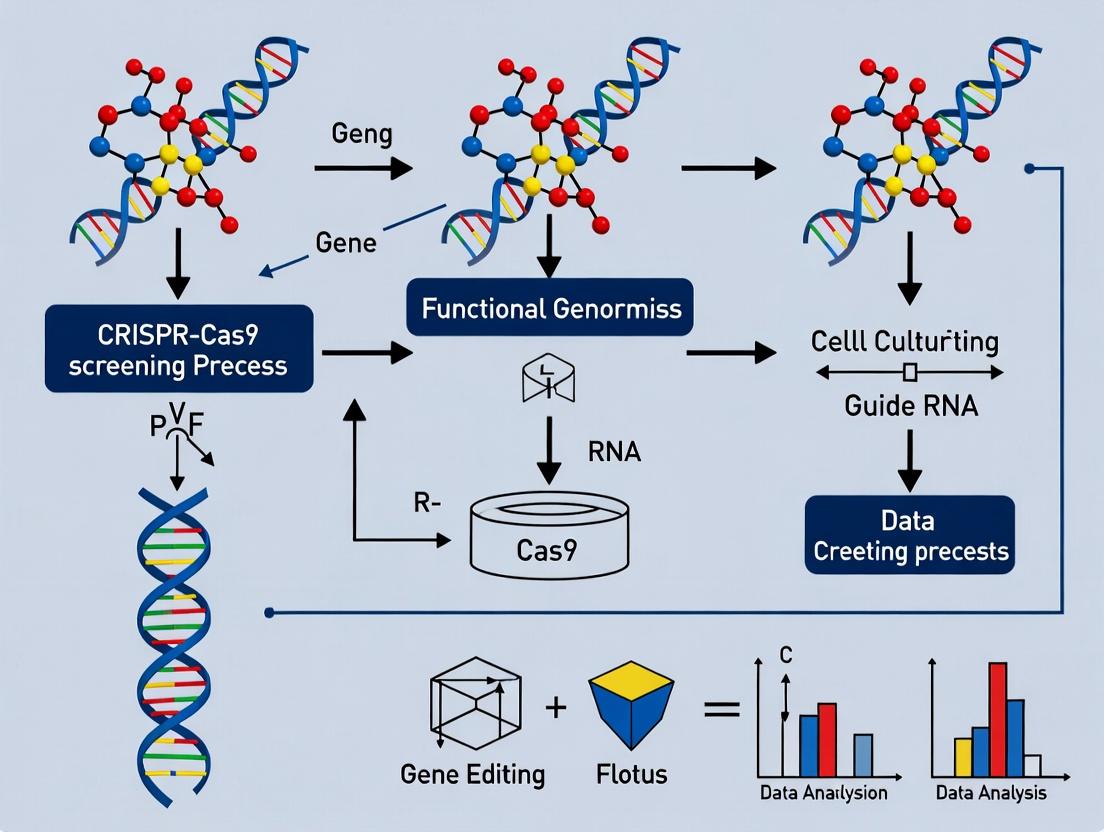
CRISPR-Cas9 Functional Genomics: A Complete Guide to Screening, Comparative Analysis, and Biomedical Applications
This article provides a comprehensive roadmap for researchers utilizing CRISPR-Cas9 screening in functional genomics.
CRISPR-Cas9 Functional Genomics: A Complete Guide to Screening, Comparative Analysis, and Biomedical Applications
Abstract
This article provides a comprehensive roadmap for researchers utilizing CRISPR-Cas9 screening in functional genomics. It begins by establishing the core principles of CRISPR screening for gene function discovery, including library design and essential cellular processes. It then details advanced methodological workflows for pooled and arrayed screens, focusing on target identification in oncology and infectious disease. Critical troubleshooting sections address common pitfalls in screen optimization, data noise reduction, and validation of screen hits. Finally, the article offers a comparative analysis of CRISPR screening against RNAi and other genetic tools, discussing validation strategies and data integration. Aimed at scientists and drug developers, this guide synthesizes current best practices to design, execute, and interpret robust, comparative functional genomics studies.
CRISPR-Cas9 Screening Fundamentals: From Core Concepts to Exploratory Library Design
CRISPR-Cas9 technology has revolutionized functional genomics by providing a scalable, precise, and programmable system for gene editing and perturbation. The core principle enabling genome-wide interrogation is the transformation of Cas9, an RNA-guided DNA endonuclease, into a high-throughput discovery tool. This is achieved by pairing a single, constant Cas9 protein with vast libraries of single guide RNAs (sgRNAs), each designed to target a specific genomic locus. The system's simplicity allows for the simultaneous generation of thousands to millions of genetic perturbations in a pooled population of cells, enabling the systematic assessment of gene function across the entire genome.
Two primary modalities are employed: knockout screens using wild-type Streptococcus pyogenes Cas9 (SpCas9) to create disruptive insertions/deletions (indels) in coding exons, and modulation screens using modified Cas9 variants. For example, nuclease-dead Cas9 (dCas9) fused to transcriptional repressors (e.g., KRAB) or activators (e.g., VP64) enables CRISPR interference (CRISPRi) or activation (CRISPRa), respectively, allowing for reversible, tunable gene knockdown or overexpression without altering the underlying DNA sequence. The power of these screens lies in coupling each sgRNA to a heritable "barcode." By tracking sgRNA abundance before and after applying a selective pressure (e.g., drug treatment, viral infection, cell proliferation) via next-generation sequencing, researchers can identify genes essential for survival, drug resistance, or specific phenotypic outcomes.
Application Notes: Key Applications in Drug Discovery and Functional Genomics
Table 1: Quantitative Outcomes from Key CRISPR-Cas9 Screening Studies in Oncology Drug Discovery
| Study Focus (Year) | Library Size (sgRNAs) | Genes Targeted | Key Hit(s) Identified | Validation Rate | Selective Pressure | Impact Metric (e.g., Log2 Fold Change) |
|---|---|---|---|---|---|---|
| Resistance to PARP Inhibitors (2018) | ~78,000 | ~19,000 | CDK12, PAXIP1, SPRTN | >80% | Olaparib | CDK12 KO: +4.2 to +5.1 (sgRNA abundance) |
| Sensitivity to Immunotherapy (2021) | ~123,000 | ~20,000 | APLNR, JAK1, PTEN | ~70% | Co-culture with T-cells | APLNR KO: -3.8 (T-cell mediated killing) |
| Essentiality in PDAC (2022) | ~92,000 | ~18,000 | KRAS, TP53, MYC | >90% | In vivo tumor growth | KRAS: Essential (FDR < 0.01) |
| Mechanism of Targeted Therapy (2023) | ~65,000 | ~18,500 | SWI/SNF Complex | 85% | SMARCA2/4 degrader | ARID1A/B KO: -2.5 to -3.0 (cell fitness) |
Note: KO = Knockout; FDR = False Discovery Rate; PDAC = Pancreatic Ductal Adenocarcinoma.
These screens have moved beyond identifying single gene essentiality to mapping complex genetic interactions (synthetic lethality), understanding signaling pathway architecture, and discovering novel drug targets and biomarkers. The quantitative data from such screens, typically represented as log2 fold-changes in sgRNA abundance and analyzed with specialized algorithms (MAGeCK, BAGEL, CERES), provide a robust statistical framework for hit prioritization.
Detailed Protocols
Protocol 1: Pooled CRISPR-Cas9 Knockout Screening for Essential Genes
Objective: To identify genes essential for cell proliferation/survival in a given cancer cell line.
Part A: Library Design and Cloning
- Library Selection: Choose a genome-wide sgRNA library (e.g., Brunello, Brie, or Toronto KnockOut v3). The Brunello library contains ~77,441 sgRNAs targeting 19,114 human genes (~4 sgRNAs/gene) plus non-targeting controls.
- Lentiviral Vector: Use a lentiviral backbone (e.g., lentiCRISPRv2, lentiGuide-Puro) expressing the sgRNA, Cas9, and a selection marker (puromycin).
- Library Amplification: Transform the pooled sgRNA plasmid library into electrocompetent E. coli (e.g., Endura cells) to achieve at least 200x coverage of the library diversity. Ispute plasmid DNA using an endotoxin-free maxiprep kit.
Part B: Lentiviral Production and Titration
- Transfection: In a 10cm dish, co-transfect HEK293T cells with: 9 µg of sgRNA library plasmid, 6.75 µg of psPAX2 (packaging plasmid), and 2.25 µg of pMD2.G (VSV-G envelope plasmid) using a transfection reagent like PEI.
- Virus Harvest: Collect lentiviral supernatant at 48 and 72 hours post-transfection. Pool, filter through a 0.45µm PES filter, and concentrate via ultracentrifugation or PEG precipitation.
- Titration: Transduce target cells (e.g., A549) with serial dilutions of virus in the presence of polybrene (8µg/mL). Apply puromycin selection (e.g., 1-2µg/mL) 48 hours later. Calculate titer (TU/mL) based on the percentage of puromycin-resistant cells after 5-7 days.
Part C: Screen Transduction and Harvest
- Transduction at MOI 0.3-0.4: Infect cells at a low Multiplicity of Infection to ensure most cells receive only one sgRNA. Use a cell number that maintains >500x library representation.
- Selection: Begin puromycin selection 48 hours post-transduction for 5-7 days to eliminate uninfected cells.
- Harvest Timepoints:
- T0: Harvest 5x10^6 cells as the baseline reference.
- Tfinal: Passage remaining cells, maintaining >500x coverage, for ~14 population doublings (approx. 2 weeks). Harvest genomic DNA from both timepoints using a maxi-prep kit (e.g., Qiagen Blood & Cell Culture DNA Maxi Kit).
Part D: Sequencing and Data Analysis
- sgRNA Amplification: Perform a two-step PCR to amplify the sgRNA region from genomic DNA and attach Illumina sequencing adapters and sample barcodes. Use high-fidelity polymerase.
- Sequencing: Pool PCR products and sequence on an Illumina NextSeq or HiSeq platform to achieve >500 reads per sgRNA.
- Bioinformatics: Align reads to the reference sgRNA library. Count reads per sgRNA for T0 and Tfinal. Use MAGeCK (https://sourceforge.net/p/mageck/wiki/Home/) to calculate essentiality scores (e.g., robust ranking algorithm [RRA] score) and false discovery rates (FDR) for each gene.
Part E: Validation
- Perform secondary validation by individually cloning top-hit sgRNAs, transducing cells, and monitoring proliferation via competitive growth assays or real-time cell analyzers.
Visualization via Graphviz
Diagram 1: CRISPR-Cas9 Screening Workflow
Diagram 2: CRISPR-Cas9 Functional Modes
The Scientist's Toolkit: Key Research Reagent Solutions
Table 2: Essential Materials for CRISPR-Cas9 Genome-Wide Screening
| Item Name | Supplier Examples | Function in Screening |
|---|---|---|
| Genome-Wide sgRNA Libraries | Addgene (Brunello, Brie), Sigma (Mission), Cellecta | Pre-designed, cloned pools of sgRNAs targeting all annotated genes; the core screening reagent. |
| Lentiviral Packaging Plasmids | Addgene (psPAX2, pMD2.G) | Second and third-generation systems for producing safe, high-titer lentiviral particles. |
| High-Titer Lentivirus Production System | Takara (Lenti-X), Thermo (Cellvento), standard PEI/293T method | Reliable systems to generate the high-quality, concentrated virus needed for pooled transduction. |
| Cas9-Expressing Cell Line | Generated in-house or purchased (e.g., Horizon) | Stable Cas9-expressing cells simplify screening by requiring only delivery of the sgRNA library. |
| Next-Gen Sequencing Kit | Illumina (Nextera XT), NEB (NEBNext Ultra II) | For preparing sgRNA amplicon libraries from genomic DNA for high-throughput sequencing. |
| CRISPR Screen Analysis Software | Broad Institute (MAGeCK), BAGEL2 | Open-source computational tools to quantify sgRNA depletion/enrichment and identify significant hits. |
| Polybrene or Hexadimethrine Bromide | Sigma-Aldrich, Millipore | A cationic polymer that enhances lentiviral transduction efficiency. |
| Puromycin Dihydrochloride | Thermo Fisher, Invivogen | Common antibiotic for selecting successfully transduced cells expressing the sgRNA vector. |
Within the broader thesis on CRISPR-Cas9 screening for functional genomics comparisons, the initial and most critical step is defining the screening goal. This determines whether a Loss-of-Function (LoF) or Gain-of-Function (GoF) approach is optimal. Both paradigms enable systematic interrogation of gene function on a genome-wide scale but answer fundamentally different biological questions.
The Conceptual and Biological Distinction
LoF screens, utilizing nuclease-active Cas9 to create disruptive indels, identify genes whose absence confers a selective advantage or disadvantage under a specific condition. This is ideal for finding essential genes, tumor suppressors, or genes required for resistance to a therapy or pathogen infection.
GoF screens, employing modified Cas9 systems like dCas9 fused to transcriptional activators, identify genes whose overexpression drives a phenotypic change. This is crucial for discovering oncogenes, genes conferring drug resistance through overexpression, or modifiers of cellular differentiation.
Quantitative Comparison of Screening Approaches
Table 1: Core Comparative Framework for LoF vs. GoF CRISPR Screens
| Parameter | Loss-of-Function (Knockout) | Gain-of-Function (Activation) |
|---|---|---|
| Cas9 Variant | Wild-type SpCas9 (Nuclease) | dCas9-VPR (or similar) |
| Genetic Alteration | Disruptive indels (Knockout) | Transcriptional upregulation |
| Primary Goal | Identify essential/required genes | Identify sufficient/driver genes |
| Typical Phenotypes | Lethality, Sensitivity, Drop-out | Survival, Resistance, Morphology change |
| Key Library Types | Whole-genome KO, Sub-library (e.g., kinase) | CRISPRa (e.g., SAM, CRISPR-SunTag) |
| Common Analysis | Depletion of sgRNAs (Negative Selection) | Enrichment of sgRNAs (Positive Selection) |
| Off-Target Concerns | DSB-dependent indels at off-target sites | dCas9 binding & transcriptional noise at off-targets |
Table 2: Example Quantitative Outcomes from Parallel Screening Studies
| Study Context (Example) | LoF Screen Hit (FDR<0.1) | GoF Screen Hit (FDR<0.1) | Concordance |
|---|---|---|---|
| Anti-cancer Drug Resistance | Tumor suppressor genes (e.g., TP53, PTEN) | Oncogenes (e.g., EGFR, KRAS) | Low (Complementary) |
| Viral Infection | Host dependency factors (e.g., CCR5) | Host restriction factor overexpression (e.g., IFITM3) | Low (Complementary) |
| Cell Differentiation | Genes required for lineage commitment | Genes that alone can drive differentiation | Partial Overlap |
Experimental Protocols
Protocol 1: Genome-wide Loss-of-Function Screening using Brunello Library Objective: Identify genes essential for cell proliferation in cancer cell line X.
- Library Amplification & Preparation: Amplify the Brunello human genome-wide knockout sgRNA library (4 sgRNAs/gene, ~77k sgRNAs) in Endura electrocompetent cells. Ispute plasmid DNA using an endotoxin-free maxiprep kit.
- Lentiviral Production: Co-transfect HEK293T cells with the Brunello library plasmid, psPAX2 (packaging), and pMD2.G (VSV-G envelope) plasmids using PEI transfection reagent. Harvest virus-containing supernatant at 48 and 72 hours post-transfection, concentrate via ultracentrifugation.
- Cell Transduction & Selection: Transduce target cells at an MOI of ~0.3 to ensure majority receive single sgRNA. 24 hours post-transduction, replace media with selection media containing puromycin (1-2 µg/mL). Select for 5-7 days until uninfected control cells are >99% dead.
- Phenotypic Selection: Passage selected cells, maintaining a minimum of 500x library representation at each step. Harvest genomic DNA (gDNA) from an initial reference timepoint (T0) and after 14-21 population doublings (Tend) using a blood & cell culture DNA maxi kit.
- Next-Generation Sequencing (NGS) Prep: Perform a two-step PCR to amplify the integrated sgRNA cassette from gDNA and attach Illumina adapters and sample barcodes. Purify PCR products and quantify by qPCR.
- Sequencing & Analysis: Pool and sequence on an Illumina NextSeq. Align reads to the library reference. Use MAGeCK or similar tool to compare sgRNA abundance between T0 and Tend, identifying significantly depleted genes.
Protocol 2: Gain-of-Function Screening using the SAM CRISPRa System Objective: Identify genes whose overexpression confers resistance to targeted therapy Y.
- Library Selection: Obtain the SAM library (synergistic activation mediator; 3 sgRNAs per gene targeting ~200 bp upstream of TSS).
- Lentiviral Production: Produce lentivirus as in Protocol 1, but using the SAM sgRNA library plasmid.
- Stable Cell Line Generation: Generate a stable cell line expressing the SAM machinery (MS2-p65-HSF1 activator and dCas9-VP64) via lentiviral transduction and blasticidin selection. Validate activation via qPCR of a positive control gene.
- Screening Transduction: Transduce the stable cell line with the SAM sgRNA library at MOI~0.3. Select with puromycin as in Protocol 1.
- Positive Selection: After selection, split cells and treat one population with drug Y (treatment) and another with DMSO (control). Maintain drug pressure for 14-21 days, replenishing drug with each passage.
- Harvest & Analysis: Harvest gDNA from treated and control cells at endpoint. Perform NGS library prep and sequencing as in Protocol 1. Analyze data to identify sgRNAs significantly enriched in the drug-treated population versus control.
Visualizations
Title: Loss-of-Function CRISPR Screening Workflow
Title: SAM CRISPRa System Mechanism
The Scientist's Toolkit: Research Reagent Solutions
Table 3: Essential Materials for CRISPR Screening
| Item | Function in Screening | Example/Note |
|---|---|---|
| Genome-wide sgRNA Library | Provides pooled targeting reagents for systematic gene perturbation. | Brunello (LoF), SAM (GoF). Maintain >500x coverage. |
| Lentiviral Packaging Plasmids | Required for production of sgRNA-delivering lentiviral particles. | psPAX2 (gag/pol), pMD2.G (VSV-G envelope). |
| dCas9 Activator Cell Line | For GoF screens; provides the transcriptional activation machinery. | HEK293T SAM cell line (expresses dCas9-VP64 & MS2-p65-HSF1). |
| PEI Transfection Reagent | For high-efficiency co-transfection of packaging plasmids in HEK293T cells. | Linear PEI, MW 25,000. Cost-effective and efficient. |
| Puromycin / Selection Antibiotics | Selects for cells successfully transduced with the sgRNA vector. | Concentration must be titrated for each cell line. |
| Genomic DNA Extraction Kit | High-yield, high-quality gDNA extraction from pooled cell populations. | Critical for accurate NGS representation (e.g., Qiagen Maxi Kit). |
| NGS Library Prep Kit | Amplifies and prepares sgRNA sequences for high-throughput sequencing. | Two-step PCR with indexing primers. |
| Bioinformatics Software | For statistical analysis of sgRNA abundance and hit identification. | MAGeCK, PinAPL-Py, CRISPResso2. |
Within a thesis on CRISPR-Cas9 screening for functional genomics comparisons, the strategic design of single-guide RNA (sgRNA) libraries is foundational. The choice of library—genome-wide, subset, or custom—dictates the scale, resolution, and biological focus of the screen, directly impacting the validation of comparative functional genomics hypotheses.
Genome-Wide sgRNA Libraries
Genome-wide libraries aim to target every gene in the genome, enabling unbiased discovery. They are essential for exploratory comparisons between biological states (e.g., healthy vs. diseased, treated vs. untreated).
- Application Note: Used in initial, discovery-phase screens to identify genes essential for cell viability, drug resistance, or specific phenotypic responses across different cell lineages or conditions.
- Common Designs: The Brunello (human) and Brie (mouse) libraries are current gold standards, each featuring ~4 sgRNAs/gene and improved on-target efficiency predictions.
Table 1: Current Benchmark Genome-Wide Library Designs
| Library Name | Species | Target Genes | sgRNAs per Gene | Total sgRNAs | Key Design Feature |
|---|---|---|---|---|---|
| Brunello | Human | 19,114 | 4 | ~76,456 | Optimized Rule Set 2 for improved on-target activity. |
| Brie | Mouse | 20,661 | 4 | ~82,644 | Mouse-adapted version of Brunello. |
| Human CRISPR Knockout (GeCKO) v2 | Human | 19,050 | 6 | ~114,300 | Mixed design (3 sgRNAs/gene from two algorithms). |
| Mouse CRISPR Knockout (GeCKO) v2 | Mouse | 20,611 | 6 | ~123,666 | Mixed design for broad coverage. |
Protocol 1.1: Lentiviral Production for Genome-Wide Screening
- Day 1: Seed HEK293T cells in a 10-cm dish to reach 70-80% confluence the next day.
- Day 2: Co-transfect using a polyethylenimine (PEI) protocol:
- In Tube A, mix 10 µg library plasmid (e.g., lentiCRISPRv2-Brunello), 7.5 µg psPAX2 (packaging), and 3 µg pMD2.G (VSV-G envelope) in 500 µL serum-free DMEM.
- In Tube B, mix 60 µL PEI (1 mg/mL) in 500 µL serum-free DMEM.
- Combine tubes, vortex, incubate 15 min at RT.
- Add dropwise to cells. Replace medium after 6-8 hours.
- Day 3 & 4: Harvest viral supernatant at 48 and 72 hours post-transfection. Pool, filter through a 0.45 µm PES filter, and concentrate using centrifugal filter units (100kDa MWCO). Aliquot and store at -80°C. Determine titer via transduction of target cells with a serial dilution of virus and puromycin selection.
Protocol 1.2: Cell Transduction and Screening at Genome-Wide Scale
- Titration: Transduce target cells (e.g., 10 million) at a low MOI (<0.3) with varying virus volumes to ensure most cells receive only one sgRNA. Include a non-transduced control.
- Selection: 24 hours post-transduction, apply appropriate antibiotic (e.g., 2 µg/mL puromycin) for 5-7 days to eliminate untransduced cells.
- Screening: Maintain the selected cell population (maintaining >500x library coverage) under the experimental condition (e.g., drug treatment, hypoxia) vs. control for 14-21 population doublings.
- Harvest & Sequencing: Harvest genomic DNA from ≥50 million cells per condition (Qiagen Maxi Prep). Amplify integrated sgRNA sequences via a two-step PCR, adding sample barcodes and Illumina adapters. Pool and sequence on an Illumina NextSeq or HiSeq platform.
Subset (Focused) sgRNA Libraries
Focused libraries target a predefined set of genes (e.g., kinase family, epigenetic regulators, candidate genes from prior omics data). They enable higher sgRNA density per gene and deeper interrogation within a specific biological context.
- Application Note: Ideal for hypothesis-driven comparative research within a thesis, such as comparing the essential kinome across multiple cancer subtypes or validating hits from a prior genome-wide screen under new conditions.
Table 2: Comparison of Subset Library Applications
| Library Focus | Typical Gene Count | sgRNAs/Gene | Primary Application in Comparative Research |
|---|---|---|---|
| Druggable Genome | 5,000 - 7,000 | 6 - 10 | Identify novel therapeutic targets across disease models. |
| Specific Pathway (e.g., Apoptosis) | 100 - 500 | 8 - 12 | Dissect pathway-specific genetic interactions in different cellular backgrounds. |
| Gene Family (e.g., GPCRs) | 800 - 1,500 | 6 - 10 | Functional deorphanization and comparison of family roles. |
| Custom Candidate List | 10 - 500 | 10 - 20 | High-confidence validation and mechanistic follow-up. |
Protocol 2.1: Designing and Cloning a Focused Library
- sgRNA Selection: Using tools like CRISPRko (Broad Institute), input your gene list. Select top-ranked sgRNAs (6-12 per gene) based on efficiency and specificity scores. Include non-targeting control sgRNAs (~5% of total).
- Oligo Pool Synthesis: Order the sgRNA sequences (including 5' and 3' cloning overhangs) as a pooled oligonucleotide library.
- Cloning into Lentiviral Vector: a. Digest the lentiviral backbone (e.g., lentiGuide-Puro) with BsmBI. b. Gel-purify the linearized vector. c. Perform a Golden Gate assembly of the oligo pool with the vector using T4 DNA ligase and BsmBI. d. Electroporate the assembly reaction into Endura electrocompetent cells. Plate on large bioassay dishes to ensure >200x coverage of the library. e. Harvest plasmid DNA via Maxiprep. This is your cloned subset library for viral production (follow Protocol 1.1).
Custom sgRNA Libraries
Custom libraries are bespoke designs for non-standard applications, including targeted non-coding regions, specific isoforms, or introducing precise mutations via base or prime editing.
- Application Note: Enables precise comparative questions, such as screening enhancer regions identified in differential ATAC-seq studies or comparing the functional impact of specific single nucleotide variants (SNVs) across cell lines.
Protocol 3.1: Screening Non-Coding Regulatory Elements
- Design: Use tools like CRISPOR or CHOPCHOP to design tiling sgRNAs (~5-10 sgRNAs per kilobase) across genomic regions of interest (e.g., promoter, enhancer). Include flanking regions.
- Cloning & Production: Follow Protocol 2.1 for oligo pool cloning and viral production.
- Phenotypic Readout: Use a reporter assay (e.g., GFP expression from a targeted enhancer) or a survival/proliferation-based screen. For enhancer screens, transduce cells, sort based on reporter signal (High vs. Low), and sequence sgRNAs from sorted populations to identify regulators.
The Scientist's Toolkit: Research Reagent Solutions
| Item | Function in CRISPR Screening |
|---|---|
| Lentiviral sgRNA Vector (e.g., lentiGuide-Puro) | All-in-one plasmid expressing sgRNA and selection marker (Puromycin R) for stable integration. |
| Lentiviral Packaging Plasmids (psPAX2, pMD2.G) | Required for production of replication-incompetent, high-titer lentiviral particles. |
| Polyethylenimine (PEI), Linear | High-efficiency, low-cost transfection reagent for viral production in HEK293T cells. |
| Puromycin Dihydrochloride | Antibiotic for selecting successfully transduced cells post-lentiviral delivery. |
| Endura Electrocompetent E. coli | High-efficiency bacteria for transforming large, complex sgRNA library plasmid pools. |
| Nextera XT Index Kit (Illumina) | For attaching dual indices and adapters during PCR preparation of sgRNA amplicons for NGS. |
| MAGeCK (Computational Tool) | Standard algorithm for robust identification of enriched/depleted sgRNAs and genes from screen data. |
| Cell Titer Glo | Luminescent assay to measure cell viability/cytotoxicity as a screening readout. |
Visualizations
Title: sgRNA Library Selection Workflow for Comparative Studies
Title: Core Workflow for a Comparative CRISPR Knockout Screen
The advent of CRISPR-Cas9 screening has revolutionized functional genomics, enabling systematic, genome-wide interrogation of gene function. The choice of biological model—immortalized cell lines versus primary cells—is a critical determinant of a screen’s physiological relevance, translational impact, and technical success. This article, framed within a broader thesis on CRISPR-Cas9 screening for functional genomics comparisons, provides detailed application notes and protocols for model selection, considering their respective advantages, limitations, and applications.
Model Comparison: Key Considerations
The selection between cell lines and primary cells involves trade-offs between experimental tractability and biological fidelity. Key parameters are summarized in the table below.
Table 1: Quantitative and Qualitative Comparison of Cell Models for CRISPR Screening
| Parameter | Immortalized Cell Lines | Primary Cells |
|---|---|---|
| Availability & Cost | High, low cost (< $500/vial typical) | Limited, high cost (can be > $1000/donor) |
| Genetic Stability | High karyotypic instability (aneuploidy common) | Normal, diploid genome (subject to donor variation) |
| Proliferative Capacity | Unlimited (easy expansion for library-scale screens) | Finite (3-10 passages typical, limits screen scale) |
| Physiological Relevance | Low; adapted to in vitro conditions, may lack tissue-specific functions | High; retain native phenotype, signaling, and differentiation state |
| Donor/Clonal Variation | Low (clonal population) | High (inter-donor genetic and epigenetic diversity) |
| Transfection/Transduction Efficiency | Typically high (>70% for lentiviral transduction common) | Often low and variable; may require optimized methods |
| Experimental Reproducibility | High (consistent genetic background) | Lower (requires multiple donors for robust conclusions) |
| Typical Screening Applications | Target identification/validation, mechanistic studies, toxicology | Pathway analysis in native context, translational biomarker discovery, immunotherapy (e.g., T-cell screens) |
Detailed Experimental Protocols
Protocol 3.1: CRISPR-Cas9 Knockout Screening in Immortalized Cell Lines
Objective: To perform a pooled, genome-wide CRISPR knockout screen in a human cancer cell line (e.g., HEK293T, HeLa, or a cancer-relevant line) to identify genes essential for cell proliferation under a specific selective pressure.
Materials (Research Reagent Solutions):
- Lentiviral sgRNA Library: (e.g., Brunello, Brie, or GeCKO v2). Contains ~70,000 sgRNAs targeting human genes.
- Lentiviral Packaging Plasmids: psPAX2 (packaging) and pMD2.G (VSV-G envelope).
- Transfection Reagent: Polyethylenimine (PEI) or Lipofectamine 3000.
- Antibiotics: Puromycin for selection, Penicillin-Streptomycin.
- Cell Culture Media: Appropriate media (e.g., DMEM + 10% FBS).
- Cas9-Expressing Cell Line: Or a plasmid for generating one (e.g., lentiCas9-Blast).
- DNA Extraction Kit: For genomic DNA extraction from >1e7 cells.
- PCR Reagents: For sgRNA amplification and indexing.
- Next-Generation Sequencing (NGS) Platform: For sgRNA readout.
Methodology:
- Generate Cas9-Expressing Cells: Stably transduce your cell line of interest with a lentiviral Cas9 construct (e.g., lentiCas9-Blast). Select with blasticidin (e.g., 10 µg/mL for 7 days) and validate Cas9 activity via a surrogate reporter assay.
- Library Virus Production: In a 10cm dish, co-transfect HEK293T cells (70% confluent) with the sgRNA library plasmid (10 µg), psPAX2 (7.5 µg), and pMD2.G (2.5 µg) using PEI. Harvest viral supernatant at 48 and 72 hours post-transfection, concentrate via ultracentrifugation, and titer on target cells.
- Cell Line Transduction & Selection: Transduce Cas9-expressing cells at a low Multiplicity of Infection (MOI ~0.3) to ensure most cells receive only one sgRNA. Include a minimum of 500 cells per sgRNA in the library to maintain representation (e.g., for a 70,000-sgRNA library, transduce >35 million cells). 24h post-transduction, add puromycin (e.g., 2 µg/mL) for 5-7 days to select transduced cells.
- Screen Passage & Harvest: Passage cells every 3-4 days, maintaining a minimum of 500X library coverage. Harvest genomic DNA from a) the initial selected cell population (Day 0/T0 control) and b) the final population after ~14-21 population doublings (or under selective pressure, e.g., drug treatment).
- NGS Library Preparation & Analysis: PCR-amplify integrated sgRNA sequences from genomic DNA using barcoded primers. Pool and sequence on an NGS platform. Align reads to the reference sgRNA library and count sgRNA abundances. Use analysis pipelines (e.g., MAGeCK, BAGEL) to compare T0 vs. final timepoint, identifying significantly depleted or enriched sgRNAs and their target genes.
Diagram Title: CRISPR Screen in Cell Lines Workflow
Protocol 3.2: CRISPR Screening in Primary Human T Cells
Objective: To perform a targeted CRISPR screen in isolated primary human CD4+ or CD8+ T cells to identify genes regulating T-cell activation or exhaustion.
Materials (Research Reagent Solutions):
- Primary Cells: Human PBMCs or isolated T cells from leukopaks.
- T Cell Activation Kit: Anti-CD3/CD28 beads or antibodies.
- Cell Culture Media: X-VIVO 15 or RPMI-1640 + 10% human serum + IL-2 (100-300 IU/mL).
- CRISPR RNP Complex Components: Recombinant S.p. Cas9 protein, synthetic sgRNA(s).
- Electroporation System: Lonza 4D-Nucleofector or Neon Transfection System.
- Electroporation Kit: P3 Primary Cell 4D-Nucleofector Kit or similar.
- Phenotyping Antibodies: Anti-CD3, CD4, CD8, CD69, PD-1 for flow cytometry.
- NGS Library Prep Kit: For targeted amplicon sequencing.
Methodology:
- Primary T Cell Isolation & Activation: Isolate CD4+ or CD8+ T cells from PBMCs using negative selection magnetic beads. Activate cells with anti-CD3/CD28 beads (1 bead:2 cells) in media containing IL-2 (100 IU/mL) for 48 hours.
- CRISPR RNP Complex Formation: For a targeted sgRNA library (e.g., 100-500 sgRNAs), complex each individual synthetic sgRNA with recombinant Cas9 protein at a molar ratio of 1:2 (sgRNA:Cas9) in a buffer to form ribonucleoprotein (RNP) complexes. Pool equal amounts of each RNP.
- Electroporation: Harvest activated T cells, wash, and resuspend in electroporation buffer. Mix 1-2 million cells with the pooled RNP complexes (e.g., 2 µg Cas9 per 100k cells). Electroporate using a primary cell-optimized protocol (e.g., Lonza 4D-Nucleofector, program EO-115). Immediately transfer cells to pre-warmed, cytokine-supplemented media.
- Post-Editing Culture & Assay: Culture cells for 5-10 days, maintaining IL-2. Apply relevant assay pressure (e.g., repetitive antigen stimulation to model exhaustion). Harvest cells at multiple time points for phenotypic analysis (e.g., flow cytometry for activation/exhaustion markers) and genomic DNA extraction.
- Screen Readout & Analysis: Amplify the integrated sgRNA region from genomic DNA using barcoded primers for each sample. Perform NGS. Analyze sgRNA frequencies across conditions (e.g., high-PD-1 vs. low-PD-1 sorted populations) to identify regulators of the phenotype.
Diagram Title: Primary T Cell CRISPR Screen Workflow
The Scientist's Toolkit: Essential Reagents & Materials
Table 2: Key Research Reagent Solutions for CRISPR Screening in Different Models
| Item | Function | Recommended for Cell Lines | Recommended for Primary Cells |
|---|---|---|---|
| Lentiviral sgRNA Library | Delivers heritable, stable genomic integration of sgRNA for long-term screens. | Yes (standard) | Limited (low efficiency, risk of insertional mutagenesis). |
| CRISPR RNP Complexes | Pre-formed complexes of Cas9 protein and sgRNA; transient, high-activity editing. | For difficult-to-transduce lines. | Yes (gold standard). Fast, efficient, minimizes off-target integration. |
| Recombinant Cas9 Protein | High-purity, ready-to-use protein for RNP formation. | Optional. | Essential. |
| Electroporation System | Device for physical delivery of RNPs or plasmids into cells. | Optional (lipofection often sufficient). | Critical. Nucleofection is most efficient method for many primary cells. |
| Cytokines/Growth Factors | Maintain cell viability, proliferation, and native state. | Seldom required (in serum). | Essential (e.g., IL-2 for T cells, M-CSF for macrophages). |
| Cell Activation Beads/Antibodies | Mimic physiological stimulation for immune cells. | Not applicable. | Essential for functional screens in lymphocytes. |
| Genomic DNA Cleanup Kit | High-yield gDNA isolation from large cell numbers. | Yes (for library-scale prep). | Yes (often from fewer cells). |
| NGS Indexing Primers | Add sample-specific barcodes for multiplexed sequencing. | Yes (for pooled library screens). | Yes (for targeted amplicon sequencing). |
The choice between cell lines and primary cells is not binary but strategic. For initial, large-scale, mechanistic discovery screens where scale, cost, and reproducibility are paramount, immortalized cell lines remain the workhorse. For follow-up validation, studying specific human biology, immune-oncology, or translational research where physiological context is non-negotiable, primary cell screens are indispensable despite their technical challenges. A robust functional genomics thesis will often employ a phased strategy: discovery in tractable cell line models, followed by targeted validation in primary cell systems, thereby balancing discovery power with biological relevance.
Within functional genomics CRISPR-Cas9 screening, three primary genetic interaction readouts are critical for target discovery: essential genes, fitness genes, and synthetic lethal (SL) interactions. Essential genes are required for cellular survival under standard conditions. Fitness genes, when disrupted, cause a measurable growth defect but are not lethal. SL interactions occur when the disruption of two genes together is lethal, while disruption of either alone is not, offering high therapeutic potential for selective targeting of diseased cells. This application note details protocols and analyses for deriving these key readouts, framed within a thesis on comparative functional genomics.
Key Readout Definitions and Data Analysis
The core quantitative outputs from a CRISPR screening campaign are summarized in the following tables.
Table 1: Classification and Characteristics of Key Genetic Readouts
| Readout Type | Definition | Primary Screening Approach | Typical Hit Threshold (Gene Effect Score*) | Therapeutic Implication |
|---|---|---|---|---|
| Essential Genes | Required for fundamental cellular proliferation/survival. | Negative selection screen in a reference cell line. | ≤ -0.5 (Strongly Depleting) | Potential anti-cancer or anti-proliferative targets; often toxic. |
| Fitness Genes | Confer a growth disadvantage but not cell death upon loss. | Negative selection screen. | -0.5 to -0.2 (Moderately Depleting) | Modulators of cellular fitness; context-dependent targets. |
| Synthetic Lethal (SL) Pairs | Combined loss of Gene A & Gene B is lethal; loss of either alone is viable. | Differential screening (e.g., treated vs. untreated, isogenic pairs). | Differential Gene Effect (∆) ≤ -0.6 & FDR < 0.05 | High selectivity for targeting genetic vulnerabilities (e.g., BRCA-PARP1). |
*Gene Effect scores are normalized, where 0 represents no effect and -1 represents a strong loss-of-fitness effect akin to core essential genes (e.g., DepMap standard).
Table 2: Example Quantitative Data from a Representative CRISPR SL Screen (BRCA1-WT vs. BRCA1-Mutant Context)
| Gene Targeted | Gene Effect (BRCA1-WT) | Gene Effect (BRCA1-Mutant) | Differential Gene Effect (∆) | Adjusted p-value | Classification in Mutant Context |
|---|---|---|---|---|---|
| PARP1 | -0.05 (Neutral) | -1.12 (Lethal) | -1.07 | 1.2e-08 | Validated SL Hit |
| Gene X | -0.61 (Essential) | -0.59 (Essential) | +0.02 | 0.87 | Pan-essential, not SL |
| Gene Y | -0.10 (Neutral) | -0.35 (Fitness) | -0.25 | 0.04 | Contextual Fitness Gene |
| POLQ | +0.01 (Neutral) | -0.82 (Lethal) | -0.83 | 3.5e-06 | Potential SL Hit |
Experimental Protocols
Protocol 1: Genome-Wide CRISPR Knockout for Essential & Fitness Gene Identification
Objective: Identify genes essential for proliferation/survival in a given cell line. Materials: See "The Scientist's Toolkit" below. Workflow:
- Library Design & Production: Use a genome-wide lentiviral sgRNA library (e.g., Brunello, 4 sgRNAs/gene).
- Cell Transduction: Infect target cells at a low MOI (~0.3) to ensure single integration. Maintain >500x representation of each sgRNA.
- Selection & Expansion: Treat with puromycin (2 µg/mL, 48-72h) 24h post-transduction. Harvest initial reference sample (T0).
- Proliferation Passaging: Culture cells for ~14-21 population doublings, passaging to maintain representation. Harvest final sample (T_end).
- Genomic DNA (gDNA) Extraction & NGS Prep: Isolate gDNA (Qiagen Maxi Prep). Amplify integrated sgRNA sequences via PCR with indexed primers.
- Sequencing & Analysis: Sequence on Illumina platform. Align reads to library reference. Calculate depletion scores (MAGeCK, CERES) to identify essential (strongly depleted) and fitness (moderately depleted) genes.
Protocol 2: Differential Screening for Synthetic Lethal Interactions
Objective: Identify genes specifically essential in a defined genetic or treatment context (e.g., mutant vs. wild-type, drug-treated vs. control). Materials: As in Protocol 1, plus isogenic cell line pairs or compound of interest. Workflow:
- Parallel Screening: Perform Protocol 1 steps 1-3 in two conditions in parallel (e.g., BRCA1-mutant and BRCA1-WT cells).
- Contextual Challenge: After puromycin selection, split and maintain the two conditions. For drug SL, add appropriate dose of compound (e.g., PARP inhibitor) to the treated arm.
- Harvest Samples: Collect T0 and T_end samples from both conditions.
- NGS & Bioinformatic Analysis: Process all samples for sequencing. Use differential analysis algorithms (MAGeCK RRA, DrugZ) to compare sgRNA depletion between conditions. Prioritize hits with significant differential depletion (e.g., ∆ ≤ -0.6, FDR < 0.05) in the test context but neutral in the control.
Visualizing Screening Workflows and Concepts
CRISPR Negative Selection Screening Workflow
Concept of Context-Dependent Synthetic Lethality
PARP1-BRCA1 Synthetic Lethality Pathway
The Scientist's Toolkit
Table 3: Essential Research Reagent Solutions for CRISPR-Cas9 Screening
| Reagent/Material | Function & Critical Notes | Example Product/Supplier |
|---|---|---|
| Genome-wide sgRNA Library | Pre-designed, cloned lentiviral library targeting all human genes. Defines screen scope. | Brunello (Addgene #73179), Human CRISPR Knockout Pooled Library (Sigma). |
| Lentiviral Packaging Plasmids | For production of infectious, replication-incompetent lentiviral particles. | psPAX2 (Addgene #12260), pMD2.G (Addgene #12259). |
| HEK293T Cells | Highly transferable cell line for high-titer lentivirus production. | ATCC CRL-3216. |
| Target Cell Line | The cell model for screening. Must be highly infectable and proliferative. | Relevant cancer or disease model (e.g., A549, RPE1). |
| Polybrene (Hexadimethrine Bromide) | Cationic polymer that enhances viral transduction efficiency. | Use at 4-8 µg/mL during transduction. |
| Puromycin (or appropriate antibiotic) | Selects for cells successfully transduced with the sgRNA/Cas9 construct. | Concentration must be pre-titrated for each cell line. |
| Genomic DNA Isolation Kit | High-yield, high-purity gDNA extraction from large cell pellets (>100e6 cells). | Qiagen Blood & Cell Culture DNA Maxi Kit. |
| High-Fidelity PCR Mix | For accurate, unbiased amplification of integrated sgRNA sequences from gDNA. | KAPA HiFi HotStart ReadyMix. |
| Next-Generation Sequencer | Platform for deep sequencing of sgRNA amplicons. | Illumina NextSeq 500/550 or NovaSeq. |
| Bioinformatics Pipeline | Software for quantifying sgRNA reads and calculating gene-level scores. | MAGeCK (Broad), PinAPL-Py, CERES (for copy-number correction). |
Advanced CRISPR Screening Protocols: Methodologies and Translational Applications in Biomedicine
CRISPR-Cas9 screening is a cornerstone of modern functional genomics, enabling systematic interrogation of gene function. The choice between pooled and arrayed screening formats is critical and depends on the specific research question, assay type, available infrastructure, and desired data output. This application note, framed within a thesis on CRISPR-Cas9 for functional genomics comparisons, delineates the core considerations, protocols, and tools for selecting and executing the optimal screening workflow.
Core Concepts and Comparative Analysis
Defining the Formats
- Pooled Screening: A single population of cells is transduced with a lentiviral library containing a complex mix of single guide RNAs (sgRNAs). The screen is read out by sequencing genomic DNA to quantify sgRNA abundance changes over time or after a selection pressure.
- Arrayed Screening: Each well of a multi-well plate contains cells transfected or transduced with a single, known genetic perturbation (e.g., one sgRNA, one siRNA). Phenotypes are measured per well using high-content imaging, fluorescence, or luminescence.
Quantitative Comparison of Screening Formats
Table 1: Strategic Comparison of Pooled vs. Arrayed CRISPR Screening
| Parameter | Pooled Screening | Arrayed Screening |
|---|---|---|
| Primary Application | Positive/Negative selection screens (e.g., viability, drug resistance). | Complex phenotypic screens (e.g., morphology, spatial signaling, multi-parameter imaging). |
| Theoretical Library Size | Very High (10^5 - 10^6 elements). | Limited by plate format (10^3 - 10^4). |
| Perturbation Delivery | Lentiviral transduction (stable integration). | Transient transfection (RNAi), lentivirus, or electroporation (RNP). |
| Readout Method | Next-Generation Sequencing (NGS) of sgRNA abundance. | Per-well assay: HCS imaging, fluorescence, luminescence, absorbance. |
| Key Advantage | Scalability, cost-effectiveness for genome-scale screens, simple deconvolution. | Direct genotype-phenotype linkage, immediate hit identification, compatibility with complex assays. |
| Key Limitation | Restricted to bulk, population-level phenotypes that can be linked to fitness. | Lower throughput, higher reagent cost, requires sophisticated automation. |
| Data Output | Relative sgRNA enrichment/depletion scores. | Rich, multi-dimensional phenotypic data per perturbation. |
| Optimal For Thesis Context | Comparing gene essentiality across cell lines or conditions at genome scale. | Deep functional genomics comparisons of specific pathways using multi-parametric phenotyping. |
Table 2: Typical Experimental Metrics and Resource Requirements
| Metric | Pooled Screening Protocol | Arrayed Screening Protocol |
|---|---|---|
| Cells per sgRNA | 200 - 1000 | 1000 - 5000 |
| Total Cells for Genome-wide Screen | ~5 x 10^8 | ~5 x 10^7 (but in 10^4 wells) |
| Time to Hit Identification | Weeks (after sequencing & bioinformatics). | Days (immediate from plate readout). |
| Primary Cost Driver | NGS sequencing depth. | Automation, assay reagents, plates. |
| Key Instrumentation | Sequencer, liquid handler for library prep. | High-content imager, plate washer, automated dispenser. |
Detailed Experimental Protocols
Protocol 1: Pooled CRISPR-Cas9 Negative Selection Screen
Objective: To identify genes essential for cell proliferation in a specific cell line. Materials: See "The Scientist's Toolkit" below.
Procedure:
- Library Amplification & Lentivirus Production:
- Transform the sgRNA plasmid library (e.g., Brunello) into competent E. coli and culture on large LB agar plates. Pool colonies and maxi-prep DNA.
- In a HEK293T producer cell line, co-transfect the sgRNA library plasmid, psPAX2 (packaging), and pMD2.G (envelope) plasmids using PEI.
- Harvest lentiviral supernatant at 48 and 72 hours post-transfection, concentrate via ultracentrifugation, and titre on target cells.
- Cell Transduction & Selection:
- Seed target cells expressing Cas9. Transduce at a low MOI (<0.3) to ensure most cells receive ≤1 sgRNA. Include a non-targeting control sgRNA virus.
- At 48 hours post-transduction, add puromycin (or relevant antibiotic) for 5-7 days to select for successfully transduced cells.
- Screen Passage & Harvest:
- Maintain transduced cells at a minimum coverage of 500 cells per sgRNA. Passage cells every 3-4 days for 14-21 population doublings.
- Harvest cell pellets (at least 1e7 cells) at the initial timepoint (T0) after selection and at the final endpoint (T_end). Store at -80°C.
- NGS Library Prep & Analysis:
- Extract genomic DNA from T0 and T_end pellets using a maxi-prep kit.
- Amplify integrated sgRNA sequences via a two-step PCR: (1) Amplify sgRNA region with indexed primers. (2) Add Illumina adapters and barcodes.
- Sequence on an Illumina NextSeq. Align reads to the sgRNA library, count reads per sgRNA, and use algorithms like MAGeCK or BAGEL to identify significantly depleted sgRNAs.
Protocol 2: Arrayed CRISPR-Cas9 High-Content Imaging Screen
Objective: To compare changes in nuclear morphology upon perturbation of DNA damage pathway genes. Materials: See "The Scientist's Toolkit" below.
Procedure:
- sgRNA Array Plate Preparation:
- Obtain a commercial arrayed sgRNA library in a 384-well plate format, or prepare one by dispensing individual sgRNA expression plasmids (e.g., in lentiGuide-Puro backbone) into wells.
- Reverse Transfection in Assay Plate:
- In a 384-well imaging plate, pre-dispense 5 µL of opti-MEM containing 0.1 µL of Lipofectamine L3000 reagent per well.
- Using an acoustic liquid handler, transfer 20 ng of each sgRNA plasmid from the source plate to the corresponding assay well. Incubate 20 min.
- Seed 1500 Cas9-expressing cells in 40 µL of complete medium per well. Centrifuge briefly and incubate for 72h.
- Phenotypic Induction and Staining:
- Optionally treat cells with a DNA damaging agent (e.g., 1µM Camptothecin) for 6 hours.
- Fix cells with 4% PFA for 15 min, permeabilize with 0.1% Triton X-100, and stain with DAPI (nuclei) and an antibody against a DNA damage marker (e.g., γH2AX).
- Image Acquisition & Analysis:
- Image plates using a 20x objective on a high-content imager (e.g., ImageXpress). Acquire 4 fields per well.
- Use integrated software (e.g., MetaXpress, CellProfiler) to segment nuclei based on DAPI, measure intensity (γH2AX), and extract morphological features (area, roundness, texture).
- Normalize data per plate, and use Z-score or B-score normalization to identify phenotypic outliers (hits) per sgRNA.
Workflow and Pathway Diagrams
(Decision Tree for Screening Format Selection)
(Pooled CRISPR Screening Workflow)
(Arrayed CRISPR Screening Workflow)
(Simplified DNA Damage Response Pathway)
The Scientist's Toolkit: Research Reagent Solutions
Table 3: Essential Materials for CRISPR-Cas9 Screening
| Item | Function | Example (Provider) |
|---|---|---|
| Genome-wide sgRNA Library | Pre-designed, cloned sets of sgRNAs targeting all human/mouse genes. | Brunello human library (Addgene), Mouse Brie library (Addgene). |
| Arrayed sgRNA Library | Individual sgRNAs or gene-targeting sets in multi-well plates. | Dharmacon Edit-R arrayed sgRNA libraries (Horizon). |
| Lentiviral Packaging Plasmids | For producing replication-incompetent lentivirus to deliver sgRNAs. | psPAX2, pMD2.G (Addgene). |
| Cas9-Expressing Cell Line | Stable cell line expressing SpCas9, enabling rapid sgRNA action. | HEK293T-Cas9, A375-Cas9 (ATCC, commercial sources). |
| Transfection Reagent | For arrayed delivery of sgRNA plasmids or RNPs. | Lipofectamine CRISPRMAX (Invitrogen), Lipofectamine L3000. |
| Selection Antibiotic | To select for cells successfully transduced with sgRNA vectors. | Puromycin, Blasticidin. |
| NGS Library Prep Kit | For amplifying and barcoding sgRNA sequences from genomic DNA. | NEBNext Ultra II DNA Library Prep Kit (NEB). |
| High-Content Imaging System | Automated microscope for capturing complex phenotypes in multi-well plates. | ImageXpress Micro Confocal (Molecular Devices), Opera Phenix (Revvity). |
| Analysis Software | For quantifying sgRNA depletion (pooled) or extracting features (arrayed). | MAGeCK (pooled), CellProfiler (arrayed). |
Within the broader thesis of CRISPR-Cas9 screening for functional genomics comparisons, pooled knockout screens represent a cornerstone methodology. They enable the systematic, genome-wide interrogation of gene function in a high-throughput, cost-effective manner. By transducing a complex population of cells with a pooled lentiviral guide RNA (gRNA) library, applying a selective pressure (e.g., drug treatment, cell fitness, or fluorescence), and quantifying gRNA abundance via Next-Generation Sequencing (NGS), researchers can identify genes essential for specific biological processes. This protocol details the end-to-end workflow for conducting such a screen, from library design to data analysis.
The Scientist's Toolkit: Key Research Reagent Solutions
| Item | Function in Pooled CRISPR Screen |
|---|---|
| Genome-wide gRNA Library | A pooled lentiviral plasmid library containing ~3-10 gRNAs per gene and non-targeting controls. Enables simultaneous targeting of thousands of genes. |
| Lentiviral Packaging Mix | Plasmid mix (e.g., psPAX2, pMD2.G) for producing replication-incompetent lentivirus to deliver the gRNA library and Cas9. |
| Cas9-Expressing Cell Line | Stable cell line expressing the Streptococcus pyogenes Cas9 nuclease. Essential for gRNA-mediated DNA cleavage. |
| Polybrene / Hexadimethrine Bromide | A cationic polymer that enhances viral transduction efficiency by neutralizing charge repulsion between virus and cell membrane. |
| Puromycin (or other Antibiotics) | Selection antibiotic to eliminate untransduced cells after library delivery, ensuring a pure population of gRNA-containing cells. |
| Genomic DNA Extraction Kit | For high-yield, high-quality gDNA extraction from large cell populations (≥ 10⁷ cells). Critical for PCR amplification of integrated gRNA sequences. |
| High-Fidelity PCR Master Mix | For accurate, unbiased amplification of gRNA sequences from genomic DNA prior to NGS library preparation. |
| NGS Indexing Primers | Dual-indexed primers to multiplex multiple samples in a single NGS run, reducing cost and enabling comparison of pre- and post-selection samples. |
| Illumina-Compatible Sequencing Kit | For preparation and sequencing of the gRNA amplicon library, typically on an Illumina MiSeq, HiSeq, or NextSeq platform. |
Experimental Protocol: Detailed Methodology
Part 1: Library Production & Cell Transduction
Library Amplification & Virus Production:
- Transform the purified, plasmid-based gRNA library into competent E. coli and plate at low density to maintain complexity. Pool all colonies and maxi-prep plasmid DNA.
- Co-transfect HEK293T cells with the pooled library plasmid, psPAX2 (packaging), and pMD2.G (VSV-G envelope) plasmids using a transfection reagent (e.g., PEI).
- Harvest lentiviral supernatant at 48 and 72 hours post-transfection, concentrate via ultracentrifugation, and titer on target cells.
Cell Line Preparation & Transduction:
- Culture your Cas9-expressing cell line in appropriate media. Ensure >90% viability.
- Perform a pilot transduction to determine the viral volume needed for a Multiplicity of Infection (MOI) of ~0.3-0.4, ensuring most cells receive ≤1 viral particle.
- For the main screen, transduce ≥ 1x10⁷ cells (≥1000x library coverage) in the presence of polybrene (e.g., 8 µg/mL).
- 24 hours post-transduction, replace media with fresh media containing puromycin (or relevant antibiotic). Select for 3-7 days until all cells in a non-transduced control are dead.
Part 2: Screen Conduct & Sample Harvest
- Baseline & Experimental Arms:
- At the end of antibiotic selection, harvest a baseline sample (T0; ≥ 1x10⁷ cells, representing ≥1000x library coverage). Pellet and store at -80°C for gDNA extraction.
- Split the remaining cell population into experimental and control arms (e.g., drug-treated vs. DMSO control). Passage cells, maintaining ≥1000x library coverage at all times.
- Culture for 14-21 population doublings to allow phenotypic manifestation.
- Harvest final cell pellets from all arms. Store at -80°C.
Part 3: NGS Library Preparation & Sequencing
- Genomic DNA Extraction & gRNA Amplification:
- Extract genomic DNA from all cell pellets using a large-scale kit. Quantify using a fluorometric method.
- Perform a two-step PCR.
- PCR1 (gRNA Recovery): Using a high-fidelity polymerase, amplify the integrated gRNA cassette from 5-20 µg of gDNA per sample. Use sample-specific forward primers containing partial Illumina adapter sequences and a common reverse primer.
- PCR2 (Indexing): Use a second set of primers to append full Illumina adapters and unique dual indices to the PCR1 product. Purify the final library using size-selection beads.
- Sequencing:
- Quantify libraries by qPCR, pool equimolar amounts, and sequence on an Illumina platform. Aim for ≥500 reads per gRNA in the baseline sample.
Data Presentation: Key Quantitative Benchmarks
Table 1: Critical Experimental Parameters for a Genome-Wide Screen
| Parameter | Recommended Value | Rationale |
|---|---|---|
| Library Coverage | ≥ 500 cells/gRNA (≥1000x ideal) | Minimizes stochastic gRNA dropout. |
| Transduction MOI | 0.3 - 0.4 | Maximizes fraction of cells with a single gRNA integration. |
| Post-Selection Cell Number | ≥ 1x10⁷ | Maintains high library coverage for statistics. |
| gDNA per PCR | 5 - 20 µg | Ensures sufficient template to maintain library diversity. |
| Sequencing Depth | ≥ 500 reads/gRNA (T0 sample) | Enables precise fold-change calculation. |
Table 2: Example NGS Read Distribution Analysis
| Sample | Total Reads (M) | gRNAs Detected (% of Library) | Mean Reads/gRNA | CV of Reads (Non-Targeting Controls) |
|---|---|---|---|---|
| T0 Baseline | 50 | 99.5% | 625 | 18% |
| Control (Day 21) | 45 | 99.3% | 563 | 22% |
| Treated (Day 21) | 48 | 98.8% | 600 | 25% |
Visualization: Experimental Workflow & Analysis
Workflow for Pooled CRISPR Screen
Bioinformatics Analysis Pipeline
CRISPR-Cas9 screening has revolutionized functional genomics by enabling systematic, genome-scale knockout studies to identify genes essential for specific biological processes. Within the broader thesis of using CRISPR for functional genomics comparisons, this application note focuses on its pivotal role in oncology. By performing parallel genetic screens in cancer cell lines under different selective pressures—such as drug treatment—researchers can directly compare genetic dependencies. This comparative approach uncovers not only novel therapeutic targets but also the complex molecular networks that drive drug resistance, a major clinical challenge.
Key Applications & Quantitative Findings
Uncovering Synthetic Lethal Interactions
A core application is identifying synthetic lethal partners of oncogenic mutations or known drug targets. Recent pooled CRISPR knockout screens compare viability in isogenic cell lines with and without a specific genetic lesion (e.g., BRCA1 mutation) or in the presence/absence of a targeted therapy.
Table 1: Top Synthetic Lethal Hits from Recent CRISPR Screens
| Target Gene (Knockout) | Context (Oncogene/Drug) | Cancer Type | Fold Depletion (KO vs Control) | Validation Method |
|---|---|---|---|---|
| PARP1 | BRCA1/2 mutation | Ovarian, Breast | 15.2 - 22.7x | Clonal Competition Assay |
| WEE1 | MYC amplification | Small Cell Lung Cancer | 8.5 - 12.1x | In vivo Xenograft |
| ATR | ATM loss | Colorectal Cancer | 10.3 - 18.4x | Organoid Viability |
| POLQ | HRD phenotypes | Multiple | 6.8 - 9.9x | Colony Formation |
Mapping Resistance Mechanisms
CRISPR knockout and activation (CRISPRa) screens are deployed to identify genes whose loss or overexpression confers resistance to a chemotherapeutic or targeted agent.
Table 2: Clinically Relevant Resistance Mechanisms Identified via CRISPR
| Drug/Therapy | Cancer Type | Screen Type | Key Resistance Gene(s) | Proposed Mechanism |
|---|---|---|---|---|
| Vemurafenib (BRAFi) | Melanoma | Knockout | MED12, NF1, CUL3 | Reactivation of MAPK Pathway |
| Olaparib (PARPi) | Ovarian | Activation | ABCB1, 53BP1 loss | Drug Efflux; Restoration of HR |
| EGFR Inhibitors | Lung | Knockout | AXL, MYC | Activation of Bypass Pathways |
| Immune Checkpoint Blockade | Various | Knockout | PTEN, APLNR | Alteration of Tumor Microenvironment |
Detailed Protocols
Protocol: Pooled CRISPR-Cas9 Dropout Screen for Drug Target Identification
Objective: Identify genes essential for cell survival in the presence of an oncogenic driver. Materials: See "Scientist's Toolkit" below.
Procedure:
- Library Design & Lentiviral Production: Use a genome-wide sgRNA library (e.g., Brunello). Produce high-titer lentivirus in HEK293T cells.
- Cell Line Engineering & Screening: Infect target cancer cell line (e.g., a BRCA1 mutant line) at low MOI (<0.3) to ensure single integration. Select with puromycin for 72h. Split cells into two arms: Control Arm (DMSO) and Experimental Arm (treated with drug, e.g., PARP inhibitor).
- Passaging & Harvest: Maintain cells for 14-21 population doublings, keeping ≥500 cells/sgRNA representation. Harvest genomic DNA from 50-100 million cells per arm at the endpoint.
- NGS Library Prep & Sequencing: Amplify integrated sgRNA sequences via PCR using indexed primers. Sequence on an Illumina platform to ~500 reads/sgRNA.
- Data Analysis: Align reads to the sgRNA library reference. Calculate depletion/enrichment scores (e.g., MAGeCK or BAGEL algorithm) for each gene by comparing sgRNA abundance in the Experimental vs. Control arm.
Protocol: CRISPR Activation Screen for Resistance Gene Discovery
Objective: Identify genes whose overexpression confers resistance to a therapeutic agent. Materials: CRISPRa sgRNA library (e.g., Calabrese), dCas9-VPR expressing cell line.
Procedure:
- Generate Stable dCas9 Cell Line: Lentivirally transduce the cancer cell line with dCas9-VPR and select with blasticidin.
- Activation Screen: Infect the dCas9-VPR line with the CRISPRa sgRNA library. After puromycin selection, split into vehicle and drug-treated cohorts. Culture under drug selection for 14+ days.
- Harvest & Sequencing: Harvest genomic DNA and prepare sequencing libraries as in Protocol 3.1.
- Analysis: Identify sgRNAs enriched in the drug-treated arm versus vehicle control. Genes targeted by multiple enriched sgRNAs are candidate resistance drivers.
Diagrams
Title: CRISPR-Cas9 Pooled Screening Workflow
Title: Drug Sensitivity and Resistance Pathways
The Scientist's Toolkit: Key Research Reagent Solutions
| Reagent / Material | Function & Application in CRISPR Screening |
|---|---|
| Genome-wide sgRNA Libraries (e.g., Brunello, GeCKOv2) | Pre-defined pools of sgRNAs targeting every gene in the genome; the foundational reagent for pooled screens. |
| CRISPR Activation Libraries (e.g., Calabrese, SAM) | sgRNA libraries targeting promoter regions for gene overexpression screens to find resistance drivers. |
| Lentiviral Packaging Mix (psPAX2, pMD2.G) | Plasmids for producing the 2nd/3rd generation lentivirus used to deliver sgRNAs and Cas9. |
| Cas9-Expressing Cell Lines | Stable, clonal cell lines expressing Cas9 or dCas9-effectors, ensuring uniform cutting/activation baseline. |
| Next-Generation Sequencing Kits (Illumina) | For preparing sequencing libraries from amplified sgRNA inserts to quantify guide abundance. |
| Bioinformatics Pipelines (MAGeCK, BAGEL, PinAPL-Py) | Essential software for statistical analysis of screen data, identifying significantly enriched/depleted genes. |
| Validated sgRNAs & Controls | Positive/negative control sgRNAs for assay optimization and validation of screening hits. |
| Pooled Library Lentivirus | Ready-to-use, QC'd lentiviral particles of common sgRNA libraries, saving time on production. |
Within the broader thesis on CRISPR-Cas9 screening for functional genomics comparisons, host-factor screening represents a pivotal application. It enables the systematic, genome-wide identification and comparison of host cellular genes essential for viral entry, replication, and pathogenesis. This approach shifts the therapeutic target paradigm from the pathogen to the host, offering potential for broad-spectrum antiviral strategies and a deeper understanding of infectious disease mechanisms.
Key Quantitative Data from Recent Studies
Table 1: Key Host Factors Identified via CRISPR-Cas9 Screens in Virology (2022-2024)
| Virus / Pathogen | Target Cell Line | Primary Host Factor(s) Identified | Gene Function | Phenotype (KO Effect) | Key Reference (PMID) |
|---|---|---|---|---|---|
| SARS-CoV-2 | A549-ACE2, Calu-3 | ACE2, TMPRSS2, CTSL | Receptor, Serine Protease, Cathepsin | Abolished viral entry | 35042227, 36774580 |
| Influenza A (IAV) | A549, HAP1 | SLC35A1, CIC | Nucleotide Sugar Transporter, Transcriptional Repressor | Reduced viral replication & gene expression | 36261528 |
| Human Cytomegalovirus (HCMV) | Human Fibroblasts | EGFR, PDGFRα | Receptor Tyrosine Kinases | Impaired viral entry & signaling | 36399521 |
| Zika Virus (ZIKV) | Huh7, Neural Progenitors | AXL, MYRF, SLC38A5 | Receptor, Transcription Factor, Amino Acid Transporter | Reduced infection & virion production | 37295433 |
| Mycobacterium tuberculosis | THP-1 Macrophages | IRGM, SPNS1 | Immunity-related GTPase, Sphingolipid Transporter | Enhanced intracellular bacterial growth | 37388792 |
Table 2: Comparative Performance of CRISPR Screening Modalities
| Screening Modality | Throughput | Typical Readout | Key Advantage for Host-Factor Screening | Main Limitation |
|---|---|---|---|---|
| Arrayed CRISPRi/a | Low-Medium | Imaging, Luminescence | Single-cell resolution, complex phenotypes | Cost, scale |
| Pooled CRISPR-KO (GeCKO, Brunello) | Very High | NGS (sgRNA abundance) | Genome-wide, cost-effective for entry/replication | Bulky DNA double-strand break artifacts |
| Pooled CRISPRi (dCas9-KRAB) | Very High | NGS (sgRNA abundance) | Tunable, reversible knock-down; fewer artifacts | Potential incomplete silencing |
| CRISPRa (dCas9-VPR) | Very High | NGS (sgRNA abundance) | Gain-of-function; identify restricting factors | Risk of non-physiological overexpression |
Detailed Experimental Protocol: Pooled CRISPR-KO Screen for SARS-CoV-2 Host Factors
A. Library Amplification & Lentivirus Production
- Library: Use the Brunello human genome-wide KO library (≈77,441 sgRNAs).
- Amplification: Transform library DNA into Endura ElectroCompetent cells. Plate on large LB-ampicillin bioassay dishes. Harvest plasmid DNA via maxiprep.
- Virus Production: Co-transfect HEK293T cells (in 15cm dishes) with:
- 22.5 µg lentiviral library plasmid
- 16.5 µg psPAX2 packaging plasmid
- 6 µg pMD2.G VSV-G envelope plasmid Use polyethylenimine (PEI) transfection. Replace media after 6-8 hours.
- Harvest: Collect lentiviral supernatant at 48h and 72h post-transfection. Pool, filter (0.45µm), concentrate via PEG-it virus precipitation solution. Titer on target cells (e.g., A549-ACE2).
B. Cell Line Engineering & Screening
- Transduction: Transduce A549-ACE2 cells at an MOI of ~0.3 to ensure majority single integration. Use 8µg/mL polybrene. Spinfect at 1000xg for 1h at 32°C.
- Selection: Begin puromycin selection (1-2µg/mL) 48h post-transduction. Maintain for 7 days to select successfully transduced cells.
- Infection Challenge:
- Split cells into two arms: Infected and Control.
- Infect the Infected arm with SARS-CoV-2 (WA1 strain) at an MOI of 0.5-1.0 for 48-72h. Perform all work in BSL-3 containment.
- Maintain the Control arm in parallel without virus.
- Harvest genomic DNA from a minimum of 20 million cells per arm at the end point.
C. Next-Generation Sequencing (NGS) & Data Analysis
- PCR Amplification of sgRNAs: Amplify integrated sgRNA sequences from genomic DNA in two-step PCR.
- PCR1: Use primers adding partial Illumina adapters. Run 12-14 cycles.
- PCR2: Use indexing primers to add full Illumina adapters and sample barcodes. Run 12-14 cycles.
- Purify PCR products and quantify via Qubit.
- Sequencing: Pool samples and sequence on an Illumina MiSeq or NextSeq (150bp single-end, minimum 50-100 reads per sgRNA).
- Bioinformatics:
- Alignment: Align reads to the reference sgRNA library using MAGeCK-VISPR or Bowtie2.
- Hit Calling: Use MAGeCK (Model-based Analysis of Genome-wide CRISPR-Cas9 Knockout) to calculate sgRNA depletion/enrichment. Compare Infected vs. Control arm abundances.
- Scoring: Rank genes based on robust rank aggregation (RRA) scores and false discovery rate (FDR). Genes with significant negative selection (FDR<0.05, RRA score < 0) are candidate essential host factors.
Visualizations
Title: CRISPR-Cas9 Host Factor Screening Workflow
Title: Host Factor Roles in SARS-CoV-2 Entry Pathway
The Scientist's Toolkit: Key Research Reagent Solutions
Table 3: Essential Materials for CRISPR Host-Factor Screening
| Item / Reagent | Function / Role in Protocol | Example Product / Provider |
|---|---|---|
| Genome-wide sgRNA Library | Targets all human genes for systematic knockout; foundation of the screen. | Brunello Human KO Library (Addgene #73178) |
| Lentiviral Packaging Plasmids | Provide viral structural proteins (psPAX2) and envelope (pMD2.G) for virus production. | psPAX2 (Addgene #12260), pMD2.G (Addgene #12259) |
| Polyethylenimine (PEI) | High-efficiency transfection reagent for plasmid delivery into HEK293T packaging cells. | Linear PEI, MW 40,000 (Polysciences) |
| Polybrene | Cationic polymer that enhances viral transduction efficiency by neutralizing charge repulsion. | Hexadimethrine bromide (Sigma) |
| Puromycin Dihydrochloride | Antibiotic for selecting cells successfully transduced with the lentiviral sgRNA library. | Puromycin (Gibco) |
| Pathogen of Interest | The viral or bacterial agent used to challenge the modified cell population. | e.g., SARS-CoV-2 (BEI Resources) |
| Next-Gen Sequencing Kit | For preparation and barcoding of sgRNA amplicons for deep sequencing. | Illumina Nextera XT DNA Library Prep Kit |
| Bioinformatics Pipeline | Software to align sequences, count sgRNAs, and identify significantly enriched/depleted genes. | MAGeCK (https://sourceforge.net/p/mageck) |
Functional genomics, particularly through CRISPR-Cas9 screening, has revolutionized the drug development pipeline. This approach enables the systematic interrogation of gene function on a genome-wide scale, directly linking genetic perturbations to phenotypic outcomes in disease-relevant models. Within the context of a thesis focused on CRISPR-Cas9 for functional genomics comparisons, this document outlines critical applications and protocols that bridge foundational research to therapeutic discovery. The workflow progresses from unbiased identification of novel drug targets and mechanisms of action (MoA) to the rational design of synergistic combination therapies, thereby de-risking and accelerating preclinical development.
Table 1: Quantitative Outcomes of Functional Genomics Screens in Drug Development
| Application Phase | Typical Screen Type | Key Metric | Representative Value | Outcome/Impact |
|---|---|---|---|---|
| Target Identification | Pooled Knockout (Viability) | Hit Genes (FDR < 1%) | 50-200 genes | Prioritization of essential genes in cancer cell lines over normal cells. |
| Mechanism of Action | Arrayed Knockout/Synthetic Lethality | Synergy Score (ZIP) | >10 | Identification of 3-5 high-confidence synthetic lethal partners for a target of interest. |
| Resistance Mechanisms | Pooled Knockout (Resistance Selection) | Enriched gRNAs (Log2 Fold Change) | > 2.5 | Discovery of 10-30 genetic modifiers conferring resistance to Drug A. |
| Combination Therapy Discovery | Dual-gRNA Combinatorial Screen | Effective Combination Rate | 0.5%-2% of tested pairs | Validation of 1-3 novel, synergistic drug-gene or drug-drug combinations. |
| Biomarker Discovery | CRISPRi/a (Transcriptional Perturbation) | Differential Expression Genes | 100-500 genes | Definition of a 5-gene signature predictive of drug response (AUC > 0.85). |
Detailed Experimental Protocols
Protocol 1: Genome-wide Pooled CRISPR Knockout Screen for Target Identification
Objective: To identify genes essential for the survival/proliferation of a specific cancer cell line. Materials: See "Research Reagent Solutions" (Section 5). Workflow:
- Library Design & Production: Use the Brunello (human) or Brie (mouse) genome-wide sgRNA library. Produce high-titer lentivirus.
- Cell Infection & Selection: Infect target cells at an MOI of ~0.3 to ensure single integration. Select with puromycin (2 µg/mL) for 5-7 days.
- Screen Passage & Harvest: Passage cells for 14-21 population doublings, maintaining >500x library representation at each step. Harvest genomic DNA (gDNA) from the initial (T0) and final (Tend) cell pellets.
- NGS Library Prep & Sequencing: Amplify sgRNA sequences from gDNA via a two-step PCR. Pool and sequence on an Illumina platform to obtain >500 reads per sgRNA.
- Data Analysis: Align reads, count sgRNA abundances. Use MAGeCK or CERES algorithms to calculate gene-level essentiality scores (beta score, p-value, FDR). Essential genes are those significantly depleted in the Tend vs. T0 sample.
Protocol 2: Arrayed CRISPR-Cas9 Synthetic Lethality Screen
Objective: To find genes whose knockout specifically sensitizes cells to a drug of interest. Materials: Arrayed sgRNA library (e.g., in 96/384-well plates), reverse transfection reagents, cell viability assay kit. Workflow:
- Plate Preparation: Aliquot individual sgRNAs (or pools of 3-4) into assay plates using liquid handling robotics.
- Reverse Transfection: Complex sgRNAs with Cas9-expressing cells using lipid-based transfection. Incubate for 72h to allow gene editing.
- Drug Treatment: Add the investigational drug at IC50 concentration. Include DMSO-only control wells. Incubate for 5-7 days.
- Viability Assessment: Quantify cell viability using ATP-based luminescence (CellTiter-Glo).
- Data Analysis: Normalize luminescence to non-targeting sgRNA controls. Calculate a synergy score (e.g., Zero Interaction Potency - ZIP score) to identify significant sensitizers (synergy score > 10, p < 0.01).
Pathway & Workflow Visualizations
Title: Functional Genomics Screening Workflow
Title: Synthetic Lethality: PARP & BRCA
Research Reagent Solutions
Table 2: Essential Toolkit for CRISPR-Cas9 Functional Genomics Screens
| Reagent/Material | Function | Example/Note |
|---|---|---|
| Genome-wide sgRNA Library | Provides comprehensive gene targeting reagents. | Brunello (4 sgRNAs/gene), optimized for minimal off-target effects. |
| Lentiviral Packaging System | Produces high-titer virus for efficient sgRNA delivery. | 2nd/3rd generation systems (psPAX2, pMD2.G). |
| Cas9-Expressing Cell Line | Provides the endonuclease for DNA cleavage. | Stable cell lines (e.g., Cas9-HeLa, Cas9-HEK293T) ensure uniform activity. |
| Puromycin/Selection Antibiotic | Selects for cells successfully transduced with sgRNA vectors. | Critical for pooled screens; concentration must be pre-titrated. |
| NGS Library Prep Kit | Amplifies and prepares sgRNA sequences for sequencing. | Must include unique molecular identifiers (UMIs) for accurate counting. |
| Cell Viability Assay (Arrayed) | Quantifies phenotypic outcome in high-throughput. | Luminescent (CellTiter-Glo) or fluorescent assays. |
| Bioinformatics Pipeline Software | Analyzes NGS data to identify hit genes. | MAGeCK-VISPR, CRISPResso2, BAGEL2 for essentiality. |
Troubleshooting CRISPR Screens: Optimizing Efficiency and Mitigating Common Pitfalls
Within functional genomics research, CRISPR-Cas9 pooled screening is a cornerstone for identifying genes essential for specific biological processes or drug responses. A successful screen depends on high-quality library representation and efficient delivery. Screen failures, characterized by low infection efficiency and poor sgRNA representation, compromise statistical power and obscure true biological signals, leading to inconclusive or misleading results for comparative genomics studies. This protocol details diagnostic steps and remedies.
Table 1: Common Failure Points and Diagnostic Thresholds
| Parameter | Optimal Range | Warning Range | Failure Threshold | Measurement Method |
|---|---|---|---|---|
| Infection Efficiency (MOI=0.3-0.5) | 30-50% GFP+ cells | 20-30% GFP+ cells | <20% GFP+ cells | Flow cytometry 72h post-transduction |
| Library Coverage | >95% of sgRNAs | 80-95% of sgRNAs | <80% of sgRNAs | NGS of plasmid & post-infection library |
| Reads per sgRNA (Minimum) | 200-500 | 50-200 | <50 | NGS sequencing depth analysis |
| Cell Viability Post-Selection | >70% relative to control | 50-70% | <50% | Trypan Blue exclusion 7 days post-puromycin |
| PCR Cycle Number (for NGS lib prep) | 12-18 cycles | 18-22 cycles | >22 cycles (saturation risk) | qPCR monitoring during amplification |
Table 2: Troubleshooting Guide for Poor Representation
| Symptom | Potential Root Cause | Diagnostic Experiment | Recommended Solution |
|---|---|---|---|
| Low overall sgRNA reads | Insufficient starting cells | Count cells pre-infection; quantify library DNA. | Scale up infection; ensure ≥200x library coverage (e.g., 1000x for 500 sgRNA library). |
| Skewed sgRNA distribution (some missing) | Inefficient lentiviral transduction | Titer virus on target cells; check polybrene/hexadimethrine bromide concentration. | Re-titer virus; optimize spinfection (e.g., 1000g, 90 min, 32°C); use fresh polybrene (8 µg/mL). |
| Overrepresentation of non-targeting controls | High cell death or inefficient Cas9 activity | Assess Cas9 activity via GFP reporter assay; check puromycin kill curve. | Use Cas9-expressing cell line with >95% cutting efficiency; re-optimize selection drug concentration & duration. |
| High PCR cycles required | Low sgRNA integration or poor PCR efficiency | Run agarose gel on amplified product; check PCR reagent freshness. | Optimize genomic DNA isolation; use high-fidelity, high-processivity polymerase; avoid over-cycling. |
Detailed Diagnostic Protocols
Protocol 3.1: Accurate Measurement of Infection Efficiency
Objective: Determine the percentage of cells successfully transduced with the lentiviral sgRNA library. Materials: Target cells, lentiviral supernatant, polybrene, flow cytometer with GFP filter set. Procedure:
- Day 0: Seed 2e5 cells per well in a 12-well plate.
- Day 1: Prepare serial dilutions of virus with 8 µg/mL polybrene. Infect cells. Include a no-virus control.
- Day 2: Replace medium with fresh growth medium.
- Day 3 (72h post-infection): Harvest cells, wash with PBS.
- Analysis: Resuspend in PBS + 2% FBS. Analyze by flow cytometry for GFP positivity. Calculate infection efficiency:
(% GFP+ in test) - (% GFP+ in control). - Target: An infection efficiency of 30-50% at an MOI of ~0.3 ensures most cells receive a single sgRNA.
Protocol 3.2: Assessing sgRNA Library Representation by NGS
Objective: Quantify the representation of each sgRNA in the plasmid library and the transduced cell pool. Materials: QIAamp DNA Blood Maxi Kit, Herculase II Fusion DNA Polymerase, NEBNext Ultra II FS DNA Library Prep Kit, Illumina-compatible indexing primers. Procedure:
- Sample Collection: Harvest a minimum of 1e7 cells 48-72h post-puromycin selection. Extract genomic DNA (gDNA).
- Amplification of sgRNA Cassettes: Set up 100µL PCR reactions per sample (enough for 500x coverage). Use primers that add partial Illumina adapters.
- Cycle: 98°C 2min; [98°C 20s, 60°C 20s, 72°C 30s] x Cycle Number (see Table 1); 72°C 5min.
- Purification & Indexing: Purify PCR product with AMPure XP beads. Perform a second, limited-cycle (4-6 cycles) PCR to add full Illumina adapters and dual index barcodes.
- Sequencing: Pool libraries and sequence on an Illumina platform. Aim for >50 reads per sgRNA for the initial pool.
- Analysis: Use fastp for adapter trimming. Align reads to the sgRNA library reference file using Bowtie2. Count reads per sgRNA. Analyze distribution (e.g., using Python Pandas). Poor representation is indicated if >20% of sgRNAs have <30 reads.
Visualization: Workflows & Pathways
Diagram Title: CRISPR Screen Failure Diagnostic Workflow
Diagram Title: Root Causes of Low Infection & Poor Representation
The Scientist's Toolkit: Research Reagent Solutions
Table 3: Essential Reagents for Robust CRISPR Screening
| Reagent/Material | Function & Role in Screen Quality | Example Product/Note |
|---|---|---|
| High-Titer Lentiviral Preps | Ensures efficient gene delivery without requiring high viral volumes, which can be cytotoxic. Critical for achieving optimal MOI. | Lenti-X Concentrator; 3rd generation packaging plasmids (psPAX2, pMD2.G). |
| Polybrene (Hexadimethrine Bromide) | A cationic polymer that enhances viral adhesion to the cell membrane, boosting transduction efficiency. | Use at 4-8 µg/mL; toxicity varies by cell line—test first. |
| Puromycin Dihydrochloride | Selective antibiotic for cells expressing the sgRNA vector's resistance marker. Ensures population is transduced. | Perform a kill curve (0.5-10 µg/mL) for 3-5 days pre-screen to determine minimal 100% killing concentration. |
| High-Fidelity DNA Polymerase | For accurate, unbiased amplification of sgRNA sequences from gDNA prior to NGS. Prevents PCR-induced skewing. | Herculase II Fusion, KAPA HiFi HotStart. Avoid polymerases with high GC bias. |
| AMPure XP Beads | Magnetic beads for size selection and purification of PCR-amplified sgRNA libraries. Removes primers and primer dimers. | Critical for clean NGS library prep. Ratio of beads:sample determines size cutoff. |
| Validated Cas9-Expressing Cell Line | A cell line with stable, high-level Cas9 expression ensures consistent cutting efficiency across the screen. | Use lines with >95% cutting efficiency as measured by T7E1 or NGS assay on a control target. |
| Next-Gen Sequencing Kit | For preparing the amplified sgRNA pool for Illumina sequencing. Adds full adapters and sample indexes. | NEBNext Ultra II FS DNA Library Prep Kit. Ensures high complexity sequencing libraries. |
Within functional genomics comparisons using CRISPR-Cas9 screening, the reliability of phenotypic data hinges on the specificity and potency of the genetic perturbation. False positives arise from off-target effects, where unintended genomic loci are cleaved, leading to misleading phenotypic associations. False negatives stem from ineffective sgRNAs that fail to knockout the target gene, obscuring its true biological function. This Application Note details protocols and considerations to mitigate these critical issues, thereby strengthening the validity of comparative functional genomics studies.
Table 1: Common Sources of False Results in CRISPR-Cas9 Screens
| Source | Leads to False... | Primary Cause | Typical Impact* | ||
|---|---|---|---|---|---|
| Off-Target Cleavage | Positive | sgRNA sequence homology to non-target loci; High nuclease persistence. | Can exceed 50% of identified hits in poorly designed libraries. | ||
| Ineffective sgRNA | Negative | Poor chromatin accessibility; Low sequence specificity/activity. | 10-30% of sgRNAs in a library may show negligible activity. | ||
| Copy Number Effects | Positive/Negative | Essential gene false negatives in amplified regions; false positives in deletions. | Log2 fold-change skew > | 1 | in aneuploid regions. |
| Phenotypic Buffering | Negative | Genetic redundancy or compensatory pathways. | Gene-dependent; can mask knockout phenotypes entirely. | ||
| DNA Damage Response | Positive | p53-mediated cell cycle arrest/apoptosis independent of target gene function. | Enrichment p-value < 0.01 in viability screens. |
*Impact estimates aggregated from recent literature (2023-2024).
Table 2: sgRNA Design Rules for Enhanced Fidelity & Efficacy
| Design Parameter | Optimal Characteristic | Rationale & Effect |
|---|---|---|
| On-Target Score | > 0.6 (e.g., using Doench ‘16 or ‘22 algorithms) | Predicts high knockout efficacy; reduces false negatives. |
| Off-Target Score | Max of 3 mismatches in seed region (positions 1-12). | Minimizes risk of off-target binding and cleavage. |
| Genomic Context | Avoids repetitive elements, high GC (>70%) or low GC (<30%) content. | Improves specificity and sgRNA accessibility. |
| Poly-T Tracts | Avoids 4 or more consecutive T's. | Prevents premature Pol III transcription termination. |
| Predicted Chromatin | Prefers open chromatin regions (e.g., DNase I hypersensitive sites). | Increases Cas9 binding probability, improving efficacy. |
Experimental Protocols
Protocol 3.1: In Silico sgRNA Library Design for High Specificity
Objective: Design a genome-scale sgRNA library minimizing off-target potential. Materials: Design software (CRISPick, CHOPCHOP), reference genome (e.g., GRCh38), target gene list. Procedure:
- Input: Provide a list of all protein-coding genes or specific pathways of interest for your comparative study.
- sgRNA Generation: For each gene, use software to generate 5-10 candidate sgRNAs targeting early constitutive exons.
- Filtering: Apply stringent filters:
- Accept only sgRNAs with a Doench CFD efficiency score > 0.6.
- Reject any sgRNA with ≤3 mismatches to any other genomic locus, especially within the seed sequence (bases 1-12 adjacent to PAM).
- Cross-reference with datasets from genome-wide off-target prediction tools (e.g., Cas-OFFinder).
- Control Inclusion: Include:
- Non-targeting controls (NTCs): ≥1000 sgRNAs with no homology to the genome. Essential for false positive assessment.
- Essential gene controls: sgRNAs targeting core essential genes (e.g., ribosomal proteins). Essential for false negative assessment and screen QC.
- Final Array: Synthesize library as an oligo pool.
Protocol 3.2: Empirical Off-Target Validation (GUIDE-seq)
Objective: Experimentally identify off-target sites for a subset of high-priority sgRNAs. Materials: Cells of interest, Cas9/gRNA expression construct, GUIDE-seq oligonucleotide tag, NGS library prep kit, bioinformatics pipeline (GUIDE-seq software). Procedure:
- Transfection: Co-transfect 1x10⁶ cells with 2 µg of Cas9 expression plasmid, 2 µg of sgRNA expression plasmid, and 100 pmol of blunt-ended, phosphorylated GUIDE-seq oligonucleotide tag using an optimized method (e.g., nucleofection).
- Genomic DNA Extraction: Harvest cells 72 hours post-transfection. Extract gDNA using a column-based method.
- Library Preparation & Sequencing:
- Shear 5 µg of gDNA to ~500 bp fragments.
- Prepare an NGS library with adapters. Perform two nested PCRs using primers specific to the GUIDE-seq tag and the adapter to selectively amplify tag-integrated off-target sites.
- Purify and sequence the amplicons on a high-throughput sequencer (2x150 bp).
- Bioinformatic Analysis:
- Align reads to the reference genome.
- Use the GUIDE-seq software to identify genomic sites with tag integration, generating a list of off-target loci with read counts.
- Validate top off-target sites (<10 mismatches) by amplicon sequencing in original samples.
Protocol 3.3: Assessing sgRNA Efficacy via NGS-based Indel Analysis
Objective: Quantify the on-target knockout efficiency of individual sgRNAs. Materials: Genomic DNA from edited cells, PCR primers flanking target site, high-fidelity polymerase, NGS platform. Procedure:
- Amplicon Generation: Design primers ~150-200 bp upstream/downstream of the sgRNA cut site. Perform PCR on 100 ng of gDNA.
- NGS Library Prep: Barcode amplicons from different samples/targets. Pool and purify for sequencing (minimum 10,000x read depth per amplicon).
- Analysis:
- Align reads to the reference amplicon sequence.
- Use indel quantification tools (e.g., CRISPResso2) to calculate the percentage of reads containing insertions or deletions (indels) at the cut site.
- Efficacy Threshold: sgRNAs with <20% indel formation are considered low-efficacy and potential sources of false negatives.
Visualization
Title: Workflow for Minimizing False Results in CRISPR Screens
Title: Molecular Origins of False Positives from Off-Target Effects
The Scientist's Toolkit: Research Reagent Solutions
Table 3: Essential Reagents for Optimized CRISPR-Cas9 Screening
| Reagent / Material | Function & Rationale | Example Product/Type |
|---|---|---|
| High-Fidelity Cas9 Variant | Reduces off-target cleavage while maintaining robust on-target activity. Critical for lowering false positives. | HiFi Cas9, eSpCas9(1.1), SpCas9-HF1 |
| Validated Genome-Wide sgRNA Library | Pre-designed libraries with optimized on-target scores and minimal off-targets. Saves time and improves reliability. | Brunello, Brie, TKOv3 libraries |
| Non-Targeting Control sgRNA Pool | A large pool (≥1000) of sgRNAs with no genomic target. Essential for defining baseline signal and identifying false hits. | Custom or commercial NTC pools |
| Cas9 Stable Cell Line | Provides uniform, consistent Cas9 expression, reducing variability in sgRNA efficacy across the screen. | Cell lines with Dox-inducible Cas9 |
| Next-Gen Sequencing Kits | For deep sequencing of guide abundance (screen deconvolution) and amplicons for indel analysis (efficacy validation). | Illumina Nextera, Twist NGS kits |
| Bioinformatics Software | For sgRNA design, off-target prediction, and screen data analysis with robust statistical frameworks. | CRISPick, MAGeCK-VISPR, CRISPResso2 |
| Positive Control sgRNAs | Targeting core essential genes. Used to monitor screen dynamic range and identify false negatives due to technical issues. | sgRNAs vs. RPL5, PSMA1, etc. |
This application note details protocols for generating high-quality, stable CRISPR-Cas9 knockout cell lines, a critical prerequisite for robust functional genomics screening. Within the context of a thesis on comparative functional genomics using CRISPR-Cas9 screening, the efficiency and consistency of stable line generation directly impact screening data quality, library representation, and the validity of cross-cell line or cross-condition comparisons. The focus is on optimizing lentiviral transduction, the most common delivery method for single-guide RNA (sgRNA) libraries, to achieve high efficiency with low cytotoxicity.
Key Quantitative Parameters for Transduction Optimization
Optimization involves titrating critical variables to achieve a high percentage of transduced cells (efficiency) while maintaining cell health and ensuring single-copy viral integration to prevent multiple sgRNAs per cell. The target Multiplicity of Infection (MOI) for library transduction is typically 0.3-0.4 to ensure most infected cells receive a single viral particle.
Table 1: Critical Variables for Lentiviral Transduction Optimization
| Variable | Typical Test Range | Optimal Goal (for stable line generation) | Impact on Outcome |
|---|---|---|---|
| Multiplicity of Infection (MOI) | 0.1, 0.3, 0.5, 1, 2, 5 | 0.3 - 0.5 (for screening libraries) | Controls viral copy number per cell. Low MOI ensures single integration. |
| Polybrene Concentration | 0 - 8 µg/ml | 4 - 6 µg/ml (for adherent lines) | Enhances viral adhesion to cell membrane. Can be cytotoxic. |
| Hexadimethrine Bromide Alternative | 5 - 10 µg/ml | Optimize per cell line (e.g., 8 µg/ml) | Often less toxic than polybrene for sensitive cells. |
| Spinoculation Speed & Time | 400 - 1200 x g, 30-120 min | 1000 x g, 60-90 min at 32°C | Increases infection efficiency 2-10x for many cell types. |
| Cell Density at Transduction | 20% - 50% confluency | 30% - 40% confluency (adherent) | Affects cell health and accessibility for viral particles. |
| Time of Virus Exposure | 6 - 24 hours | 12 - 16 hours (overnight) | Balances efficiency with cytotoxicity from media components. |
Table 2: Comparison of Transduction Enhancers
| Enhancer | Mechanism | Best For | Considerations |
|---|---|---|---|
| Polybrene | Cationic polymer, reduces charge repulsion. | Robust adherent lines (HEK293T, HeLa). | Cytotoxic; avoid for sensitive primary cells. |
| Hexadimethrine Bromide | Similar cationic polymer. | Many immortalized cell lines. | Often reported as less toxic than polybrene. |
| Protamine Sulfate | 4-8 µg/ml, cationic agent. | Hematopoietic and suspension cells. | Effective alternative for blood-derived cells. |
| LentiBOOST / ViroBoost | Synthetic polymers, non-cytotoxic. | Sensitive & primary cells, stem cells. | High cost, but superior for difficult cells. |
| Spinoculation | Centrifugation enhances contact. | Most cell types, especially refractory lines. | Requires specific centrifuges/rotors. |
Detailed Protocols
Protocol 3.1: Determining Lentiviral Titer (Functional TU/ml)
Objective: Measure the functional titer (Transducing Units per ml, TU/ml) of your lentiviral prep for accurate MOI calculation. Materials: Target cells (e.g., HEK293T), viral supernatant, polybrene, puromycin or relevant antibiotic, culture media. Procedure:
- Day 1: Seed 1 x 10^5 target cells per well in a 12-well plate in complete medium. Prepare enough wells for a dilution series (e.g., 1:10^2, 1:10^3, 1:10^4, 1:10^5) and a no-virus control.
- Day 2: Replace medium with fresh medium containing polybrene (e.g., 6 µg/ml). Add the appropriate volume of virus for each dilution. Include a no-virus control well.
- Day 3: Replace medium with fresh complete medium (no polybrene/virus).
- Day 4: Begin antibiotic selection (e.g., 1-2 µg/ml puromycin). Maintain selection for 5-7 days, replacing medium with antibiotic every 2-3 days.
- Day 10-11: Stain colonies with crystal violet or count resistant colonies manually.
- Calculation: Titer (TU/ml) = (Number of colonies) x (Dilution Factor) / (Volume of virus in ml). Example: 50 colonies from 10 µl of a 1:10^4 dilution → Titer = 50 x 10^4 / 0.01 = 5 x 10^7 TU/ml.
Protocol 3.2: Optimized Transduction for Stable Cell Line Generation
Objective: Transduce target cells at a low MOI to generate a polyclonal stable Cas9-expressing or sgRNA-expressing cell pool. Materials: Lentiviral supernatant (titer known), target cells, polybrene/transduction enhancer, appropriate selection antibiotic. Procedure:
- Day 0: Cell Seeding. Trypsinize and count target cells. Seed cells at 30-40% confluency (e.g., 3-4 x 10^4 cells/cm²) in a 6-well plate with complete medium. Incubate overnight.
- Day 1: Transduction. a. Prepare transduction cocktail: Fresh complete medium, transduction enhancer (e.g., 6 µg/ml polybrene), and lentivirus calculated for the desired MOI. MOI Calculation: Volume (µl) = (MOI x Number of cells) / (Viral Titer in TU/ml x 10^-3). b. Remove medium from cells and add the 1-2 ml of transduction cocktail per well. c. (Optional but recommended) Spinoculation: Secure plate lid and centrifuge at 1000 x g for 60-90 minutes at 32°C. Return to 37°C incubator. d. Incubate for 12-16 hours (overnight).
- Day 2: Virus Removal. Aspirate virus-containing medium and replace with fresh, warm complete medium.
- Day 3: Begin Selection. Start antibiotic selection (e.g., 1 µg/ml puromycin for lentiviral vectors with puromycin resistance). Determine the minimum lethal concentration for your cell line via kill curve assay prior.
- Days 4-10: Maintain Selection. Culture cells under selection for at least 5-7 days, changing medium with antibiotic every 2-3 days until all cells in the control (non-transduced) well are dead.
- Day 11+: Expand Pool. Passage the polyclonal stable cell pool and expand for downstream validation (genomic DNA extraction for PCR, Western blot for Cas9 expression, or sequencing of sgRNA region).
Protocol 3.3: Validation of Stable Cas9 Cell Line Functionality (Knockout Efficiency)
Objective: Confirm the generated stable Cas9 cell line has functional CRISPR activity before proceeding with genome-wide library transduction. Materials: Stable Cas9 cell pool, control sgRNA targeting a well-characterized locus (e.g., AAVS1, HPRT1), transfection reagent, genomic DNA extraction kit, Surveyor or T7E1 assay kit or materials for next-generation sequencing (NGS). Procedure:
- Transfect the stable Cas9 cell pool with a plasmid expressing a validated sgRNA and a fluorescent marker (e.g., GFP).
- 72 hours post-transfection, harvest cells. For FACS-based methods, sort GFP+ cells.
- Extract genomic DNA from the sorted/transfected population and the parental control.
- PCR-amplify the target genomic region surrounding the sgRNA cut site.
- Assess Indel Formation:
- T7E1/Surveyor Nuclease Assay: Denature and reanneal PCR products to form heteroduplexes. Digest with mismatch-cleaving nuclease. Run products on gel; cleavage bands indicate indel mutations. Estimate efficiency by band intensity.
- NGS Validation (Gold Standard): Subject PCR amplicons to deep sequencing. Analyze reads for insertions/deletions around the cut site. Functional lines should show >70-80% editing efficiency for a positive control sgRNA.
Diagrams
Diagram Title: CRISPR-Cas9 Stable Line Generation Workflow
Diagram Title: Lentiviral Integration & CRISPR Component Expression
The Scientist's Toolkit: Essential Research Reagent Solutions
Table 3: Key Reagents for Lentiviral CRISPR-Cas9 Work
| Item | Function & Role in Protocol | Key Considerations |
|---|---|---|
| Third-Generation Lentiviral Packaging Plasmids (psPAX2, pMD2.G) | Provide essential viral proteins (Gag/Pol) and envelope (VSV-G) for producing replication-incompetent, high-titer virus. | Standard for safety and efficiency. Use with transfer plasmid (e.g., lentiCRISPRv2). |
| LentiCRISPRv2 or lentiGuide-Puro Transfer Plasmid | All-in-one vector expressing sgRNA, Cas9 (v2), and a selection marker (Puromycin R). Backbone for constructing sgRNA libraries. | Barcode and sequencing primer sites are critical for library deconvolution. |
| Polybrene (Hexadimethrine bromide) | Cationic polymer that neutralizes charge repulsion between viral particles and cell membrane, increasing transduction efficiency. | Cytotoxic; optimal concentration is cell line-specific. |
| LentiBOOST/ViroBoost | Non-cytotoxic, synthetic transduction enhancers. Often used for sensitive/primary cells where polybrene is toxic. | Significantly increases titer in difficult cells; higher cost. |
| Puromycin Dihydrochloride | Antibiotic selection agent for cells transduced with puromycin-resistance containing vectors. Kills non-transduced cells. | A kill curve to determine minimum effective concentration is mandatory for each new cell line. |
| T7 Endonuclease I (T7E1) or Surveyor Nuclease Assay Kit | Enzymes that cleave DNA heteroduplexes formed by reannealing of wild-type and mutant PCR strands. Quick, cost-effective validation of editing efficiency. | Less quantitative than NGS. May not detect small indels efficiently. |
| Next-Generation Sequencing (NGS) Library Prep Kit for Amplicons | For precise, quantitative measurement of indel frequencies at target loci. Gold standard for validation. | Required for confirming high editing efficiency (>80%) in stable Cas9 pools. |
| High-Efficiency Competent Cells (e.g., Stbl3) | For transformation of large, repetitive lentiviral plasmid DNA, reducing recombination during library amplification. | Essential for maintaining diversity of sgRNA library plasmid stocks. |
| PEG-it Virus Precipitation Solution | Concentrates lentiviral supernatant, increasing effective titer and allowing storage at -80°C. | Useful when direct supernatant titer is low or for long-term storage of valuable preps. |
CRISPR-Cas9 pooled screening has revolutionized functional genomics by enabling genome-scale interrogation of gene function. However, the accurate identification of true “hits” (genes whose perturbation affects a phenotype of interest) is confounded by multiple sources of noise. This application note details best practices for experimental design and data analysis to mitigate noise, enhance reproducibility, and enable robust hit calling within comparative functional genomics research.
Major contributors to noise include:
- Technical Variability: Library amplification biases, uneven viral transduction, and sequencing depth.
- Biological Variability: Clonal heterogeneity, cell cycle effects, and variable Cas9 cutting efficiency.
- Experimental Noise: Bottleneck effects during cell passaging, reagent batch variability, and cell culture conditions.
Best Practice Protocols
Experimental Design for Noise Reduction
Protocol: Designing a Robust Pooled Screen with Replicates
- Objective: To minimize variance and allow statistical separation of true signals from noise.
- Materials: See "Research Reagent Solutions" table.
- Procedure:
- Library Design: Use a validated, genome-wide library (e.g., Brunello, Brie). Include a minimum of 4-6 sgRNAs per gene.
- Biological Replicates: Perform at least three independent biological replicates. A biological replicate is defined as a screen initiated from an independently transduced and selected cell population, cultured separately.
- Cell Coverage: Maintain a minimum representation of 500 cells per sgRNA at the time of library transduction to prevent stochastic dropout.
- Harvest Timepoints: Collect genomic DNA (gDNA) at the initial timepoint (T0) post-selection and at the experimental endpoint (Tend).
- PCR Amplification: Amplify sgRNA sequences from gDNA in triplicate PCR reactions for each sample, then pool PCR products for sequencing to reduce amplification bias.
- Data Output: sgRNA count tables for T0 and Tend for each biological replicate.
Utilization of Controls
Protocol: Implementing Non-Targeting and Essential/Non-Essential Controls
- Objective: To normalize data and estimate false discovery rates (FDR).
- Procedure:
- Non-Targeting Controls (NTCs): The library should contain at least 100 sgRNAs targeting no genomic locus. These model the null distribution of sgRNA abundance changes.
- Core Essential Gene Controls: Include sgRNAs targeting genes required for cellular proliferation (e.g., RPA3, PSMC1). These should consistently drop out in positive selection screens.
- Non-Essential Gene Controls: Include sgRNAs targeting genes with no phenotype in most cell types (e.g., AAVS1). These should remain stable.
- Reference Sample: Always sequence a plasmid library sample as a reference for sgRNA representation prior to biological selection.
- Analysis Integration: Control sgRNAs are used for data normalization (e.g., median normalization to NTCs) and in gene ranking algorithms (e.g., BAGEL, which uses essential/non-essential sets for Bayesian classification).
Data Analysis & Hit Calling Workflow
Protocol: From Read Counts to Ranked Hit Lists
- Objective: To statistically identify genes significantly enriched or depleted.
- Software Tools: MAGeCK, CERES, or PinAPL-Py.
- Procedure (using MAGeCK as example):
- Count Normalization: Normalize read counts using the median count of all sgRNAs or NTCs.
- Calculate Enrichment Scores: For each sgRNA, compute a log2 fold change (LFC) between Tend and T0.
- Gene-Level Summary: Aggregate sgRNA LFCs to the gene level using a robust statistical method (e.g., MAGeCK's robust rank aggregation, or RRA).
- Statistical Testing: Calculate p-values and False Discovery Rates (FDR) for each gene by comparing its sgRNA distribution to the null (NTC) distribution.
- Apply Thresholds: Define hits based on a combined threshold (e.g., FDR < 0.05 & absolute LFC > 0.5).
- Validation: Top hits should be validated using individual sgRNAs or siRNA in a secondary, orthogonal assay.
Diagram 1: CRISPR Screen Workflow & Hit Calling
Table 1: Impact of Replicate Number on Hit Confidence
| Biological Replicates | Typical Minimum Correlation (R) Between Replicates | Approx. False Positive Rate Reduction* | Recommended Use Case |
|---|---|---|---|
| 2 | 0.85 - 0.95 | 2-5x | Pilot screens, resource-limited studies |
| 3 | 0.90 - 0.98 | 5-10x | Standard genome-wide screens |
| 4+ | >0.95 | >10x | High-confidence discovery, complex phenotypes |
*Compared to a single replicate screen.
Table 2: Common Controls for CRISPR Screens
| Control Type | Example Genes/Loci | Primary Function | Optimal Number in Library |
|---|---|---|---|
| Non-Targeting (Negative) | N/A (designed against no locus) | Model null distribution, normalize counts | 100 - 1000 sgRNAs |
| Core Essential (Positive) | RPA3, PSMC1, POLR2A | Confirm screen activity; training set for algorithms (BAGEL) | 50 - 100 genes |
| Non-Essential (Neutral) | AAVS1, HPRT1 | Training set for algorithms; assess background | 50 - 100 genes |
| Plasmid Reference | Library plasmid prep | Baseline for sgRNA representation | 1 sample per library |
The Scientist's Toolkit: Research Reagent Solutions
Table 3: Essential Materials for Robust CRISPR Screening
| Item | Function | Example Product/Details |
|---|---|---|
| Validated sgRNA Library | Ensures on-target activity and minimal off-target effects. | Brunello (human), Brie (mouse) genome-wide libraries. |
| High-Titer Lentivirus | Enables efficient, uniform library transduction. | Produced via 3rd-gen packaging system (psPAX2, pMD2.G). |
| Puromycin/Selection Agent | Selects for successfully transduced cells. | Critical for establishing T0 population. |
| PCR Reagents for NGS Prep | Amplifies sgRNA region from genomic DNA for sequencing. | KAPA HiFi HotStart ReadyMix for high-fidelity amplification. |
| Unique Dual-Indexed Primers | Enables multiplexed sequencing of multiple samples. | TruSeq or Nextera-style indices. |
| Cell Viability Stain | Enables FACS-based sorting for viability/phenotype screens. | Propidium Iodide, DAPI for dead cell exclusion. |
| Data Analysis Pipeline | Performs read counting, normalization, and statistical testing. | MAGeCK (0.5.9+), PinAPL-Py, or custom R/python scripts. |
Diagram 2: Noise Sources vs. Mitigation Strategies
Within the broader thesis on CRISPR-Cas9 screening for functional genomics comparisons, primary screening data is inherently noisy. Identifying true positive hits requires a robust, multi-stage validation pipeline. This Application Note details the critical transition from analyzing pooled screening data to validating individual gene candidates using single sgRNA strategies and orthogonal functional assays, ensuring robust conclusions for downstream target prioritization in drug discovery.
Stage 1: From Pooled Screen to Candidate Gene List
Primary pooled CRISPR screens generate quantitative phenotype data (e.g., dropout enrichment) for each sgRNA in a library.
Quantitative Analysis & Hit Calling: The primary analysis involves normalizing sequencing read counts, calculating fold-changes, and applying statistical models (e.g., MAGeCK, BAGEL2) to rank genes based on their essentiality or phenotype score. Key metrics are summarized below:
Table 1: Representative Hit-Calling Metrics from a Pooled Proliferation Screen (Example Data)
| Gene | MAGeCK Beta Score | p-value | FDR (q-value) | # Significant sgRNAs (p<0.05) | Interpretation |
|---|---|---|---|---|---|
| POLR2A | -2.51 | 2.4e-08 | 5.1e-06 | 4/4 | Core Essential (Positive Control) |
| Candidate A | -1.78 | 6.7e-05 | 0.012 | 3/4 | High-Confidence Hit |
| Candidate B | -1.21 | 0.023 | 0.18 | 2/4 | Moderate Hit / Require Validation |
| Negative Ctrl | 0.05 | 0.81 | 0.99 | 0/4 | Non-Targeting Controls |
Protocol 1.1: Primary Screen Data Analysis with MAGeCK MLE
- Data Preparation: Prepare
count.txtfiles with raw sgRNA read counts for all samples (initial plasmid and post-selection replicates). - Normalization & Modeling: Run MAGeCK MLE to model gene essentiality.
- Hit Selection: Filter genes based on a combination of FDR (< 0.1 for discovery, < 0.05 for validation), beta score magnitude, and consistency across individual sgRNA phenotypes.
Title: Primary Analysis from Pooled Screen to Gene List
Stage 2: Single sgRNA Validation
Validating hits requires moving from pooled libraries to focused, individual sgRNA experiments.
Protocol 2.1: Cloning and Production of Lentiviral Vectors for Single sgRNAs
- sgRNA Selection: For each candidate gene, select 2-3 top-performing sgRNAs from the primary screen and 1-2 newly designed sgRNAs.
- Cloning: Clone individual sgRNA sequences into a lentiviral vector (e.g., lentiCRISPRv2, pLKO5) via BsmBI digestion and ligation.
- Virus Production: In HEK293T cells, co-transfect the sgRNA vector with packaging plasmids (psPAX2, pMD2.G) using PEI transfection reagent. Harvest supernatant at 48 and 72 hours, concentrate via ultracentrifugation, and titer on target cells.
Protocol 2.2: Competitive Proliferation Assay with Individual sgRNAs
- Infection & Selection: Infect target cells (MOI ~0.3-0.5) with individual sgRNA viruses. Select with appropriate antibiotic (e.g., puromycin, 1-2 µg/mL) for 5-7 days.
- Timepoint Sampling: Harvest cells at Day 3 (post-selection baseline, T0) and at regular intervals (e.g., Day 7, 10, 14; T1, T2).
- Genomic DNA Extraction & Analysis: Isolate gDNA (e.g., Qiagen DNeasy). Perform PCR to amplify the integrated sgRNA region. Quantify sgRNA abundance via qPCR (using probe-based assays) or NGS of the amplicon.
- Data Normalization: Calculate ∆∆Cq or log2 fold-change relative to T0 and to non-targeting control sgRNAs processed in parallel.
Table 2: Example Single sgRNA Validation Data (qPCR, Day 10)
| Target Gene | sgRNA ID | ∆∆Cq (vs. NT Ctrl) | Fold Depletion | Validation Outcome |
|---|---|---|---|---|
| Non-Targeting | NT_1 | 0.00 | 1.00 | Control |
| Candidate A | A_sg1 | 3.32 | 0.10 | Confirmed |
| Candidate A | A_sg2 | 2.85 | 0.14 | Confirmed |
| Candidate B | B_sg1 | 0.51 | 0.70 | Not Confirmed |
| Candidate B | B_sg3 | 0.92 | 0.53 | Inconclusive |
Title: Single sgRNA Validation Workflow
Stage 3: Orthogonal Functional Validation
Orthogonal validation uses a different methodological principle to rule out false positives from CRISPR-specific artifacts (e.g., off-target effects).
Protocol 3.1: siRNA-Based Knockdown Validation
- Design: Select 2-3 distinct siRNA duplexes targeting the candidate gene's mRNA.
- Transfection: Reverse-transfect target cells with 20-50 nM siRNA using a lipid-based transfection reagent (e.g., Lipofectamine RNAiMAX).
- Assessment:
- Molecular: At 48-72h, assess mRNA knockdown via RT-qPCR (using GAPDH/ACTB normalization).
- Functional: Measure the relevant phenotypic endpoint (e.g., cell viability via CellTiter-Glo, apoptosis via caspase-3/7 assay, or migration/invasion).
Protocol 3.2: Small Molecule/Pharmacological Inhibition (If Applicable)
- Compound Sourcing: Identify a known chemical inhibitor for the candidate gene product (e.g., kinase inhibitor).
- Dose-Response: Treat cells with a range of compound concentrations (e.g., 1 nM to 10 µM) for 5-7 days.
- Phenotypic Analysis: Measure the primary screen's phenotype (e.g., viability). Calculate IC50 values. Correlate sensitivity with genetic knockout/knockdown effect.
Table 3: Orthogonal Validation Results Matrix
| Candidate Gene | CRISPR Depletion (Fold) | siRNA KD (% mRNA remaining) | Phenotype Concordance? | Pharmacologic Inhibitor IC50 | Final Validation Status |
|---|---|---|---|---|---|
| Candidate A | 0.10 | 25% | Yes (Synergistic) | 150 nM (Potent) | Strongly Validated |
| Candidate B | 0.70 | 85% | No | >10 µM (Inactive) | False Positive |
| Candidate C | 0.20 | 30% | Yes | N/A | Genetically Validated |
Title: Orthogonal Validation Pathways for a Hit
The Scientist's Toolkit
Table 4: Essential Research Reagent Solutions
| Reagent/Material | Provider Examples | Function in Validation Pipeline |
|---|---|---|
| LentiCRISPRv2 Vector | Addgene #52961 | All-in-one vector for sgRNA & Cas9 expression in single sgRNA validation. |
| Lentiviral Packaging Plasmids (psPAX2, pMD2.G) | Addgene #12260, #12259 | Essential for producing recombinant lentivirus. |
| Polybrene (Hexadimethrine Bromide) | Sigma-Aldrich, Millipore | Enhances lentiviral infection efficiency. |
| Puromycin Dihydrochloride | Thermo Fisher, Sigma-Aldrich | Selective antibiotic for cells expressing sgRNA/Cas9 constructs. |
| Lipofectamine RNAiMAX | Thermo Fisher | Lipid-based transfection reagent for high-efficiency siRNA delivery. |
| CellTiter-Glo Luminescent Viability Assay | Promega | Orthogonal assay to measure cell viability/proliferation based on ATP content. |
| NucleoSpin Tissue / DNeasy Blood & Tissue Kits | Macherey-Nagel / Qiagen | Reliable gDNA extraction for sgRNA abundance quantification. |
| SsoAdvanced Universal SYBR / TaqMan Gene Expression Master Mix | Bio-Rad / Thermo Fisher | For qPCR-based quantification of sgRNA abundance or mRNA knockdown. |
Validation and Comparative Analysis: Benchmarking CRISPR Against RNAi and Multi-Omics Integration
Within a broader thesis on CRISPR-Cas9 screening for functional genomics comparisons, this document provides detailed application notes and protocols to guide researchers in selecting and implementing CRISPR-Cas9 versus RNAi screening technologies. Both are foundational for large-scale loss-of-function studies but differ fundamentally in mechanism, application, and output.
Comparative Analysis: Key Parameters
Table 1: Core Technological Comparison
| Parameter | CRISPR-Cas9 (Knockout) | RNAi (Knockdown) |
|---|---|---|
| Molecular Mechanism | DNA cleavage → Indels → Frameshift knockout | mRNA degradation/dilution via RISC → Transcript knockdown |
| Targeting | Genomic DNA (exons, introns, regulatory) | mRNA (typically 3' UTR or coding sequence) |
| Duration of Effect | Permanent, stable (cell division inherited) | Transient (days to a week, depending on reagent) |
| Typical Efficiency | High (>80% indel formation common) | Variable (40-80% knockdown, target-dependent) |
| Primary Artifact Source | Off-target DNA cleavage | Seed-sequence mediated off-target transcript repression |
| Screening Timeframe | Longer (requires DNA repair, stable knockout) | Shorter (rapid protein depletion) |
| Best For | Essential genes, non-coding regions, gain-of-function (activation), stringent phenotype discovery. | Hypomorphic phenotypes, essential gene phenocopy, acute protein depletion studies. |
Table 2: Performance Metrics from Recent Studies (2023-2024)
| Metric | CRISPR-Cas9 Screening | RNAi Screening |
|---|---|---|
| Typical False Negative Rate | Lower (~10-15%) | Higher (~25-40%) |
| Typical False Positive Rate | Lower (mainly from off-target cleavage) | Higher (primarily from seed-based off-targets) |
| Hit Concordance (Essential Genes) | High (aligned with gold-standard sets) | Moderate (misses some deep essential genes) |
| Library Size (Human Genome) | ~60,000 sgRNAs (4-10 guides/gene) | ~90,000 shRNAs (5-10 shRNAs/gene) |
| Reproducibility (Inter-study) | High (Pearson r > 0.8) | Moderate (Pearson r ~ 0.6-0.7) |
Detailed Protocols
Protocol 1: Arrayed CRISPR-Cas9 Knockout Screening
Application: Validating hits from pooled screens or studying complex phenotypes (imaging, high-content).
I. Materials & Pre-Screening
- Cell Line: HeLa or HEK293T, validated for high transfection efficiency.
- CRISPR Components: Arrayed sgRNAs (lyophilized in 96/384-well plates), Alt-R S.p. Cas9 Nuclease V3, Lipofectamine CRISPRMAX.
- Controls: Non-targeting sgRNA, sgRNA targeting essential gene (e.g., POLR2A), and a transfection control (e.g., GFP plasmid).
II. Reverse Transfection in Arrayed Format
- Complex Formation: Dilute Lipofectamine CRISPRMAX in Opti-MEM (25 µL/well). In separate tubes, dilute Cas9 nuclease and sgRNA (final: 50 nM sgRNA, 20 nM Cas9). Combine diluted Cas9 and sgRNA, incubate 5 min. Add this to diluted lipid, incubate 20 min at RT.
- Cell Seeding: Trypsinize and resuspend cells in complete medium without antibiotics. Add cell suspension (containing ~1500 cells in 75 µL) directly to lipid/RNP complexes in assay plate.
- Incubation: Centrifuge briefly, incubate at 37°C, 5% CO₂.
III. Phenotype Assay & Analysis
- Timing: Assay at 5-7 days post-transfection for knockout.
- Viability Assay: Add CellTiter-Glo 3D reagent, shake, incubate 10 min, record luminescence.
- Analysis: Normalize luminescence values to non-targeting control wells. Calculate % viability. Z'-factor > 0.5 indicates robust assay.
Protocol 2: Pooled shRNA Screening for Hypomorphic Phenotypes
Application: Identifying genes where partial knockdown induces a selectable phenotype.
I. Lentiviral shRNA Library Transduction
- Library: Use DECIPHER shRNA library (Cellecta) pool in pRSI9-U6-(sh)-UbiC-TagRFP-2A-Puro.
- Virus Production: In a 10cm dish, co-transfect HEK293T cells with 9 µg library plasmid, 9 µg psPAX2, and 1.8 µg pMD2.G using PEI. Harvest supernatant at 48h and 72h, concentrate via ultracentrifugation.
- Cell Transduction: Seed target cells (e.g., A549, 200k/well in 6-well). Titrate virus with 8 µg/mL polybrene to achieve MOI ~0.3 (ensuring most cells receive 1 shRNA). Spinfect at 1000 x g, 32°C for 90 min.
II. Selection & Phenotype Propagation
- Selection: Begin puromycin selection (cell line-dependent concentration, e.g., 1 µg/mL) 24h post-transduction. Maintain selection for 5-7 days until non-transduced control is dead.
- Phenotype Application: Passage cells, maintaining representation of >500 cells per shRNA. Apply selective pressure (e.g., drug treatment) or harvest control and experimental arms at designated time points (typically 14-21 days post-transduction).
III. Next-Generation Sequencing & Hit Identification
- Genomic DNA Extraction: Use QIAamp DNA Blood Maxi Kit. Isolate DNA from ≥1e7 cells per sample.
- PCR Amplification of shRNA Barcodes: Perform 2-step PCR. First PCR (25 cycles) amplifies integrated shRNA constructs using primers homologous to the vector backbone. Second PCR (8 cycles) adds Illumina adapters and sample indexes.
- Sequencing & Analysis: Pool libraries, sequence on Illumina NextSeq 500 (75bp single-end). Align reads to shRNA library reference. Use DESeq2 or edgeR to identify statistically enriched or depleted shRNAs between conditions.
The Scientist's Toolkit: Key Research Reagent Solutions
Table 3: Essential Materials for Functional Genomic Screens
| Reagent / Material | Function & Application |
|---|---|
| Alt-R S.p. Cas9 Nuclease V3 (IDT) | High-fidelity Cas9 protein for RNP formation; reduces off-target effects in CRISPR screens. |
| CRISPRko Library (Brunello) | Optimized human sgRNA library (4 guides/gene); high on-target efficiency for knockout screens. |
| DECIPHER shRNA Library (Cellecta) | barcoded shRNA library in a modular vector; enables complex pooled screens with NGS readout. |
| Lipofectamine CRISPRMAX (Thermo Fisher) | Lipid-based transfection reagent specifically optimized for RNP delivery. |
| Lentiviral Packaging Mix (psPAX2/pMD2.G) | Standard 2nd/3rd generation system for producing replication-incompetent lentivirus. |
| CellTiter-Glo 3D (Promega) | Luminescent ATP assay for viability measurement in 2D or 3D cultured cells. |
| NextSeq 500/550 High Output Kit v2.5 (Illumina) | Sequencing reagent for deep sequencing of shRNA/sgRNA barcodes from pooled screens. |
| MAGeCK-VISPR Computational Pipeline | Open-source software for robust statistical analysis of CRISPR and RNAi screen NGS data. |
Visualizations
Title: Decision Flowchart: CRISPR vs RNAi Screening
Title: Molecular Mechanisms of CRISPR-Cas9 vs RNAi
Within the broader thesis on CRISPR-Cas9 screening for functional genomics, a critical challenge is moving from a list of putative hit genes to a mechanistic understanding of their function. Isolating CRISPR screening data provides a phenotypic readout (e.g., cell proliferation, drug resistance) but lacks molecular context. Correlating these functional genetic hits with orthogonal transcriptomic and proteomic datasets is essential for validating targets, understanding modes of action (e.g., synthetic lethality, pathway modulation), and identifying biomarkers. This integration confirms that genetic perturbation leads to expected molecular changes, reveals compensatory mechanisms, and prioritizes hits with coherent multi-omic signatures for downstream drug development.
Table 1: Comparison of Multi-Omic Integration Methods for CRISPR Hit Follow-Up
| Method | Primary Readout | Throughput | Key Metric for Correlation | Typical Timeline | Primary Utility |
|---|---|---|---|---|---|
| CRISPR-sci-RNA-seq | Single-cell Transcriptomics | High (10,000s cells) | Differential expression per sgRNA | 5-7 days | Uncovering heterogeneous transcriptional responses & cell states. |
| CRISPR-seq/Perturb-seq | Single-cell Transcriptomics | High (10,000s cells) | Gene expression signatures per knockout | 5-7 days | Mapping gene regulatory networks at scale. |
| CRISPR + Bulk RNA-seq | Population Transcriptomics | Medium (10s of samples) | Fold-change in pathway enrichment scores | 3-5 days | Validating consistent transcriptional pathways. |
| CRISPR + Mass Spec (e.g., Perseus) | Global Proteomics | Low-Medium (4-12 samples) | Protein abundance fold-change (LFQ intensity) | 1-2 weeks | Direct measurement of protein-level effects, PTMs. |
| CRISPR + RPPA | Targeted Proteomics | High (100s of samples, 100s of antibodies) | Phospho-protein or total protein signal | 2-4 days | Quantifying specific signaling pathway activities. |
Table 2: Example Correlation Outcomes from Integrated Data
| CRISPR Hit Gene | Phenotype (Screen) | Transcriptomic Change | Proteomic Change | Integrated Interpretation |
|---|---|---|---|---|
| KEAP1 | Resistance to Oxidative Stress Inducers | ↑ NRF2 pathway genes (HMOX1, NQO1) | ↑ NRF2 protein stability; ↓ KEAP1 protein | Confirms hit; validates on-target knockout and expected pathway activation. |
| MCL1 | Sensitivity to Chemotherapy | ↑ Pro-apoptotic genes (BAX, PMAIP1) | ↓ MCL1 protein; ↑ Cleaved PARP | Confirms mechanism of cell death via apoptosis pathway. |
| Unknown Kinase X | Resistance to Targeted Inhibitor Y | ↑ Bypass signaling pathway (e.g., MAPK) | ↑ Phospho-ERK/phospho-AKT | Identifies potential compensatory resistance mechanism for combination therapy. |
Detailed Experimental Protocols
Protocol 3.1: Correlating Pooled CRISPR Screens with Bulk RNA-Seq
Objective: To validate CRISPR screen hits by assessing consistent transcriptomic alterations in polyclonal knockout pools. Materials: Puromycin, TRIzol, NextSeq 550 System, CRISPR library-transduced cell pools (7 days post-transduction). Procedure:
- Cell Collection & Sorting: For a resistance screen, treat transduced cell pools with compound or DMSO for 10-14 days. Harvest cells and isolate genomic DNA (gDNA) for NGS of the sgRNA locus. In parallel, isolate total RNA from an aliquot of the same cell population using TRIzol.
- sgRNA Abundance Quantification: Amplify the sgRNA region from gDNA via a two-step PCR, add Illumina adaptors/indexes, and sequence on a MiSeq. Calculate log2(fold-change) and p-value for each sgRNA using model-based analysis (e.g., MAGeCK).
- Transcriptomic Profiling: Prepare RNA-seq libraries from total RNA using a poly-A selection kit (e.g., NEBNext Ultra II). Sequence on a NextSeq platform to a depth of 25-30 million reads per sample.
- Data Integration: Perform differential expression analysis (DESeq2) on RNA-seq data. Conduct Gene Set Enrichment Analysis (GSEA) using hits from the CRISPR screen (e.g., genes with depleted sgRNAs) as a custom gene set against the ranked transcriptomic data.
Protocol 3.2: Integrating CRISPR Hits with Reverse-Phase Protein Array (RPPA)
Objective: To quantify changes in signaling protein and phospho-protein levels following knockout of hit genes. Materials: RPPA-compatible cell lysate buffer, nitrocellulose-coated slides, automated arrayer, validated primary antibodies, near-infrared fluorescent scanners. Procedure:
- Knockout Generation & Lysate Prep: Generate monoclonal or polyclonal cell lines with knockout of top hit genes using validated sgRNAs. Grow cells to 80% confluency, wash with PBS, and lyse directly in plates using RPPA lysis buffer containing protease/phosphatase inhibitors. Normalize lysates by total protein concentration.
- Array Printing: Spot normalized lysates in technical replicates onto nitrocellulose slides using an automated arrayer. Include a dilution series for each sample for linearity assessment.
- Immunostaining & Scanning: Perform automated immunostaining using a robotic platform. Incubate arrays with a single validated primary antibody per slide, followed by fluorescently labeled secondary antibodies. Scan slides using a laser scanner (e.g., Azure Sapphire).
- Data Analysis & Correlation: Quantify spot intensity using specialized software (e.g., SuperCurve). Normalize data across arrays and perform supervised analysis comparing knockouts to controls. Correlate significant protein/phospho-protein changes (p<0.05, fold-change >1.5) with the original CRISPR phenotype.
Diagrams & Visualizations
Title: Multi-Omic CRISPR Hit Validation Workflow
Title: KEAP1 KO Multi-Omic Validation Pathway
The Scientist's Toolkit: Research Reagent Solutions
Table 3: Essential Reagents & Tools for Integrated CRISPR Multi-Omics
| Item | Function & Application |
|---|---|
| LentiCRISPRv2 or sgRNA Library Pool | Delivery vehicle for stable expression of Cas9 and sgRNAs. Essential for generating knockout populations. |
| MAGeCK or BAGEL2 Software | Computational tools for analyzing CRISPR screen NGS data to identify significantly enriched/depleted sgRNAs/genes. |
| NEBNext Ultra II RNA Library Prep Kit | Robust, high-yield kit for preparing sequencing-ready RNA-seq libraries from total RNA. |
| DESeq2 / Limma R Packages | Standard statistical software for differential expression analysis of RNA-seq or proteomics data. |
| Cell Signaling Technology RPPA Kit | Provides validated antibody collections, protocols, and analysis services for targeted proteomics via RPPA. |
| TMT or LFQ Reagents (Thermo Fisher) | Isobaric or label-free mass spectrometry reagents for multiplexed, quantitative global proteomics. |
| GSEA Software (Broad Institute) | Enables pathway enrichment analysis by correlating gene-level CRISPR hits with pre-ranked transcriptomic datasets. |
Within the context of CRISPR-Cas9 functional genomics screens for comparative research, the initial identification of gene "hits" is merely the starting point. The validation cascade is a critical, multi-tiered process designed to separate true biological effects from false positives, ultimately confirming a target's role in a phenotype of interest. This document provides detailed application notes and protocols for secondary assays and phenotypic confirmation, forming the essential bridge between screening data and high-confidence targets for drug development.
The Validation Cascade: A Tiered Workflow
Cascade Logic and Decision Points
The validation process follows a sequential, hypothesis-testing framework where the stringency of evidence increases at each tier.
Title: CRISPR Hit Validation Cascade Logic Flow
Quantitative Hit Selection Criteria from Primary Screen
Table 1 summarizes common statistical and effect-size thresholds used to prioritize hits from a primary pooled CRISPR screen for entry into the validation cascade.
Table 1: Primary Hit Selection Metrics for Validation
| Metric | Typical Threshold | Purpose | Tool/Software |
|---|---|---|---|
| MAGeCK RRA p-value | < 0.01 | Ranks sgRNA enrichment/depletion. | MAGeCK, PinAPL-Py |
| MAGeCK β Score | > 2 (Positive) or < -2 (Negative) | Log-transformed phenotypic score. | MAGeCK |
| STARS Score | > 0.25 (Top 25% of hits) | Ranks genes by sgRNA consistency. | STARS |
| False Discovery Rate (FDR) | < 5% (q < 0.05) | Corrects for multiple testing. | CRISPResso2, edgeR |
| Fold Change (Log2) | > 1 or < -1 | Minimum effect size. | Custom Scripts, DESeq2 |
| sgRNA Consistency | ≥ 3/4 sgRNAs agree | Confirms on-target effect. | Primary Screen Data |
Tier 1: Hit Retest in Arrayed Format
Protocol: Arrayed CRISPR-Cas9 Validation Assay
Objective: To individually re-test each primary hit gene using an arrayed library of sgRNAs in a biologically relevant cellular model.
Materials & Reagents:
- Cell line of interest (e.g., A549, Jurkat).
- Arrayed sgRNA library (4-5 sgRNAs/gene in separate wells).
- Lentiviral packaging plasmids (psPAX2, pMD2.G).
- Polybrene (8 µg/mL).
- Puromycin (concentration determined by kill curve).
- CellTiter-Glo 2.0 Assay kit.
Procedure:
- Virus Production: For each sgRNA, produce lentivirus in HEK293T cells via co-transfection of the sgRNA vector, psPAX2, and pMD2.G using PEI transfection reagent. Collect supernatant at 48h and 72h post-transfection.
- Cell Infection: Seed target cells in 96-well plates. Add virus supernatant and polybrene. Spinoculate at 1000 × g for 1h at 32°C. Incubate for 24h.
- Selection: Replace medium with puromycin-containing medium. Select cells for 3-5 days.
- Phenotype Assay: At 7-10 days post-infection, assay the phenotype (e.g., viability using CellTiter-Glo 2.0). Include non-targeting control (NTC) sgRNAs and essential gene (e.g., RPA3) positive controls.
- Analysis: Normalize luminescence to NTC controls. Calculate Z'-factor for plate quality control (Z' > 0.5 is acceptable). Perform a t-test comparing each gene's sgRNAs to the NTC pool.
Expected Outcome: ~60-70% of primary hits will show a significant (p < 0.05) and directionally consistent phenotype in the arrayed retest.
Tier 2: Orthogonal Validation
Protocol: siRNA-Mediated Gene Knockdown Confirmation
Objective: To confirm the phenotype using an independent gene perturbation modality (siRNA), reducing the risk of CRISPR-specific artifacts.
Procedure:
- Reverse Transfection: Using a lipid-based transfection reagent, reverse-transfect cells in a 96-well format with a pool of 3-4 siRNAs targeting the hit gene. Include non-targeting siRNA and a positive control siRNA (e.g., PLK1).
- Incubation: Assay the phenotype 72-96 hours post-transfection.
- QC: Measure knockdown efficiency for a subset of targets via qRT-PCR (target: >70% mRNA reduction).
Table 2: Orthogonal Validation Success Rates
| Perturbation Method | Typical Concordance with CRISPR | Key Advantage | Common Reagents |
|---|---|---|---|
| siRNA Pool | 50-80% | Rapid, eliminates CRISPR off-target concerns | Dharmacon ON-TARGETplus, Lipofectamine RNAiMAX |
| shRNA | 60-70% | Enables long-term studies & in vivo validation | TRC shRNA library, Mission shRNA |
| CRISPR-Cas12a/Cas13 | 70-85% | Different PAM/sequence requirement, rules out sgRNA-specific effects | Alt-R Cas12a, CRISPR-Cas13d systems |
Tier 3: Phenotypic Confirmation & Rescue
Protocol: cDNA Complementation Rescue Experiment
Objective: To definitively link the observed phenotype to the targeted gene by expressing a wild-type cDNA version that is resistant to the sgRNA/siRNA.
Procedure:
- Design: Clone the target gene's ORF into a lentiviral expression vector. Introduce silent mutations in the PAM site or sgRNA target sequence to confer resistance.
- Generate Stable Cell Line: Infect Cas9-expressing cells with the rescue construct and select with appropriate antibiotic (e.g., blasticidin).
- Perturb and Measure: Knock out the endogenous gene using the original validated sgRNA in both parental and rescue cell lines.
- Analysis: A successful rescue is confirmed when the phenotype (e.g., cell death) is reversed specifically in the rescue cell line but remains in the parental line.
Title: Genetic Rescue Experiment Design
Tier 4: Mechanistic & Pathway Analysis
Signaling Pathway Integration
Confirming a hit's mechanism involves placing it within a known signaling pathway. The diagram below illustrates a generic pro-survival pathway where a validated hit might function.
Title: Example Pro-Survival Pathway with Validated Hit
The Scientist's Toolkit: Research Reagent Solutions
Table 3: Essential Reagents for the Validation Cascade
| Reagent Category | Specific Product/Example | Function in Validation | Key Provider(s) |
|---|---|---|---|
| Arrayed CRISPR Libraries | Brunello 4-plex sgRNA library, Calabrese genome-wide library | Tier 1 retest with high-quality, validated sgRNAs. | Addgene, Sigma-Aldrich (Mission), Horizon Discovery |
| Orthogonal Knockdown Reagents | Dharmacon ON-TARGETplus siRNA SMARTpools, TRC shRNA | Tier 2 confirmation independent of CRISPR. | Horizon Discovery, Sigma-Aldrich |
| Rescue Cloning Systems | LentiORF cDNA clones, pHAGE-EF1a vectors | Tier 3 genetic rescue with sgRNA-resistant cDNA. | Addgene, DNASU Plasmid Repository |
| Phenotypic Assay Kits | CellTiter-Glo 2.0 (Viability), Incucyte Caspase-3/7 Dye | Quantitative readout of functional phenotype. | Promega, Sartorius |
| Next-Gen Sequencing Kits | Illumina Nextera XT, SMARTer CRISPR sequencing kit | Off-target analysis & sgRNA abundance quantification. | Illumina, Takara Bio |
| CRISPR-Cas Variants | Alt-R S.p. Cas9 Nuclease V3, A.s. Cas12a (Cpf1) | Increased specificity or alternative PAM requirements. | Integrated DNA Technologies (IDT) |
| Analysis Software | MAGeCK-VISPR, CRISPResso2, GraphPad Prism | Statistical analysis, sequence verification, and graphing. | Open Source, Broad Institute, GraphPad |
This application note presents a comparative analysis of CRISPR-Cas9 knockout and RNA interference (RNAi) screening technologies for interrogating a well-characterized signaling pathway: the Epidermal Growth Factor Receptor (EGFR)-Mitogen-Activated Protein Kinase (MAPK) pathway. Framed within a broader thesis on CRISPR-Cas9 screening for functional genomics comparisons, this document provides a detailed side-by-side evaluation of the two technologies in the context of identifying genes essential for EGF-mediated cell proliferation. The EGFR-MAPK pathway is a cornerstone of oncology research, driving fundamental cellular processes, and its perturbation is a key therapeutic strategy.
The EGFR-MAPK pathway is initiated by ligand binding (e.g., EGF) to the EGFR receptor, leading to autophosphorylation and recruitment of adaptor proteins (GRB2, SOS). This activates the small GTPase KRAS, triggering a sequential phosphorylation cascade through RAF (ARAF, BRAF, RAF1), MEK1/2 (MAP2K1/2), and ERK1/2 (MAPK3/1). Phosphorylated ERK translocates to the nucleus to regulate transcription factors (e.g., ELK1, c-MYC), promoting cell cycle progression and proliferation.
For this comparative study, we selected ten core components of the pathway as targets for genetic perturbation.
Diagram: EGFR-MAPK Signaling Pathway
Quantitative Comparison: CRISPR vs. RNAi Screening Data
We conducted parallel loss-of-function screens in A549 lung adenocarcinoma cells (which harbor a KRAS G12S mutation) using a lentiviral CRISPR-Cas9 sgRNA library (Brunello) and an shRNA library (TRC-Hs 1.0). Cells were screened for essentiality in normal growth conditions and under EGF stimulation. Readout was performed via next-generation sequencing of guide abundances. Key metrics are compared below.
Table 1: Overall Screening Performance Metrics
| Metric | CRISPR-Cas9 Screen | RNAi (shRNA) Screen |
|---|---|---|
| Library Type | Brunello (4 sgRNAs/gene) | TRC-Hs 1.0 (5 shRNAs/gene) |
| Coverage | 500x per sgRNA | 500x per shRNA |
| Screening Duration | 14 days (5 population doublings) | 10 days (5 population doublings) |
| Positive Hit Rate (FDR<0.1) | 8.2% of genes | 12.5% of genes |
| Median Gene-level R² (Reproducibility) | 0.94 | 0.76 |
| Off-Target Effect Score (Predicted) | Low | Moderate to High |
Table 2: EGFR-MAPK Pathway Core Gene Results
| Gene Target | CRISPR Log2 Fold Change | CRISPR p-value | RNAi Log2 Fold Change | RNAi p-value | Agreement |
|---|---|---|---|---|---|
| EGFR | -3.21 | 1.2E-08 | -2.15 | 3.4E-05 | Yes |
| GRB2 | -2.98 | 5.5E-07 | -1.87 | 2.1E-03 | Yes |
| SOS1 | -1.15 | 0.023 | -0.92 | 0.18 | Partial |
| KRAS | -3.45 | 2.1E-09 | -1.05 | 0.045 | Partial |
| BRAF | -0.32 | 0.41 | -1.89 | 1.8E-03 | No |
| RAF1 | -0.41 | 0.35 | -1.77 | 4.2E-03 | No |
| MAP2K1 (MEK1) | -2.88 | 1.1E-06 | -2.01 | 8.9E-04 | Yes |
| MAPK3 (ERK1) | -1.02 | 0.065 | -0.88 | 0.22 | No |
| MAPK1 (ERK2) | -2.12 | 2.3E-04 | -1.45 | 0.012 | Yes |
| MYC | -3.12 | 3.7E-07 | -2.89 | 6.1E-06 | Yes |
Interpretation: CRISPR screening showed stronger, more consistent depletion signals for core oncogenes like KRAS and EGFR, aligning with its mechanism of complete gene knockout. RNAi showed significant depletion for BRAF and RAF1, likely due to stronger transcriptional dependency ("addiction") in this cell line, whereas CRISPR revealed these nodes may be bypassed due to pathway redundancy or adaptation. The lower reproducibility (R²) for RNAi highlights its higher variability.
Experimental Protocols
Protocol 4.1: Parallel Genome-Scale Loss-of-Function Screening
Objective: To identify genes essential for EGF-mediated proliferation in A549 cells using CRISPR-Cas9 and RNAi.
Part A: CRISPR-Cas9 Screening with the Brunello Library
- Cell Preparation: Generate a stable A549-Cas9 cell line via lentiviral transduction and blasticidin selection (5 µg/mL, 10 days). Confirm Cas9 activity via surveyor assay.
- Virus Production: Co-transfect HEK293T cells with the Brunello sgRNA library plasmid (Addgene #73178), psPAX2, and pMD2.G using PEI transfection reagent. Harvest lentivirus supernatant at 48 and 72 hours.
- Cell Infection & Selection: Infect A549-Cas9 cells at an MOI of ~0.3 in the presence of 8 µg/mL polybrene. At 48 hours post-infection, commence selection with 2 µg/mL puromycin for 72 hours.
- Screen Conduct: Maintain the selected cell population (minimum 500x coverage) for approximately 5 population doublings (14 days). Split cells regularly to maintain representation. Include a sample of the initial selected population (T0) for reference.
- Genomic DNA Extraction & PCR: Harvest cells at T0 and T14. Extract gDNA using a Maxi Prep kit. Amplify sgRNA sequences via a two-step PCR protocol to add Illumina adaptors and sample barcodes.
- Sequencing & Analysis: Pool PCR products and sequence on an Illumina NextSeq. Align reads to the Brunello library reference. Calculate gene essentiality scores (e.g., MAGeCK RRA algorithm) using T14 vs. T0 abundance.
Part B: RNAi Screening with the TRC shRNA Library
- Cell Preparation: Use wild-type A549 cells (no Cas9 requirement).
- Virus Production: Produce lentivirus for the TRC-Hs 1.0 shRNA library as in Part A, Step 2.
- Cell Infection & Selection: Infect A549 cells at an MOI of ~0.3. Select with puromycin (1 µg/mL) for 96 hours starting at 48 hours post-infection.
- Screen Conduct: Maintain selected cells for 5 population doublings (10 days). The shorter duration minimizes compensatory adaptation to partial knockdown.
- gDNA Extraction & PCR: Harvest T0 and T10 samples. Extract gDNA. Use a two-step PCR specific for the shRNA barcode region.
- Sequencing & Analysis: Sequence and analyze as in Part A, Step 6, using software like RNAiGEM or edgeR for shRNA analysis.
Protocol 4.2: Validation via Individual Gene Knockout/Knockdown
Objective: To validate screening hits for key nodes (e.g., KRAS, BRAF) using orthogonal methods.
- CRISPR Validation: Design and clone 2-3 independent sgRNAs per target gene into lentiCRISPRv2. Produce lentivirus and transduce A549-Cas9 cells. Perform a competitive growth assay over 14 days, tracking guide abundance by qPCR of the target region.
- RNAi Validation: Obtain 2-3 independent siRNA sequences (e.g., from Dharmacon SMARTpools) targeting each gene. Reverse-transfect A549 cells with 20 nM siRNA using a lipid-based transfection reagent. Assess knockdown efficiency at mRNA (qRT-PCR) and protein (Western blot) levels at 72 and 96 hours post-transfection.
- Phenotypic Assay: For both methods, seed validated cells in 96-well plates. Stimulate with 50 ng/mL EGF. Measure proliferation at 0, 24, 48, and 72 hours using a cell viability assay (e.g., CellTiter-Glo).
Diagram: Comparative Screening Workflow
The Scientist's Toolkit: Research Reagent Solutions
Table 3: Essential Materials for Comparative Screening
| Item | Function/Description | Example Supplier/Catalog |
|---|---|---|
| Brunello sgRNA Library | Genome-wide human CRISPR knockout library (4 sgRNAs/gene). Optimized for minimal off-target effects. | Addgene (#73178) |
| TRC shRNA Library | The RNAi Consortium's genome-wide lentiviral shRNA library. | Dharmacon / Sigma-Aldrich |
| lentiCRISPRv2 | All-in-one lentiviral vector for expressing sgRNA and Cas9. Used for validation. | Addgene (#52961) |
| psPAX2 & pMD2.G | Lentiviral packaging plasmids for producing VSV-G pseudotyped virus. | Addgene (#12260, #12259) |
| Polyethylenimine (PEI) | High-efficiency, low-cost transfection reagent for lentivirus production in HEK293T cells. | Polysciences (#24765) |
| Polybrene | Cationic polymer that enhances viral transduction efficiency. | Sigma-Aldrich (#TR-1003) |
| Puromycin | Antibiotic for selecting cells successfully transduced with lentiviral vectors carrying the puromycin resistance gene. | Thermo Fisher (#A1113803) |
| CellTiter-Glo | Luminescent assay for quantifying viable cells based on ATP content. Used for proliferation readouts. | Promega (#G7572) |
| MAGeCK Software | Computational tool for analyzing CRISPR screen data (Model-based Analysis of Genome-wide CRISPR-Cas9 Knockout). | (Open Source) |
| RNAiGEM/Cutoff Finder | Analytical tools for identifying significant hits from RNAi screening data. | (Open Source) |
This side-by-side analysis highlights the complementary strengths and inherent differences of CRISPR and RNAi technologies. CRISPR-Cas9 knockout screens provided more potent and reproducible loss-of-function effects for core essential oncogenes like KRAS and EGFR, reflecting its mechanism of inducing permanent DNA double-strand breaks. This makes CRISPR superior for identifying genes whose complete loss is lethal, with lower off-target rates in well-designed libraries.
Conversely, the RNAi screen identified BRAF and RAF1 as stronger hits, potentially revealing a "kinase addiction" where the cell is acutely sensitive to the reduced protein levels caused by knockdown, even if not completely essential. This underscores RNAi's utility in modeling pharmacological inhibition, which often leads to partial protein inhibition rather than complete ablation.
For a comprehensive functional genomics thesis, the integration of both technologies is powerful. CRISPR knockout establishes a baseline of genetic essentiality, while RNAi knockdown can model therapeutic vulnerability and reveal nodes of acute pathway sensitivity. The choice of technology should be driven by the biological question: "Is the gene required?" (CRISPR) vs. "Is the protein's activity acutely required?" (RNAi). In drug development, this combined approach can distinguish robust genetic dependencies from potentially druggable susceptibilities, streamlining target prioritization.
Within the broader thesis of CRISPR-Cas9 screening for functional genomics comparisons, traditional gene knockout via non-homologous end joining (NHEJ) has been a cornerstone. However, emerging modalities like base editing and CRISPR interference/activation (CRISPRi/a) now provide nuanced, complementary tools for probing gene function. These technologies expand the screening landscape, enabling precise single-nucleotide resolution interrogation, reversible transcriptional modulation, and the study of essential gene regions where complete knockout is lethal.
The table below summarizes the core characteristics and applications of each modality, highlighting their complementary roles.
Table 1: Comparison of CRISPR Screening Modalities
| Feature | Traditional Knockout (CRISPR-KO) | Base Editing | CRISPR Interference (CRISPRi) | CRISPR Activation (CRISPRa) |
|---|---|---|---|---|
| Primary Mechanism | NHEJ-induced indels | Chemical conversion of C•G to T•A or A•T to G•C | dCas9 fused to repressive domain (e.g., KRAB) blocks transcription | dCas9 fused to activator domains (e.g., VPR, SAM) recruits transcription machinery |
| Genetic Outcome | Frameshift mutations, gene disruption | Precise point mutations (SNPs) | Reversible gene knockdown (typically ~70-95% reduction) | Targeted gene overexpression (often 2-10x induction) |
| Key Applications | Essential gene identification, loss-of-function screens | Modeling pathogenic or protective SNPs, functional single-nucleotide variant (SNV) screening | Essential gene characterization, hypomorphic studies, non-coding element screening | Gain-of-function screens, identifying oncogenes or drug-resistance genes |
| Throughput | High (genome-wide) | High (focused or genome-wide) | High (genome-wide) | High (genome-wide) |
| Delivery System | Lentiviral sgRNA + Cas9 | Lentiviral sgRNA + Base Editor | Lentiviral sgRNA + dCas9-KRAB | Lentiviral sgRNA + dCas9-VPR |
| Typical Efficiency | High indel rate (80-95%) | Varies by base editor & locus (10-50% editing) | High repression efficiency | Moderate activation efficiency |
Application Notes
Base Editing for Functional Variant Screening
Base editors (BEs), such as cytosine base editors (CBEs) and adenine base editors (ABEs), enable direct, irreversible conversion of one DNA base pair to another without generating double-strand breaks. In screening contexts, they are invaluable for systematically interrogating the function of single-nucleotide polymorphisms (SNPs) identified in genome-wide association studies (GWAS). A 2023 study using a CBE library targeting >30,000 GWAS variants identified novel gain-of-function mutations in an oncogenic pathway, which would have been missed by traditional knockout as complete loss of the gene was lethal.
CRISPRi/a for Dosage-Sensitive and Essential Gene Studies
CRISPRi and CRISPRa modulate transcription without altering the underlying DNA sequence. This reversibility and tunability are critical for studying essential genes, where complete knockout prevents cell survival. A 2024 pooled CRISPRi screen targeting the whole genome with a hyper-accurate dCas9 (HypaCas9) variant identified ~700 core essential genes with fewer false positives compared to knockout screens, which are confounded by escapers and alternative splicing. CRISPRa screens are uniquely powerful for identifying genes whose overexpression confers a selective advantage, such as in drug resistance.
Table 2: Quantitative Outcomes from Representative Screening Studies (2023-2024)
| Study Focus | Modality | Library Size | Key Hit Count | False Discovery Rate (FDR) | Selective Pressure Duration |
|---|---|---|---|---|---|
| Oncogenic SNV Discovery | Base Editing (CBE) | 34,000 sgRNAs | 12 significant SNVs | < 1% | 14 days |
| Core Essential Genes | CRISPRi (dCas9-KRAB) | 20,000 sgRNAs | 710 genes | 5% | 21 days |
| Chemotherapy Resistance | CRISPRa (dCas9-VPR) | 18,000 sgRNAs | 45 resistance drivers | 10% | 28 days |
| Comparative Essentialome | Traditional Knockout | 19,000 sgRNAs | 1,850 essential genes | 15-20% | 14 days |
Detailed Protocols
Protocol 1: Pooled CRISPRi Knockdown Screen with dCas9-KRAB
Objective: To identify essential genes in a cancer cell line. Workflow Diagram Title: CRISPRi Pooled Screen Workflow
Materials:
- Cell Line: e.g., A549, K562.
- Lentiviral Construct: pLV-dCas9-KRAB-P2A-Puro.
- sgRNA Library: e.g., Brunello CRISPRi human library (4 sgRNAs/gene, ~20,000 total).
- Reagents: Polybrene (8 µg/mL), Puromycin (2 µg/mL), PEG-it Virus Precipitation Solution, Lenti-X GoStix, QIAamp DNA Blood Maxi Kit, Herculase II Fusion DNA Polymerase.
Procedure:
- Generate dCas9-KRAB Stable Line: Transduce cells with pLV-dCas9-KRAB-P2A-Puro lentivirus. Select with puromycin for 7 days. Validate expression via western blot (anti-FLAG for dCas9).
- Library Virus Production: Produce lentiviral library in HEK293T cells using standard 3rd-generation packaging system. Titrate using Lenti-X GoStix.
- Screen Transduction: Seed dCas9-KRAB cells at 200 cells/µL. Transduce with library virus at MOI=0.3 in the presence of 8 µg/mL Polybrene. Spinfect at 1000 × g for 30 min at 32°C.
- Selection: 24h post-transduction, begin puromycin selection (2 µg/mL) for 72 hours.
- Harvest T0: Collect a minimum of 50 million cells (~1000x library coverage). Pellet, wash with PBS, and freeze pellet at -80°C for DNA.
- Passaging: Maintain cells at a minimum 500x library coverage. Passage every 2-3 days for 14-21 population doublings.
- Harvest T1: Collect final cell pellet as in T0.
- Genomic DNA & NGS Prep: Isolate gDNA using QIAamp kit. Perform two-step PCR to amplify integrated sgRNA sequences and attach sequencing adapters/indexes.
- Sequencing & Analysis: Sequence on Illumina NextSeq (75 bp single-end). Align reads to the sgRNA library. Use MAGeCK (v0.5.9) to compare sgRNA abundance between T0 and T1, identifying significantly depleted sgRNAs/genes (FDR < 5%).
Protocol 2: Base Editing Screen for Functional SNP Interrogation
Objective: To assess the impact of specific C-to-T (or G-to-A) variants on drug resistance. Workflow Diagram Title: Base Editing Screen for SNPs
Materials:
- Base Editor: Lentiviral construct pCMV_BE4max-P2A-Puro.
- Custom sgRNA Library: Designed for target SNPs within a 5-base editing window (positions 4-8 in protospacer, relative to NG PAM for BE4max).
- Reagents: Gibco Advanced DMEM, Lipofectamine 3000, Sanger Sequencing reagents, Illumina Nextera XT for amplicon sequencing.
- Drug: e.g., 5-Fluorouracil for TS gene variant screening.
Procedure:
- sgRNA Library Design: For each target SNP, design 2-3 sgRNAs positioning the editable base within the optimal editing window (e.g., C in positions 4-8). Include non-targeting controls.
- Stable Base Editor Line: Generate cell line stably expressing BE4max as in Protocol 1, step 1.
- Library Transduction & Selection: Transduce BE4max cells with the custom SNP-targeting sgRNA library at 200x coverage. Select with puromycin.
- Apply Selection: Split cells into treated (e.g., with IC50 dose of 5-FU) and untreated control arms. Culture for 14 days.
- Dual-Readout Harvest: Collect gDNA from T0 (post-puromycin) and T1 (post-drug) populations.
- Sequencing:
- sgRNA Abundance: Follow Protocol 1, steps 8-9.
- Editing Efficiency: Amplify genomic loci surrounding each target SNP from pooled gDNA using specific primers with overhangs. Perform Nextera XT library prep and sequence (2x150bp paired-end). Analyze with CRISPResso2 to quantify base conversion percentages per target.
- Integrated Analysis: Rank sgRNAs by enrichment/depletion in drug-treated vs control. Correlate highly enriched sgRNAs with high-efficiency base conversion at the target SNP to validate functional variants.
The Scientist's Toolkit: Research Reagent Solutions
Table 3: Essential Reagents for CRISPR Screening Modalities
| Item (Vendor Example) | Function in Screening | Applicable Modality |
|---|---|---|
| Brunello CRISPRi Human Library (Addgene #73179) | Genome-wide sgRNA library (4 guides/gene) optimized for dCas9-KRAB. | CRISPRi |
| Lenti-dCas9-KRAB-blast (Addgene #89567) | All-in-one lentiviral vector for stable dCas9-KRAB expression with blasticidin resistance. | CRISPRi |
| BE4max Plasmid (Addgene #112093) | High-efficiency cytosine base editor (rat APOBEC1-nCas9-UGI) with nuclear localization signals. | Base Editing |
| All-in-One Lentiviral sgRNA(MS2)P65HSF1 (Addgene #89308) | CRISPRa activation construct (SAM system) for synergistic gene activation. | CRISPRa |
| Polybrene (Hexadimethrine Bromide) | Cationic polymer that enhances viral transduction efficiency by neutralizing charge repulsion. | All (Lentiviral) |
| Puromycin Dihydrochloride | Aminonucleoside antibiotic for selection of cells successfully transduced with puromycin resistance-containing vectors. | All (Selection) |
| Lenti-X GoStix (Takara Bio) | Rapid lateral flow test for semi-quantitative titration of lentiviral p24 antigen. | All (Viral Titer) |
| MAGeCK Software (Broad Institute) | Model-based Analysis of Genome-wide CRISPR-Cas9 Knockout; statistical tool for identifying enriched/depleted sgRNAs. | KO, CRISPRi/a, Base Editing |
| CRISPResso2 (Pinello Lab) | Software pipeline for analysis of next-generation sequencing data from base editing experiments; quantifies editing efficiency and outcomes. | Base Editing |
| Herculase II Fusion DNA Polymerase | High-fidelity polymerase for accurate amplification of sgRNA barcodes from genomic DNA prior to NGS. | All (NGS Prep) |
Conclusion
CRISPR-Cas9 screening has revolutionized functional genomics by providing a precise, scalable, and systematic platform for gene function discovery. A successful screening campaign requires a solid foundational strategy, a robust and optimized methodological pipeline, diligent troubleshooting to ensure data quality, and rigorous comparative validation. By integrating CRISPR screening data with other omics layers and comparing it to historical tools like RNAi, researchers can derive high-confidence biological insights. The future of the field lies in enhanced screening modalities like single-cell CRISPR screens, in vivo applications, and the integration of artificial intelligence for predictive modeling. These advancements will further accelerate the translation of functional genomics discoveries into novel therapeutic targets and biomarkers, solidifying CRISPR screening's pivotal role in biomedicine and personalized medicine.