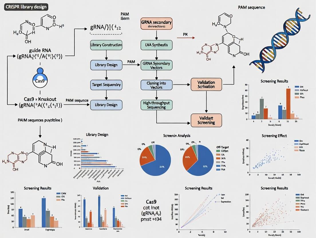
Designing CRISPR Libraries: A Complete Guide to Knockout and Activation Screens for Functional Genomics
This comprehensive guide provides researchers and drug development professionals with a detailed framework for designing and implementing CRISPR library screens for gene knockout and activation.
Designing CRISPR Libraries: A Complete Guide to Knockout and Activation Screens for Functional Genomics
Abstract
This comprehensive guide provides researchers and drug development professionals with a detailed framework for designing and implementing CRISPR library screens for gene knockout and activation. Covering foundational principles, practical methodologies, common troubleshooting strategies, and comparative validation techniques, the article synthesizes current best practices to empower robust, high-throughput functional genomics studies that accelerate target discovery and therapeutic development.
CRISPR Screens 101: Understanding Knockout vs. Activation for Functional Genomics
This whitepaper provides an in-depth technical comparison of CRISPR knockout (CRISPRko) and CRISPR activation (CRISPRa) libraries, framed within the broader thesis of library design for functional genomics screens in drug discovery and basic research. The fundamental mechanistic divergence lies in the endpoint: CRISPRko aims to permanently disrupt gene function by inducing double-strand breaks (DSBs) and leveraging error-prone non-homologous end joining (NHEJ), while CRISPRa aims to upregulate endogenous gene expression by recruiting transcriptional activators to promoter regions without damaging DNA.
Core Mechanisms & Molecular Components
CRISPR Knockout (CRISPRko): The standard CRISPRko system employs the Streptococcus pyogenes Cas9 nuclease complexed with a single guide RNA (sgRNA). The sgRNA directs Cas9 to a genomic locus complementary to its 20-nucleotide spacer sequence, adjacent to a Protospacer Adjacent Motif (PAM; NGG for SpCas9). Cas9 creates a blunt-ended DSB ~3 bp upstream of the PAM. The cell's primary repair pathway, NHEJ, often introduces small insertions or deletions (indels) during repair. When these indels occur within a protein-coding exon and shift the translational reading frame, they lead to premature stop codons and a complete loss of gene function via nonsense-mediated decay (NMD) of the mRNA or truncation of the protein.
CRISPR Activation (CRISPRa): CRISPRa fundamentally repurposes a catalytically inactive or "dead" Cas9 (dCas9). dCas9 retains its ability to bind DNA via sgRNA guidance but lacks endonuclease activity. To drive gene activation, transcriptional activation domains are tethered to dCas9. The most common systems are:
- dCas9-VP64: The minimal activator VP64 (a tetramer of VP16 peptides) is fused to dCas9.
- Synergistic Activation Mediator (SAM): A more robust system where dCas9 is fused to VP64. The sgRNA is engineered with RNA stem-loop aptamers that recruit additional activator proteins (e.g., MS2-p65-HSF1), creating a synergistic multi-component activator complex. CRISPRa sgRNAs are designed to target regions ~200 bp upstream of the transcription start site (TSS) to recruit this machinery to the promoter, thereby opening chromatin and recruiting RNA polymerase II to initiate transcription.
Quantitative Comparison of Key Parameters
Table 1: Mechanistic and Practical Comparison of CRISPRko and CRISPRa Libraries
| Parameter | CRISPRko | CRISPRa |
|---|---|---|
| Cas9 Form | Wild-type, nuclease-active Cas9 | Catalytically dead Cas9 (dCas9) |
| Primary Target | Protein-coding exons (early exons preferred) | Promoter/Enhancer regions (~200 bp upstream of TSS) |
| DNA Damage | Induces Double-Strand Breaks (DSBs) | No DSBs; Epigenetic modulation only |
| Core Mechanism | Frame-shift indels via error-prone NHEJ | Recruitment of transcriptional activators (e.g., VP64, p65, HSF1) |
| Genetic Outcome | Permanent, heritable gene disruption | Reversible transcriptional upregulation |
| Typical Fold-Change | Complete loss (100% knockdown) | 2- to 10-fold+ mRNA upregulation |
| Screen Phenotype | Loss-of-function (negative selection) | Gain-of-function (positive selection) |
| Key Design Constraint | Avoidance of off-target DSBs; PAM availability | Precise positioning relative to TSS; chromatin accessibility |
| Common Library (e.g., Human) | Brunello (4 sgRNAs/gene, ~76k sgRNAs) | Calabrese SAM (3-5 sgRNAs/gene, ~70k sgRNAs) |
Table 2: Performance Metrics in a Typical Pooled Screen
| Metric | CRISPRko Screen | CRISPRa Screen |
|---|---|---|
| Library Coverage | 3-10 sgRNAs per gene | 5-10 sgRNAs per gene (due to variable activation efficiency by target site) |
| Screen Duration | 14-21 population doublings (for depletion) | Often shorter (7-14 days) for positive selection |
| Key Readout | Depletion of sgRNAs in treated vs. control (Next-Gen Sequencing) | Enrichment of sgRNAs in selected vs. control (Next-Gen Sequencing) |
| False Positive Sources | Off-target cleavage; essential gene toxicity | Over-activation toxicity; off-target transcription |
| False Negative Sources | Inefficient indels; in-frame edits | Poor chromatin context at target site |
Detailed Experimental Protocol for a Pooled Screen
A. Library Design & Cloning
- sgRNA Design: For CRISPRko, use algorithms (e.g., from the Broad Institute's GPP Portal) to select guides with high on-target and low off-target scores targeting early constitutive exons. For CRISPRa, use tools like CRISPRa Design (from the Weissman Lab) to pick guides within -200 to -50 bp from the TSS of the annotated dominant isoform.
- Library Synthesis: Oligonucleotide pools are synthesized, PCR-amplified, and cloned via Golden Gate or Gibson assembly into the appropriate lentiviral backbone (e.g., lentiCRISPRv2 for KO; lentiSAMv2 for activation).
- Quality Control: Deep sequence the plasmid library to confirm even sgRNA representation.
B. Lentivirus Production & Cell Transduction
- Produce lentivirus in HEK293T cells by co-transfecting the library plasmid with packaging (psPAX2) and envelope (pMD2.G) plasmids.
- Titrate virus on target cells to determine the volume yielding a Multiplicity of Infection (MOI) of ~0.3-0.4, ensuring most cells receive a single sgRNA.
- Transduce >500 cells per sgRNA in the library (e.g., 50 million cells for a 100k-guide library) to maintain representation. Include a non-targeting control sgRNA pool.
C. Screen Execution & Sequencing
- Selection: Apply puromycin (or relevant antibiotic) for 3-7 days to select successfully transduced cells.
- Harvest "T0" Sample: Collect 50-100 million cells at the end of selection as the baseline reference.
- Phenotype Application: Split cells into experimental and control arms. Apply the selective pressure (e.g., drug treatment, nutrient stress) for the CRISPRko depletion screen or a growth factor/condition for the CRISPRa positive selection screen. Passage cells, maintaining >500x coverage.
- Harvest Endpoint ("Tfinal") Sample: Collect cells after ~14-21 (KO) or ~7-14 (activation) population doublings.
- Genomic DNA Extraction & NGS Prep: Isolate gDNA (Qiagen Maxi Prep). Perform a two-step PCR: (i) Amplify integrated sgRNA cassettes from gDNA using primers adding partial Illumina adapters; (ii) Add full adapters and sample indices.
D. Data Analysis
- Read Alignment & Count: Align sequencing reads to the reference sgRNA library. Count reads per sgRNA for T0 and Tfinal samples.
- Normalization & Statistical Testing: Normalize counts (e.g., to total reads). Use specialized algorithms (MAGeCK, CRISPResso2, PinAPL-Py) to calculate enrichment/depletion scores (log2 fold change) and statistical significance (p-value, FDR) for each gene.
Visualizing the Mechanistic Pathways
CRISPRko vs CRISPRa Core Mechanism Diagram
CRISPRa Synergistic Activation Mediator Complex
The Scientist's Toolkit: Essential Research Reagents
Table 3: Key Reagent Solutions for CRISPRko/CRISPRa Screens
| Reagent / Material | Function & Purpose | Example Product/Catalog |
|---|---|---|
| Validated CRISPRko Library | Pre-designed, cloned sgRNA sets targeting all annotated genes for knockout screens. Ensures high on-target efficiency. | Brunello Human CRISPR Knockout Pooled Library (Addgene #73179) |
| Validated CRISPRa Library | Pre-designed, cloned sgRNA sets targeting promoter regions for activation screens, optimized for dCas9-activator systems. | Human CRISPRa SAMv2 Library (Addgene #1000000132) |
| Lentiviral Packaging Plasmids | Second-generation system for safe, high-titer lentivirus production to deliver CRISPR libraries. | psPAX2 (Addgene #12260), pMD2.G (Addgene #12259) |
| dCas9-VP64 or SAM Vector | All-in-one lentiviral backbone expressing dCas9-activator and the modified sgRNA scaffold. | lenti-dCas9-VP64_Blast (Addgene #61425) or lenti SAMv2 (Addgene #75112) |
| Next-Generation Sequencing Kit | For preparing sgRNA amplicon libraries from genomic DNA of screen cells for deep sequencing. | Illumina Nextera XT DNA Library Prep Kit |
| Genomic DNA Isolation Kit (Large Scale) | For high-yield, high-quality gDNA extraction from millions of pelleted screen cells. | Qiagen Blood & Cell Culture DNA Maxi Kit |
| Pooled Screen Analysis Software | Computational pipeline for aligning sequencing reads, normalizing counts, and identifying significantly enriched/depleted genes. | MAGeCK (Model-based Analysis of Genome-wide CRISPR-Cas9 Knockout) |
| Cell Line with High Transduction Efficiency | A robust, rapidly dividing cell line compatible with the biological question and lentiviral transduction. | HEK293T, K562, A549, or relevant patient-derived organoids. |
The strategic deployment of CRISPR-based genetic screens—knockout (CRISPRko) and activation (CRISPRa)—has become a cornerstone of modern functional genomics. Within the broader thesis of CRISPR library design, the choice between these screens is not arbitrary but is dictated by the specific biological question, the genetic context of the target phenotype, and the desired mechanistic insight. This guide provides a technical framework for researchers to make an informed selection, ensuring library design aligns precisely with experimental goals.
Core Principles and Biological Rationale
CRISPR Knockout Screens utilize a catalytically active Cas9 nuclease (e.g., SpCas9) to create double-strand breaks in the coding exons of target genes, leading to frameshift mutations and permanent gene disruption via non-homologous end joining (NHEJ). This approach is ideal for identifying genes whose loss confers a selective advantage or disadvantage.
CRISPR Activation Screens employ a nuclease-deficient Cas9 (dCas9) fused to transcriptional activation domains (e.g., VPR, SAM system). The sgRNA guides this complex to promoter or enhancer regions, leading to targeted transcriptional upregulation. This modality is essential for identifying genes whose gain-of-function drives a phenotype.
The fundamental distinction lies in the directionality of the perturbation: loss-of-function (LOF) versus gain-of-function (GOF).
Comparative Analysis: A Decision Matrix
The decision to use a knockout or activation screen can be distilled into key comparative parameters, summarized in Table 1.
Table 1: Comparative Analysis of CRISPR Knockout vs. Activation Screens
| Parameter | CRISPR Knockout Screen (CRISPRko) | CRISPR Activation Screen (CRISPRa) |
|---|---|---|
| Cas9 Variant | Wild-type SpCas9 (Nuclease active) | dCas9 (Nuclease-dead) fused to activators (VPR, p65HSF1) |
| Primary Effect | Indels causing frameshifts & premature stop codons | Transcriptional upregulation near transcription start site (TSS) |
| Typical Phenotype | Loss-of-Function (Recessive) | Gain-of-Function (Dominant) |
| Optimal Library Size | 3-10 sgRNAs/gene; Whole-genome: ~70,000 sgRNAs | 5-10 sgRNAs/gene targeting -200 to +50 bp from TSS |
| Key Applications | Essential gene identification, resistance/sensitivity screens (e.g., drug, toxin), tumor suppressor discovery | Synthetic lethality (overexpression), drug target identification (overexpression rescue), differentiation drivers |
| Best for Genes | Haploinsufficient, tumor suppressors, essential genes | Oncogenes (where overexpression is pathogenic), redundant pathway members |
| Screen Duration | Longer (requires turnover of existing protein) | Shorter (rapid mRNA induction) |
| Common Readout | Depletion or enrichment of sgRNA counts over time | Enrichment of sgRNA counts over time |
| Major Limitation | Cannot assess GOF phenotypes; less effective for non-coding regions | Off-target transcriptional activation; position-dependent efficiency |
Detailed Experimental Protocols
Protocol for a Pooled CRISPR Knockout Screen
A. Library Design & Cloning:
- Select a validated genome-wide library (e.g., Brunello, Brie, or Toronto KnockOut). These contain ~4-10 sgRNAs per gene and ~1000 non-targeting controls.
- Amplify the plasmid library via ultra-deep sequencing (>500x coverage) to maintain diversity.
- Clone the sgRNA pool into a lentiviral backbone (e.g., lentiCRISPRv2) via Gibson Assembly or Golden Gate assembly.
B. Virus Production & Cell Transduction:
- Generate lentivirus by co-transfecting HEK293T cells with the sgRNA library plasmid, psPAX2 (packaging), and pMD2.G (envelope) plasmids using PEI transfection reagent.
- Harvest virus supernatant at 48 and 72 hours post-transfection, concentrate via ultracentrifugation.
- Titer virus on target cell line. Transduce cells at a low MOI (~0.3) to ensure most cells receive a single sgRNA. Maintain a representation of 500-1000 cells per sgRNA in the library.
C. Selection & Phenotype Induction:
- Apply puromycin selection (2-5 µg/mL, 3-7 days) to eliminate non-transduced cells.
- Passage cells for the duration of the phenotypic assay (e.g., 14-21 population doublings for a fitness screen, or apply a selective agent like a chemotherapeutic drug).
D. Genomic DNA Extraction & Sequencing:
- Harvest cells at the experimental endpoint (and at baseline, T0). Extract genomic DNA using a Maxi prep kit.
- Amplify integrated sgRNA sequences via a two-step PCR: 1st PCR with primers flanking the sgRNA scaffold, 2nd PCR to add Illumina adaptors and sample barcodes.
- Purify PCR products and sequence on an Illumina NextSeq or HiSeq platform (75bp single-end is sufficient).
E. Data Analysis:
- Align sequencing reads to the reference sgRNA library using a tool like
MAGeCKorCRISPResso2. - Count sgRNA reads for each sample (T0 and Tfinal). Normalize counts and calculate log2 fold-changes.
- Use robust rank aggregation (RRA) algorithm in MAGeCK to identify significantly enriched or depleted genes.
Protocol for a Pooled CRISPR Activation Screen
A. Library Design & Cell Engineering:
- Select a CRISPRa-optimized library (e.g., Calabrese, SAM, or CRISPRA). sgRNAs are designed to target regions -200 to +50 bp relative to the TSS.
- Prior to screening, generate a stable cell line expressing the dCas9-activator fusion protein (e.g., dCas9-VPR or dCas9-SAM component MS2-p65-HSF1). Use lentiviral transduction and blasticidin selection to create a monoclonal or polyclonal population.
- Confirm robust activation of positive control genes (e.g., CD69, MYOD1) via RT-qPCR.
B. Library Transduction & Screening:
- Produce lentivirus from the sgRNA activation library as in 4.1.B.
- Transduce the engineered dCas9-activator cell line at low MOI (~0.3), maintaining >500x coverage.
- Apply puromycin selection to select for sgRNA-expressing cells.
C. Phenotypic Selection & Analysis:
- Apply the phenotypic selection pressure (e.g., a growth factor withdrawal, a low dose of a pathway inhibitor). For a resistance screen, cells with a protective overexpressed gene will enrich.
- Harvest genomic DNA at T0 and after sufficient selection periods (often 14-21 days).
- Amplify and sequence sgRNA cassettes as in 4.1.D.
- Analyze data with tools like
MAGeCKorPinAPL-Py, identifying genes with significantly enriched sgRNAs.
Visualizing Screening Workflows and Logic
Decision Flow: CRISPRko vs. CRISPRa Screen Selection
Workflow for a Pooled CRISPR Knockout Screen
Workflow for a Pooled CRISPR Activation Screen
The Scientist's Toolkit: Essential Research Reagents
Table 2: Key Research Reagent Solutions for CRISPR Screens
| Reagent / Material | Function in Screen | Example Product/Catalog Number (Representative) |
|---|---|---|
| CRISPRko Library | Provides pooled sgRNAs for gene knockout. | Brunello Human Genome-Wide KO Library (Addgene #73179) |
| CRISPRa Library | Provides pooled sgRNAs for transcriptional activation. | Calabrese Human CRISPRa Library (Addgene #92379) |
| Lentiviral Packaging Plasmids | Required for production of lentiviral particles. | psPAX2 (Addgene #12260), pMD2.G (Addgene #12259) |
| Polyethylenimine (PEI) | High-efficiency transfection reagent for virus production in HEK293T cells. | Linear PEI, MW 40,000 (Polysciences #24765) |
| Puromycin Dihydrochloride | Selective antibiotic for cells expressing sgRNA-containing vectors. | Puromycin, 10 mg/mL Solution (Thermo Fisher #A1113803) |
| Genomic DNA Extraction Kit | For high-yield, high-quality gDNA from large cell pellets. | QIAGEN Blood & Cell Culture DNA Maxi Kit (Qiagen #13362) |
| Herculase II Fusion DNA Polymerase | High-fidelity polymerase for robust sgRNA amplicon generation for NGS. | Herculase II Fusion (Agilent #600679) |
| NGS Library Prep Kit | For attaching indices and adapters for Illumina sequencing. | NEBNext Ultra II DNA Library Prep Kit (NEB #E7645) |
| MAGeCK Software | Standard computational tool for analyzing CRISPR screen count data. | MAGeCK (Source: https://sourceforge.net/p/mageck) |
| dCas9-VPR Expression Plasmid | For constructing stable cell lines for CRISPRa screens. | lenti dCas9-VPR (Addgene #63798) |
This whitepaper details the core technical components of CRISPR library design, framed within the broader thesis of enabling robust, high-throughput genetic screens for functional genomics, with primary applications in gene knockout (CRISPRko) and activation (CRISPRa). The strategic integration of guide RNA (gRNA) design, library architecture, and delivery modality is paramount for generating high-quality, interpretable data in both discovery research and drug target identification.
Guide RNA (gRNA) Design Principles
The efficacy and specificity of a CRISPR screen are fundamentally determined by gRNA design. Modern design algorithms optimize for on-target activity and minimize off-target effects.
Key Design Parameters
- On-Target Efficiency: Predictors use machine learning models trained on empirical screen data (e.g., Rule Set 2, DeepHF, CRISPRon) to score gRNAs based on sequence features (GC content, nucleotide positions, secondary structure).
- Off-Target Specificity: Algorithms (e.g., from Benchling, IDT, Synthego) score potential off-target sites by tolerating mismatches and indels. Strict specificity filtering is critical for reducing false positives.
- Genomic Context: Target site selection relative to the transcription start site (TSS) varies by modality: for CRISPRko, target early exons of coding sequences; for CRISPRa, target regions -200 to -50 bp upstream of the TSS.
Quantitative Metrics for gRNA Design
Table 1 summarizes key performance metrics for leading gRNA design tools, based on recent benchmarking studies (2023-2024).
Table 1: Comparative Performance of gRNA Design Algorithms
| Algorithm/Tool | Primary Use Case | On-Target Prediction Accuracy (AUC) | Off-Target Consideration | Key Differentiator |
|---|---|---|---|---|
| Rule Set 3 (Azimuth) | CRISPRko | 0.79 | Mismatch/Position weighting | Industry-standard, validated on large datasets |
| CRISPRon | CRISPRa/i | 0.82 | Yes | Optimized for epigenetically defined regions |
| DeepSpCas9 | SpCas9 variants | 0.85 | Yes (CFD score) | Deep learning model for high-fidelity Cas9 |
| CHOPCHOP v3 | General design | 0.75 | Integrated Bowtie search | User-friendly, multi-species support |
| Synthego E-score | Synthetic gRNAs | Proprietary | Proprietary | Correlates with in vivo performance data |
Experimental Protocol: Validating gRNA Efficacy
Protocol: T7 Endonuclease I (T7EI) Mismatch Cleavage Assay for Indel Efficiency
- Cell Transfection: Transfect target cells with your CRISPR-Cas9 plasmid and the candidate gRNA using your preferred method (lipofection, nucleofection).
- Genomic DNA Extraction: Harvest cells 72 hours post-transfection. Extract genomic DNA using a silica-column based kit.
- PCR Amplification: Design primers flanking the gRNA target site (~500-800 bp product). Amplify the locus from purified gDNA.
- DNA Hybridization: Purify PCR products. Denature and re-anneal 200 ng of product in a thermal cycler (95°C for 5 min, ramp down to 25°C at 0.1°C/sec) to form heteroduplexes if indels are present.
- T7EI Digestion: Incubate hybridized DNA with T7 Endonuclease I (NEB) for 1 hour at 37°C. The enzyme cleaves mismatched heteroduplexes.
- Analysis: Run digested products on a 2% agarose gel. Cleavage products indicate indel formation. Quantify indel percentage using band intensity analysis (e.g., ImageJ).
Library Formats: Arrayed vs. Pooled
Library format dictates screening workflow, readout, and cost.
Comparative Analysis
Table 2: Arrayed vs. Pooled CRISPR Library Formats
| Parameter | Arrayed Library | Pooled Library |
|---|---|---|
| Format | Individual gRNAs or gRNA sets in separate wells (96/384-well plates). | A single complex pool of lentiviral vectors, each containing a unique gRNA. |
| Screening Readout | Compatible with high-content imaging, FACS, luminescence/fluorescence (e.g., viability, reporter). | Primarily NGS-based readout of gRNA abundance via genomic DNA sequencing. |
| Primary Application | Phenotypic screens requiring single-cell resolution, kinetic measurements, or complex multi-parameter assays. | Positive/Negative selection screens (e.g., cell viability, drug resistance, FACS sorting for top/bottom quantiles). |
| Throughput | Lower throughput (hundreds to thousands of genes). | Very high throughput (whole genome, ~10k-20k genes). |
| Cost & Labor | Higher reagent cost, more labor-intensive. | Lower per-gene cost, less hands-on time post-infection. |
| Hit Deconvolution | Directly known from well position. | Requires NGS and bioinformatic analysis. |
Experimental Protocol: Pooled Library Screen Workflow
Protocol: Basic CRISPRko Positive Selection Screen (e.g., for Drug Resistance)
- Library Transduction: Determine the library's MOI (Multiplicity of Infection) via pilot infection and puromycin selection to achieve ~30-40% infection efficiency, ensuring most cells receive a single gRNA. Scale up to transduce cells at a library coverage of 500-1000x (e.g., 500 cells per gRNA).
- Selection & Expansion: After puromycin selection (e.g., 2-3 days), maintain cells in culture for ≥14 population doublings under two conditions: DMSO control and drug-treated. Maintain minimum coverage at all steps.
- Genomic DNA Harvesting: Harvest at least 500 cells per original gRNA from each condition. Use a scalable gDNA extraction method (e.g., Qiagen Blood & Cell Culture Maxi Kit).
- gRNA Amplification & NGS Library Prep: Perform a two-step PCR. PCR1: Amplify the integrated gRNA cassette from gDNA using primers with partial Illumina adapter sequences. PCR2: Add full Illumina adapters and sample barcodes. Purify libraries and quantify by qPCR.
- Sequencing & Analysis: Sequence on an Illumina platform (MiSeq for small libraries, NextSeq for genome-wide). Align reads to the library manifest and use analysis tools (MAGeCK, CRISPResso2) to identify significantly enriched or depleted gRNAs.
Delivery Systems
Efficient, stable delivery is essential for introducing CRISPR components into target cells.
Delivery Modalities
- Lentiviral Vector (LV): The gold standard for pooled libraries and stable cell line generation. Provides durable, integrated expression of gRNA. Safety-modified (3rd generation, self-inactivating) vectors are standard.
- Adeno-Associated Virus (AAV): Used for in vivo delivery and primary/non-dividing cells. Limited cargo capacity (~4.7 kb) requires compact editors (e.g., SaCas9).
- Lipid Nanoparticles (LNPs) & Electroporation: For transient delivery of RNP complexes (pre-assembled Cas9 protein + gRNA). Offers rapid action, reduced off-targets, and no DNA integration. Ideal for arrayed screens in hard-to-transfect cells.
The Scientist's Toolkit: Key Research Reagent Solutions
Table 3: Essential Reagents for CRISPR Library Screens
| Item | Function & Key Consideration |
|---|---|
| Validated CRISPR Library (e.g., Brunello, Calabrese) | Pre-designed, cloned genome-wide gRNA sets for knockout or activation, with high on-target/off-target scores. |
| Lentiviral Packaging Plasmids (psPAX2, pMD2.G) | Second/third-generation systems for producing replication-incompetent lentivirus with high titer. |
| HEK293T/FT Cells | Standard cell line for high-titer lentivirus production due to high transfectability. |
| Transfection Reagent (PEI Max or Lipofectamine 3000) | For plasmid delivery into packaging cells. PEI Max is cost-effective for large-scale preps. |
| Polybrene (Hexadimethrine Bromide) | Cationic polymer that enhances viral transduction efficiency in many cell types. |
| Puromycin or Blasticidin | Selection antibiotics for cells stably expressing the gRNA vector. Critical concentration must be predetermined. |
| NGS Library Prep Kit (e.g., Nextera XT) | For efficient preparation of barcoded sequencing libraries from amplified gRNA cassettes. |
| CRISPR Analysis Software (MAGeCK, CRISPResso2) | Open-source tools for quantifying gRNA abundance and identifying significantly hit genes from screen data. |
Visualization of Workflows and Relationships
CRISPR Library Screening Decision Pathway
Title: Decision Tree for Choosing CRISPR Library Format and Delivery
Pooled CRISPRko Screen Experimental Workflow
Title: Step-by-Step Workflow for a Pooled CRISPR Screening Campaign
The precision of functional genomic screens hinges on a meticulously engineered pipeline: computationally optimized gRNAs, a library format aligned with the biological question, and a delivery system matched to the cellular model. As the field advances, integration of improved base editors, epigenetic modifiers, and single-cell readouts into these foundational frameworks will further empower researchers in mapping genetic dependencies and identifying novel therapeutic targets.
Within the comprehensive thesis on CRISPR library design for functional genomics, the primary objective of a screen is the most critical determinant of experimental architecture. This guide details the technical considerations, protocols, and analytical frameworks for three cornerstone screen goals: essential gene discovery, synthetic lethality (SL) identification, and drug resistance mechanism mapping. Each goal dictates unique library selection, control design, and validation pathways.
Core Screen Goals: Technical Specifications
The following table summarizes the key parameters defining each primary screening objective.
Table 1: Comparative Specifications for Primary CRISPR Screen Goals
| Parameter | Essential Gene Discovery | Synthetic Lethality (SL) | Drug Resistance Mapping |
|---|---|---|---|
| Primary Objective | Identify genes required for cellular proliferation/survival under baseline conditions. | Identify genes whose loss is specifically lethal in a defined genetic (e.g., oncogenic) or environmental context. | Identify gene knockouts or activations that confer survival advantage upon drug treatment. |
| Typical Library | Genome-wide (e.g., Brunello, Human CRISPR Knockout v2) | Focused (e.g., DNA damage repair, metabolic genes) or genome-wide. | Genome-wide or targeted (e.g., kinome, chromatin regulators). |
| Experimental Arms | Single cell population. | Test: Isogenic mutant or treated cell line. Control: Wild-type or untreated counterpart. | Test: Drug-treated cells. Control: Vehicle-treated (DMSO) cells. |
| Key Analytic Metric | Depletion of sgRNAs over time (fitness effect). | Differential depletion between test and control (context-specific fitness). | Enrichment of sgRNAs in test vs. control. |
| Primary Hit Class | Core cellular machinery, transcription/translation, essential metabolic pathways. | Pathway paralogs, backup pathways, compensatory networks. | Drug target, efflux pumps, activating mutations (via CRISPRa), alternative survival pathways. |
| Validation Approach | Competition assays, orthogonal siRNA/shRNA. | Selective validation in matched vs. mismatched genetic background. | Dose-response curves, resistance reversal assays. |
Detailed Experimental Protocols
Protocol for a Synthetic Lethality CRISPR Knockout Screen
This protocol is fundamental for identifying genetic vulnerabilities.
I. Library Selection & Cloning:
- Select a targeted or genome-wide knockout library (e.g., Toronto KnockOut v3).
- Amplify the sgRNA plasmid library following low-cycle PCR (18 cycles) to maintain representation. Use high-fidelity polymerase.
- Lentivirally package the library in HEK293T cells. Co-transfect the sgRNA library plasmid, psPAX2 (packaging), and pMD2.G (VSV-G envelope) at a 3:2:1 mass ratio using polyethylenimine (PEI).
- Titer the virus on target cells. Aim for a Multiplicity of Infection (MOI) of ~0.3 to ensure most cells receive a single sgRNA.
II. Cell Infection & Screening:
- Plate two isogenic cell lines: Disease Model (e.g., KRAS G12V) and Wild-Type Control.
- Infect cells at a library coverage of 500-1000x (e.g., 500 cells per sgRNA). Include non-targeting sgRNA controls.
- Select transduced cells with puromycin (1-5 µg/mL, 3-7 days).
- Harvest initial reference sample (Day 0). Split remaining cells and passage for ~14-21 population doublings. Maintain coverage throughout.
- Harvest final cell pellets from both arms for genomic DNA extraction.
III. Sequencing & Analysis:
- Extract gDNA using a maxi-prep kit. Perform two-step PCR to amplify sgRNA cassettes and add Illumina adaptors/indexes.
- Sequence on an Illumina NextSeq (Mid-Output, ~30M reads).
- Align reads to the library manifest. Calculate sgRNA read counts for Day 0 and Endpoint samples in both arms.
- Normalize counts and compute log₂ fold changes. Use statistical frameworks (e.g., MAGeCK or STARS) to rank sgRNAs by differential depletion in the disease model versus control.
Protocol for a Drug Resistance CRISPR Activation (CRISPRa) Screen
This protocol identifies gene upregulations that confer resistance.
I. Library & Cell Line Preparation:
- Select a genome-wide CRISPR activation library (e.g., Calabrese SAM v2).
- Generate a stable cell line expressing the dCas9-VP64 transcriptional activator and MS2-p65-HSF1 fusion protein. Confirm with immunoblot.
- Package and titer the sgRNA library as in 2.1.
II. Screening with Drug Challenge:
- Infect the CRISPRa cell line at high coverage (1000x). Select with puromycin/blasticidin.
- Split cells into Drug Treatment and Vehicle Control arms. Determine a sub-lethal dose (IC20-IC30) of the drug in a pilot assay.
- Treat cells with this dose, refreshing drug/vehicle every 3-4 days. Passage cells for 14-21 days.
- Harvest genomic DNA from both arms at endpoint.
III. Analysis for Enrichment:
- Amplify and sequence sgRNAs as in 2.1.
- Analyze for enriched sgRNAs in the drug-treated arm versus control. Tools like MAGeCK or drugZ are used to calculate significance.
Visualizing Screening Workflows & Pathways
Title: Synthetic Lethality CRISPR Screen Workflow
Title: PARP Inhibitor Synthetic Lethality Pathway
The Scientist's Toolkit: Research Reagent Solutions
Table 2: Essential Reagents and Materials for CRISPR Functional Screens
| Reagent/Material | Function & Purpose | Key Considerations |
|---|---|---|
| Validated CRISPR Library | Pre-designed pooled sgRNA collections for knockout (KO) or activation (a). | Select based on goal (genome-wide vs. targeted), version (improved on-target scores), and modality (KO, CRISPRa/i). |
| Lentiviral Packaging Plasmids | psPAX2 (gag/pol) and pMD2.G (VSV-G envelope) for producing replication-incompetent virus. | Use 2nd/3rd generation systems for enhanced safety. Always include a packaging-only negative control. |
| Polyethylenimine (PEI), linear | High-efficiency, low-cost cationic polymer for transient transfection of packaging cells. | Optimize PEI:DNA ratio (e.g., 3:1). Use high-concentration stocks (1 mg/mL, pH 7.0). |
| Puromycin Dihydrochloride | Selection antibiotic for cells transduced with puromycin-resistance carrying lentivectors. | Titrate to determine minimal concentration that kills all non-transduced cells within 3-5 days for your cell line. |
| Genomic DNA Extraction Kit (Maxi) | High-yield, high-purity gDNA isolation from millions of screen cells. | Scalability and removal of contaminants that inhibit PCR are critical. Spin-column or magnetic bead-based. |
| High-Fidelity PCR Master Mix | For accurate, low-bias amplification of sgRNA sequences from genomic DNA during library prep. | Essential for maintaining sgRNA representation. Use enzymes with >100x fidelity of Taq. |
| Illumina Indexed Primers | Custom primers for the two-step PCR that add sequencing adaptors and sample-specific barcodes. | Allows multiplexing of many screen arms. Must be HPLC-purified. |
| Analysis Software (MAGeCK, CRISPhieRmix) | Computational pipelines for quantifying sgRNA abundance, normalization, and statistical hit calling. | Choose based on screen type (e.g., MAGeCK for essentiality, CRISPhieRmix for resistance). |
Step-by-Step Protocol: Designing and Executing Your CRISPR Library Screen
This guide provides a technical framework for selecting between commercial and custom-designed gRNA libraries within CRISPR-based functional genomics screens. The choice impacts experimental flexibility, cost, validation burden, and ultimately, the success of knockout (CRISPRko) or activation (CRISPRa) screens central to target identification and validation in drug development.
Core Decision Factors: A Quantitative Comparison
The selection hinges on specific project parameters. The table below summarizes key quantitative and qualitative differentiators.
Table 1: Comparative Analysis of Commercial vs. Custom gRNA Libraries
| Factor | Commercial Libraries | Custom-Designed Libraries |
|---|---|---|
| Design & Content | Fixed, genome-wide (e.g., human, mouse) or focused (e.g., kinase, epigenetic) sets. Based on public algorithms (e.g., Doench '16, Hsu '13). | Fully flexible. Target any gene set, including non-standard organisms, specific isoforms, or non-coding regions. |
| Lead Time | 1-3 weeks (shipped as ready-to-use plasmids or lentiviral preps). | 4-12+ weeks (design, synthesis, cloning, validation). |
| Upfront Cost | Moderate ($2,000 - $10,000 for plasmid libraries). | High ($15,000 - $50,000+ for synthesis and cloning). |
| Validation | Extensive QC by vendor (NGS verification, titering). Minimal burden on researcher. | Requires full in-house validation: sequencing coverage, representation, viral titer. |
| Optimization | Limited to available formats. May not use latest algorithms or rules. | Can incorporate proprietary data, specific on/off-target scoring algorithms, and tailored controls. |
| Scalability | Ideal for standard, high-throughput screens. | Best for specialized, iterative, or niche target screens. |
| Best For | Standard genome-wide screens, benchmarking, labs initiating CRISPR screens. | Hypothesis-driven focused screens, non-model organisms, industrial pipeline projects. |
Critical Technical Considerations
Library Design Algorithms
gRNA efficacy predictions rely on algorithms that must be considered whether evaluating a commercial product or designing custom.
- For Knockout (Cas9): Modern libraries use rules from Doench et al. (2016) Nat Biotechnol and Moreno-Mateos et al. (2015) Nat Methods. Key features include GC content (40-80%), avoidance of homopolymers, and specific nucleotide preferences at positions 1-4 and 20.
- For Activation (dCas9-VPR): gRNAs are typically designed within -400 to -50 bp upstream of the transcription start site (TSS), as per Konermann et al. (2015) Nature.
- Controls: Essential for both types. Include non-targeting gRNAs (≥100 sequences) and positive control gRNAs (e.g., targeting essential genes).
Essential Experimental Protocols
Protocol 1: Validation of Library Representation by NGS (Pre-Screen)
- Purpose: Ensure even gRNA representation before lentiviral production.
- Steps:
- Amplify Library: Perform a limited-cycle PCR (≤20 cycles) from plasmid DNA using primers adding Illumina adapters and sample indexes.
- Purify & Quantify: Clean PCR product with SPRI beads and quantify by qPCR or bioanalyzer.
- Sequence: Run on a MiSeq or NextSeq (2x150bp) to get ≥500 reads per gRNA for a 50k-gRNA library.
- Analysis: Align reads to the library manifest. Calculate the coefficient of variation (CV) of gRNA counts. A CV < 0.5 indicates good evenness. Identify any "drop-out" gRNAs (<20 reads).
Protocol 2: Determination of Minimum Viral Titer and MOI for Screen
- Purpose: Achieve optimal infection for high-quality screen data.
- Steps:
- Produce Virus: Generate lentivirus from the library plasmid pool using a standard HEK293T transfection protocol.
- Titer Virus: Using the target cell line (e.g., HeLa), perform a puromycin (or appropriate antibiotic) kill curve to determine the minimum antibiotic concentration and duration for 100% cell death in 3-5 days.
- MOI Optimization: Infect cells at varying MOIs (e.g., 0.2, 0.5, 1.0) in technical triplicate, followed by antibiotic selection. After 5-7 days, extract genomic DNA and perform NGS as in Protocol 1.
- Analysis: Calculate the Pearson correlation of gRNA abundances between replicates. An MOI of ~0.3-0.4, yielding >500x library coverage, and a correlation >0.9 between replicates is optimal to ensure most cells receive a single gRNA.
Visualization of Key Concepts
Title: gRNA Library Selection and Screening Workflow
Title: Linking Screen Goal to gRNA Design Strategy
The Scientist's Toolkit: Research Reagent Solutions
Table 2: Essential Reagents for CRISPR Library Screens
| Reagent / Material | Function & Critical Notes |
|---|---|
| Validated gRNA Library | Commercial (e.g., Brunello, Calabrese) or custom array-synthesized oligo pool. The core reagent. Must be cloned into a lentiviral backbone (e.g., lentiGuide-Puro). |
| Lentiviral Packaging Plasmids | Typically a 2nd (psPAX2) & 3rd (pMD2.G) generation system for producing replication-incompetent virus in HEK293T cells. |
| High-Quality HEK293T Cells | Standard cell line for high-titer lentivirus production. Low passage number is critical. |
| Transfection Reagent | PEI or commercial lipid-based reagents (e.g., Lipofectamine 3000) optimized for 293T cells. |
| Target Cell Line | The biologically relevant cell line for the screen. Must be susceptible to lentiviral infection and have stable Cas9/dCas9 expression if using a two-part system. |
| Selection Antibiotic | Puromycin, blasticidin, or hygromycin for selecting successfully transduced cells. Concentration must be pre-titered on target cells. |
| NGS Library Prep Kit | Kits for amplicon sequencing (e.g., Illumina Nextera XT) to attach indexes and adapters to PCR-amplified gRNA regions from genomic DNA. |
| Genomic DNA Extraction Kit | Scalable kit for high-quality gDNA from large cell pellets (≥10^7 cells), often using silica-membrane columns. |
| Bioinformatic Pipeline | Software (e.g., MAGeCK, CERES, CRISPResso2) for quantifying gRNA abundance, normalization, and statistical analysis of enrichment/depletion. |
Within the broader thesis on CRISPR library design for functional genomics screens, the selection of optimal single guide RNAs (sgRNAs) is the foundational step determining the success of both knockout (CRISPRko) and activation (CRISPRa) screens. This guide focuses on the design rules for CRISPRko using Streptococcus pyogenes Cas9 (SpCas9), balancing maximal on-target cutting efficiency with minimal off-target effects to ensure clean, interpretable phenotypic data.
Core Principles for On-Target Efficiency
On-target efficiency is driven by sgRNA sequence features and genomic context. Key parameters are summarized below.
Table 1: Key sgRNA Sequence Features for High On-Target Efficiency
| Feature | Optimal Characteristic | Rationale & Impact |
|---|---|---|
| GC Content | 40-60% | sgRNAs with very low or high GC content show reduced stability and efficiency. |
| Polymerase III Terminator | Avoid 4+ consecutive T's | TTTT acts as a termination signal for U6 promoters, truncating sgRNA transcription. |
| Seed Region (PAM-proximal 8-12 nt) | High GC content, no secondary structure | Critical for R-loop formation; stable binding increases cleavage probability. |
| sgRNA Length | 20 nt spacer (standard) | Shorter (17-18 nt) can increase specificity but may reduce efficiency; longer may tolerate mismatches. |
| Target Position within Gene | Early constitutive exons, before functional domains | Maximizes probability of frameshift indel leading to complete loss-of-function (knockout). |
| 5' Nucleotide (for U6) | G (or A, if G not possible) | U6 promoter strongly prefers a guanosine at the transcription start site for high expression. |
Recent algorithmic predictions (e.g., from DeepCRISPR, Azimuth/Doench et al. 2016 rules) integrate these features into efficiency scores. It is critical to validate these predictions for your specific cell line, as chromatin accessibility (e.g., ATAC-seq data) and local nucleosome positioning can override sequence-based predictions.
Strategies to Minimize Off-Target Effects
Off-target cleavage remains a major concern for confident phenotype attribution.
Table 2: Strategies and Tools for Off-Target Minimization
| Strategy | Method | Key Resource/Tool |
|---|---|---|
| In Silico Prediction & Selection | Use algorithms to rank sgRNAs by predicted specificity. | CRISPick (Broad), CHOPCHOP, CRISPRitz; integrate scores like CFD (Cutting Frequency Determination) and MIT specificity scores. |
| Truncated gRNAs (tru-gRNAs) | Use 17-18 nt spacers instead of 20 nt. | Increases stringency of base-pairing required for cleavage, reducing tolerance to mismatches. |
| Modified Cas9 Variants | Use high-fidelity Cas9 nucleases. | SpCas9-HF1, eSpCas9(1.1): engineered to reduce non-specific DNA contacts. HiFi Cas9 (IDT) is a commercially available variant. |
| Dimeric CRISPR Systems | Use paired nickases (Cas9 D10A) with offset sgRNAs. | Requires two adjacent off-target sites for a double-strand break, dramatically increasing specificity. |
| Empirical Validation | Detect off-target sites via genome-wide assays. | GUIDE-seq, CIRCLE-seq, SITE-seq: Identify and quantify off-target cleavage events experimentally. |
Integrated Design and Validation Workflow
A robust sgRNA design pipeline incorporates both efficiency and specificity.
Diagram Title: Integrated sgRNA Design and Validation Workflow
Protocol 1: In Silico Design of sgRNAs for a Single Gene
- Input: Obtain the canonical transcript (e.g., from RefSeq) of your target gene.
- Generate Candidates: Use a tool like CRISPick (Broad Institute) or CHOPCHOP. Specify the target region (e.g., exons 1-3), PAM sequence (NGG for SpCas9), and sgRNA length (20nt).
- Initial Filter: Programmatically remove any candidate with 4+ consecutive T's, GC content <20% or >80%, or lacking a 5' G for U6.
- Ranking: Apply on-target efficiency (e.g., Azimuth score ≥0.5) and off-target specificity (e.g., CFD specificity score ≥60) filters. Select the top 3-4 sgRNAs targeting distinct sites within the 5' coding exons.
- BLAST: Perform a final genome-wide BLAST with the selected sequences to manually check for highly homologous off-target sites in coding regions.
Protocol 2: Experimental Validation of On-Target Editing (T7 Endonuclease I Assay)
- Transfection: Deliver your candidate sgRNAs and Cas9 (as plasmid, RNP, etc.) into your model cell line.
- Harvest Genomic DNA: 72 hours post-transfection, extract gDNA.
- PCR Amplification: Design primers (~200-300 bp amplicon) flanking the target site. Amplify the locus from purified gDNA.
- Heteroduplex Formation: Denature and reanneal PCR products: 95°C for 10 min, ramp down to 25°C at -2°C/sec.
- Digestion: Treat reannealed DNA with T7E1 enzyme (NEB) for 1 hour at 37°C. This cleaves mismatched heteroduplex DNA formed by WT and edited alleles.
- Analysis: Run products on an agarose gel. Quantify cleavage band intensities to estimate indel efficiency: % indel = 100 * (1 - sqrt(1 - (b+c)/(a+b+c))), where
ais the integrated intensity of the undigested band, andb+care the digested bands.
Table 3: Key Research Reagent Solutions for CRISPRko gRNA Design & Validation
| Item | Function/Benefit | Example Vendor/Catalog |
|---|---|---|
| High-Fidelity Cas9 Nuclease | Reduces off-target cleavage while maintaining high on-target activity. | IDT: Alt-R HiFi S.p. Cas9 Nuclease V3 |
| Synthetic sgRNA (chemically modified) | Ready-to-use, enhanced stability and RNP formation efficiency over plasmid-based systems. | Synthego (sgRNA EZ Kit), IDT (Alt-R crRNA) |
| Validated Positive Control sgRNA | Essential for optimizing delivery and confirming system functionality in your cell line. | e.g., Targeting AAVS1 or HPRT1 safe harbor loci. |
| T7 Endonuclease I | Fast, cost-effective enzyme for detecting indels via mismatch cleavage. | New England Biolabs (NEB), M0302S |
| Next-Gen Sequencing Kit for Editing Analysis | For precise, quantitative measurement of editing efficiency and spectrum. | Illumina (MiSeq), Amplicon-EZ service (Genewiz) |
| CRISPR Plasmids (All-in-One) | For stable expression from a single vector (U6-sgRNA + Cas9). | Addgene: lentiCRISPRv2 (52961) |
| Genomic DNA Extraction Kit | Rapid, high-yield gDNA isolation from cultured cells for PCR validation. | Qiagen DNeasy Blood & Tissue Kit |
For robust CRISPR library design, sgRNA selection cannot rely on a single parameter. The optimal strategy integrates computational predictions of efficiency and specificity with empirical validation in the relevant cellular context. Employing high-fidelity Cas9 variants and chemically modified sgRNAs further enhances the signal-to-noise ratio in pooled screens, ensuring that observed phenotypes are directly linked to the intended genetic perturbation. This rigorous approach to gRNA design forms the cornerstone of reliable, reproducible functional genomics research.
Within the broader scope of CRISPR library design for functional genomics, screens for gene knockout (CRISPRko) and gene activation (CRISPRa) serve as complementary pillars. This technical guide focuses on the design of CRISPR activation (CRISPRa) libraries, specifically those employing promoter-targeting guide RNAs (gRNAs) and the Synergistic Activation Mediator (SAM) system. CRISPRa enables targeted, gain-of-function screening, allowing researchers to identify genes whose overexpression drives phenotypic changes, such as drug resistance or cell differentiation. This approach is critical for drug target discovery and understanding gene regulatory networks.
Core Principles of the SAM System
The SAM system is a robust CRISPRa platform that significantly enhances transcriptional activation compared to early dCas9-VP64 fusions. It employs a tripartite mechanism:
- dCas9-VP64 Fusion: A catalytically dead Cas9 (dCas9) fused to the VP64 transcriptional activator (four copies of VP16) forms the foundation.
- MS2-P65-HSF1 (MPH) Activation Complex: The engineered gRNA contains two MS2 RNA aptamers in its tetraloop and stem-loop 2. These aptamers recruit MS2 bacteriophage coat proteins fused to a potent transcriptional activator complex: P65 and HSF1 (Heat Shock Factor 1).
- Synergistic Effect: The simultaneous recruitment of VP64 (via dCas9) and the MPH complex (via the MS2-gRNA) to a promoter region results in synergistic, high-level gene activation.
Diagram 1: SAM System Mechanism for Gene Activation
Designing Promoter-Targeting gRNAs for SAM Libraries
Effective CRISPRa requires precise gRNA placement within gene promoters. Unlike CRISPRko gRNAs that target exons, CRISPRa gRNAs must target regions upstream of the Transcription Start Site (TSS).
Key Design Rules and Quantitative Data
Target Window: The optimal region for gRNA binding is typically from -400 bp to -50 bp upstream of the TSS. Activity sharply declines beyond -400 bp and is minimal downstream of the TSS.
gRNA Length: Standard 20-nt spacer sequences are used, followed by the NGG Protospacer Adjacent Motif (PAM) for Streptococcus pyogenes Cas9 (SpCas9).
Avoidance of Epigenetic Marks: gRNAs should be designed to avoid nucleosome-occupied regions and specific repressive histone marks (e.g., H3K27me3) for optimal accessibility.
Table 1: Performance Metrics of gRNAs Targeting Different Promoter Regions
| Promoter Region (Relative to TSS) | Median Fold Activation (vs. Non-Targeting) | Success Rate* (% gRNAs with >5x activation) | Key Considerations |
|---|---|---|---|
| -50 to -150 bp | 15x | ~75% | Highest activity, potential for TSS disruption. |
| -150 to -400 bp | 12x | ~65% | Robust and reliable target window. |
| -400 to -800 bp | 5x | ~30% | Variable, enhancer regions possible. |
| Downstream of TSS | <2x | <5% | Generally ineffective for activation. |
*Success Rate: Percentage of designed gRNAs that achieve significant activation in validation assays.
Protocol: In Silico Design of a SAM gRNA Library
Step 1: Define Transcript Models. Use a reference genome (e.g., GRCh38) and an annotation database (e.g., GENCODE) to obtain precise TSS coordinates for all target genes.
Step 2: Generate Candidate gRNAs. For each gene, extract sequences from -400 to -50 bp upstream of the TSS. Identify all 20-nt sequences followed by a 5'-NGG-3' PAM on either strand.
Step 3: Filter for Specificity. Perform genome-wide alignment (using tools like Bowtie or BWA) to exclude gRNAs with significant off-target matches (allowing ≤3 mismatches). Tools like CHOPCHOP or CRISPick are commonly used.
Step 4: Rank and Select. Rank remaining gRNAs using an on-target scoring algorithm optimized for CRISPRa (e.g., CRISPRa scores from the Weissman or Gilbert labs). Select the top 3-5 gRNAs per gene for a pooled library to ensure robustness through redundancy.
Step 5: Incorporate SAM Scaffold. Append the specific gRNA scaffold sequence containing the two MS2 aptamers (e.g., the sequence from Konermann et al., 2015) to each selected 20-nt spacer.
Diagram 2: In Silico gRNA Library Design Workflow
Experimental Protocol: Performing a CRISPRa Screen with a SAM Library
Materials and Library Cloning
- SAM Plasmid System: Typically a 2-plasmid system: 1) lenti-dCas9-VP64Blast, and 2) lenti-MS2-P65-HSF1sgRNA_Puro.
- Pooled gRNA Library: A synthesized oligo pool containing 90-nt oligos (20-nt spacer + 70-nt constant scaffold with MS2 aptamers), cloned into the sgRNA backbone via Golden Gate assembly.
- Cells: A cell line relevant to the biological question (e.g., HEK293T, K562, primary T cells). Must be transducible and have high efficiency.
Procedure
Day 1-3: Generate Lentiviral Library. Co-transfect HEK293T packaging cells with the SAM sgRNA library plasmid, psPAX2, and pMD2.G. Harvest virus-containing supernatant at 48 and 72 hours.
Day 4: Determine Viral Titer. Transduce target cells with a dilution series of the virus and select with puromycin. Calculate the Multiplicity of Infection (MOI) to achieve ~30% infection, ensuring most cells receive a single gRNA.
Day 5: Bulk Transduction. Infect a large population of target cells (library coverage >500x) at MOI~0.3. Include a non-transduced control.
Day 6-8: Selection. Begin puromycin selection (e.g., 1-2 µg/mL) for 3-7 days to eliminate non-transduced cells.
Day 9-30: Screening. Apply the phenotypic selection pressure (e.g., drug treatment, FACS sorting for a surface marker, growth competition). Passage cells as needed, maintaining >500x coverage.
Day X: Harvest and Sequencing. Harvest genomic DNA from the selected population and a reference pre-selection population. PCR amplify the integrated gRNA sequences using flanking primers, add Illumina adapters/indexes, and sequence on a NextSeq or HiSeq platform.
Analysis: Align sequencing reads to the library manifest. Use MAGeCK or similar tools to compare gRNA abundance between selected and control populations, identifying significantly enriched or depleted gRNAs and, by extension, hit genes.
The Scientist's Toolkit: Essential Research Reagents
Table 2: Key Reagent Solutions for SAM CRISPRa Screens
| Reagent / Material | Function in SAM Screen | Example/Notes |
|---|---|---|
| lenti-dCas9-VP64_Blast | Stably expresses the dCas9-VP64 fusion protein. Provides the DNA-targeting foundation. | Addgene #61425 (pLV dCas9-VP64_Blast). Selection with blasticidin. |
| lenti-sgRNA(MS2)_Puro | Backbone for cloning the pooled gRNA library. Expresses the MS2-aptamer-containing sgRNA. | Addgene #73795 (lenti sgRNA(MS2) zsGreen Puro). Selection with puromycin. |
| lenti-MS2-P65-HSF1_Hygro | Stably expresses the MPH transcriptional activator complex. Recruited by the MS2-gRNA. | Addgene #89308 (lenti MPH v2). Selection with hygromycin. |
| Pooled gRNA Oligo Library | Defines the target genes for the screen. Synthesized as an oligo pool. | Custom-designed and ordered from vendors like Twist Bioscience or Agilent. |
| psPAX2 & pMD2.G | Lentiviral packaging plasmids. Required for production of infectious viral particles. | Addgene #12260 and #12259. |
| Polybrene (Hexadimethrine Bromide) | A cationic polymer that enhances viral transduction efficiency. | Typically used at 4-8 µg/mL during infection. |
| Next-Generation Sequencing Kit | For preparing gRNA amplicons from genomic DNA for abundance quantification. | Illumina Nextera XT or equivalent. |
| MAGeCK Software | Computational tool for analyzing gRNA read counts and identifying significantly enriched/depleted genes. | https://sourceforge.net/p/mageck/wiki/Home/ |
Diagram 3: SAM CRISPRa Screening Experimental Workflow
The design of effective CRISPRa libraries for the SAM system requires careful consideration of gRNA placement within a narrow promoter window, stringent off-target filtering, and the use of redundant gRNAs per gene. When combined with a robust experimental protocol for pooled screening, this approach provides a powerful platform for systematic gain-of-function genetics. Integrating insights from both CRISPRa and CRISPRko screens offers a comprehensive view of gene function, accelerating the discovery of novel therapeutic targets and biological mechanisms in drug development.
Within the broader thesis on CRISPR library design for functional genomics, this guide details the end-to-end experimental pipeline required to perform pooled knockout (CRISPRko) or activation (CRISPRa) screens. The robustness of this workflow directly impacts screen quality, data reproducibility, and the validity of downstream hit identification in drug target discovery.
The core process involves transitioning from a designed plasmid library to phenotypically screened cells, with lentivirus serving as the delivery vehicle. The following diagram outlines the key stages.
Title: CRISPR Pooled Screen Workflow from Cloning to Analysis
Detailed Methodologies & Protocols
Library Cloning into Lentiviral Backbone
Objective: Insert the synthesized pool of sgRNA expression cassettes into a lentiviral transfer plasmid (e.g., lentiCRISPRv2, lentiGuide-puro).
Protocol:
- Restriction Digest: Digest 5 µg of the lentiviral backbone with a high-fidelity enzyme (e.g., BsmBI-v2 for Addgene vectors) at 55°C for 2 hours. Purify the linearized vector via gel extraction.
- Gibson Assembly: Assemble the reaction using a 1:3 molar ratio of vector to insert (pooled sgRNA oligo duplexes or pre-annealed fragments). Use 50 ng of vector DNA in a 10 µl reaction with NEBuilder HiFi DNA Assembly Master Mix. Incubate at 50°C for 60 minutes.
- Bacterial Transformation: Desalt the assembly reaction and transform into highly competent E. coli (e.g., Endura ElectroCompetent Cells) via electroporation (2.5 kV, 1 mm cuvette). Recover cells in 1 ml SOC medium at 37°C for 1 hour.
- Library Amplification: Plate the entire recovery onto five 245 mm x 245 mm bioassay dishes with selective antibiotic (e.g., 100 µg/ml ampicillin). Grow at 32°C for 16-20 hours to minimize recombination. Harvest colonies via scraping.
- Plasmid Maxiprep: Isolate the pooled plasmid library using an endotoxin-free maxiprep kit. Elute in TE buffer. Critical: Determine library complexity by titering transformations and ensuring >200x coverage of the sgRNA library.
Lentiviral Production (HEK293T/17 Transfection)
Objective: Produce high-titer, replication-incompetent lentiviral particles.
Protocol:
- Cell Seeding: Seed 8 x 10⁶ HEK293T/17 cells per 15 cm dish in 20 ml DMEM + 10% FBS (no antibiotics) the day before transfection. Aim for 70-80% confluency.
- Calcium Phosphate Transfection (per dish):
- Prepare Solution A: Mix 22.5 µg library plasmid, 16.5 µg psPAX2 (packaging), and 6 µg pMD2.G (VSV-G envelope) in 1.35 ml of sterile 0.1x TE buffer.
- Prepare Solution B: 1.35 ml of 0.25 M CaCl₂.
- Add Solution B to Solution A dropwise while vortexing. Incubate at room temperature for 10-20 minutes until a faint precipitate forms.
- Add the 2.7 ml mixture dropwise to the dish. Gently swirl.
- Medium Change & Harvest: At 8-12 hours post-transfection, replace medium with 20 ml fresh, pre-warmed medium. Collect viral supernatant at 48 and 72 hours post-transfection. Pool harvests, filter through a 0.45 µm PES filter, and aliquot. Store at -80°C. Note: Commercially available transfection reagents (e.g., polyethylenimine, PEI) are widely used as an alternative.
Viral Titering & Target Cell Transduction
Objective: Determine viral functional titer and infect target cells at low Multiplicity of Infection (MOI) to ensure single sgRNA integration per cell.
Protocol for Functional Titer (in HeLa or HEK293T):
- Seed 1 x 10⁵ cells/well in a 12-well plate.
- Prepare serial dilutions (e.g., 10⁻¹ to 10⁻⁴) of viral supernatant in medium containing 8 µg/ml polybrene.
- Infect cells. After 24 hours, replace with fresh medium.
- At 72 hours post-infection, apply appropriate selection (e.g., 2 µg/ml puromycin). Maintain selection for 5-7 days, changing medium every 2-3 days.
- Count surviving colonies or assess viability. Calculate titer:
Titer (TU/ml) = (Number of resistant colonies * Dilution Factor * 1000) / Volume of virus (ml).
Protocol for Library Transduction:
- Scale Transduction: Perform a pilot transduction to determine the volume of virus required to achieve an MOI of ~0.3, ensuring <40% infection efficiency as measured by a fluorescent or antibiotic resistance marker.
- Bulk Transduction: Transduce the minimum number of cells required to maintain >200x library representation (e.g., for a 50,000 sgRNA library, transduce at least 10 million cells). Use polybrene (6-8 µg/ml) or protamine sulfate (4-8 µg/ml).
- Selection: Begin antibiotic selection (e.g., puromycin, 1-5 µg/ml) 24-48 hours post-transduction. Maintain selection until all cells in a non-transduced control well are dead (typically 5-7 days).
Screening & Sample Preparation for NGS
Objective: Apply selective pressure and harvest genomic DNA for sgRNA abundance quantification.
Protocol for a Positive Selection Proliferation Screen:
- Cell Passaging: After selection, expand cells to maintain >1000x library coverage at each passage. Count cells at each split.
- Time Points: Harvest a baseline sample (T0) immediately after selection. Continue passaging the remaining population. Harvest endpoint samples (e.g., T14, T21) after the phenotype manifests.
- Genomic DNA Extraction: Harvest at least 1 x 10⁷ cells per sample. Use a large-scale gDNA extraction kit (e.g., Qiagen Blood & Cell Culture Maxi Kit). Elute in TE buffer. Quantify by fluorometry.
- sgRNA Amplification & Sequencing: Perform a two-step PCR to add sequencing adapters and sample barcodes to the sgRNA region.
- PCR1 (from gDNA): Use 100 µg gDNA per sample as template in 50 µl reactions with primers amplifying the sgRNA scaffold. Pool reactions and purify.
- PCR2 (add indices/adapters): Use 5-10 ng of purified PCR1 product as template to attach full Illumina P5/P7 flow cell adapters and dual index barcodes. Purify final library, quantify, and sequence on an Illumina NextSeq or HiSeq platform (75 bp single-end is standard).
Successful execution requires monitoring key quantitative benchmarks.
Table 1: Critical Quality Control Metrics in a Pooled CRISPR Screen Workflow
| Stage | Parameter | Target Value | Purpose |
|---|---|---|---|
| Library Cloning | Plasmid DNA Yield | > 100 µg | Sufficient material for viral production and sequencing. |
| Bacterial Colony Coverage | > 200x library size | Maintains library complexity, prevents bottlenecking. | |
| Lentiviral Production | Functional Titer (HeLa) | > 1 x 10⁷ TU/ml | Enables efficient transduction at low MOI. |
| Cell Transduction | Infection Efficiency (Pilot) | 30-40% | Maximizes cells with single integrations (MOI ~0.3-0.4). |
| Post-Selection Cell Number | > 1000x library coverage | Prevents stochastic loss of sgRNAs. | |
| Sequencing | Read Depth per Sample | > 500 reads per sgRNA | Enables accurate fold-change calculation. |
| Bioinformatics | Pearson Correlation (Reps) | R² > 0.9 | Indicates high technical reproducibility. |
The Scientist's Toolkit: Essential Research Reagents
Table 2: Key Reagent Solutions for CRISPR Pooled Screening
| Reagent / Material | Function / Purpose | Example Product/Type |
|---|---|---|
| Lentiviral Transfer Vector | Backbone for sgRNA expression; contains antibiotic resistance for selection. | lentiCRISPRv2 (for KO), lentiSAMv2 (for activation) |
| Packaging Plasmids | Provide viral structural proteins (psPAX2) and envelope glycoprotein (pMD2.G) for particle production. | psPAX2, pMD2.G |
| HEK293T/17 Cells | Production cell line for generating high-titer lentivirus due to high transfectability. | ATCC CRL-11268 |
| Polyethylenimine (PEI) | Cationic polymer transfection reagent for efficient plasmid delivery into HEK293T cells. | Linear PEI, MW 25,000 |
| Polybrene | Cationic polymer that enhances viral transduction efficiency by neutralizing charge repulsion. | Hexadimethrine bromide |
| Puromycin Dihydrochloride | Selection antibiotic; kills non-transduced cells post-infection. | Cell culture grade, soluble in water. |
| Next-Generation Sequencer | Platform for high-throughput sequencing of sgRNA amplicons to determine abundance. | Illumina NextSeq 550/2000 |
| sgRNA Library Design Software | In-silico tool for designing specific, efficient, and minimal off-target sgRNAs. | Broad Institute GPP, CHOPCHOP, CRISPick |
| Screen Analysis Pipeline | Bioinformatics software to calculate sgRNA depletion/enrichment and perform statistical hit calling. | MAGeCK, CERES, PinAPL-Py |
Pathway: Lentiviral Transduction and sgRNA Action
The following diagram illustrates the mechanistic steps from viral entry to functional gene modulation in target cells.
Title: Mechanism of Lentiviral CRISPR Delivery and Gene Modulation
In large-scale CRISPR library screens for gene knockout (CRISPRko) or activation (CRISPRa), the accurate quantification of guide RNA (gRNA) abundance before and after a selection pressure is paramount. The core thesis—that optimized library design and precise gRNA tracking are critical for determining gene function and identifying therapeutic targets—rests on robust NGS data generation. This guide details the technical pipeline for amplifying and sequencing gRNA libraries from genomic DNA to generate the quantitative count data essential for screen analysis.
PCR Amplification Strategy for NGS Library Preparation
The goal is to amplify the integrated gRNA sequence from genomic DNA and attach sequencing adapters and sample indices (barcodes) for multiplexed NGS. A two-step PCR protocol is standard.
Protocol 1: Primary PCR (Amplification of gRNA Locus)
- Objective: Amplify the gRNA cassette from purified genomic DNA with primers adding partial adapter sequences.
- Reagents: High-fidelity DNA polymerase (e.g., Q5, KAPA HiFi), dNTPs, genomic DNA (≥ 1 µg per library sample).
- Primer Design:
- Forward Primer (Library-specific): Targets the constant promoter region upstream of the gRNA scaffold (e.g., U6 promoter).
- Reverse Primer (Library-specific): Targets the constant scaffold region downstream of the variable gRNA spacer.
- Note: These primers contain 5' overhangs with the partial Illumina i5 (Forward) and i7 (Reverse) adapter sequences.
- Cycling Conditions:
- 98°C for 30s (initial denaturation)
- 98°C for 10s (denaturation)
- 65°C for 30s (annealing – temperature must be optimized for primer Tm)
- 72°C for 20s (extension)
- Repeat steps 2-4 for 18-22 cycles (minimize over-amplification to preserve diversity)
- 72°C for 2m (final extension)
- Clean-up: Purify the PCR product using magnetic beads (e.g., SPRIselect) at a 0.8x bead-to-sample ratio.
Protocol 2: Secondary PCR (Indexing and Full Adapter Addition)
- Objective: Attach full dual indices and P5/P7 flow cell binding sites.
- Reagents: Purified Primary PCR product, high-fidelity polymerase, Illumina indexing primers (i5 and i7).
- Procedure: The purified primary PCR product serves as the template. Universal primers that bind the partial adapters added in step 1 are used to complete the adapter sequences and add unique dual indices.
- Cycling Conditions: Use a similar cycle profile as Primary PCR but limit to 6-10 cycles.
- Clean-up & Quantification: Perform a double-sided SPRI bead clean-up (e.g., 0.8x ratio, then 0.9x ratio). Quantify the final library by fluorometry (e.g., Qubit dsDNA HS Assay). Validate library size (~280-350 bp) via capillary electrophoresis (e.g., Bioanalyzer/Tapestation).
NGS Sequencing Considerations
- Sequencing Platform: Illumina NextSeq or NovaSeq series are typical for high-throughput screens.
- Read Configuration: A single-read (SR) run of 75-150 bp is sufficient, as the gRNA spacer (typically 20 bp) is located at a fixed distance from the constant primer binding site.
- Sequencing Depth: Critical for statistical power.
- Minimum: 50-100 reads per gRNA for the initial library.
- Recommended: 500-1000 reads per gRNA for each screen sample (T0 and TEnd) to robustly detect ~5-fold depletion/enrichment.
- PhiX Spike-in: Recommended at 5-10% to add diversity during initial cycles.
Table 1: Recommended NGS Sequencing Parameters for CRISPR Screens
| Parameter | Recommended Specification | Rationale |
|---|---|---|
| Read Length | SR75 - SR150 | Ample to cover variable spacer + constant scaffold. |
| Reads per gRNA (T0/TEnd) | ≥ 500 | Ensures statistical power to detect meaningful fold-changes. |
| Sequencing Coverage | 300-1000x Library Complexity | Oversampling to ensure all gRNAs are counted. |
| PhiX Spike-in | 5-10% | Mitigates low-diversity issues from short amplicons. |
| Q30 Score | > 80% | Ensures high base-call accuracy for gRNA identification. |
Table 2: Common Issues and Troubleshooting in gRNA NGS Library Prep
| Issue | Potential Cause | Solution |
|---|---|---|
| Low Library Complexity | Excessive PCR cycles in Primary PCR | Reduce Primary PCR cycles; use sufficient genomic DNA input. |
| Size Distribution Shift | Primer dimer or non-specific amplification | Optimize annealing temperature; titrate primer concentration; use bead clean-up. |
| Low Yield | Inefficient bead clean-up or PCR inhibition | Re-quantify gDNA; ensure bead freshness and correct ratios. |
| Index Misassignment | Excessive cluster density on flow cell | Dilute library appropriately; lower loading concentration. |
Visualization of Workflows
Title: gRNA Quantification NGS Library Prep Workflow
Title: Primer Design for gRNA Amplification
The Scientist's Toolkit: Research Reagent Solutions
Table 3: Essential Reagents for gRNA NGS Library Construction
| Item | Function & Critical Feature | Example Product(s) |
|---|---|---|
| High-Fidelity DNA Polymerase | Amplifies gRNA locus with minimal bias and error. Essential for maintaining library representation. | NEB Q5, KAPA HiFi HotStart, Herculase II. |
| SPRIselect Magnetic Beads | Size-selective purification of PCR amplicons and cleanup. Ratios (e.g., 0.8x) are critical for removing primer dimers. | Beckman Coulter SPRIselect, AMPure XP. |
| Illumina-Compatible Index Primers | Dual-unique indices allow multiplexing of many samples. Must be compatible with your sequencer's chemistry. | Illumina TruSeq CD Indexes, IDT for Illumina UD Indexes. |
| Fluorometric DNA Quant Kit | Accurate quantification of low-concentration libraries. More precise than absorbance (A260). | Invitrogen Qubit dsDNA HS Assay, Promega QuantiFluor. |
| Library Size Analyzer | Assesses final library fragment size distribution and detects adapter dimer contamination. | Agilent Bioanalyzer/Tapestation, FEMTO Pulse. |
| High-Quality Genomic DNA Kit | Produces pure, high-molecular-weight gDNA from screened cells. Integrity and purity are vital for PCR efficiency. | Qiagen Blood & Cell Culture DNA Maxi Kit, PureLink Genomic DNA Kit. |
Solving Common Problems: Optimizing Screen Performance and Data Quality
Within the paradigm of CRISPR functional genomics for gene knockout (CRISPRko) and activation (CRISPRa) screens, screen efficiency is the paramount determinant of data quality and biological discovery. The broader thesis of modern library design asserts that predictability and robustness are achieved not merely by optimal guide RNA (gRΝA) design, but by ensuring each target cell receives a single, functional CRISPR ribonucleoprotein complex. Low screen efficiency—manifested as low fold-changes, high noise, poor gene hit concordance, and high false-negative rates—most frequently originates from suboptimal Multiplicity of Infection (MOI) and inefficient viral transduction. This guide details the technical strategies to address these core bottlenecks.
Quantitative Foundations: The Impact of MOI on Screen Outcomes
The Poisson distribution dictates the probability of a cell receiving k viral particles when the average MOI is m: P(k) = (e^-m * m^k) / k!. The critical metrics for screen quality are derived from this.
Table 1: Poisson-Derived Cell Outcomes at Varying MOIs
| Average MOI | % Uninfected Cells (0 gRNAs) | % Cells with 1 gRNA | % Cells with >1 gRNA | Theoretical Screen Efficiency* |
|---|---|---|---|---|
| 0.3 | 74.1% | 22.2% | 3.7% | Low |
| 0.5 | 60.7% | 30.3% | 9.0% | Moderate |
| 0.7 | 49.6% | 34.7% | 15.7% | High (Optimal) |
| 1.0 | 36.8% | 36.8% | 26.4% | High but increased multiplicity |
| 3.0 | 5.0% | 14.9% | 80.1% | Unacceptable |
*Efficiency defined as maximum signal-to-noise and minimal confounding from multiple gRNAs per cell.
An MOI of 0.3-0.5 is often targeted to minimize multi-hit cells, but this comes at the cost of a high uninfected population, which dilutes signal. An MOI of ~0.7 balances a high rate of single-gRNA infection (desired) with a tolerable level of multi-hit cells.
Core Protocol: Determining Functional Lentiviral Titer and Optimizing MOI
Objective: To empirically determine the viral titer that yields the desired MOI for a specific cell line and screen format (e.g., antibiotic selection or FACS sorting for a fluorescent marker).
Materials:
- Producer cell line (e.g., HEK293T) and target screen cell line.
- Lentiviral transfer plasmid (e.g., lentiCRISPRv2, lentiGuide-Puro, with GFP/PuroR).
- Packaging plasmids (psPAX2, pMD2.G).
- Transfection reagent (e.g., polyethylenimine (PEI)).
- Polybrene (hexadimethrine bromide).
- Appropriate selection agent (e.g., Puromycin) or access to FACS.
Procedure: A. Virus Production (in Producer Cells):
- Seed HEK293T cells in a 6-well plate to reach 70-80% confluence at transfection.
- Co-transfect with transfer plasmid (e.g., 1 µg), psPAX2 (0.75 µg), and pMD2.G (0.25 µg) using PEI (3:1 ratio, PEI:total DNA).
- Replace media 6-8 hours post-transfection with fresh growth medium.
- Harvest viral supernatant at 48 and 72 hours post-transfection. Pool, filter through a 0.45 µm PVDF filter, and aliquot. Store at -80°C.
B. Functional Titer Determination (in Target Screen Cells):
- Day 0: Seed your target cell line in a 24-well plate at 2x10^4 cells/well in growth medium with polybrene (8 µg/mL).
- Day 1: Prepare a serial dilution of viral supernatant (e.g., undiluted, 1:10, 1:100) in medium with polybrene. Apply to cells.
- Day 2: Replace with fresh medium without virus.
- For Antibiotic Selection (e.g., Puromycin):
- Day 3: Begin selection with predetermined lethal concentration of puromycin.
- Day 7-10: Stain surviving colonies with crystal violet or count using an automated cell counter.
- Calculate TU/mL: (Number of colonies * Dilution Factor * 1000) / Volume of virus in mL.
- For Fluorescent Marker (e.g., GFP):
- Day 4-5: Analyze by flow cytometry to determine % GFP+ cells.
- Calculate TU/mL: (%GFP+ / 100) * (Cell number at infection * Dilution Factor * 1000) / Virus volume (mL).
C. MOI Calibration & Infection for Screen:
- Calculate virus volume needed:
Volume (mL) = (Desired MOI * Number of Target Cells) / (TU/mL). - For a pooled screen, infect at least 200-1000 cells per gRNA in the library to maintain representation. Using the calculated volume, infect cells in the presence of polybrene.
- Apply selection or sort 72 hours post-infection. The resulting population is your screen-ready, transduced pool.
Transduction Enhancement Strategies
When functional titer is low, these strategies can improve transduction efficiency without increasing multi-hit risk.
Table 2: Transduction Enhancement Reagents and Methods
| Strategy | Mechanism | Protocol Adjustment | Consideration |
|---|---|---|---|
| Polycation Additives (Polybrene, Protamine Sulfate) | Neutralizes charge repulsion between viral envelope and cell membrane. | Add to infection medium at 4-8 µg/mL (Polybrene). | Can be toxic to sensitive cells; titrate. |
| Spinoculation | Centrifugal force increases virus-cell contact. | Plate cells/virus in plate, centrifuge at 800-1000 x g for 30-60 min at 32°C. | Standard for refractory cell lines (e.g., primary T cells). |
| Envelope Pseudotyping (VSV-G) | VSV-G binds ubiquitous LDL receptor for broad tropism. | Use pMD2.G (VSV-G) plasmid as standard. | Gold standard for most mammalian cells. |
| Alternative Pseudotypes (RD114, GALV) | Bind different receptors; can improve transduction in specific lineages (e.g., hematopoietic). | Replace pMD2.G with alternative envelope plasmid during production. | Requires cell line-specific receptor expression. |
| Adhesion Promoters (RetroNectin, Fibronectin) | Coats plate, binding both virus and cell integrins to co-localize. | Coat plate overnight (5-20 µg/cm²), block, then add virus and cells. | Essential for many primary and stem cells. |
The Scientist's Toolkit: Key Research Reagent Solutions
Table 3: Essential Materials for MOI Optimization & Transduction
| Item | Function | Example Product/Catalog # |
|---|---|---|
| Lentiviral Packaging Plasmids | Provide gag/pol and envelope proteins for viral particle production. | psPAX2 (gag/pol/rev), pMD2.G (VSV-G) |
| Polycation Transduction Reagent | Enhances viral adsorption to cell surface. | Polybrene (Hexadimethrine bromide), H9268 (Sigma) |
| Recombinant Fibronectin Fragment | Enhances transduction of hematopoietic cells via co-localization. | Retronectin (Takara Bio), T100B |
| Selectable Marker | Enriches for successfully transduced cells. | Puromycin dihydrochloride, A1113803 (Thermo) |
| Fluorescent Reporter Plasmid | Enables titer determination and FACS sorting via marker expression. | lentiCRISPRv2-Blast-EGFP, Addgene #82416 |
| Concentration Reagent | Increases effective viral titer for low-titer supernatants. | Lenti-X Concentrator (Takara Bio), 631231 |
Visualizing Core Concepts and Workflows
Diagram 1: MOI Impact on Screen Cell Population Distribution (Max Width: 760px)
Diagram 2: Functional Titer to Screen-Ready Pool Workflow (Max Width: 760px)
Achieving high-efficiency CRISPR screens is a function of precise viral dosage and robust transduction. By rigorously determining functional titer, targeting an MOI of ~0.7, and implementing tailored enhancement strategies, researchers can transform low-efficiency screens into powerful, reproducible discovery engines. This optimization is not a preliminary step but the foundational pillar of the thesis that robust library design must account for delivery efficiency with the same rigor as guide design efficacy.
Within the thesis on CRISPR library design for functional genomics screens, achieving reliable results hinges on minimizing erroneous hits. False positives (genes incorrectly identified as hits) and false negatives (true hits missed) are pervasive challenges that can derail research and drug development. This guide provides a technical framework for mitigating these errors through rigorous library design, sufficient coverage, and experimental replication, focusing on CRISPR knockout (CRISPRko) and activation (CRISPRa) screens.
The Core Principles: Coverage and Replication
The statistical power to detect true phenotypes depends fundamentally on two parameters: the number of single guide RNAs (sgRNAs) per gene and the number of biological replicates. Insufficient values for either inflate both false positive and negative rates.
Quantitative Foundations
The following table summarizes key parameters and recommendations derived from current literature and statistical modeling.
Table 1: Guidelines for Library Coverage and Replication
| Parameter | Minimum Recommendation (Genome-wide) | Optimal Recommendation (Focused) | Rationale & Impact on Error Rates |
|---|---|---|---|
| sgRNAs per gene | 3-4 | 5-10 | Reduces false negatives from ineffective sgRNAs; enables robust statistical ranking via median/mean aggregation. |
| Library Representation (Coverage) | 200-500x | 500-1000x | Ensures each sgRNA is adequately represented in the screened population, preventing stochastic dropout (false negatives). |
| Biological Replicates | 3 | 4-6 | Essential for estimating experimental variance; critical for distinguishing technical noise from biological signal (reduces both false positives & negatives). |
| Minimum Read Count per sgRNA (Pre-screen) | 50-100 | >200 | Low starting counts increase sampling noise and risk of effective "dropout," leading to false negatives. |
| Fold-Change Threshold (Log2) | ±0.5 - ±1.0 | ±1.0 - ±2.0 | Context-dependent. Must be combined with statistical significance (p-value, FDR) to filter false positives. |
Detailed Methodologies for Key Experiments
Protocol 1: Determining Optimal Library Coverage (Transduction & Harvest)
Objective: To ensure each sgRNA in the pooled library is represented in a sufficient number of cells at the start of the screen (T0).
- Library Amplification & Preparation: Amplify the pooled lentiviral sgRNA library (e.g., Brunello, Calabrese) using low-cycle PCR and purify. Quantify via fluorometry.
- Virus Production & Titering: Produce lentivirus in HEK293T cells. Determine functional titer (TU/mL) on target cells using a fluorescent (e.g., GFP) or puromycin-resistance marker.
- Transduction at Low MOI: Transduce target cells at a low Multiplicity of Infection (MOI ≤ 0.3) to ensure most cells receive only one sgRNA. Include a non-targeting control sgRNA pool.
- Selection & Expansion: Apply selection (e.g., puromycin) for 3-7 days. Harvest a pre-selection sample (
T_pre) and a post-selection, pre-screen sample (T0). - Sequencing Library Prep: Extract genomic DNA from ≥ 1e7 cells for
T0. Amplify the integrated sgRNA sequences using indexed PCR, adding Illumina adapters. Pool and purify amplicons. - Quantitative Analysis: Sequence the
T0library to a depth of at least 50 reads per sgRNA in the sample. Calculate coverage:- Coverage = (Number of Transduced Cells at T0) / (Number of Unique sgRNAs in Library)
- Ensure coverage meets targets in Table 1. Discard screens where >15% of sgRNAs have <30 reads at T0.
Protocol 2: Implementing Biological Replication for Robust Hit Calling
Objective: To account for biological variability and enable rigorous statistical testing.
- Independent Cell Culture: Initiate at least three separate cultures of the target cell line from frozen stocks at least one passage apart.
- Independent Transduction & Selection: Treat each replicate culture as an entirely independent screen. Perform viral transduction (using the same virus batch is acceptable, but cell handling must be separate) and selection.
- Parallel Processing: Maintain and passage replicates separately throughout the screen duration (e.g., during proliferation or selection pressure).
- Endpoint Harvest & Sequencing: Harvest genomic DNA from each replicate's final timepoint (
T_final_rep1,T_final_rep2, etc.) and the sharedT0sample independently. Prepare sequencing libraries with unique sample indexes for each replicate. - Statistical Analysis: Use tools like
MAGeCK,CRISPRcleanR, orPinAPL-Pyto process counts. Essential steps include:- Normalize read counts across samples (e.g., median normalization).
- Test for differential abundance of each sgRNA/gene between
T_finalandT0within each replicate. - Perform robust rank aggregation (RRA) or linear modeling across replicates to generate a consensus p-value and false discovery rate (FDR) for each gene.
Visualizing Workflows and Relationships
Title: CRISPR Screen Workflow for Robust Results
Title: Causes and Mitigations for False Positives & Negatives
The Scientist's Toolkit: Research Reagent Solutions
Table 2: Essential Materials for CRISPR Pooled Screens
| Item | Function & Rationale | Example/Details |
|---|---|---|
| Validated Genome-wide CRISPR Library | Provides comprehensive, pre-designed sgRNAs with known efficiency and minimal off-target predictions. Essential for baseline reliability. | Brunello (KO), Calabrese (Activation) from Addgene. |
| Lentiviral Packaging Mix (2nd/3rd Gen) | Produces high-titer, replication-incompetent lentivirus for stable sgRNA delivery. A consistent system is critical for reproducibility. | psPAX2 & pMD2.G, or commercial kits (e.g., Lenti-X). |
| Next-Generation Sequencing Platform | For deep sequencing of sgRNA barcodes pre- and post-screen to quantify abundance changes. | Illumina NextSeq 500/550 for mid/high-throughput. |
| Genomic DNA Isolation Kit (Scalable) | High-yield, high-quality gDNA extraction from large cell pools (1e7 to 1e8 cells) is non-negotiable for even representation. | Qiagen Blood & Cell Culture DNA Maxi Kit. |
| PCR Additives for High GC-Content | sgRNA amplicons from genomic loci can be GC-rich. Additives improve amplification uniformity during NGS library prep. | Q5 High-Fidelity 2X Master Mix, DMSO, or GC Enhancer. |
| Analysis Software Suite | Specialized tools for count normalization, statistical testing, and hit ranking across replicates. | MAGeCK (Broad Institute), CRISPRcleanR. |
| Validated Positive Control sgRNAs/Perturbations | Essential for benchmarking screen performance and identifying technical failure. | sgRNAs targeting essential genes (e.g., RPA3) for dropout controls. |
| Pooled Non-Targeting Control sgRNAs | A large set (>100) of sgRNAs with no known targets. Crucial for modeling null distribution and calculating FDRs. | Included in most validated libraries. |
CRISPR activation (CRISPRa) screens are pivotal for discovering genes that confer phenotypes when overexpressed. However, a critical confounding factor is the "essential gene toxicity" or "gRNA dropout" phenomenon, where sgRNAs targeting essential genes cause proliferation defects, leading to their depletion independent of the intended activation phenotype. This guide details methods to identify, quantify, and correct for this bias, framed within the broader thesis that optimized CRISPR library design must account for both loss-of-function (knockout) and gain-of-function (activation) confounders to ensure clean genetic screening data.
The Mechanism of Essential Gene Toxicity in CRISPRa
In CRISPRa, a nuclease-dead Cas9 (dCas9) is fused to transcriptional activation domains (e.g., VPR, SAM). While designed to upregulate target gene expression, sgRNAs targeting essential genes can lead to toxic overexpression, mimicking a knockout phenotype. This is distinct from CRISPR knockout screens, where dropout is due to loss of gene function. The core hypothesis is that overexpression of certain essential genes (e.g., core cell cycle regulators) disrupts cellular homeostasis.
Signaling Pathway of CRISPRa Toxicity
The diagram below illustrates the proposed mechanistic pathway leading to proliferation defects from essential gene activation.
Title: Proposed Pathway for CRISPRa Essential Gene Toxicity
Identifying gRNA Dropout Signals: A Comparative Data Analysis
Quantitative data from recent studies comparing CRISPR-KO and CRISPRa screens highlight the dropout phenomenon. The table below summarizes key metrics from a synthetic analysis of such studies.
Table 1: Comparative Analysis of gRNA Depletion in Essential vs. Non-Essential Genes
| Gene Category | CRISPR-KO Screen (Log2 Fold Change)* | CRISPRa Screen (Log2 Fold Change)* | False Positive Rate in CRISPRa (without correction) | Primary Proposed Mechanism |
|---|---|---|---|---|
| Core Essential (e.g., PCNA) | -3.5 ± 0.8 | -2.1 ± 0.9 | 85% | Toxic overexpression disrupting stoichiometry |
| Common Essential | -2.8 ± 0.7 | -1.5 ± 1.0 | 70% | Overexpression-induced stress or apoptosis |
| Non-Essential | 0.2 ± 0.5 | 0.3 ± 0.6 | 5% | Baseline noise |
| Cell-Type Specific | -1.5 ± 1.2 | 1.8 ± 1.1 (Hit) | N/A | Valid activation phenotype |
*Negative values indicate gRNA depletion. Data is a composite from recent literature.
Experimental Protocol for Dropout Assessment and Correction
A robust workflow is required to distinguish true CRISPRa hits from false positives due to toxicity.
Title: Workflow for gRNA Dropout Analysis & Correction
Detailed Protocol: Paired CRISPR-KO/CRISPRa Screening for Dropout Identification
Objective: To generate paired datasets enabling the quantification of essential gene toxicity in CRISPRa. Materials: See "The Scientist's Toolkit" below. Procedure:
- Cell Line Preparation: Generate stable cell lines expressing dCas9-VPR (for CRISPRa) and wild-type Cas9 (for CRISPR-KO). Use the same parental line and ensure similar Cas9/dCas9 expression levels.
- Library Transduction: Transduce each cell line with a genome-wide sgRNA library (e.g., Calabrese et al., Nat Methods, 2023) at a low MOI (<0.3) to ensure single integration. Include a minimum of 500 cells per sgRNA during infection.
- Sample Harvesting: Harvest cells at baseline (T0, ~72h post-transduction) and at the experimental endpoint (Tfinal, typically 14-21 population doublings later). For proliferation-sensitive screens, include intermediate time points.
- Sequencing Library Prep: Amplify integrated sgRNA sequences from genomic DNA using a two-step PCR protocol. Use indexed primers for multiplexing.
- First PCR: Amplify sgRNA region (20-25 cycles). Use high-fidelity polymerase.
- Second PCR: Add Illumina adapters and sample indexes (10-12 cycles).
- Sequencing: Pool libraries and sequence on an Illumina platform to achieve >500x coverage per sgRNA.
- Data Processing:
- Read Alignment: Map reads to the sgRNA library reference using
bowtie2or a custom script. - Count Normalization: Normalize raw counts using median-of-ratios method (e.g., DESeq2) or total count normalization.
- Fitness Score Calculation: Compute gene-level log2 fold changes and statistical significance using dedicated tools:
- For CRISPR-KO: Use
MAGeCKorMAGeCK-VISPR. - For CRISPRa: Use
DrugZorMAGeCK-MLE.
- For CRISPR-KO: Use
- Read Alignment: Map reads to the sgRNA library reference using
- Dropout Analysis: Perform linear regression of CRISPRa gene scores against CRISPR-KO gene scores using a defined set of core essential genes (from DepMap). A significant positive correlation indicates a strong dropout signal.
Protocol: Correcting CRISPRa Scores Using a Regression Model
Objective: To subtract the toxicity-driven dropout signal from the CRISPRa results. Procedure:
- Model Building: Fit a linear model:
CRISPRa_Score ~ β * CRISPR-KO_Score + ε, using only genes classified as "common essential" in the DepMap database. - Parameter Estimation: Derive the slope (
β) which represents the fraction of the CRISPR-KO dropout effect that is recapitulated in the CRISPRa screen. - Score Adjustment: For every gene
iin the CRISPRa screen, calculate the corrected score:Corrected_CRISPRa_Score_i = Observed_CRISPRa_Score_i - (β * CRISPR-KO_Score_i) - Re-evaluate Hit Calling: Re-rank genes based on corrected scores. Genes whose significance is greatly diminished after correction are likely toxicity false positives.
The Scientist's Toolkit: Research Reagent Solutions
Table 2: Essential Materials for gRNA Dropout Analysis
| Item | Function/Description | Example Product/Catalog |
|---|---|---|
| CRISPRa Cell Line | Stable cell line expressing dCas9-activator fusion (e.g., dCas9-VPR, SAM). Required for gain-of-function screening. | Custom generated or commercial (e.g., Thermo Fisher A35371). |
| CRISPR-KO Cell Line | Stable cell line expressing wild-type Cas9 nuclease. Paired control for essential gene identification. | Custom generated or commercial (e.g., Synthego modified cell lines). |
| Genome-wide sgRNA Libraries | Lentiviral pools targeting all human genes. Libraries should be designed for both KO and activation. | KO: Brunello or TorontoKO. CRISPRa: Calabrese (Addgene #163064) or SAM (Addgene #1000000079). |
| Next-Gen Sequencing Kit | For preparing sgRNA amplicon libraries from genomic DNA. | Illumina Nextera XT, NEBNext Ultra II. |
| gRNA Read Alignment Software | Tool to process raw sequencing files into sgRNA count tables. | MAGeCKFlute (R package), bowtie2 aligner. |
| Screen Analysis Pipeline | Software to calculate gene fitness scores and significance. | MAGeCK (command line), CRISPRanalyzeR (web tool). |
| Essential Gene Reference | Curated list of core/common essential genes to calibrate dropout signal. | DepMap Portal (Broad Institute) Achilles Project data. |
| Proliferation Assay Kit | To validate toxicity of candidate sgRNAs (e.g., cell counting, ATP levels). | CellTiter-Glo (Promega G7570). |
Integrating gRNA dropout analysis into the CRISPRa screening workflow is essential for accurate hit identification. By performing parallel CRISPR-KO screens and applying statistical corrections, researchers can deconvolute toxicity-driven false positives from true activation phenotypes. This approach refines the thesis on CRISPR library design, arguing that future activation libraries should incorporate predictive models of essential gene toxicity at the design stage, potentially by excluding or flagging sgRNAs with high predicted dropout risk. This leads to more efficient screens and more reliable target discovery for drug development.
Batch Effect Correction and Normalization Strategies for Robust Hit Calling
Within the context of CRISPR library design for gene knockout and activation screens, robust hit calling is the critical process of distinguishing true biological signals from technical noise. Batch effects—systematic non-biological variations introduced by experimental factors such as different reagent lots, personnel, sequencing runs, or time—can severely compromise screen integrity. This guide details advanced correction and normalization strategies essential for ensuring reliable identification of essential genes, synthetic lethal interactions, or potent activators.
Batch effects manifest at multiple stages of a CRISPR screen workflow.
Table 1: Common Sources of Batch Effects in CRISPR Screens
| Source | Stage Introduced | Typical Manifestation | Impact on Readout |
|---|---|---|---|
| Library Transduction | Viral production, MOI variance | Differential sgRNA representation pre-selection | Skewed initial abundance |
| Cell Passaging & Selection | Antibiotic selection duration, cell density | Variation in selection efficiency across plates | Altered sgRNA dropout rates |
| Genomic DNA Harvesting | Lysis efficiency, extraction kit lot | Variable sgRNA recovery & PCR bias | Inconsistent count depth |
| PCR Amplification | Primer efficiency, cycle number, polymerase lot | Over-amplification, chimeras, index hopping | Amplification noise, mis-assignment |
| Next-Generation Sequencing | Lane/flow cell, cluster density, reagent kit | Differential sequencing depth & quality scores | Coverage bias, increased missing data |
Normalization Strategies: From Raw Counts to Analyzable Data
Normalization adjusts raw sgRNA read counts to enable meaningful comparison across samples.
Core Normalization Methods
- Total Count Normalization: Scales counts by the total library size (e.g., counts per million). Assumes most sgRNAs are unchanged, which can fail in strong positive selection screens.
- Median Ratio Normalization (DESeq2): Calculates a size factor for each sample as the median of the ratios of sgRNA counts to their geometric mean across all samples. Robust to differentially abundant sgRNAs.
- Trimmed Mean of M-values (TMM): Trims extreme log-fold-changes and abundances before calculating a scaling factor. Effective for screens with many neutral sgRNAs.
- Upper Quartile (UQ) Normalization: Scales counts using the 75th percentile of counts, excluding zeros. More robust than total count to highly abundant sgRNAs.
Table 2: Comparison of Core Normalization Methods
| Method | Key Principle | Pros | Cons | Best For |
|---|---|---|---|---|
| Total Count | Simple scaling by sum | Simple, fast | Biased by highly abundant sgRNAs | Pilot studies, quality control |
| Median Ratio | Median of count ratios | Robust to many DE sgRNAs | Sensitive to many zero counts | Knockout screens (many neutrals) |
| TMM | Trimmed mean of log ratios | Robust to outliers & composition bias | Computationally heavier | Comparisons with moderate effects |
| Upper Quartile | Scaling by 75th percentile | Resists top-count influence | May under-correct if upper quartile is unstable | Screens with clear positive controls |
Experimental Protocol: Performing Median Ratio Normalization
Objective: To generate normalized sgRNA count data from raw sequencing FASTQ files.
Input: Raw count matrix (sgRNAs x samples).
Software: R with DESeq2 package.
- Import Data: Create a
DESeqDataSetobject from the count matrix and sample information table. - Estimate Size Factors: Run
estimateSizeFactors()on the dataset object. This function calculates the median ratio for each sample. - Retrieve Normalized Counts: Use
counts(dds, normalized=TRUE)to extract the normalized count matrix, where counts are divided by the sample-specific size factor. - Validation: Plot PCA on normalized counts pre- and post-normalization to visualize reduction in sample-centric clustering.
Batch Effect Correction Algorithms
Post-normalization, dedicated algorithms model and remove residual batch variance.
Key Algorithms
- ComBat (sva package): Uses an empirical Bayes framework to adjust for known batch covariates. It estimates batch-specific location (mean) and scale (variance) parameters and shrinks them toward the global mean.
- Remove Unwanted Variation (RUV): Uses control sgRNAs (e.g., targeting non-essential genes or intergenic regions) to estimate factors of unwanted variation.
RUVseqoffers multiple methods (RUVg, RUVs, RUVr). - Limma
removeBatchEffect: Fits a linear model to the data and removes the component due to specified batch effects. Does not adjust for batch-by-condition interactions.
Table 3: Batch Effect Correction Algorithm Comparison
| Algorithm | Model Type | Requires Controls | Handles Unknown Factors | Output |
|---|---|---|---|---|
| ComBat | Empirical Bayes | No (uses known batches) | No | Batch-adjusted counts |
| RUV (e.g., RUVs) | Factor Analysis | Yes (negative controls) | Yes | Residuals or adjusted counts |
Limma removeBatchEffect |
Linear Model | No (uses known batches) | No | Batch-adjusted log2(CPM) |
Experimental Protocol: Batch Correction with ComBat-seq
Objective: Correct for known batch effects (e.g., sequencing date) in a normalized count matrix.
Input: Normalized count matrix, batch covariate vector, optional model matrix for biological conditions.
Software: R with sva package.
- Prepare Data: Ensure count matrix is not logged. Define a batch vector (e.g.,
batch <- c("A","A","B","B")). - Run ComBat-seq: Use
ComBat_seq(count_matrix, batch=batch, group=condition)whereconditionis the biological group (e.g., treatment vs control). Thegroupparameter preserves biological signal. - Assess Correction: Generate PCA plots on the
ComBat_seqoutput. Successful correction is indicated by samples clustering by biological condition rather than batch.
Integrated Workflow for Robust Hit Calling
A standardized pipeline integrates normalization and correction.
Title: CRISPR Screen Data Analysis Workflow
The Scientist's Toolkit: Research Reagent Solutions
Table 4: Essential Reagents & Tools for Batch-Robust Screens
| Item | Function | Consideration for Batch Control |
|---|---|---|
| CRISPR Library (e.g., Brunello, Calabrese) | Defined pool of sgRNAs targeting the genome. | Use a single high-quality plasmid prep for all screens; aliquot to avoid freeze-thaw. |
| Viral Packaging Plasmids (psPAX2, pMD2.G) | Produce lentiviral particles for library delivery. | Use a single master stock; titrate consistently across batches. |
| Polybrene / Hexadimethrine Bromide | Enhances viral transduction efficiency. | Use the same concentration and source; prepare fresh working solutions. |
| Puromycin / Selection Antibiotic | Selects for successfully transduced cells. | Determine kill curve for each new batch; use consistent concentration and duration. |
| Cell Culture Media & Sera | Supports growth of screening cell lines. | Use the same lot for an entire screen; pre-test for performance. |
| gDNA Extraction Kit (e.g., Qiagen Blood & Cell Culture Maxi) | High-yield genomic DNA extraction from pooled cells. | Use the same kit lot; standardize cell input and elution volume. |
| PCR Enzymes for Library Prep (e.g., Kapa HiFi) | Amplifies sgRNA region from gDNA with high fidelity. | Use a single master mix lot; optimize and fix cycle numbers. |
| Dual-Indexing Primers (i7/i5) | Adds unique sample barcodes for multiplex sequencing. | Use balanced, unique dual indices to prevent index hopping and batch-confounding. |
| Negative Control sgRNAs | Target safe-harbor or non-functional genomic loci. | Essential for RUV normalization and assessing false discovery rate. |
| Positive Control sgRNAs | Target essential genes (e.g., RPA3) or known hits. | Monitor screen performance and batch-to-batch efficacy. |
Visualization of Batch Effect Correction Impact
Title: Batch Effect Separates Treatment Groups
Title: Correction Reveals True Biological Signal
Implementing a rigorous pipeline combining appropriate normalization, such as median ratio methods, with robust batch correction algorithms like ComBat-seq, is non-negotiable for confident hit calling in CRISPR screens. This is especially critical in complex research streams involving library design for gene knockout and activation, where the fidelity of results directly informs target validation and drug discovery. Proactive experimental design—using standardized reagents, incorporating controls, and randomizing samples—minimizes batch effects at the source and ensures the robustness required for translational science.
Within the framework of CRISPR-based functional genomics screens for gene knockout (CRISPRko) or activation (CRISPRa), rigorous experimental design is paramount. A core tenet of this design is the strategic incorporation of control gRNAs. This technical guide details the implementation of two critical control classes: non-targeting gRNAs and gRNAs targeting core essential genes or pseudogenes. These controls are indispensable for normalizing screen data, assessing assay quality, and minimizing false discoveries, thereby ensuring the biological validity and reproducibility of screening outcomes.
The Role of Control gRNAs in Screen Analysis
Non-Targeting Control (NTC) gRNAs
NTCs are designed with sequences that lack perfect complementarity to any genomic locus in the target organism. They control for non-specific cellular responses to the Cas9/gRNA complex and transduction.
Primary Functions:
- Background Noise Estimation: Establish the distribution of phenotypes caused by the screening process itself (e.g., viral transduction, Cas9 expression).
- False Discovery Rate (FDR) Control: Serve as a null distribution for statistical testing to identify hits with phenotypes significantly different from "no effect."
- Normalization Anchor: Used in data normalization algorithms (e.g., median normalization, MAGeCK, BAGEL) to center read count distributions.
Targeting Controls: Essential Genes and Pseudogenes
These are gRNAs with known, expected phenotypes, providing internal benchmarks for screen performance.
- Core Essential Gene Controls: Target genes universally required for cellular proliferation or survival (e.g., RPL9, PSMB2). In a dropout screen, their gRNAs should be significantly depleted, validating screen sensitivity and dynamic range.
- Pseudogene or Safe-Harbor Locus Controls: Target genomically "neutral" sites (e.g., AAVS1, HPRT1 pseudogene regions, ROSA26). Their gRNAs should remain at stable abundance, similar to NTCs, controlling for sequence-specific off-target effects and validating screen specificity.
Design and Implementation Strategies
Design Principles & Quantitative Benchmarks
Table 1: Control gRNA Design Specifications and Benchmarks
| Control Type | Recommended Quantity per Screen | Design Principle | Expected Phenotype (Proliferation Screen) | Quality Metric (Post-Screen) |
|---|---|---|---|---|
| Non-Targeting (NTC) | 50 - 1,000 (≥5% of library) | No significant homology to genome (BLASTn; ≤17-nt contiguous match). Scrambled or designed against non-existent sequences. | Neutral (No depletion/enrichment). Log2 fold-change (LFC) ~0. | Tight distribution of LFCs (low median absolute deviation). Separation from essential gene signals. |
| Core Essential Gene | 50 - 500 (Targeting 5-20 genes) | Target multiple sites per gene. Use high-activity, validated gRNAs from reference sets (e.g., Dolcetto, Brunello libraries). | Strong depletion. Negative LFC > -2 to -4. | Clear, significant depletion (FDR < 0.001). Used in BAGEL2 for Bayes Factor calculation. |
| Pseudogene / Safe Harbor | 20 - 100 | Target loci with no known function in the cell type used. Validate neutrality in pilot assays. | Neutral (LFC ~0, matching NTCs). | Abundance stable relative to NTCs. Confirms lack of position effect. |
Sources: Recent analyses from publications using Brunello/Kosuke libraries (2023-2024) recommend higher NTC counts (>500) for robust statistical power in complex phenotypes. The DepMap consortium routinely uses ~1000 NTCs in genome-wide screens.
Experimental Protocol: Integrating Controls into a CRISPR Screen Workflow
Protocol: Library Construction and Screening with Integrated Controls
I. Library Design & Cloning
- Select Control gRNAs: Curate NTCs from established library resources (e.g., Addgene #1000000052 for Brunello NTCs). Select essential gene gRNAs from benchmark sets (Hart et al., 2017; Doench et al., 2016).
- Proportional Mixing: Combine control gRNAs with your target gene gRNA pool at the predetermined percentages (see Table 1). For a 5,000-gRNA library with 5% controls, include 250 control gRNAs.
- Oligo Pool Synthesis & Cloning: Synthesize the full oligo pool and clone into your lentiviral CRISPR backbone (e.g., lentiCRISPRv2, lentiGuide-Puro) via Golden Gate or Gibson assembly. Critical Step: Sequence the plasmid pool to confirm representation.
II. Lentivirus Production & Cell Transduction
- Produce lentivirus from the pooled plasmid library in HEK293T cells using standard protocols (psPAX2/pMD2.G).
- Transduce target cells at a low Multiplicity of Infection (MOI ~0.3) to ensure most cells receive a single gRNA. Include a non-transduced control.
- Apply selection (e.g., puromycin) for 3-7 days post-transduction to eliminate non-infected cells.
III. Screening & Sequencing
- Timepoint Harvest: Harvest genomic DNA (gDNA) from a) the plasmid pool (T0), b) post-selection cells (T1), and c) endpoint cells after the phenotypic selection (e.g., 14-21 population doublings for dropout, drug treatment for resistance/sensitivity).
- gRNA Amplification: Perform a two-step PCR on gDNA to add Illumina sequencing adapters and sample barcodes. Use a high-fidelity polymerase and minimize PCR cycles (≤20) to avoid skewing representation.
- High-Throughput Sequencing: Pool and sequence amplified libraries on an Illumina NextSeq or HiSeq platform to achieve >500x coverage per gRNA across all samples.
Data Analysis Workflow Utilizing Controls
Protocol: Control-Based Screen Data Analysis with MAGeCK
- Read Count Alignment: Align sequencing reads to your library's gRNA reference list using
magck count. - Normalization: Use the
--control-sgrnaparameter to specify your NTCs. MAGeCK will normalize read counts across samples based on the median count of these controls. - Beta Score Calculation: Run
magck testcomparing endpoint (Tfinal) to initial (T0 or T1) samples. The algorithm uses the distribution of NTCs and essential gene controls to model the null and essential distributions, respectively. - Quality Assessment:
- Plot the log2 fold-change (LFC) of all gRNAs. NTCs should center at zero.
- Essential gene controls should form a distinct, depleted population.
- Calculate the Gini Index of gRNA-level LFCs per gene. A low Gini index (<0.2) for essential controls indicates consistent on-target activity.
- Use tools like BAGEL2 which explicitly requires a reference set of core essential and non-essential (NTC/pseudogene) genes to compute a Bayes Factor for each target gene's essentiality.
Visualizing Experimental and Analytical Workflows
Title: CRISPR Screen Workflow with Control Integration
Title: Control-Based Data Analysis Pipeline
The Scientist's Toolkit: Research Reagent Solutions
Table 2: Essential Reagents and Resources for Control Implementation
| Item | Function & Description | Example Source/Product |
|---|---|---|
| Curated Control gRNA Libraries | Pre-designed, validated sets of NTCs and targeting controls for immediate use. | Addgene: Brunello NTCs (#1000000052), Dolcetto library (Essential/NT controls). |
| Lentiviral CRISPR Backbone | Plasmid for gRNA expression, often with Cas9 (KO) or dCas9-activator (a). | lentiCRISPRv2 (KO), lentiSAMv2 (a), lentiGuide-Puro (for stable Cas9 lines). |
| Packaging Plasmids | For production of replication-incompetent lentivirus. | psPAX2 (gag/pol), pMD2.G (VSV-G envelope). |
| High-Fidelity Polymerase | For accurate amplification of gRNA representation from genomic DNA prior to sequencing. | Q5 Hot Start (NEB), KAPA HiFi HotStart. |
| gRNA Read Alignment & Analysis Software | Open-source tools that incorporate control-based normalization and statistics. | MAGeCK, BAGEL2, PinAPL-Py. |
| Core Essential Gene Reference Sets | Consensus lists of genes essential across many cell lines, for control selection. | DepMap (Broad Institute), Hart et al. (2015) gene lists. |
| Next-Generation Sequencer | Platform for high-depth sequencing of gRNA amplicons to quantify abundance. | Illumina NextSeq 500/1000, NovaSeq. |
| Cell Line of Interest | The biological system for the screen, with validated Cas9/dCas9 expression and sgRNA delivery. | Various ATCC/ECACC lines, or custom-engineered lines. |
Beyond the Screen: Validating Hits and Comparing CRISPR Tools
This whitepaper details a critical phase within the broader thesis of CRISPR library design and implementation for functional genomics. Following a primary pooled screen, the transition from high-throughput data to validated hits is a major bottleneck. A robust hit validation pipeline is essential to confirm phenotype causality, minimize false positives from screening noise and off-target effects, and generate high-confidence leads for downstream drug discovery. This guide outlines the systematic progression from initial gRNA deconvolution through to rigorous individual gene verification.
Phase 1: gRNA Deconvolution & Hit Identification
The initial step analyzes sequencing data from the pooled screen to identify gRNAs and, by extension, target genes, whose abundance significantly changes between experimental conditions (e.g., treatment vs. control, survival vs. death).
Core Analysis Workflow:
- Sequence Demultiplexing & Alignment: Raw FASTQ files are demultiplexed by sample. gRNA sequences are extracted and aligned to the library reference.
- Read Count Quantification: The number of reads per gRNA per sample is tallied.
- Normalization: Read counts are normalized (e.g., using median ratio or TMM) to account for differences in sequencing depth.
- Statistical Analysis: Enrichment/depletion scores are calculated. Common tools and methods include:
- MAGeCK (Model-based Analysis of Genome-wide CRISPR-Cas9 Knockout): Uses a negative binomial model and robust rank aggregation (RRA) to score genes.
- CRISPResso2: For base-editing screens, quantifying allele frequencies.
- Custom R/Python Pipelines: Utilizing DESeq2 or edgeR for differential abundance analysis.
Key Data Output Table: Table 1: Example Top Hit Candidates from Primary Screen Analysis (MAGeCK RRA Output)
| Gene Symbol | Neg | score | Neg | p-value | Neg | FDR | Pos | score | Pos | p-value | Pos | FDR | Associated Phenotype |
|---|---|---|---|---|---|---|---|---|---|---|---|---|---|
| MYC | -5.32 | 2.1e-06 | 0.0012 | 1.01 | 0.45 | 0.78 | Essential | ||||||
| CDK9 | -4.87 | 7.8e-06 | 0.0031 | 0.98 | 0.48 | 0.80 | Essential | ||||||
| VHL | 1.15 | 0.12 | 0.45 | 4.95 | 3.5e-05 | 0.022 | Resistance Factor | ||||||
| BRCA2 | -5.01 | 5.5e-06 | 0.0025 | 1.22 | 0.11 | 0.52 | Essential |
Experimental Protocol 1: Primary Screen Data Processing with MAGeCK
- Prepare Count Files: Create a raw count matrix (gRNAs x samples) from sequencing data.
- Run MAGeCK COUNT:
mageck count -l library.csv -n output_prefix --sample-label L1,L2 --fastq sample1.fastq sample2.fastq - Run MAGeCK RRA:
mageck test -k count_matrix.txt -t treatment_sample -c control_sample -n output_prefix --norm-method total - Interpret Output:
gene_summary.txtfile contains RRA scores, p-values, and FDRs for each gene.
Phase 2: Secondary Validation with Arrayed Format
Top candidate genes from Phase 1 must be tested in an arrayed, low-throughput format to confirm the phenotype independent of library context and competition.
Key Research Reagent Solutions: Table 2: Essential Reagents for Arrayed Validation
| Reagent/Solution | Function in Validation |
|---|---|
| Arrayed gRNA/SynthRNA Libraries | Pre-cloned, individual gRNAs in plasmid or lentiviral format for transfection/transduction. |
| CRISPR-Cas9 Cell Lines | Stable Cas9-expressing cells (e.g., Cas9-EGFP) for rapid knockout studies. |
| CRISPRa/i SAM or dCas9-VPR/dCas9-KRAB Cells | Stable cell lines for activation (a/i) or inhibition (i) screens. |
| Fluorescence/Luminescence Viability Assays (CellTiter-Glo, Annexin V) | Quantify cell proliferation or apoptosis in response to gene perturbation. |
| High-Content Imaging Systems | Multiparametric phenotypic analysis (e.g., cell morphology, biomarker intensity). |
Experimental Protocol 2: Arrayed Proliferation Assay
- Seed Cells: Plate stable Cas9-expressing cells in 96-well plates (500-2000 cells/well).
- Transfect/Transduce: Deliver individual arrayed gRNAs (3-4 per gene) using a suitable method (lipofection, lentivirus). Include non-targeting control (NTC) and essential gene positive control (e.g., POLR2A) gRNAs.
- Incubate: Culture cells for 5-7 population doublings (typically 5-10 days).
- Assay Viability: Add CellTiter-Glo reagent, incubate, and measure luminescence.
- Analyze: Normalize luminescence of test wells to the NTC wells. A significant reduction (p<0.01) across multiple gRNAs confirms a hit.
Phase 3: Orthogonal Verification at Gene & Protein Level
Phenotypic confirmation must be coupled with molecular verification of the intended genetic perturbation.
Key Verification Techniques:
- Next-Generation Sequencing (NGS) of Target Locus: PCR-amplify the genomic target region from validated cell pools and sequence to confirm insertion/deletion (indel) mutations.
- Western Blot or Flow Cytometry: Directly measure protein-level knockdown or, for CRISPRa, overexpression.
- RT-qPCR: Quantify mRNA-level changes, especially for activation/knockdown screens.
Diagram 1: Core Hit Validation Pipeline Workflow
Diagram 2: Orthogonal Verification Methods
A stringent, multi-phase hit validation pipeline is the cornerstone for translating high-throughput CRISPR screen data into reliable biological insights. This process directly tests and reinforces the hypotheses generated by the initial library design—whether for identifying synthetic lethal interactions, resistance mechanisms, or novel therapeutic targets. By systematically deconvoluting gRNA signals, confirming phenotypes in an arrayed format, and providing orthogonal molecular verification, researchers can confidently advance a shortlist of high-probability targets into mechanistic studies and preclinical drug development, ensuring the integrity and impact of their functional genomics research.
Within the framework of CRISPR library design for functional genomics screens, the selection of perturbation modality—CRISPR knockout (CRISPRko), CRISPR activation (CRISPRa), or CRISPR interference (CRISPRi)—is a foundational decision. Each technology leverages the programmability of the CRISPR-Cas9 system but achieves distinct transcriptional outcomes, leading to unique experimental profiles. This guide provides a comparative analysis of these core modalities, focusing on their mechanisms, applications in pooled screening, and practical considerations for library design and implementation in drug discovery and basic research.
Core Mechanisms & Components
CRISPRko (Knockout): Utilizes wild-type Streptococcus pyogenes Cas9 (spCas9) or Cas12a (Cpf1) to generate double-strand breaks (DSBs) within the coding exons of a target gene. Repair via error-prone non-homologous end joining (NHEJ) leads to insertion/deletion (indel) mutations that disrupt the open reading frame, resulting in a permanent, complete loss-of-function.
CRISPRa (Activation): Employs a catalytically "dead" Cas9 (dCas9), fused to transcriptional activation domains. The dCas9-VPR system (VP64, p65, Rta) is a common configuration. Guided to promoter or enhancer regions, the complex recruits RNA polymerase II and co-activators to drive robust, tunable gene overexpression.
CRISPRi (Interference): Uses dCas9 fused to a transcriptional repressor domain, such as the Krüppel-associated box (KRAB). When targeted to a transcription start site (TSS), the dCas9-KRAB complex induces heterochromatin formation (e.g., H3K9 trimethylation) and blocks transcriptional initiation, leading to potent, reversible gene knockdown.
Comparative Strengths and Weaknesses
Table 1: Head-to-Head Comparison of Modalities
| Feature | CRISPRko | CRISPRa | CRISPRi |
|---|---|---|---|
| Primary Molecular Target | Coding exon | Promoter/Enhancer (200 bp upstream of TSS) | Transcription Start Site (TSS; -50 to +300 bp) |
| Cas9 Form | Wild-type (nuclease active) | dCas9 fused to activator (e.g., VPR) | dCas9 fused to repressor (e.g., KRAB) |
| Transcriptional Outcome | Permanent knockout | Gain-of-function (overexpression) | Reversible knockdown (typically 70-95% reduction) |
| Key Strength | Complete, permanent loss-of-function; gold standard for essentiality screens. | Enables gain-of-function and suppressor screens; studies gene dosage effects. | High specificity, minimal off-target transcription; tunable, reversible. |
| Key Weakness/Limitation | Can be confounded by NHEJ escape, alternative splicing, or truncated protein function. | Overexpression can be non-physiological; positional sensitivity for gRNA design. | Knockdown is incomplete; potential for "leaky" expression. |
| Typical Efficacy | >90% frameshift indels in bulk populations. | Up to 10-1000x mRNA upregulation, depending on target. | 70-95% mRNA knockdown, depending on target. |
| Pleiotropy/Off-Targets | DNA-level off-target DSBs; possible p53 activation. | Transcriptional "squelching" from strong activators; fewer DNA lesions. | Minimal DNA damage; possible off-target repression. |
| Optimal Library Design | 3-6 gRNAs/gene targeting early exons; Brunello, Brie, and similar libraries. | 3-10 gRNAs/gene targeting -200 bp upstream of TSS; Calabrese, SAM libraries. | 3-10 gRNAs/gene targeting TSS; Dolcetto, CRISPRi-v2 libraries. |
| Primary Screening Application | Essential gene identification, loss-of-function phenotypic screens. | Gene overexpression screens, resistance/suppressor screens, differentiation studies. | Essential gene identification (esp. in diploid cells), hypomorphic studies, synthetic lethality. |
Table 2: Quantitative Performance Metrics in a Standard Pooled Screen
| Metric | CRISPRko (Brunello) | CRISPRa (SAM) | CRISPRi (Dolcetto) |
|---|---|---|---|
| Average gRNAs per Gene | 4 | 3-5 | 3-10 |
| Typical Library Size (Human) | ~77,000 gRNAs (19k genes) | ~93,000 gRNAs (23k genes) | ~102,000 gRNAs (20k genes) |
| Screen Noise (Pearson R²)* | 0.85 - 0.95 | 0.75 - 0.90 | 0.90 - 0.98 |
| Optimal MOI (Lentivirus) | 0.3 - 0.5 | 0.3 - 0.5 | 0.3 - 0.5 |
| Critical Cell Coverage | >500 cells/gRNA | >1000 cells/gRNA | >500 cells/gRNA |
| Typical Screening Duration | 14-21 population doublings | 7-14 days post-transduction | 14-21 population doublings |
| *Noise refers to replicate concordance in negative control (non-targeting) gRNA abundance. |
Detailed Methodologies for Key Experiments
Protocol 1: Pooled Lentiviral Library Production for CRISPRko/a/i Screens
- Library Reconstitution: Transform high-complexity plasmid library (e.g., Addgene) into electrocompetent E. coli (≥50x coverage). Plate on large LB-ampicillin bioassay dishes. Scrape and maxiprep.
- Lentiviral Packaging: Co-transfect HEK293T cells (in 10-cm dish) using PEI or lipid-based method:
- 10 µg Library plasmid (sgRNA backbone).
- 7.5 µg psPAX2 (packaging plasmid).
- 2.5 µg pMD2.G (VSV-G envelope plasmid).
- Harvest & Concentration: Collect supernatant at 48h and 72h post-transfection. Filter (0.45 µm). Concentrate via PEG-it virus precipitation solution or ultracentrifugation.
- Titration: Transduce target cells (e.g., K562, HeLa) with serial dilutions of virus + polybrene (8 µg/mL). After 48h, begin puromycin selection (1-3 µg/mL). Calculate titer (TU/mL) based on percent surviving cells and dilution factor.
Protocol 2: Essential Gene Screen with CRISPRko or CRISPRi
- Cell Line Preparation: Culture target cells (≥20 million) in appropriate medium. Confirm >90% viability.
- Library Transduction: Infect cells at an MOI of 0.3-0.5 to ensure most cells receive ≤1 gRNA. Maintain >500x library coverage (e.g., for 77k library, use ~40 million cells). Include polybrene (4-8 µg/mL) or equivalent enhancer.
- Selection & Expansion: Begin puromycin selection 24-48h post-transduction. Maintain for 3-7 days until all non-transduced control cells are dead. Expand population, maintaining >500x coverage at all steps.
- Phenotype Induction & Harvest: Split cells into replicate populations (T0 control and experimental arms, e.g., drug-treated vs. DMSO). Culture for 14-21 population doublings, harvesting ≥500 cells/gRNA at each time point for genomic DNA extraction.
- gRNA Amplification & Sequencing: Perform a two-step PCR to add sequencing adapters and sample barcodes to the integrated sgRNA cassette. Purify amplicons and sequence on an Illumina NextSeq or HiSeq platform (≥50 reads/gRNA).
- Analysis: Align reads to the library reference. Use MAGeCK or PinAPL-Py to calculate gRNA depletion/enrichment and gene-level significance scores (RRA, p-value).
Visualizations
Title: Core Mechanisms of CRISPRko, CRISPRa, and CRISPRi
Title: Pooled CRISPR Library Screen Workflow
The Scientist's Toolkit: Key Research Reagents & Solutions
Table 3: Essential Materials for CRISPR Functional Genomics Screens
| Item | Function & Specification | Example Product/Catalog |
|---|---|---|
| Validated sgRNA Library | Pre-designed, sequence-verified plasmid pools for specific modalities (ko/a/i) and genomes. | Addgene: Brunello (ko), Calabrese (a), Dolcetto (i) |
| Lentiviral Packaging Plasmids | For producing replication-incompetent, high-titer lentivirus. | psPAX2 (packaging), pMD2.G (VSV-G envelope) |
| High-Efficiency Competent Cells | For amplifying plasmid libraries with minimal bias. | NEB Stable or Endura Electrocompetent E. coli |
| Polyethylenimine (PEI) | Cost-effective transfection reagent for viral packaging in HEK293T cells. | Polysciences, linear PEI (MW 25,000) |
| Polybrene (Hexadimethrine Bromide) | Cationic polymer that enhances viral transduction efficiency. | Sigma-Aldrich, 8 mg/mL stock solution |
| Puromycin Dihydrochloride | Selection antibiotic for cells transduced with puromycin-resistance carrying vectors. | Thermo Fisher Scientific; cell line-specific titration required. |
| Genomic DNA Extraction Kit | For high-yield, high-quality gDNA from large cell pellets (≥10^7 cells). | Qiagen Blood & Cell Culture DNA Maxi Kit |
| Herculase II Fusion DNA Polymerase | High-fidelity polymerase for robust 2-step PCR amplification of sgRNA sequences from gDNA. | Agilent Technologies |
| SPRIselect Beads | For size selection and clean-up of PCR amplicons prior to sequencing. | Beckman Coulter |
| Analysis Software | Computational pipeline for quantifying sgRNA abundance and gene-level statistics. | MAGeCK, PinAPL-Py, CRISPRcleanR |
Within the broader thesis on CRISPR library design for functional genomics, a critical challenge lies in interpreting phenotypic screening results. A CRISPR screen, whether for knockout (CRISPRko) or activation (CRISPRa), identifies genes whose perturbation impacts a cellular phenotype. However, this is often a starting point. True mechanistic understanding and target validation require integrating the primary screen hits with downstream molecular consequences measured by transcriptomics (RNA-seq) and proteomics (mass spectrometry). This multi-omics integration bridges the gap between genotype and phenotype, revealing regulatory networks, signaling pathways, and potential compensatory mechanisms.
Experimental Workflow for Multi-Omic Integration
A cohesive experimental design is paramount. The following workflow ensures data compatibility and robust correlation.
Diagram: Multi-Omic Integration Workflow
Table 1: Key Experimental Stages & Objectives
| Stage | Primary Objective | Key Output |
|---|---|---|
| CRISPR Screen | Identify genes modulating phenotype | Gene essentiality scores (e.g., log2 fold-change, p-value) |
| Transcriptomics | Measure gene expression changes post-perturbation | Differential expression (DE) matrix |
| Proteomics | Measure protein abundance & modification changes | Protein abundance fold-changes |
| Integration | Correlate genetic perturbation with molecular outcomes | Gene-protein regulatory maps, pathway enrichments |
Detailed Methodologies
CRISPR Screen Followed by Multi-Omic Profiling
- Protocol: Following a pooled CRISPR screen (e.g., using the Brunello or Calabrese library), cells are transduced at a low MOI to ensure single-guide integration. After phenotypic selection (e.g., drug treatment, FACS sorting), genomic DNA is extracted for NGS to calculate guide depletion/enrichment. In parallel, sister cultures from the same transduced pool are harvested for omics analysis. For transcriptomics, total RNA is extracted and prepared for bulk or single-cell RNA-seq. For proteomics, cells are lysed, proteins digested, and peptides analyzed by LC-MS/MS using label-free (LFQ) or tandem mass tag (TMT) quantification.
- Critical Control: Include samples transduced with non-targeting control (NTC) guides for each omics assay as the baseline.
Data Processing & Normalization Pipelines
- CRISPR Screen Data: Process sequencing reads with
MAGeCKorCRISPRcleanR. Generate gene-level beta scores (β) or log2(fold-change) representing phenotypic impact. - Transcriptomics Data: Align RNA-seq reads (e.g., with
STAR), quantify gene counts (e.g., withfeatureCounts), and perform differential expression analysis (e.g., withDESeq2orlimma-voom). Output is a matrix of log2 fold-changes for each gene per perturbation. - Proteomics Data: Process raw MS files with
MaxQuantorFragPipe. Normalize protein intensities and perform differential analysis withLimma. Output is a matrix of protein log2 fold-changes.
Correlation & Integration Strategies
Statistical integration is the core of this approach, moving from lists to networks.
Diagram: Data Integration Logic
Table 2: Quantitative Correlation Metrics (Hypothetical Data)
| Perturbed Gene (Hit) | CRISPR β score | mRNA log2FC | Protein log2FC | Phenotype-mRNA Correlation (r) | Phenotype-Protein Correlation (r) |
|---|---|---|---|---|---|
| Gene A | -2.1 (Essential) | -0.8 | -1.5 | 0.91 (Strong) | 0.95 (Strong) |
| Gene B | 1.8 (Enriched) | 0.5 | 0.9 | 0.72 (Moderate) | 0.68 (Moderate) |
| Gene C | -1.5 (Essential) | 2.3 (Up) | 0.1 (Flat) | -0.65 (Anti) | 0.10 (None) |
- Direct Correlation: Calculate pairwise correlation (Spearman) between the phenotypic score of perturbing a gene and the expression/abundance change of other genes across perturbations. This identifies consistent downstream effects.
- Joint Consistency Filtering: For a given perturbation, the mRNA and protein fold-changes of downstream genes are evaluated for concordance (e.g., both up, both down). Genes with consistent changes are higher-confidence effectors.
- Pathway Enrichment Analysis: Input correlated gene lists into tools like
GSEA,Enrichr, orPANTHERto identify affected biological pathways (e.g., "Apoptosis," "MTOR signaling").
The Scientist's Toolkit: Research Reagent Solutions
Table 3: Essential Materials for Multi-Omic CRISPR Integration
| Item | Function & Application | Example |
|---|---|---|
| CRISPR Library | Defines the set of genetic perturbations for screening. | Brunello (CRISPRko), Calabrese (CRISPRa) |
| NGS Kit for Guide Quantification | Prepares sequencing libraries from amplified gDNA to count guide abundance. | Illumina Nextera XT |
| RNA-seq Library Prep Kit | Converts isolated RNA into sequence-ready cDNA libraries. | Illumina Stranded mRNA Prep |
| Proteomics Sample Prep Kit | Facilitates cell lysis, protein digestion, and peptide cleanup for MS. | S-Trap Micro Columns |
| Mass Tag Reagents | Multiplex samples for quantitative proteomics. | TMTpro 16-plex |
| Alignment/Analysis Software | Processes raw sequencing or spectrometry data into analyzable matrices. | MAGeCK, DESeq2, MaxQuant |
| Integration & Visualization Tool | Performs statistical integration and generates network diagrams. | R/Bioconductor (ggplot2, pheatmap), Cytoscape |
Interpretation & Application in Library Design
Integrating multi-omics data validates primary screen hits and reveals indirect effects. For instance, a weak phenotypic hit whose perturbation drastically alters a key pathway may be a high-value target. Discrepancies between mRNA and protein changes (as with Gene C in Table 2) highlight post-transcriptional regulation. These insights directly feed back into the thesis on library design: future libraries can be augmented with guides targeting identified downstream effectors or compensatory nodes, creating more comprehensive and hypothesis-driven screening resources for the drug development pipeline. This iterative loop between screening, multi-omic integration, and library refinement is the future of functional genomics.
Within the broader thesis on optimizing CRISPR library design for gene knockout and activation screens, benchmarking the performance of available libraries is a critical step. This technical guide provides an in-depth analysis of core performance metrics for popular genome-wide CRISPR libraries, including Brunello (for knockout), Calabrese (for activation), and the Synergistic Activation Mediator (SAM) library. We present current quantitative data, detailed experimental protocols for benchmarking, and essential resources for researchers and drug development professionals engaged in functional genomic screening.
The selection of a CRISPR library directly impacts the sensitivity, specificity, and reproducibility of high-throughput screens. This whitepaper evaluates libraries based on key performance indicators: on-target efficacy, minimal off-target effects, library completeness, and screen performance metrics (e.g., Z-prime, hit consistency). The analysis is contextualized within the practical demands of both loss-of-function (KO) and gain-of-function (GOF) screens in therapeutic target discovery.
Quantitative Performance Metrics of Featured Libraries
The following tables summarize the core design and performance characteristics of the featured libraries, compiled from recent publications and vendor specifications.
Table 1: Core Design Specifications
| Library Name | Primary Use | Target Species | # of sgRNAs/Gene | Total sgRNAs | Core Design Principle | Reference |
|---|---|---|---|---|---|---|
| Brunello | Knockout | Human | 4 | 77,441 | Optimized SpCas9 sgRNAs from Doench et al. (2016) ruleset | Doench, J.G. et al. Nat Biotechnol. 2016 |
| Calabrese | Activation | Human | 4-5 (per enhancer) | 57,830 sgRNAs targeting ~2,000 enhancers | Targets putative enhancers with SAM-compatible sgRNA design | Simeonov, D.R. et al. Nature. 2017 |
| SAM (CRISPRa) | Activation | Human | 3-10 | 70,290 (v1, genome-wide) | MS2-P65-HSF1 (MPH) activator fused to dCas9; specific sgRNA 5' extension | Konermann, S. et al. Nature. 2015 |
Table 2: Benchmarking Performance Metrics (Typical Screen Results)
| Metric | Brunello (KO) | Calabrese (Enhancer) | SAM (Genome-wide Act.) |
|---|---|---|---|
| On-target Efficacy | High (>80% gene knockout) | Variable; context-dependent | High, strong transcriptional activation |
| Off-target Score (Predicted) | Low (optimized design) | Not primary concern (enhancer-specific) | Moderate (prolonged dCas9 binding) |
| Screen Dynamic Range | High (strong negative selection) | Moderate to High | High (positive selection) |
| Hit Reproducibility (Pearson R²) | >0.8 (between replicates) | ~0.7-0.8 | >0.8 |
| Typical Z-prime Factor | >0.5 (in robust assays) | >0.4 | >0.5 |
| Key Validation Rate | 70-90% (top hits) | 50-70% (enhancer-gene links) | 70-85% (top activating hits) |
Experimental Protocols for Benchmarking Library Performance
Protocol for Assessing On-target Knockout Efficacy (Brunello)
Objective: Quantify the gene knockout efficiency of a subset of Brunello sgRNAs.
- sgRNA Selection: Select 20-30 sgRNAs targeting essential genes and 10 targeting non-essential controls from the Brunello library.
- Lentiviral Production: Clone sgRNAs into lentiviral backbone (e.g., lentiGuide-Puro). Produce virus in HEK293T cells.
- Cell Infection & Selection: Infect a well-characterized cell line (e.g., A375) at a low MOI (<0.3) to ensure single integration. Select with puromycin (1-2 µg/mL) for 5-7 days.
- Efficacy Measurement (Cell Titer-Glo): Plate cells in 96-well format. 5-7 days post-selection, measure cell viability using CellTiter-Glo assay. Normalize viability of essential gene-targeting wells to non-essential controls. Efficacy = 1 - (normalized viability).
Protocol for Benchmarking Activation Dynamics (SAM/Calabrese)
Objective: Measure the transcriptional activation strength and specificity.
- Reporter Cell Line Generation: Stably integrate a dCas9-VP64 or dCas9-MPH (for SAM) expressing construct into your cell line. Include a fluorescent reporter (e.g., GFP) under a minimal promoter with a targetable site.
- sgRNA Transfection: Transfect sgRNAs from the SAM or Calabrese library (cloned into appropriate MS2-containing vector for SAM) targeting the reporter site or endogenous loci of known genes.
- Output Quantification: 72 hours post-transfection:
- For reporter: Analyze GFP fluorescence via flow cytometry.
- For endogenous genes: Perform RT-qPCR on target genes (e.g., IL1RN for a known SAM target). Calculate fold-change relative to non-targeting control sgRNAs.
Protocol for Full-Library Screen Quality Control
Objective: Determine the robustness of a genome-wide screen using standard metrics.
- Screen Execution: Perform the screen in biological triplicate with proper controls (non-targeting sgRNAs, essential gene targeting sgRNAs). Maintain >500x library representation at all steps.
- Data Processing: Sequence the sgRNA barcodes at T0 and Tfinal. Align reads and count sgRNA abundances.
- Quality Metric Calculation:
- Z-prime Factor: Using essential and non-essential gene sgRNA abundances. Formula: Z' = 1 - [3*(σp + σn) / |μp - μn|], where p=positive controls (non-essential), n=negative controls (essential).
- Reproducibility: Calculate Pearson correlation coefficient (R²) between log2(fold-change) of all genes across replicate screens.
- Gini Index: Assess library dropout evenness. A lower Gini index (<0.2) indicates good representation.
Visualizations
Title: Workflow for CRISPR Library Screen & Benchmarking
Title: SAM CRISPRa Complex Mechanism
The Scientist's Toolkit: Key Research Reagent Solutions
| Item | Function & Description | Example Vendor/Catalog |
|---|---|---|
| Lentiviral Packaging Plasmids | Second/third-gen systems for safe, high-titer virus production of sgRNA libraries. | psPAX2 (packaging), pMD2.G (VSV-G envelope) |
| lentiGuide-Puro | Backbone vector for cloning Brunello and other sgRNA libraries; confers puromycin resistance. | Addgene #52963 |
| lentiSAMv2 | All-in-one vector for SAM activation screens; contains dCas9-VP64, MS2-P65-HSF1, and sgRNA scaffold. | Addgene #75112 |
| Polybrene (Hexadimethrine Bromide) | Cationic polymer that enhances viral transduction efficiency. | Sigma-Aldrich H9268 |
| Puromycin Dihydrochloride | Selection antibiotic for cells transduced with puromycin-resistant vectors. | Thermo Fisher A1113803 |
| CellTiter-Glo Luminescent Assay | Measures ATP concentration to quantify viable cells for knockout efficacy checks. | Promega G7571 |
| NextSeq 500/550 High Output Kit | NGS reagents for sequencing the sgRNA region from harvested genomic DNA post-screen. | Illumina 20024906 |
| MAGeCK (Bioinformatics Tool) | Model-based Analysis of Genome-wide CRISPR/Cas9 Knockout; standard for screen analysis. | Source: https://sourceforge.net/p/mageck |
| CRISPick (Design Tool) | Web tool for designing and selecting optimized sgRNAs; hosts the Brunello library designs. | Website: https://portals.broadinstitute.org/gppx/crispick/public |
Within the broader thesis on CRISPR library design for functional genomics screens, this article presents case studies demonstrating the direct application of CRISPR knockout (CRISPRko) and activation (CRISPRa) screens in identifying and validating novel drug targets across three therapeutic areas. The precision of modern, optimally designed sgRNA libraries is foundational to these successes, enabling systematic interrogation of gene function in disease-relevant models.
Case Study 1: Oncology – Identifying Synthetic Lethal Interactions
Research Context: The discovery of synthetic lethal partners for tumor suppressor genes (e.g., BRCA1, PTEN) has yielded paradigm-shifting therapies like PARP inhibitors. CRISPRko screens are now accelerating the discovery of next-generation targets.
Featured Study: Identification of WRN as a synthetic lethal target in microsatellite unstable (MSI) cancers.
- Library: Genome-wide CRISPRko (e.g., Brunello or Toronto KnockOut) library.
- Screening Model: MSI-high vs. microsatellite stable (MSS) isogenic human colorectal cancer cell lines.
- Protocol:
- Transduction: Cells are transduced with the lentiviral sgRNA library at a low MOI to ensure single integration, with sufficient coverage (≥ 500 cells per sgRNA).
- Selection & Expansion: Puromycin selection is applied for 3-5 days. Cells are then passaged for ~14 population doublings.
- Sample Collection: Genomic DNA is harvested at the initial (T0) and final (Tend) time points.
- NGS & Analysis: sgRNA sequences are PCR-amplified and deep-sequenced. Depleted sgRNAs in MSI vs. MSS lines are identified using MAGeCK or similar algorithms, pinpointing genes essential specifically in the MSI context.
- Key Finding: The helicase gene WRN was identified as a top-scoring selective essential gene in MSI models. Validation confirmed that loss of WRN causes double-strand break accumulation and cell death in MSI but not MSS cells.
- Drug Discovery Impact: This discovery has spurred the development of WRN inhibitors, with several candidates now in preclinical development.
Table 1: Key Quantitative Outcomes from Oncology CRISPR Screens
| Target Gene | Cancer Type | Genetic Context | Screen Type | Hit Validation (Cell Viability % Reduction) | Development Stage |
|---|---|---|---|---|---|
| WRN | Colorectal | MSI-High | CRISPRko | 70-80% | Preclinical |
| RNF43 | Pancreatic | Wnt-dependent | CRISPRko | 60-70% | Target Validation |
| MCL1 | AML | FLT3-ITD | CRISPRko | >80% | Clinical Trials |
Case Study 2: Immunology – Unraveling Checkpoint Regulation
Research Context: While CTLA-4 and PD-1 are established checkpoints, CRISPR screens are identifying novel immune regulators to overcome resistance or expand therapeutic utility.
Featured Study: Discovery of CISH as a negative regulator of CD8+ T cell tumor infiltration and cytotoxicity.
- Library: Custom CRISPRko library focused on immune signaling genes.
- Screening Model: Primary murine or human CAR-T cells co-cultured with target tumor cells.
- Protocol:
- Primary Cell Activation: T cells are activated with anti-CD3/CD28 beads.
- CRISPR Engineering: Activated T cells are transduced with lentiviral sgRNA library via spinfection.
- Functional Selection: Engineered T cells are co-cultured with antigen-positive tumor cells over multiple cycles. sgRNAs enriched in "winner" T cell populations are identified.
- In Vivo Selection: Engineered T cells are adoptively transferred into tumor-bearing mice. sgRNA abundance in tumor-infiltrating lymphocytes vs. input is sequenced.
- Key Finding: sgRNAs targeting CISH (cytokine-inducible SH2 protein) were highly enriched in tumor-infiltrating T cells. CISH knockout enhanced T cell sensitivity to IL-2, boosting proliferation, cytokine production, and tumor clearance.
- Drug Discovery Impact: CISH deletion or inhibition represents a strategy to enhance adoptive cell therapies, moving toward combination clinical strategies.
Diagram 1: CISH knockout potentiates IL-2 signaling in T-cells.
Case Study 3: Neurobiology – Targeting Neurodegenerative Drivers
Research Context: Complex, multifactorial diseases like Alzheimer's (AD) require systematic genetic dissection to pinpoint the most tractable therapeutic nodes.
Featured Study: A CRISPRa screen to identify modifiers of tau protein toxicity.
- Library: CRISPR activation library (e.g., SAM) targeting ~1,000 neuronal and stress-related genes.
- Screening Model: Human iPSC-derived neurons expressing a pathological tau transgene, with a fluorescent tau aggregation reporter.
- Protocol:
- Stable Line Generation: iPSC-derived neural progenitor cells are engineered to stably express dCas9-VP64 (for CRISPRa) and the tau reporter.
- Pooled Activation: Cells are transduced with the sgRNA activation library and differentiated into mature neurons.
- Phenotypic Sorting: After a period of tau expression, neurons are sorted via FACS based on the aggregation reporter signal (Low vs. High tau aggregation).
- Hit Deconvolution: Genomic DNA is extracted from each population, sgRNAs are sequenced, and their enrichment in the "Low aggregation" population is calculated.
- Key Finding: Overexpression of several genes, including RPS23 and HRD1, was found to significantly reduce tau aggregation. HRD1, an E3 ubiquitin ligase, was shown to promote tau clearance via the proteasome.
- Drug Discovery Impact: This nominates HRD1 and its pathway as a potential target for small-molecule enhancers (proteostasis regulators) for tauopathies.
Table 2: Key Reagents & Solutions for Featured CRISPR Screens
| Research Reagent | Function in Experiment | Example Product/Catalog |
|---|---|---|
| Genome-wide CRISPRko Library | Delivers sgRNAs for loss-of-function screening | "Brunello" Human CRISPR Knockout Library |
| CRISPR Activation (SAM) Library | Delivers sgRNAs for gain-of-function screening | SAM Human sgRNA Library (CRISPRa) |
| Lentiviral Packaging Plasmids | Produces lentiviral particles for sgRNA delivery | psPAX2, pMD2.G |
| Polybrene (Hexadimethrine Bromide) | Enhances lentiviral transduction efficiency | TR-1003-G |
| Puromycin Dihydrochloride | Selects for cells successfully transduced with sgRNA vector | ant-pr-1 |
| Next-Generation Sequencing Kit | For sgRNA amplicon sequencing from genomic DNA | Illumina Nextera XT |
| MAGeCK Software Tool | Statistical analysis of CRISPR screen NGS data | https://sourceforge.net/p/mageck |
Diagram 2: Workflow for a CRISPRa screen in iPSC-derived neurons.
These case studies underscore that well-designed CRISPR libraries are not merely research tools but engines for therapeutic discovery. They enable definitive genetic target identification within native disease pathophysiology—in cancer cells, immune cells, and even patient-derived neurons—de-risking the early pipeline and providing a clear genetic rationale for drug development. The continued evolution of library design, including improved on-target efficiency and expanded gene coverage, will further accelerate the translation of screen hits into novel clinical candidates across these complex diseases.
Conclusion
Effective CRISPR library design is the cornerstone of successful functional genomics screens, demanding a careful balance between strategic planning, technical execution, and rigorous validation. This guide has underscored that choosing between knockout and activation screens must be driven by specific biological questions, and that success hinges on optimized gRNA design, meticulous screen execution, and robust downstream analysis. As the field evolves, future directions point toward the integration of single-cell readouts, in vivo screening capabilities, and base-editing libraries, which will further refine phenotypic resolution. For biomedical research, mastering these approaches translates directly into accelerated identification of novel therapeutic targets, biomarkers, and mechanisms of disease, ultimately bridging the gap between basic discovery and clinical application.