From Gene to Trait: Decoding the Genotype-Phenotype Relationship for Precision Medicine and Drug Development
This article provides a comprehensive analysis of the complex relationship between genotype and phenotype, a cornerstone concept in genetics with profound implications for biomedical research and therapeutic development.
From Gene to Trait: Decoding the Genotype-Phenotype Relationship for Precision Medicine and Drug Development
Abstract
This article provides a comprehensive analysis of the complex relationship between genotype and phenotype, a cornerstone concept in genetics with profound implications for biomedical research and therapeutic development. Tailored for researchers, scientists, and drug development professionals, it explores foundational principles, from the historical distinction made by Wilhelm Johannsen to the modern understanding of how genetic makeup, environmental factors, and epigenetic modifications interact to produce observable traits. The scope extends to cutting-edge methodological applications, including the use of artificial intelligence and machine learning for phenotype prediction. It critically examines challenges in the field, such as data heterogeneity and model interpretability, and offers a comparative evaluation of computational and experimental validation strategies. The synthesis of these elements provides a roadmap for leveraging genetic insights to advance precision medicine, improve diagnostic accuracy, and accelerate targeted drug discovery.
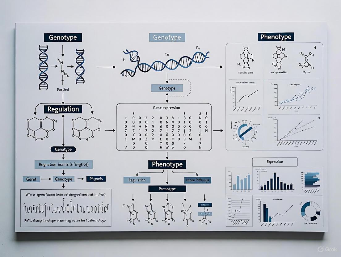
The Biological Blueprint: Foundational Concepts and Complexity in Genotype-Phenotype Dynamics
The genotype–phenotype distinction represents one of the conceptual pillars of twentieth-century genetics and remains a fundamental framework in modern biological research [1]. First proposed by the Danish scientist Wilhelm Johannsen in 1909 and further developed in his 1911 seminal work, "The Genotype Conception of Heredity," this distinction provided a revolutionary departure from prior conceptualizations of heredity [1] [2] [3]. Johannsen's terminology offered a new lexicon for genetics, introducing not only the genotype-phenotype dichotomy but also the term "gene" as a unit of heredity free from speculative material connotations [4] [3]. This conceptual framework emerged from Johannsen's meticulous plant breeding experiments and was instrumental in refuting the "transmission conception" of heredity, which presumed that parental traits were directly transmitted to offspring [1] [2]. Within the context of contemporary research on genotype-phenotype relationships, understanding this historical foundation is crucial for appreciating how genetic variability propagates across biological levels to influence disease manifestation, therapeutic responses, and complex traits—a central challenge in precision medicine and functional genomics.
Historical Context and Intellectual Genesis
The Scientific Landscape circa 1900
Johannsen's work emerged during a period of intense debate within evolutionary biology regarding the mechanisms of heredity and variation [1] [3]. The scientific community was divided between Biometricians, who followed Darwin in emphasizing continuous variation and the efficacy of natural selection, and Mendelians, who argued for discontinuous evolution through mutational leaps [1] [3]. This controversy was exacerbated by the rediscovery of Gregor Mendel's work in 1900 [3]. Competing theories included:
- Darwin's theory of pangenesis, which proposed that characteristics acquired during an organism's lifetime could be inherited [1] [5].
- August Weismann's germ-plasm theory, which postulated a strict separation between germ cells and somatic cells [1] [5].
- Galton's law of ancestral heredity, which stated that offspring tend to exhibit the average of their racial type rather than parental characteristics [3].
Johannsen's genius lay in his ability to transcend these debates through carefully designed experiments that differentiated between hereditary and non-hereditary variation.
Johannsen's Pure Line Experiments
Johannsen's conceptual breakthrough stemmed from his pure-line breeding experiments conducted on self-fertilizing plants, particularly the princess bean (Phaseolus vulgaris) [1] [4] [3]. His experimental protocol can be summarized as follows:
Table: Johannsen's Pure Line Experimental Protocol
| Experimental Phase | Methodology | Key Observations |
|---|---|---|
| Population Selection | Selected 5,000 beans from a genetically heterogeneous population; measured and recorded individual seed weights [3]. | Found continuous variation in seed weight across the population [3]. |
| Line Establishment | Created pure lines through repeated self-fertilization of individual plants, ensuring each line was genetically homozygous [1] [4]. | Established that each pure line had a characteristic average seed weight [1]. |
| Selection Application | Within each pure line, selected and planted the heaviest and lightest seeds over multiple generations [1] [3]. | Demonstrated that selection within pure lines produced no hereditary change in seed weight; offspring regressed to the line's characteristic mean [1] [3]. |
| Statistical Analysis | Employed statistical methods to compare weight distributions between generations and across different pure lines [1]. | Distinguished between non-heritable "fluctuations" (within lines) and heritable differences (between lines) [1] [5]. |
Conceptual Formalization
From these experiments, Johannsen formalized his core concepts in his 1909 textbook Elemente der exakten Erblichkeitslehre (The Elements of an Exact Theory of Heredity) [1] [3]:
- Phenotype: "All 'types' of organisms, distinguishable by direct inspection or only by finer methods of measuring or description" [5]. The phenotype represents the observable characteristics of an organism resulting from the interaction between its genotype and environmental factors during development [1] [2].
- Genotype: "The sum total of all the 'genes' in a gamete or in a zygote" [5]. The genotype constitutes the hereditary constitution of an organism, which Johannsen considered particularly stable and immune to environmental influences [1].
- Gene: A term Johannsen coined from de Vries' "pangene," defining it as a unit free from any hypothesis about its material nature, representing only the "securely ascertained fact" that many organismal properties are conditioned by separable factors in the gametes [4].
The following diagram illustrates the workflow and logical relationships of Johannsen's pure line experiment and the conceptual distinctions it revealed:
The Original Meaning and Its Evolution
Johannsen's Holistic Interpretation
Contrary to modern genocentric views, Johannsen maintained a holistic interpretation of the genotype [4]. He viewed the genotype not merely as a collection of discrete genes but as an integrated complex system. In his conception:
- The genotype represented the entire developmental potential of an organism [4].
- Genes were abstractly defined as "calculation units" or accounting devices to explain hereditary patterns, without commitment to their physical nature [4] [3].
- He explicitly rejected what he called the "transmission conception" of heredity, which assumed that parental traits were directly transmitted to offspring [1] [2].
- He equated the genotype with Richard Woltereck's concept of Reaktionsnorm (norm of reaction), emphasizing the range of potential phenotypic expressions across different environments [1].
This holistic view contrasted sharply with the increasingly reductionist direction that genetics would take in subsequent decades, particularly with the rise of the chromosome theory [4].
Contrast with Modern Meanings
Table: Evolution of Genotype-Phenotype Terminology
| Concept | Johannsen's Original Meaning (1909-1911) | Predominant Modern Meaning |
|---|---|---|
| Genotype | The class identity of a group of organisms sharing the same hereditary constitution; a holistic concept [2] [4]. | The specific DNA sequence inherited from parents [2]. |
| Phenotype | The variable appearances of individuals within a genotype, influenced by environment [2] [3]. | The observable physical and behavioral traits of an organism [2]. |
| Gene | A unit of calculation for hereditary patterns; explicitly non-hypothetical about material basis [4] [3]. | A specific DNA sequence encoding functional product [4]. |
| Primary Application | Describing differences between pure lines or populations [2]. | Describing individuals and their specific genetic makeup [2]. |
Philosophical and Scientific Implications
Johannsen's distinction carried profound implications for biological thought:
- It established an "ahistorical" conception of heredity, wherein the genotype passed unchanged from generation to generation, immune to environmental influences experienced by the organism during its lifetime [1].
- It created a conceptual framework that explicitly separated the study of heredity from the study of development [1]. Developmental biology became the study of how genotypes give rise to phenotypes, contrary to embryologists who viewed heredity and development as inseparable processes [1].
- It provided a resolution to the debate between Biometricians and Mendelians by demonstrating that continuous variation could contain both heritable and non-heritable components [1] [5].
- It limited the power of natural selection to "sort out pre-existing genotypes within heterogeneous natural populations" rather than creating new variation [1].
Modern Research Applications and Methodologies
Contemporary Genotype-Phenotype Correlation Studies
Modern research has dramatically expanded the scope and methodology of genotype-phenotype mapping, particularly in medical genetics. Current approaches include:
4.1.1 Cross-Sectional Studies of Genetic Syndromes Recent investigations into Noonan syndrome (NS) and Noonan syndrome with multiple lentigines (NSML) demonstrate sophisticated genotype-phenotype correlation methods [6]. These studies:
- Recruit individuals with specific genetic variants (e.g., in PTPN11, SOS1, RAF1 genes)
- Use standardized behavioral questionnaires (e.g., SRS-2 for social responsiveness, CBCL for emotional problems)
- Correlise specific mutations with biochemical profiling (e.g., SHP2 enzyme activity assays)
- Employ logistic regression to quantify how specific genetic functional changes (e.g., fold activation of SHP2) increase the likelihood of particular phenotypic traits (e.g., restricted and repetitive behaviors) [6]
4.1.2 Large-Scale Database Integration The development of specialized databases addresses the challenge of interpreting numerous genetic variants:
- NMPhenogen: A comprehensive database for neuromuscular genetic disorders (NMGDs) correlating genotypes from 747 nuclear and mitochondrial genes with detailed phenotypic information [7].
- Utilizes the American College of Medical Genetics and Genomics (ACMG) guidelines for standardized variant classification [7].
- Integrates population data, computational predictions, and functional assays to establish clinically relevant correlations [7].
4.1.3 Functional Genomics and Spatial Transcriptomics Cutting-edge approaches now enable high-resolution mapping:
- PERTURB-CAST: A method integrating combinatorial genetic perturbations with spatial transcriptomics to decode genotype-phenotype relationships in tumor ecosystems [8].
- CHOCOLAT-G2P: A scalable framework for studying higher-order combinatorial perturbations that mimic tumor heterogeneity [8].
- These technologies allow researchers to investigate how complex genetic alterations manifest in tissue-level phenotypes while preserving spatial context [8].
The Multi-Level Phenotype Concept
The modern conception of phenotype has expanded dramatically beyond Johannsen's original definition. Today, phenotypes are investigated at multiple biological levels:
This expansion means that important genetic and evolutionary features can differ significantly depending on the phenotypic level considered, with variation at one level not necessarily propagating predictably to other levels [5].
The Scientist's Toolkit: Essential Research Reagents and Materials
Table: Key Research Reagents and Materials for Genotype-Phenotype Studies
| Research Tool | Function/Application | Field Example |
|---|---|---|
| Pure Lines | Genetically homogeneous populations for distinguishing hereditary vs. environmental variation [1]. | Johannsen's bean lines; inbred model organisms [1] [4]. |
| Standardized Behavioral Assessments | Quantitatively measure behavioral phenotypes for correlation with genetic variants [6]. | SRS-2 for autism-related traits; CBCL for emotional problems [6]. |
| Spatial Transcriptomics Platforms | Map gene expression patterns within tissue architecture to understand phenotypic consequences [8]. | 10X Visium used in PERTURB-CAST for tumor ecosystem analysis [8]. |
| Functional Assay Reagents | Measure biochemical consequences of genetic variants in experimental systems [6]. | SHP2 activity assays for PTPN11 variants in Noonan syndrome [6]. |
| Curated Variant Databases | Classify and interpret pathogenicity of genetic variants using standardized criteria [7]. | ACMG guidelines implementation in NMPhenogen for neuromuscular disorders [7]. |
| Combinatorial Perturbation Systems | Study complex genetic interactions that mimic disease heterogeneity [8]. | CHOCOLAT-G2P framework for investigating higher-order combinatorial mutations [8]. |
Wilhelm Johannsen's genotype-phenotype distinction established a conceptual foundation that continues to shape biological research more than a century after its introduction. While the predominant meanings of these terms have evolved—particularly with the molecular biological revolution that identified DNA as the material basis of heredity—the essential framework remains remarkably prescient [2]. Johannsen's insight that the genotype represents potentialities whose expression depends on developmental processes and environmental contexts anticipated modern concepts of phenotypic plasticity, genetic canalization, and norm of reaction [1] [9].
In contemporary research, particularly in the age of precision medicine and functional genomics, the relationship between genotype and phenotype remains a central research program. The challenge has expanded from Johannsen's statistical analysis of seed weight variations to understanding how genetic variation propagates across multiple phenotypic levels—from molecular and cellular traits to organismal and clinical manifestations—to ultimately shape fitness and disease susceptibility [5]. Johannsen's holistic perspective on the genotype as an integrated system rather than a mere collection of discrete genes has regained relevance as researchers grapple with polygenic inheritance, epistasis, and the complexities of gene regulatory networks [4].
The enduring utility of Johannsen's conceptual framework lies in its ability to accommodate increasingly sophisticated methodologies while maintaining the essential distinction between hereditary potential and manifested characteristics. As modern biology develops increasingly powerful tools for probing the genotype-phenotype relationship—from single-cell omics to spatial transcriptomics and genome editing—the foundational concepts articulated by Johannsen continue to provide the "conceptual pillars" upon which our understanding of heredity and biological variation rests [1].
The relationship between genotype and phenotype represents one of the most fundamental concepts in biology. Traditionally viewed through a lens of direct causality, this paradigm has undergone substantial revision with increasing appreciation for the complex interplay between genetic information and environmental influences. Phenotypic plasticity—the ability of a single genotype to produce multiple phenotypes in response to environmental conditions—has emerged as a critical mechanism enabling organisms to cope with environmental variation [10] [11]. This capacity for responsive adaptation operates across all domains of life, from the decision between lytic and lysogenic cycles in bacteriophages to seasonal polyphenisms in butterflies and acclimation responses in plants [11] [12].
Contemporary research frameworks now recognize that the developmental trajectory from genetic blueprint to functional organism involves sophisticated regulatory processes that integrate environmental signals. As West-Eberhard articulated, the origin of novelty often begins with environmentally responsive, developmentally plastic organisms [11]. This perspective does not diminish the importance of genetic factors but rather emphasizes how environmental influences act through epigenetic mechanisms to shape phenotypic outcomes, creating a more dynamic and responsive relationship between genes and traits. Understanding these mechanisms is particularly relevant for drug development professionals seeking to comprehend individual variation in treatment response and for researchers investigating complex disease etiologies that cannot be explained by genetic variation alone [13].
Biological Mechanisms: Epigenetic Pathways and Environmental Sensing
Molecular Mechanisms of Epigenetic Regulation
Epigenetic regulation comprises molecular processes that modulate gene expression without altering the underlying DNA sequence. These mechanisms provide the molecular infrastructure for phenotypic plasticity by translating environmental experiences into stable cellular phenotypes [13]. The major epigenetic pathways include:
- DNA methylation: The addition of methyl groups to cytosine bases, primarily at CpG dinucleotides, typically associated with transcriptional repression when occurring in promoter regions. This modification represents a high-energy carbon-carbon bond that can be stable through cell divisions [13] [14].
- Histone modifications: Post-translational alterations to histone proteins including acetylation, methylation, phosphorylation, ubiquitination, and ADP-ribosylation. These modifications influence chromatin structure and DNA accessibility [13]. For example, acetylation of lysine residues on histone H3 (K14, K9) is generally associated with active transcription, while methylation of H3-K9 is linked to transcriptional silencing [13].
- Non-coding RNAs: RNA molecules that regulate gene expression at transcriptional and post-transcriptional levels, including microRNAs (miRNAs), small interfering RNAs (siRNAs), and long non-coding RNAs (lncRNAs) [13] [15].
These mechanisms function interdependently; for instance, methyl-CpG binding proteins (MeCP2) can recruit histone deacetylases (HDACs) to establish repressive chromatin states, demonstrating how DNA methylation and histone modifications act synergistically [13].
Environmental Sensing and Response
Epigenetic mechanisms serve as molecular interpreters that translate environmental signals into coordinated gene expression responses. This environmental sensing capacity enables phenotypic adjustments across diverse timescales, from rapid physiological responses to transgenerational adaptations [10] [16]. Examples include:
- Nutritional sensing: Dietary composition influences digestive enzyme plasticity through epigenetic mechanisms, as demonstrated in house sparrows that show increased maltase activity when transitioning from insect-based to seed-based diets [12].
- Thermal acclimation: Ectothermic organisms adjust membrane lipid composition through epigenetic regulation to maintain fluidity across temperature ranges [12].
- Stress responses: Maternal care behaviors in rats program offspring stress responses through epigenetic modification of glucocorticoid receptor genes, particularly in the hippocampal region [14].
The reliability of environmental cues significantly determines whether plastic responses prove adaptive. When environmental signals become unreliable due to anthropogenic change, formerly adaptive plasticity can become maladaptive, creating ecological traps [10].
Empirical Evidence: From Model Systems to Human Health
Plant and Animal Model Systems
Research in model systems has been instrumental in elucidating the mechanisms and evolutionary consequences of phenotypic plasticity:
Table 1: Empirical Evidence of Phenotypic Plasticity Across Taxa
| Organism | Plastic Trait | Environmental Cue | Mechanism | Reference |
|---|---|---|---|---|
| Ludwigia arcuata (aquatic plant) | Leaf morphology (aerial vs. submerged) | Air/water contact | ABA and ethylene hormone signaling | [12] |
| Acyrthosiphon pisum (pea aphid) | Reproductive mode (asexual/sexual), wing development | Population density | Unknown developmental switch | [12] |
| Pristimantis mutabilis (mutable rain frog) | Skin texture | Unknown | Rapid morphological change | [12] |
| Theodoxus fluviatilis (snail) | Osmolyte concentration | Water salinity | Stress-induced epigenetic modifications | [16] |
| Drosophila melanogaster (fruit fly) | Metabolic traits (triglyceride levels) | Parental environment | Parent-of-origin effects | [10] |
| House sparrows | Digestive enzyme activity | Dietary composition (insect vs. seed) | Modulation of maltase and aminopeptidase-N | [12] |
These examples demonstrate the taxonomic breadth of phenotypic plasticity and highlight how different organisms have evolved specialized mechanisms to respond to environmental challenges.
Human Health and Disease Implications
In humans, epigenetic mechanisms mediate gene-environment interactions that influence disease susceptibility and developmental outcomes [13] [17]. The Developmental Origins of Health and Disease (DOHaD) hypothesis posits that early-life environmental exposures program long-term health trajectories through epigenetic mechanisms [17]. Key evidence includes:
- Maternal care effects: In rodent models, variations in maternal licking and grooming behavior produce stable epigenetic modifications in offspring, affecting glucocorticoid receptor expression and stress responsiveness throughout life [14].
- Metabolic disease: Both maternal undernutrition and overnutrition during critical developmental windows can produce epigenetic changes that increase susceptibility to obesity and type 2 diabetes in adulthood [13].
- Mental health: Early-life adversity associates with persistent epigenetic changes in genes regulating stress response, neurotransmitter function, and neural plasticity, potentially mediating risk for psychiatric disorders [14].
These findings have profound implications for preventive medicine and therapeutic development, suggesting that epigenetic biomarkers could identify individuals at elevated risk for certain conditions and that interventions targeting epigenetic mechanisms might reverse or mitigate the effects of adverse early experiences.
Methodologies: Investigating Plasticity and Epigenetic Regulation
Experimental Approaches and Workflows
Research in phenotypic plasticity and epigenetics employs specialized methodologies to disentangle genetic, environmental, and epigenetic contributions to phenotypic variation:
Table 2: Key Methodological Approaches in Plasticity Research
| Method Category | Specific Techniques | Application | Considerations |
|---|---|---|---|
| Epigenetic Profiling | Whole Genome Bisulfite Sequencing (WGBS), Reduced Representation Bisulfite Sequencing (RRBS), MeDIP-Seq | Genome-wide DNA methylation mapping | Bisulfite conversion efficiency; cell type heterogeneity impacts data quality [15] |
| Chromatin Analysis | ChIP-Seq, ATAC-Seq, Hi-C | Histone modifications, chromatin accessibility, 3D genome architecture | Antibody specificity; cross-linking artifacts |
| Transcriptomics | RNA-Seq, Single-Cell RNA-Seq | Gene expression responses to environmental variation | Batch effects; normalization methods |
| Population Genomics | GWAS, pangenome graphs, structural variant analysis [18] | Identifying genetic loci underlying plastic responses | Sample size requirements; variant annotation |
| Experimental Designs | Common garden, reciprocal transplant, cross-fostering | Disentangling genetic and environmental effects | Logistic constraints; timescales needed |
Recent methodological advances include telomere-to-telomere genome assemblies that enable comprehensive characterization of structural variants, which have been shown to contribute substantially to phenotypic variation—accounting for an additional 14.3% heritability on average compared to SNP-only analyses in yeast models [18]. Population epigenetic approaches that apply population genetic theory to epigenetic variation are also emerging as powerful tools to understand the evolutionary dynamics of epigenetic variation [15].
The Scientist's Toolkit: Essential Research Reagents
Table 3: Essential Research Reagents for Plasticity and Epigenetics Studies
| Reagent Category | Specific Examples | Function/Application | Technical Considerations |
|---|---|---|---|
| DNA Methylation Inhibitors | 5-azacytidine, zebularine | DNA methyltransferase inhibition; experimental epigenome manipulation | Cytotoxic effects; incomplete specificity |
| Histone Modifiers | Trichostatin A (HDAC inhibitor), JQ1 (BET bromodomain inhibitor) | Altering histone acetylation patterns; probing chromatin function | Pleiotropic effects; dosage optimization |
| Bisulfite Conversion Kits | EZ DNA Methylation kits, MethylCode kits | DNA treatment for methylation detection | Incomplete conversion; DNA degradation |
| Antibodies for Chromatin Studies | Anti-5-methylcytosine, anti-H3K27ac, anti-H3K4me3 | Chromatin immunoprecipitation; epigenetic mark detection | Specificity validation; lot-to-lot variability |
| Environmental Chambers | Precision growth chambers, aquatic systems | Controlled environmental manipulation | Parameter stability; microenvironment variation |
| Epigenetic Editing Tools | CRISPR-dCas9 fused to DNMT3A/TET1, KRAB repressors | Locus-specific epigenetic manipulation | Off-target effects; persistence of modifications |
Data Presentation: Quantitative Evidence and Heritability
Quantitative Patterns in Phenotypic Plasticity Research
Rigorous quantitative analysis is essential for interpreting plasticity research. Key datasets include:
Table 4: Quantitative Evidence for Epigenetic and Plasticity Phenomena
| Phenomenon | System | Effect Size | Statistical Evidence | Source |
|---|---|---|---|---|
| Structural Variant Impact | S. cerevisiae (1,086 isolates) | 14.3% average increase in heritability with SV inclusion | SVs more frequently associated with traits than SNPs | [18] |
| Transgenerational Plasticity | Stickleback fish | Context-dependent: beneficial only when offspring environment matched parental | Significant G×E interaction (p<0.05) | [10] |
| DNA Methylation Stability | Natural populations | Varies by taxa: higher in plants/fungi than animals | Measures of epimutation rates | [15] |
| Maternal Care Effects | Rat model | 2-fold difference in glucocorticoid receptor mRNA | p<0.001 between high vs low LG offspring | [14] |
| Digestive Plasticity | House sparrow | 2-fold increase in maltase activity with diet change | Significant diet effect (p<0.01) | [12] |
These quantitative findings demonstrate the substantial contributions of epigenetic mechanisms and plasticity to phenotypic diversity. The large-scale yeast genomic study highlights how previously underexplored genetic elements, particularly structural variants, contribute significantly to trait variation [18]. Meanwhile, the context-dependency of transgenerational effects emphasizes that the adaptive value of plasticity depends on environmental predictability [10].
Current Challenges and Future Directions
Despite significant advances, the field faces several methodological and conceptual challenges that require innovative solutions:
Methodological Limitations
Current limitations in plasticity and epigenetics research include:
- Cell type heterogeneity: Epigenetic patterns are highly cell-type-specific, and studies using heterogeneous tissues (e.g., whole blood) may confound true signals with changes in cell population composition [15]. Only 44% of studies in a recent review adequately controlled for this factor [15].
- Technical variability: Bisulfite conversion methods vary in efficiency and can introduce biases, while antibody-based approaches face specificity challenges [15]. Technical replication is rarely implemented (only 12% of studies) due to cost-benefit tradeoffs [15].
- Sample size constraints: The average sample size in epigenetic studies is approximately 18 per group, substantially underpowered for detecting small-effect loci given the multiple testing burden in genome-wide analyses [15].
- Transgenerational evidence: Only 11% of studies assess epigenetic stability beyond the F3 generation, which is essential for establishing evolutionary relevance [15].
Conceptual Framework Advances
Future research directions will need to address several conceptual gaps:
- Distinguishing adaptation from fortuity: A critical challenge lies in determining whether observed plasticity represents an adaptation shaped by natural selection or merely a fortuitous response to environmental conditions [10] [11].
- Plasticity's evolutionary role: Debate continues regarding whether plasticity generally promotes or hinders genetic adaptation, with evidence supporting both facilitation (by buying time for genetic adaptation) and hindrance (by shielding genetic variation from selection) [10].
- Timescale integration: Different plastic responses operate across vastly different timescales, from rapid behavioral adjustments to transgenerational epigenetic inheritance, requiring theoretical frameworks that integrate these diverse temporal dimensions [10].
Future research should prioritize multi-generational studies with adequate sample sizes, controlled cell type composition, and integrated genomic-epigenomic analyses to fully resolve the role of plasticity in evolution and disease.
The relationship between genotype and phenotype is fundamentally shaped by two pervasive biological phenomena: genetic heterogeneity, where different genetic variants lead to the same clinical outcome, and pleiotropy, where a single genetic variant influences multiple, seemingly unrelated traits. Advances in genomic technologies and analytical frameworks are rapidly elucidating the complex mechanisms underlying these phenomena. This whitepaper explores the latest methodological breakthroughs for dissecting heterogeneity and pleiotropy, including structural causal models, techniques for disentangling pleiotropy types, and single-cell resolution approaches. The insights gained are critical for refining disease nosology, identifying therapeutic targets, and informing drug development strategies for complex human diseases.
The initial promise of the genome era was that complex diseases and traits would be mapped to a manageable set of genetic variants. Instead, research has revealed a landscape of overwhelming complexity, dominated by genetic heterogeneity and pleiotropy. Genetic heterogeneity manifests when the same phenotype arises from distinct genetic mechanisms across different individuals or populations. Conversely, pleiotropy occurs when a single genetic locus influences multiple, often disparate, phenotypic outcomes [19] [20].
These phenomena are not merely statistical curiosities; they represent fundamental challenges and opportunities for genomic medicine. For drug development, a variant with pleiotropic effects might simultaneously influence a target disease and unintended side effects. Understanding heterogeneity is equally crucial, as a treatment effective for a genetically defined patient subgroup may fail in a broader population with phenotypically similar but genetically distinct disease. This whitepaper synthesizes current research to provide a technical guide for navigating this complexity, offering robust analytical frameworks and experimental protocols to advance genotype-phenotype research.
Methodological Frameworks for Dissecting Genetic Architecture
The Causal Pivot Model for Addressing Heterogeneity
A primary challenge in analyzing genetically heterogeneous diseases is that standard association tests can fail to detect causal variants when their effects are masked by other, stronger risk factors. The Causal Pivot (CP) is a structural causal model (SCM) designed to overcome this by leveraging established causal factors to detect the contribution of additional candidate causes [21] [22].
The model typically uses a polygenic risk score (PRS) as a known cause and evaluates rare variants (RVs) or RV ensembles as candidate causes. A key innovation is its handling of outcome-induced association by conditioning on disease status. The method derives a conditional maximum-likelihood procedure for binary and quantitative traits and develops the Causal Pivot likelihood ratio test (CP-LRT) to detect causal signals [22].
Table 1: Key Applications of the Causal Pivot Model
| Disease Analyzed | Known Cause (PRS) | Candidate Cause (Rare Variants) | CP-LRT Result |
|---|---|---|---|
| Hypercholesterolemia (HC) | UK Biobank-derived PRS | Pathogenic/likely pathogenic variants in LDLR | Significant signal detected |
| Breast Cancer (BC) | UK Biobank-derived PRS | Loss-of-function mutations in BRCA1 | Significant signal detected |
| Parkinson Disease (PD) | UK Biobank-derived PRS | Pathogenic variants in GBA1 | Significant signal detected |
Experimental Protocol: Implementing the Causal Pivot Likelihood Ratio Test
The following workflow provides a detailed protocol for applying the CP-LRT, as implemented in UK Biobank analyses [21] [22]:
- Phenotype Definition: Define case-control status or quantitative trait values using precise criteria (e.g., ICD-10 codes for diseases, lab values like LDL cholesterol ≥4.9 mmol/L for hypercholesterolemia).
- Genetic Data Processing:
- PRS Calculation: Compute polygenic risk scores for all individuals using effect sizes from large, independent GWAS summary statistics.
- Rare Variant Calling: Identify rare variants (e.g., from whole-exome or whole-genome sequencing data). Focus on functionally consequential variants, such as ClinVar-classified pathogenic/likely pathogenic variants or loss-of-function mutations in disease-relevant genes.
- Ancestry Adjustment: Account for population stratification using methods like genetic matching, inverse probability weighting, regression adjustment, or doubly robust methods.
- Model Fitting and Testing:
- Fit the conditional maximum-likelihood model, conditioning on disease status and including the PRS as a covariate.
- Test the null hypothesis that the candidate rare variant (or variant ensemble) has no effect on the disease trait using the CP-LRT.
- Perform control analyses, such as testing the candidate variant in cross-disease analyses or using synonymous variants, to confirm specificity.
Diagram 1: Causal Pivot Analysis Workflow.
Disentangling Horizontal and Vertical Pleiotropy
Pleiotropy is not a monolithic concept. The Horizontal and Vertical Pleiotropy (HVP) model is a novel statistical framework designed to disentangle these two distinct forms, which is critical for understanding biological mechanisms and planning interventions [23] [24].
- Horizontal Pleiotropy: A genetic variant independently influences two or more traits via distinct biological pathways. This reflects shared genetic etiology.
- Vertical Pleiotropy (Mediated Pleiotropy): A genetic variant influences one trait, which in turn causally affects a second trait. The genetic association with the second trait is indirect.
The HVP model is a bivariate linear mixed model that simultaneously considers both pathways. It can be represented as [24]: [ \begin{cases} \mathbf{y} = \mathbf{c}\tau + \boldsymbol{\alpha} + \mathbf{e} \ \mathbf{c} = \boldsymbol{\beta} + \boldsymbol{\epsilon} \end{cases} ] Here, (\mathbf{y}) and (\mathbf{c}) are the two traits, (\tau) is the fixed causal effect of trait (\mathbf{c}) on trait (\mathbf{y}), and (\boldsymbol{\alpha}) and (\boldsymbol{\beta}) are the random genetic effects for each trait. The model uses a combination of GREML (Genomic-Relatedness-Based Restricted Maximum Likelihood) and Mendelian randomization (MR) approaches to obtain unbiased estimates of the causal effect (\tau) and the genetic correlation due to horizontal pleiotropy [24].
Table 2: Distinguishing Horizontal and Vertical Pleiotropy with the HVP Model
| Trait Pair | Primary Driver of Genetic Correlation | Biological and Clinical Implication |
|---|---|---|
| Metabolic Syndrome (MetS) & Type 2 Diabetes | Horizontal Pleiotropy | Suggests shared genetic biology; CRP is a useful biomarker but not a causal target. |
| Metabolic Syndrome (MetS) & Sleep Apnea | Horizontal Pleiotropy | Suggests shared genetic biology. |
| Body Mass Index (BMI) & Metabolic Syndrome (MetS) | Vertical Pleiotropy | Lowering BMI is likely to directly reduce MetS risk. |
| Metabolic Syndrome (MetS) & Cardiovascular Disease | Vertical Pleiotropy | MetS is a causal mediator; interventions on MetS components may reduce CVD risk. |
Advanced Multi-Trait and Multi-Locus Association Methods
Beyond pairwise pleiotropy, new methods are emerging to detect genetic variants influencing a multitude of traits. The Multivariate Response Best-Subset Selection (MRBSS) method is designed for this purpose, treating high-dimensional genotypic data as response variables and multiple phenotypic data as predictor variables [25].
The core model is: [ \mathbf{Y\Delta} = \mathbf{X\Theta} + \mathbf{\varepsilon\Delta} ] where (\mathbf{Y}) is the genotype matrix, (\mathbf{X}) is the phenotype matrix, (\mathbf{\Delta}) is a diagonal matrix whose elements indicate whether a SNP is active (associated with at least one phenotype), and (\mathbf{\Theta}) is the regression coefficient matrix. The method converts the variable selection problem into a 0-1 integer optimization, efficiently identifying the subset of "active" SNPs from a large candidate set [25].
Cutting-Edge Experimental Approaches and Protocols
Single-Cell Resolution of Context-Specific Genetic Effects
Bulk RNA sequencing masks cellular heterogeneity, limiting the detection of context-specific genetic regulation. Response expression quantitative trait locus (reQTL) mapping at single-cell resolution overcomes this by modeling the per-cell perturbation state, dramatically enhancing the detection of genetic variants whose effect on gene expression changes under stimulation [26].
A key innovation is the use of a continuous perturbation score, derived via penalized logistic regression, which quantifies each cell's degree of response to an experimental perturbation (e.g., viral infection). This continuous score is used in a Poisson mixed-effects model (PME) to test for interactions between genotype and perturbation state [26].
Table 3: Single-Cell reQTL Mapping Power and Findings
| Perturbation | reQTLs Detected (2df-model) | Percentage Increase Over Discrete Model | Example of a Discovered reQTL |
|---|---|---|---|
| Influenza A Virus (IAV) | 166 | ~37% | PXK (Decreased eQTL effect post-perturbation) |
| Candida albicans (CA) | 770 | ~37% | RPS26 (Stronger effect in B cells) |
| Pseudomonas aeruginosa (PA) | 594 | ~37% | SAR1A (rs15801 in CD8+ T cells after CA perturbation) |
| Mycobacterium tuberculosis (MTB) | 646 | ~37% | MX1 (rs461981 in CD4+ T cells after IAV perturbation) |
Experimental Protocol: Mapping reQTLs with a Continuous Perturbation Score
This protocol outlines the steps for identifying context-dependent genetic regulators using single-cell RNA sequencing data from perturbation experiments [26]:
- Perturbation Experiment and scRNA-seq: Isolate peripheral blood mononuclear cells (PBMCs) from dozens to hundreds of donors. Split cells from each donor, exposing one pool to a perturbation (e.g., IAV, CA, PA, MTB) and keeping another pool as an unstimulated control. Perform single-cell RNA sequencing on all cells.
- Data Pre-processing: Perform standard scRNA-seq quality control, normalization, and batch effect correction. Calculate corrected expression principal components (hPCs).
- Calculate Continuous Perturbation Score:
- Use a penalized logistic regression model with the hPCs as independent variables to predict the log odds of a cell belonging to the perturbed pool.
- The resulting score for each cell serves as a surrogate for its degree of response to the perturbation.
- Map Response eQTLs:
- Select a set of a priori eQTLs for testing.
- Fit a Poisson mixed-effects model where single-cell gene expression is a function of:
- Genotype (G)
- Interaction between genotype and discrete perturbation state (GxDiscrete)
- Interaction between genotype and the continuous perturbation score (GxScore)
- Include technical and biological covariates (e.g., sequencing depth, donor, cell cycle score).
- Statistical Testing: Use a 2-degree-of-freedom likelihood ratio test (LRT) to assess the joint significance of the GxDiscrete and GxScore interaction terms against a null model with no interactions. Apply false discovery rate (FDR) correction.
Diagram 2: Single-Cell reQTL Mapping Pipeline.
Table 4: Key Reagents and Resources for Genetic Heterogeneity and Pleiotropy Studies
| Resource / Reagent | Function and Utility | Example Use Case |
|---|---|---|
| UK Biobank (UKB) | A large-scale biomedical database containing deep genetic, phenotypic, and health record data from ~500,000 participants. | Discovery and validation cohort for applying CP-LRT and HVP models to human diseases [21] [27] [23]. |
| Electronic Health Records (EHRs) | Provide extensive, longitudinal phenotype data for large patient cohorts, often linked to biobanks. | Source for defining disease cases and controls using ICD codes for PheWAS and genetic correlation studies [19] [27]. |
| Polygenic Risk Scores (PRS) | A composite measure of an individual's genetic liability for a trait, calculated from GWAS summary statistics. | Serves as the "known cause" in the Causal Pivot model to detect additional variant contributions [21] [22]. |
| ClinVar Database | A public archive of reports of the relationships among human variations and phenotypes, with supporting evidence. | Curated source for identifying pathogenic/likely pathogenic rare variants for candidate causal analysis [21] [22]. |
| GWAS Summary Statistics | Publicly available results from genome-wide association studies, including effect sizes and p-values for millions of variants. | Used to calculate PRS and to perform genetic correlation analyses (e.g., with LD Score Regression) [20]. |
| Perturbation scRNA-seq Datasets | Single-cell transcriptomic data from controlled stimulation experiments on primary cells from multiple donors. | Enables mapping of context-specific genetic effects (reQTLs) and modeling of perturbation heterogeneity [26]. |
The intricate interplay between genetic heterogeneity and pleiotropy is a central theme in understanding the relationship between genotype and phenotype. The analytical frameworks and experimental protocols detailed here—including the Causal Pivot, the HVP model, and single-cell reQTL mapping—provide researchers with powerful tools to dissect this complexity. These approaches move beyond simple association to infer causality, distinguish between types of genetic effects, and capture dynamic regulation in specific cellular contexts.
For the field of drug development, these advances are transformative. They enable the stratification of patient populations based on underlying genetic etiology, even for phenotypically similar diseases, paving the way for more targeted and effective therapies. Furthermore, by clarifying whether a pleiotropic effect is vertical or horizontal, these methods help in assessing whether a potential drug target might influence a single pathway or have unintended consequences across multiple biological systems. As genomic biobanks continue to expand and single-cell technologies become more accessible, the integration of these sophisticated analytical frameworks will be indispensable for unraveling the genetic basis of human disease and translating these discoveries into precision medicine.
Pediatric dilated cardiomyopathy (DCM) represents a severe myocardial disorder characterized by left ventricular dilation and impaired systolic function, serving as a leading cause of heart failure and sudden cardiac death in children. This case study examines the complex relationship between genetic determinants and clinical manifestations in pediatric DCM, highlighting the substantial genetic heterogeneity observed in this population. Through analysis of current literature and emerging methodologies, we demonstrate how precision medicine approaches are transforming diagnosis, prognosis, and treatment strategies for this challenging condition. The integration of advanced genetic testing with functional validation and multi-omics technologies provides unprecedented opportunities to decode genotype-phenotype relationships, enabling improved risk stratification and targeted therapeutic interventions for pediatric patients with DCM.
Clinical Significance and Genetic Landscape
Pediatric dilated cardiomyopathy is a severe myocardial disease characterized by enlargement of the left ventricle or both ventricles with impaired contractile function. This condition can lead to adverse clinical consequences including heart failure, sudden death, thromboembolism, and arrhythmias [28]. The annual incidence of pediatric cardiomyopathy is approximately 1.13 per 100,000 children, with DCM representing one of the most common forms [29]. Over 100 genes have been linked to DCM, creating substantial diagnostic challenges but also opportunities for precision medicine approaches [28] [29].
The genetic architecture of pediatric DCM markedly differs from adult forms, characterized by early onset, rapid disease progression, and poorer prognosis [28]. Children with DCM display high genetic heterogeneity, with pathogenic variants identified in up to 50% of familial cases [29]. The major functional domains affected by these mutations include calcium handling, the cytoskeleton, and ion channels, which collectively disrupt normal cardiac function and structure [28].
Purpose and Scope
This case study aims to dissect the complex relationship between genetic variants and their clinical manifestations in pediatric DCM, framed within the broader context of genotype-phenotype relationship research. We will explore how genetic insights are transforming diagnostic approaches, prognostic stratification, and therapeutic development for this challenging condition. By examining current evidence and emerging methodologies, this analysis seeks to provide clinicians and researchers with a comprehensive framework for understanding and investigating genetic determinants of pediatric DCM.
Genetic Architecture of Pediatric DCM
Spectrum of Pathogenic Variants
The genetic landscape of pediatric DCM demonstrates considerable heterogeneity, with mutations identified across numerous genes encoding critical cardiac proteins. Current evidence indicates that disease-associated genetic variants play a significant role in the development of approximately 30-50% of pediatric DCM cases [30]. The table below summarizes the key genetic associations and their frequencies in pediatric DCM populations.
Table 1: Genetic Variants in Pediatric Dilated Cardiomyopathy
| Gene | Protein Function | Frequency in Pediatric DCM | Associated Clinical Features |
|---|---|---|---|
| MYH7 | Sarcomeric β-myosin heavy chain | 34.2% of genotype-positive cases [31] | Mixed cardiomyopathy phenotypes, progressive heart failure |
| MYBPC3 | Cardiac myosin-binding protein C | 12.2% of genotype-positive cases [31] | Hypertrophic features, arrhythmias |
| LMNA | Nuclear envelope protein (Lamin A/C) | Common in cardioskeletal forms [30] | Conduction system disease, skeletal myopathy, rapid progression |
| TTN | Sarcomeric scaffold protein | Significant proportion of familial cases [28] | Variable expressivity, age-dependent penetrance |
| DES | Muscle-specific intermediate filament | Associated with cardioskeletal myopathy [30] | Myopathy with cardiac involvement, conduction abnormalities |
Distinct Genetic Features in Pediatric Populations
Pediatric DCM demonstrates unique genetic characteristics that distinguish it from adult-onset disease. Children with DCM are more likely to have homozygous or compound heterozygous mutations, reflecting more severe genetic insults that manifest earlier in life [30]. Additionally, syndromic, metabolic, and neuromuscular causes represent a substantial proportion of pediatric cases, necessitating comprehensive evaluation for extracardiac features [30].
The genetic architecture of pediatric DCM also includes a higher prevalence of de novo mutations and variants in genes associated with severe, early-onset disease. For example, mutations in LMNA, which encodes a nuclear envelope protein, are frequently identified in pediatric DCM patients with associated skeletal myopathy and conduction system disease [30]. These mutations initially manifest with conduction abnormalities before progressing to DCM, illustrating the temporal dimension of genotype-phenotype correlations [30].
Methodological Approaches for Genotype-Phenotype Correlation Studies
Variant Identification and Interpretation
Establishing accurate genotype-phenotype correlations begins with comprehensive genetic testing and precise variant interpretation. Current guidelines by the American College of Medical Genetics and Genomics (ACMG) emphasize incorporating genetic evaluation into the standard care of pediatric cardiomyopathy patients [29]. The workflow for genetic variant analysis involves multiple validation steps to establish pathogenicity and clinical significance.
Table 2: Essential Methodologies for Genetic Analysis in Pediatric DCM
| Methodology | Application | Key Outputs | Considerations |
|---|---|---|---|
| Next-generation sequencing panels | Simultaneous analysis of 100+ cardiomyopathy-associated genes [28] | Identification of P/LP variants, VUS | Coverage of known genes, but may miss novel associations |
| Whole exome sequencing | Broad capture of protein-coding regions beyond targeted panels [32] | Detection of variants in non-cardiomyopathy genes explaining syndromic features | Higher rate of VUS, increased interpretation challenges |
| ACMG/AMP guidelines | Standardized framework for variant classification [32] [29] | Pathogenic, Likely Pathogenic, VUS, Benign classifications | Requires integration of population data, computational predictions, functional data, segregation evidence |
| Familial cosegregation studies | Tracking variant inheritance in affected and unaffected family members [28] | Supports or refutes variant-disease association | Particularly important for VUS interpretation; may be limited by small family size |
| Transcriptomics and functional assays | Validation of putative pathogenic mechanisms [33] [34] | Evidence of RNA expression changes, protein alterations, cellular dysfunction | Provides mechanistic insights but requires specialized expertise |
The following workflow diagram illustrates the comprehensive process for variant identification and interpretation in pediatric DCM research:
Multi-Omics Integration Approaches
Advanced methodologies integrating multiple data layers are increasingly critical for elucidating genotype-phenotype relationships in pediatric DCM. Mendelian randomization (MR) combined with Bayesian co-localization and single-cell RNA sequencing represents a powerful approach for identifying causal drug targets and molecular pathways [34]. This multi-omics framework enables researchers to move beyond association studies toward establishing causal relationships between genetic variants and disease mechanisms.
The integration of tissue-specific cis-expression quantitative trait loci (eQTL) and protein quantitative trait loci (pQTL) datasets from heart and blood tissues with genome-wide association studies (GWAS) data allows for robust identification of genes whose expression is causally associated with DCM [34]. Single-cell transcriptomic analysis further enables resolution of these associations at cellular levels, revealing cell-type-specific expression patterns in DCM hearts compared to controls [34].
The following diagram illustrates this integrated multi-omics approach:
Key Experimental Protocols
Systematic Variant Reinterpretation Framework
Variant reinterpretation has emerged as a critical component of genotype-phenotype correlation studies, with recent evidence demonstrating that approximately 21.6% of pediatric cardiomyopathy patients experience clinically meaningful changes in variant classification upon systematic reevaluation [32]. This protocol outlines a standardized approach for variant reassessment.
Protocol: Variant Reinterpretation Using Updated ACMG/AMP Guidelines
Data Collection: Compile original genetic test reports, clinical laboratory classifications, and patient phenotypic data.
Evidence Review:
- Query updated population frequency databases (gnomAD v4.1.0)
- Review recent ClinVar submissions (current through most recent release)
- Evaluate gene-disease validity using Clinical Genome Resource (ClinGen) curations
- Assess mutational hotspot and critical functional domain locations
Bioinformatic Analysis:
- Apply computational prediction tools (SIFT, PolyPhen-2, REVEL)
- Analyze conservation scores across species
- Evaluate splice site predictions
Segregation Analysis:
- Review familial genetic testing results when available
- Assess for de novo occurrence where parental testing exists
- Calculate logarithm of the odds (LOD) scores for large pedigrees
Functional Evidence Integration:
- Incorporate transcriptomic or proteomic data demonstrating molecular impact
- Consider relevant animal or cellular model data
- Evaluate therapeutic implications of variant reclassification
This systematic approach revealed that 10.9% of previously classified P/LP variants were downgraded to VUS, while 13.6% of VUS were upgraded to P/LP in pediatric cardiomyopathy cases [32]. The leading criteria for downgrading were high population allele frequency and variant location outside mutational hotspots or critical functional domains, while upgrades were primarily driven by variant location in mutational hotspots and deleterious in silico predictions [32].
Transcriptomic Analysis for Drug Repurposing
Transcriptomic profiling enables identification of gene expression signatures associated with specific genetic variants in pediatric DCM, creating opportunities for therapeutic repurposing. The following protocol outlines an approach combining in silico analysis with in vitro validation using patient-derived cells.
Protocol: Transcriptomic Analysis for Therapeutic Discovery
Gene Expression Profiling:
- RNA sequencing of patient-derived cardiomyocytes (LMNA-mutant vs. controls)
- Differential expression analysis (adjusted p-value < 0.05, fold-change > 1.5)
- Pathway enrichment analysis (Gene Ontology, KEGG, Reactome)
Computational Drug Screening:
- Query the Library of Integrated Network-based Cellular Signatures (LINCS)
- Identify compounds with inverse gene expression signatures to disease profile
- Prioritize FDA-approved drugs for repurposing candidates
In Vitro Validation:
- Generate patient-specific induced pluripotent stem cell-derived cardiomyocytes (iPSC-CMs)
- Treat with identified candidate compounds (e.g., Olmesartan for LMNA-DCM)
- Assess functional endpoints: contractility, calcium handling, arrhythmia burden
- Measure reversal of disease-associated gene expression changes
This approach successfully identified Olmesartan as a candidate therapy for LMNA-associated DCM, demonstrating improved cardiomyocyte function, reduced abnormal rhythms, and restored gene expression in patient-derived cells [33].
Research Reagent Solutions
Table 3: Essential Research Reagents for Pediatric DCM Investigations
| Reagent/Category | Specific Examples | Research Application | Key Considerations |
|---|---|---|---|
| Cardiomyopathy Gene Panels | Clinically validated multi-gene panels (100+ genes) [28] | Initial genetic screening | Comprehensive coverage of established cardiomyopathy genes; may miss novel associations |
| Whole Exome/Genome Sequencing | Illumina platforms, Oxford Nanopore | Discovery of novel variants beyond panel genes | Higher VUS rate; requires robust bioinformatic pipeline |
| iPSC Differentiation Kits | Commercial cardiomyocyte differentiation kits | Generation of patient-specific cardiac cells | Variable efficiency across cell lines; require functional validation |
| Single-cell RNA Sequencing Platforms | 10X Genomics, Smart-seq2 | Cell-type-specific transcriptomic profiling | Cell dissociation effects on gene expression; computational expertise required |
| CRISPR-Cas9 Gene Editing Systems | SpCas9, base editors, prime editors | Functional validation of variants in cellular models | Off-target effects; require careful design and validation |
| Cardiac Functional Assays | Calcium imaging dyes, contractility measurements | Assessment of cardiomyocyte functional deficits | Technical variability; require appropriate controls |
| Bioinformatic Tools for Variant Interpretation | ANNOVAR, InterVar, VEP | Standardized variant classification | Dependence on updated databases; computational resource requirements |
Clinical Implications and Therapeutic Applications
Impact on Diagnosis and Prognosis
Establishing precise genotype-phenotype correlations in pediatric DCM has profound implications for clinical management. Genetic findings directly influence diagnostic accuracy, prognostic stratification, and family screening protocols. Studies demonstrate that children with hypertrophic cardiomyopathy and a positive genetic test experience worse outcomes, including higher rates of extracardiac manifestations (38.1% vs. 8.3%), more frequent need for implantable cardiac defibrillators (23.8% vs. 0%), and higher transplantation rates (19.1% vs. 0%) compared to genotype-negative patients [31].
The high prevalence of variant reclassification (affecting 21.6% of patients) underscores the importance of periodic reevaluation of genetic test results [32]. These reinterpretations directly impact clinical care, requiring modification of family screening protocols through either initiation or discontinuation of clinical surveillance for genotype-negative family members [32].
Emerging Targeted Therapies
Advances in understanding genotype-phenotype relationships are driving the development of targeted therapies for genetic forms of DCM. Current approaches include:
Small Molecule Inhibitors: Mavacamten (Camzyos), a first-in-class cardiac myosin inhibitor, represents the first precision medicine for hypertrophic cardiomyopathy, demonstrating that targeting sarcomere proteins can normalize cardiac function [35]. This approach is being explored for specific genetic forms of DCM.
Gene Therapy Strategies: Multiple gene therapy programs are advancing toward clinical application:
- TN-201: AAV9-based gene therapy for MYBPC3-associated hypertrophic cardiomyopathy currently in Phase 1b/2 trials (MyPEAK-1) [36]
- TN-401: AAV9-based gene therapy for PKP2-associated arrhythmogenic cardiomyopathy in Phase 1b trials (RIDGE-1) [36]
- RP-A501: AAV9-based gene therapy for Danon disease [37]
- NVC-001: AAV-based gene therapy for LMNA-related dilated cardiomyopathy with planned Phase 1/2 trial [37]
Drug Repurposing Approaches: Transcriptomic analysis has identified Olmesartan as a potential therapy for LMNA-associated DCM, demonstrating that existing medications may be redirected to treat specific genetic forms of cardiomyopathy [33].
The dissection of genotype-phenotype correlations in pediatric dilated cardiomyopathy represents a cornerstone of precision cardiology. Through systematic genetic testing, vigilant variant reinterpretation, and integration of multi-omics data, clinicians and researchers can unravel the complex relationship between genetic determinants and clinical manifestations in this heterogeneous disorder. These advances are already transforming clinical practice through improved diagnostic accuracy, refined prognostic stratification, and emerging targeted therapies. Future research must focus on functional validation of putative pathogenic variants, development of gene-specific therapies, and resolution of variants of uncertain significance, particularly in underrepresented populations. The continued integration of genetic insights into clinical management promises to improve outcomes for children with this challenging condition.
Bridging the Gap: AI, Machine Learning, and Novel Methodologies for Phenotype Prediction
The quest to quantitatively predict complex traits and diseases from genetic information represents a cornerstone of modern biology and precision medicine. For decades, the relationship between genotype and phenotype remained largely correlative, with limited predictive power for complex traits influenced by numerous genetic loci and environmental factors. The field has undergone a transformative evolution, moving from traditional statistical models to sophisticated artificial intelligence approaches. This paradigm shift began with the establishment of Genomic Best Linear Unbiased Prediction (GBLUP) as a robust statistical framework for genomic selection and has accelerated toward deep neural networks capable of modeling non-linear genetic architectures. The central challenge in this domain lies in developing models that can accurately capture the intricate relationships between high-dimensional genomic data and phenotypic outcomes, which may be influenced by epistatic interactions, pleiotropic effects, and complex biological pathways.
This technical guide examines the theoretical foundations, methodological advancements, and practical implementations of predictive modeling in genomics. By providing a comprehensive analysis of both established and emerging approaches, we aim to equip researchers with the knowledge necessary to select appropriate modeling strategies for specific genotype-phenotype prediction tasks across biological domains including plant and animal breeding, human genetics, and disease risk assessment.
Theoretical Foundations: From GBLUP to Deep Learning
Genomic BLUP: The Established Benchmark
Genomic Best Linear Unbiased Prediction (GBLUP) has served as a fundamental methodology in genomic prediction since its introduction. The method operates on a mixed linear model framework: y = 1μ + g + ε, where y represents the vector of phenotypes, μ is the overall mean, g is the vector of genomic breeding values, and ε represents residual errors [38]. The genomic values are assumed to follow a multivariate normal distribution g ~ N(0, Gσ²g), where G is the genomic relationship matrix derived from marker data and σ²g is the genetic variance [38] [39].
The genomic relationship matrix G is constructed from marker genotypes, typically using the method described by VanRaden (2008), where G = ZZ′ / 2∑pk(1-pk), with Z representing a matrix of genotype scores centered by allele frequencies pk [38]. This relationship matrix enables GBLUP to capture three distinct types of quantitative-genetic information: linkage disequilibrium (LD) between quantitative trait loci (QTL) and markers, additive-genetic relationships between individuals, and cosegregation of linked loci within families [38] [40].
GBLUP's strength lies in its statistical robustness, computational efficiency, and interpretability, particularly for traits governed primarily by additive genetic effects [41] [39]. However, its linear assumptions limit its ability to capture complex non-linear genetic interactions, prompting the exploration of more flexible modeling approaches.
Deep Neural Networks: Modeling Complexity
Deep learning approaches, particularly multilayer perceptrons (MLPs) and convolutional neural networks (CNNs), offer a powerful alternative for genomic prediction tasks. The fundamental MLP architecture for a univariate response can be represented as:
Yi = w₀⁰ + W₁⁰xᵢᴸ + ϵᵢ
where xᵢˡ = gˡ(w₀ˡ + W₁ˡxᵢˡ⁻¹) for l = 1,...,L, with xᵢ⁰ = xᵢ representing the input vector of markers for individual i [41]. The function gˡ denotes the activation function for layer l (typically ReLU for hidden layers), with w₀ˡ and W₁ˡ representing the bias vectors and weight matrices for each layer [41].
Unlike GBLUP, deep learning models can automatically learn hierarchical representations of genomic data and capture non-linear relationships and interactions without explicit specification [41] [42]. This flexibility makes them particularly suitable for traits with complex genetic architectures involving epistasis and gene-environment interactions. However, this increased modeling capacity comes with requirements for careful hyperparameter tuning and potential challenges in interpretation [41] [42].
Table 1: Core Methodological Comparison Between GBLUP and Deep Learning Approaches
| Characteristic | GBLUP | Deep Neural Networks |
|---|---|---|
| Theoretical Foundation | Linear mixed models | Multi-layer hierarchical representation learning |
| Genetic Architecture | Additive effects | Additive, epistatic, and non-linear effects |
| Computational Complexity | Lower (inversion of G-matrix) | Higher (gradient-based optimization) |
| Interpretability | High (variance components, breeding values) | Lower (black-box nature) |
| Data Requirements | Effective with moderate sample sizes | Generally requires larger training sets |
| Handling of Non-linearity | Limited | Excellent |
Comparative Performance Analysis
Empirical Evidence Across Biological Systems
Recent large-scale comparative studies have provided insights into the performance characteristics of GBLUP versus deep learning approaches across diverse genetic architectures and sample sizes. A comprehensive analysis across 14 real-world plant breeding datasets demonstrated that deep learning models frequently provided superior predictive performance compared to GBLUP, particularly in smaller datasets and for traits with suspected non-linear genetic architectures [41]. However, neither method consistently outperformed the other across all evaluated traits and scenarios, highlighting the importance of context-specific model selection [41].
In simulation studies with cattle SNP data, deep learning approaches demonstrated advantages for specific scenarios. A stacked kinship CNN approach showed 1-12% lower root mean squared error compared to GBLUP for additive traits and 1-9% lower RMSE for complex traits with dominance and epistasis [39]. However, GBLUP maintained higher Pearson correlation coefficients (0.672 for GBLUP vs. 0.505 for DNN in fully additive cases) [39], suggesting that the optimal metric for evaluation may influence model preference.
For human gene expression-based phenotype prediction, deep neural networks outperformed classical machine learning methods including SVM, LASSO, and random forests when large training sets were available (>10,000 samples) [42]. This performance advantage increased with training set size, highlighting the data-hungry nature of deep learning approaches.
Table 2: Performance Comparison Across Studies and Biological Systems
| Study Context | Dataset Characteristics | GBLUP Performance | Deep Learning Performance | Key Findings |
|---|---|---|---|---|
| Plant Breeding (14 datasets) [41] | Diverse crops; 318-1,403 lines; 2,038-78,000 SNPs | Variable across traits | Variable across traits; advantage in smaller datasets | Performance dependent on trait architecture; DL required careful parameter optimization |
| Cattle Simulation [39] | 1,033 Holstein Friesian; 26,503 SNPs; simulated traits | Correlation: 0.672 (additive) RMSE: Benchmark | Correlation: 0.505 (additive) RMSE: 1-12% lower | DL better RMSE, GBLUP better correlation; trade-offs depend on evaluation metric |
| Human Disease Prediction [42] | 54,675 probes; 27,887 tissues (cancer/non-cancer) | Not assessed | Accuracy advantage with large training sets (>10,000) | DL outperformed classical ML with sufficient data; interpretation challenges noted |
| Multi-omics Prediction [43] | Blood gene expression + methylation; 2,940 samples | Not assessed | AUC: 0.95 (smoking); Mean error: 5.16 years (age) | Interpretable DL successfully integrated multi-omics data |
Factors Influencing Model Performance
Several key factors emerge as critical determinants of predictive performance across modeling approaches:
Trait Complexity: Deep learning models demonstrate particular advantages for traits with non-additive genetic architectures, including epistatic interactions and dominance effects [39]. GBLUP remains highly effective for primarily additive traits.
Sample Size: The performance advantage of deep learning increases with training set size [42]. For moderate sample sizes, GBLUP often provides robust and competitive performance [41].
Marker Density: High marker density enables more accurate estimation of genomic relationships in GBLUP and provides richer feature representation for deep learning models [41] [39].
Data Representation: Innovative data representations, such as stacked kinship matrices transformed into image-like formats for CNN input, can enhance deep learning performance [39].
Methodological Implementation
Experimental Workflows and Protocols
Standard GBLUP Implementation
The implementation of GBLUP follows a well-established statistical workflow:
Genotype Quality Control: Filter markers based on call rate, minor allele frequency, and Hardy-Weinberg equilibrium [39]. For cattle data, Wientjes et al. applied thresholds of call rate >95% and MAF >0.5% [39].
Genomic Relationship Matrix Calculation: Compute the G matrix using the method of VanRaden: G = ZZ′ / 2∑pk(1-pk), where Z is the centered genotype matrix [38] [39].
Phenotypic Data Preparation: Adjust phenotypes for fixed effects and experimental design factors. In plant breeding applications, Best Linear Unbiased Estimators (BLUEs) are often computed to remove environmental effects [41].
Variance Component Estimation: Use restricted maximum likelihood (REML) to estimate genetic and residual variance components [38].
Breeding Value Prediction: Solve the mixed model equations to obtain genomic estimated breeding values (GEBVs) for selection candidates [38].
Deep Learning Pipeline for Genomic Prediction
The implementation of deep learning models for genomic prediction requires careful attention to data preparation and model architecture:
Data Preprocessing:
Model Architecture Design:
Model Training:
Model Interpretation:
Visualization of Methodological Workflows
The following diagram illustrates the comparative workflows for GBLUP and deep learning approaches in genomic prediction:
Advanced Applications and Hybrid Approaches
Multi-omics Integration
The integration of multiple omics data types represents a promising application for advanced neural network architectures. Visible neural networks, which incorporate biological prior knowledge into their architecture, have demonstrated success in multi-omics prediction tasks [43]. These approaches connect molecular features to genes and pathways based on existing biological annotations, enhancing interpretability.
For example, in predicting smoking status from blood-based transcriptomics and methylomics data, a visible neural network achieved an AUC of 0.95 by combining CpG methylation sites with gene expression through gene-annotation layers [43]. This integration outperformed single-omics approaches and provided biologically plausible interpretations, highlighting known smoking-associated genes like AHRR, GPR15, and LRRN3 [43].
Interpretable Deep Learning
Addressing the "black box" nature of deep learning models remains an active research area. Several approaches have emerged to enhance interpretability in genomic contexts:
Visible Neural Networks: Architectures that incorporate biological hierarchies (genes → pathways → phenotypes) to maintain interpretability [43].
Gradient-Based Interpretation Methods: Techniques like Layerwise Relevance Propagation (LRP) and Integrated Gradients that quantify feature importance by backpropagating output contributions [42].
Biological Validation: Functional enrichment analysis of important features identified by models to verify biological relevance [42].
These approaches facilitate the extraction of biologically meaningful insights from complex deep learning models, bridging the gap between prediction and mechanistic understanding.
Research Reagent Solutions
Table 3: Essential Research Reagents and Computational Tools for Genomic Prediction
| Category | Specific Tools/Reagents | Function/Application | Considerations |
|---|---|---|---|
| Genotyping Platforms | Illumina BovineSNP50 BeadChip [39], Affymetrix microarrays [42] | Genome-wide marker generation | Density, species specificity, cost |
| Sequencing Technologies | Next-generation sequencing (NGS) [45] [46] | Variant discovery, sequence-based genotyping | Coverage, read length, error profiles |
| Data Processing Tools | PLINK, GCTA, TASSEL | Quality control, relationship matrix calculation | Data format compatibility, scalability |
| Deep Learning Frameworks | TensorFlow [42], PyTorch | Model implementation and training | GPU support, community resources |
| Biological Databases | KEGG [43], GO, gnomAD [45] | Functional annotation, prior knowledge | Currency, species coverage, accessibility |
Future Directions and Implementation Recommendations
The field of genomic prediction continues to evolve rapidly, with several promising research directions emerging. The development of hybrid models that combine the statistical robustness of GBLUP with the flexibility of deep learning represents a particularly promising avenue [41] [39]. Such approaches could leverage linear methods for additive genetic components while using neural networks to capture non-linear residual variation.
Transfer learning approaches, where models pre-trained on large genomic datasets are fine-tuned for specific applications, may help address the data requirements of deep learning while maintaining performance on smaller datasets [41]. Similarly, the incorporation of biological prior knowledge through visible neural network architectures shows promise for enhancing both performance and interpretability [43] [42].
For researchers implementing these approaches, we recommend the following strategy:
- Begin with GBLUP as a robust baseline, particularly for moderate-sized datasets and primarily additive traits [41].
- Explore deep learning approaches when non-linear genetic architectures are suspected or when integrating diverse data types [41] [43].
- Prioritize interpretability through biological validation and gradient-based interpretation methods, particularly for medical applications [42].
- Consider computational resources and expertise when selecting modeling approaches, as deep learning requires significant infrastructure and technical knowledge [41] [42].
The following diagram illustrates the decision process for selecting appropriate modeling strategies based on research context:
As genomic technologies continue to advance, producing increasingly large and complex datasets, the evolution of predictive modeling approaches will remain essential for unlocking the relationship between genotype and phenotype. The complementary strengths of statistical and deep learning approaches provide a powerful toolkit for researchers addressing diverse challenges across biological domains.
The relationship between genotype (an organism's genetic makeup) and phenotype (its observable traits) represents one of the most fundamental challenges in modern biology. Understanding this relationship is particularly critical for advancing drug discovery, developing personalized treatments, and unraveling the mechanisms of complex diseases. Traditional statistical methods have provided valuable insights but often struggle to capture the nonlinear interactions, high-dimensional nature, and complex architectures of biological systems [44] [47]. Machine learning (ML) has emerged as a transformative toolkit capable of addressing these challenges by detecting intricate patterns in large-scale biological datasets that conventional approaches might miss [48].
Machine learning approaches are especially valuable for integrating multimodal data—including genomic, transcriptomic, proteomic, and clinical information—to build predictive models that bridge the gap between genetic variation and phenotypic expression [47]. The application of ML in genotype-phenotype research spans multiple domains, from identifying disease-associated genetic variants to predicting drug response and optimizing therapeutic interventions [44] [49]. This technical guide examines how supervised, unsupervised, and deep learning methodologies are being leveraged to advance our understanding of genotype-phenotype relationships, with particular emphasis on applications in pharmaceutical research and development.
Machine Learning Fundamentals in Biological Context
Core Paradigms and Definitions
In the context of genotype-phenotype research, machine learning algorithms can be categorized into several distinct paradigms, each with specific applications and strengths:
Supervised Learning operates on labeled datasets where each input example is associated with a known output value. In biological applications, this typically involves using genomic features (e.g., SNP arrays, sequence data) to predict phenotypic outcomes (e.g., disease status, drug resistance, quantitative traits) [48]. Common algorithms include random forests, support vector machines, and regularized regression models, which are particularly valuable for classification tasks (e.g., case vs. control) and regression problems (e.g., predicting continuous physiological measurements) [48] [50].
Unsupervised Learning identifies inherent structure in data without pre-existing labels. These methods are particularly valuable for exploratory data analysis in genomics, where they can reveal novel subtypes of diseases, identify co-regulated gene clusters, or reduce dimensionality for visualization and further analysis [48]. Techniques such as clustering (k-means, hierarchical clustering) and dimensionality reduction (principal component analysis) help researchers discover patterns in genomic data that may reflect underlying biological mechanisms [50].
Deep Learning utilizes multi-layered neural networks to learn hierarchical representations of data. This approach excels at capturing non-linear relationships and interaction effects between multiple genetic variants and phenotypic outcomes [48] [47]. Deep learning architectures including convolutional neural networks (CNNs), recurrent neural networks (RNNs), and generative adversarial networks (GANs) have demonstrated remarkable performance in analyzing complex biological data such as DNA sequences, protein structures, and medical images [49] [51].
Critical Considerations for Biological Applications
Several unique challenges must be addressed when applying machine learning to genotype-phenotype problems:
The Curse of Dimensionality: Genomic datasets typically contain vastly more features (e.g., SNPs, genes) than samples (e.g., patients, organisms), creating statistical challenges for model training and validation [48]. Dimensionality reduction techniques and feature selection methods are essential for mitigating this issue.
Data Quality and Availability: The performance of ML models is heavily dependent on the quality and quantity of training data. In biological domains, labeled data can be particularly scarce due to experimental costs, ethical constraints, and data sharing barriers [48].
Interpretability and Biological Insight: While complex models like deep neural networks often achieve high predictive accuracy, their "black box" nature can limit biological interpretability [49] [47]. Developing methods to extract meaningful biological insights from these models remains an active research area.
Confounding and Spurious Correlations: Population structure, batch effects, and technical artifacts can create spurious genotype-phenotype associations [52]. Careful study design and appropriate normalization techniques are essential to avoid these pitfalls.
Supervised Learning Applications
Genotype to Phenotype Prediction
Supervised learning approaches have been successfully applied to predict phenotypic outcomes directly from genotypic information. For example, the deepBreaks framework utilizes multiple ML algorithms to identify important positions in sequence data associated with phenotypic traits [44]. The methodology involves:
- Data Preprocessing: Handling missing values, ambiguous reads, and dropping zero-entropy columns from multiple sequence alignments.
- Feature Selection: Clustering correlated positions using DBSCAN and selecting representative features to reduce collinearity.
- Model Training and Selection: Comparing multiple ML algorithms (e.g., AdaBoost, Decision Tree, Random Forest) using cross-validation to identify the best-performing model.
- Interpretation: Using feature importance metrics from the top-performing model to prioritize genetic variants associated with the phenotype [44].
This approach has demonstrated effectiveness in identifying genotype-phenotype associations in both nucleotide and amino acid sequence data, with applications ranging from microbial genomics to complex human diseases [44].
Whole-Genome Prediction in Bacterial Genomics
In bacterial genomics, supervised learning methods face unique challenges due to linkage disequilibrium, limited sampling, and spurious associations that can corrupt model interpretations [52]. Despite these challenges, ML models have achieved high accuracy in predicting bacterial phenotypes such as antibiotic resistance and virulence from whole-genome sequence data. However, extracting biologically meaningful insights from these predictive models requires careful consideration of potential confounders and rigorous validation [52].
Table 1: Performance Metrics of Supervised Learning Algorithms in Genotype-Phenotype Prediction
| Algorithm | Application Domain | Key Strengths | Limitations |
|---|---|---|---|
| Random Forest | Microbial GWAS, Crop Improvement [52] [50] | Handles high-dimensional data, Provides feature importance metrics | Can be biased toward correlated features, Limited extrapolation |
| Support Vector Machines | Disease Classification [48] [51] | Effective in high-dimensional spaces, Versatile kernel functions | Memory intensive, Black box interpretations |
| Regularized Regression (LASSO, Ridge) | Polygenic Risk Scores [47] | Feature selection inherent in LASSO, Stable coefficients in Ridge | Assumes linear relationships, May miss interactions |
| Gradient Boosting (XGBoost, LightGBM) | Genomic Selection [50] | High predictive accuracy, Handles mixed data types | Computational complexity, Hyperparameter sensitivity |
Experimental Protocol: Genotype-Phenotype Association Study
A typical workflow for supervised genotype-phenotype association analysis involves the following key steps:
Sample Collection and Genotyping: Collect biological samples from individuals with recorded phenotypic measurements. Perform whole-genome sequencing or SNP array genotyping to obtain genetic data.
Quality Control and Imputation: Apply quality filters to remove low-quality variants and samples. Impute missing genotypes using reference panels.
Feature Engineering: Convert genetic variants to a numerical representation (e.g., one-hot encoding for sequences, dosage for SNPs). Perform linkage disequilibrium pruning or clustering to reduce feature collinearity.
Model Training with Cross-Validation: Split data into training and test sets. Train multiple ML algorithms using k-fold cross-validation to optimize hyperparameters and prevent overfitting.
Model Evaluation and Interpretation: Assess model performance on held-out test data using appropriate metrics (e.g., AUC-ROC for classification, R² for regression). Compute feature importance scores to identify genetic variants most predictive of the phenotype.
This protocol emphasizes the critical importance of proper validation to ensure that models generalize to new data and avoid overfitting, particularly given the high-dimensional nature of genomic data [48] [52].
Unsupervised Learning Approaches
Discovering Latent Structures in Genomic Data
Unsupervised learning techniques excel at identifying inherent patterns in genomic data without pre-specified phenotypic labels. These methods are particularly valuable for exploratory analysis of high-dimensional biological datasets, where they can reveal novel disease subtypes, identify co-regulated gene modules, or detect population stratification that might confound association studies [48].
In genotype-phenotype research, clustering algorithms such as k-means and hierarchical clustering have been applied to group individuals based on genetic similarity, potentially revealing subpopulations with distinct phenotypic characteristics [50]. Similarly, dimensionality reduction techniques like principal component analysis (PCA) are routinely used to visualize population structure and control for confounding in genome-wide association studies [50].
Integration of Multi-Omics Data
Unsupervised methods facilitate the integration of diverse data types, helping researchers understand how variation at different molecular levels (genomics, transcriptomics, epigenomics) collectively influences phenotype. Approaches such as MOFA (Multi-Omics Factor Analysis) use Bayesian frameworks to decompose variation across multiple data modalities and identify latent factors that capture coordinated biological signals [47].
This integrated perspective is particularly important for understanding complex genotype-phenotype relationships, as phenotypic outcomes often emerge from interactions between multiple molecular layers rather than from genetic variation alone.
Table 2: Unsupervised Learning Techniques in Genotype-Phenotype Research
| Technique | Algorithm Type | Key Applications | Biological Insights Generated |
|---|---|---|---|
| Principal Component Analysis (PCA) | Dimensionality Reduction | Population Stratification, Batch Effect Detection [50] | Reveals genetic relatedness, Technical artifacts |
| K-means Clustering | Clustering | Patient Subtyping, Gene Expression Patterns [51] | Identifies disease subtypes, Co-regulated genes |
| Hierarchical Clustering | Clustering | Phylogenetic Analysis, Functional Module Discovery | Evolutionary relationships, Biological pathways |
| MOFA | Multi-View Learning | Multi-Omics Integration [47] | Cross-modal regulatory relationships |
Experimental Protocol: Unsupervised Discovery of Disease Subtypes
A typical workflow for unsupervised discovery of disease subtypes from genomic data includes:
Data Collection: Assemble multi-omics data (e.g., genotype, gene expression, epigenomic markers) from patient cohorts.
Data Normalization and Batch Correction: Apply appropriate normalization methods for each data type. Correct for technical artifacts using methods like ComBat.
Feature Selection: Filter features (genes, variants) based on quality metrics and variance to reduce noise.
Dimensionality Reduction: Apply PCA or non-linear methods (t-SNE, UMAP) to project data into lower-dimensional space.
Cluster Analysis: Perform clustering on the reduced dimensions to identify putative disease subtypes.
Validation and Biological Characterization: Validate clusters using stability measures. Characterize identified subtypes through enrichment analysis, clinical variable association, and functional genomics.
This approach has proven valuable for identifying molecularly distinct forms of diseases that may require different treatment strategies, advancing the goals of precision medicine [48].
Deep Learning Architectures
Advanced Neural Networks for Complex Predictions
Deep learning methods have demonstrated remarkable success in modeling complex genotype-phenotype relationships that involve non-linear effects and higher-order interactions. Several specialized architectures have been developed for biological applications:
Convolutional Neural Networks (CNNs) excel at detecting local patterns in biological sequences. They have been successfully applied to predict phenotypic outcomes from DNA and protein sequences by learning informative motifs and spatial hierarchies [49] [51].
Recurrent Neural Networks (RNNs), particularly Long Short-Term Memory (LSTM) networks, are well-suited for sequential data where context and long-range dependencies are important. These have been used to model temporal phenotypic data and analyze sequential patterns in genomics [51].
Graph Neural Networks (GNNs) can incorporate biological network information (e.g., protein-protein interaction networks, gene regulatory networks) into predictive models, enabling more biologically informed predictions [47].
Interpretable Deep Learning with Biological Constraints
A significant challenge in applying deep learning to genotype-phenotype research is the trade-off between model complexity and interpretability. Methods like DeepGAMI address this by incorporating biological prior knowledge to guide network architecture and improve interpretability [47].
DeepGAMI utilizes functional genomic information (e.g., eQTLs, gene regulatory networks) to constrain neural network connections, making the models more biologically plausible and interpretable. The framework also includes an auxiliary learning approach for cross-modal imputation, enabling phenotype prediction even when some data modalities are missing [47].
This approach has demonstrated superior performance in classifying complex brain disorders and cognitive phenotypes, while simultaneously prioritizing disease-associated variants, genes, and regulatory networks [47].
Experimental Protocol: Deep Learning for Multimodal Data Integration
Implementing deep learning for genotype-phenotype prediction with multimodal data involves:
Data Preparation and Normalization: Process each data modality separately with appropriate normalization. Handle missing data through imputation or specific architectural choices.
Network Architecture Design: Design modality-specific input branches that capture unique data characteristics. Incorporate biological constraints (e.g., known gene-regulatory relationships) to guide connections between layers.
Auxiliary Task Formulation: Define auxiliary learning tasks that support the primary phenotype prediction objective, such as cross-modal imputation or reconstruction.
Model Training with Regularization: Implement training with strong regularization (dropout, weight decay) to prevent overfitting. Use early stopping based on validation performance.
Interpretation and Biological Validation: Apply interpretation techniques (integrated gradients, attention mechanisms) to identify important features. Validate findings through enrichment analysis and comparison to established biological knowledge.
This protocol emphasizes the importance of incorporating biological domain knowledge throughout the modeling process, not just as a post-hoc interpretation step [47].
Applications in Drug Discovery and Development
AI-Driven Platforms in Pharmaceutical Research
Machine learning has dramatically transformed drug discovery pipelines, with numerous AI-driven platforms demonstrating substantial reductions in development timelines and costs. Companies such as Exscientia, Insilico Medicine, and BenevolentAI have developed integrated platforms that leverage ML across the drug discovery continuum, from target identification to clinical trial optimization [53] [49].
These platforms have generated impressive results, with Exscientia reporting the identification of clinical candidates after synthesizing only 136 compounds—far fewer than the thousands typically required in traditional medicinal chemistry campaigns [53]. Similarly, Insilico Medicine developed a preclinical candidate for idiopathic pulmonary fibrosis in approximately 18 months, compared to the 3-6 years typical of conventional approaches [49] [54].
Target Identification and Validation
Supervised learning approaches play a crucial role in target identification by integrating multi-omics data to prioritize disease-relevant molecular targets. For example, BenevolentAI used its platform to predict novel targets in glioblastoma by integrating transcriptomic and clinical data, identifying promising leads for further validation [49].
Deep learning models can analyze protein-protein interaction networks to highlight novel therapeutic vulnerabilities and identify potential drug targets that might be overlooked in conventional approaches [49].
Compound Design and Optimization
Generative deep learning models have revolutionized compound design by enabling de novo molecular generation. Techniques such as generative adversarial networks (GANs) and variational autoencoders (VAEs) can create novel chemical structures with optimized pharmacological properties [49] [51].
These approaches allow researchers to explore chemical space more efficiently, generating compounds with desired target engagement profiles while minimizing off-target effects and toxicity risks. AI-driven design cycles have been reported to be approximately 70% faster and require 10× fewer synthesized compounds than industry norms [53].
Table 3: AI-Driven Drug Discovery Platforms and Their Applications
| Platform/Company | Core AI Technologies | Key Applications | Reported Impact |
|---|---|---|---|
| Exscientia [53] | Generative AI, Automated Design-Make-Test Cycles | Small Molecule Design, Lead Optimization | 70% faster design cycles, 10x fewer compounds synthesized |
| Insilico Medicine [49] | Generative Adversarial Networks, Reinforcement Learning | Target Identification, Novel Compound Design | Preclinical candidate in 18 months vs. 3-6 years typically |
| BenevolentAI [49] | Knowledge Graphs, Machine Learning | Target Discovery, Drug Repurposing | Identified baricitinib as COVID-19 treatment candidate |
| Schrödinger [53] | Physics-Based Simulations, Machine Learning | Molecular Modeling, Binding Affinity Prediction | Accelerated virtual screening of compound libraries |
Research Reagent Solutions
The successful implementation of machine learning in genotype-phenotype research relies on both computational tools and experimental resources. The following table outlines key reagents and data resources essential for this field.
Table 4: Essential Research Reagents and Resources for Genotype-Phenotype Studies
| Resource Type | Specific Examples | Function in Research | Considerations for Use |
|---|---|---|---|
| Reference Genomes | GRCh38 (human), GRCm39 (mouse), strain-specific references | Baseline for variant calling and sequence alignment | Ensure consistency across samples; Use lineage-appropriate references |
| Multiple Sequence Alignment Tools | MAFFT, MUSCLE, Clustal Omega | Alignment of homologous sequences for comparative genomics | Choice affects downstream variant calling and evolutionary inferences |
| Genomic Databases | dbSNP, gnomAD, dbGaP, ENA [52] | Variant frequency data, population references, archived datasets | Address population biases in reference data; Consider data sovereignty |
| Functional Genomic Annotations | GENCODE, Ensembl, Roadmap Epigenomics | Gene models, regulatory elements, epigenetic markers | Version control critical for reproducibility |
| Cell Line Resources | ENCODE cell lines, HipSci iPSC lines, CCLE cancer models | Standardized models for experimental validation | Account for genetic drift and authentication issues |
| Multi-Omics Data Portals | TCGA, GTEx, PsychENCODE, HuBMAP | Integrated molecular and clinical data for model training | Harmonize data across sources; Address batch effects |
| ML-Ready Biological Datasets | MoleculeNet, OpenML.org, PMLB | Curated datasets for benchmarking ML algorithms | Ensure biological relevance of benchmark tasks |
Future Directions and Challenges
Emerging Trends and Opportunities
The field of machine learning in genotype-phenotype research continues to evolve rapidly, with several emerging trends poised to shape future research:
Multi-Modal Learning: Approaches that integrate diverse data types (genomics, transcriptomics, proteomics, imaging, clinical records) will provide more comprehensive models of biological systems and disease processes [47].
Federated Learning: Privacy-preserving approaches that train models across multiple institutions without sharing raw data can overcome data governance barriers while leveraging larger, more diverse datasets [49].
Causal Inference Methods: Moving beyond correlation to causal understanding represents a critical frontier, with methods like Mendelian randomization and causal neural networks gaining traction [52] [47].
Explainable AI (XAI): Developing methods that provide biologically interpretable insights from complex models remains an active research area, essential for building trust and generating testable hypotheses [49] [47].
Persistent Challenges and Limitations
Despite considerable progress, significant challenges remain:
Data Quality and Bias: Biased training data can lead to models that perform poorly on underrepresented populations, potentially exacerbating health disparities [48] [49].
Validation and Reproducibility: The complexity of ML workflows creates challenges for reproducibility, while the publication bias toward positive results can distort perceptions of model performance [52].
Regulatory and Ethical Considerations: As AI-derived discoveries move toward clinical application, regulatory frameworks must adapt to address unique challenges around validation, explainability, and accountability [53] [49].
Integration into Workflows: Successful implementation requires not just technical solutions but also cultural shifts among researchers, clinicians, and regulators who may be skeptical of AI-derived insights [49].
Addressing these challenges will require collaborative efforts across computational, biological, and clinical domains to fully realize the potential of machine learning in advancing our understanding of genotype-phenotype relationships and translating these insights into improved human health.
The integration of genomics, transcriptomics, and proteomics represents a paradigm shift in biological research, enabling a systems-level understanding of how genetic information flows through molecular layers to manifest as phenotype. This technical guide examines established and emerging methodologies for multi-omic data integration, focusing on computational frameworks, experimental designs, and visualization strategies that bridge traditional omics silos. By synthesizing data across biological scales, researchers can unravel the complex interplay between genotype and phenotype, accelerating discoveries in functional genomics, disease mechanisms, and therapeutic development.
Comprehensive understanding of human health and diseases requires interpretation of molecular intricacy and variations at multiple levels including genome, transcriptome, and proteome [55]. The central dogma of biology outlines the fundamental flow of genetic information from DNA to RNA to protein, yet the relationships between these layers are far from linear. Non-genetic factors, regulatory mechanisms, and post-translational modifications create a complex network of interactions that collectively determine phenotypic outcomes. Multi-omics approaches address this complexity by combining data from complementary molecular layers, providing unprecedented opportunities to trace the complete path from genetic variation to functional consequence [56].
The fundamental premise of multi-omics integration is that biologically different signals across complementary omics layers can reveal the intricacies of interconnections between multiple layers of biological molecules and identify system-level biomarkers [57]. This holistic perspective is particularly valuable for understanding complex diseases, where multiple genetic and environmental factors interact through diverse molecular pathways. As high-throughput technologies become more accessible, the research community is transitioning from single-omics studies to integrated approaches that provide a more comprehensive understanding of biological systems [55].
Large-scale consortia have generated extensive multi-omics datasets that serve as valuable resources for the research community. These repositories provide standardized, well-annotated data that facilitate integrative analyses. Key resources include:
Table 1: Major Public Repositories for Multi-Omic Data
| Repository | Disease Focus | Data Types Available | Sample Scope |
|---|---|---|---|
| The Cancer Genome Atlas (TCGA) | Cancer | RNA-Seq, DNA-Seq, miRNA-Seq, SNV, CNV, DNA methylation, RPPA | >20,000 tumor samples across 33 cancer types [55] |
| International Cancer Genomics Consortium (ICGC) | Cancer | Whole genome sequencing, somatic and germline mutations | 20,383 donors across 76 cancer projects [55] |
| Clinical Proteomic Tumor Analysis Consortium (CPTAC) | Cancer | Proteomics data corresponding to TCGA cohorts | Matched proteogenomic samples [55] |
| Cancer Cell Line Encyclopedia (CCLE) | Cancer models | Gene expression, copy number, sequencing data, drug response | 947 human cancer cell lines [55] |
| Omics Discovery Index (OmicsDI) | Consolidated data from 11 repositories | Genomics, transcriptomics, proteomics, metabolomics | Unified framework for cross-dataset analysis [55] |
These resources enable researchers to access large-scale multi-omics datasets without generating new experimental data, facilitating method development and meta-analyses. The availability of matched multi-omics data from the same samples is particularly valuable for vertical integration approaches that examine relationships across molecular layers [55].
Methodological Frameworks for Data Integration
Horizontal versus Vertical Integration Strategies
Multi-omics data integration strategies can be classified into two primary categories based on their objectives and analytical approaches:
Horizontal (within-omics) integration combines multiple datasets from the same omics type across different batches, technologies, or laboratories. This approach addresses the challenge of batch effects—systematic technical variations that can confound biological signals. Effective horizontal integration requires sophisticated normalization and batch correction methods to generate robust, combined datasets for downstream analysis [57].
Vertical (cross-omics) integration combines diverse datasets from multiple omics types derived from the same set of biological samples. This approach aims to identify relationships across different molecular layers, such as how genetic variants influence gene expression, which in turn affects protein abundance. Vertical integration faces unique challenges, including differing statistical properties across omics types, varying numbers of features per platform, and distinct noise structures that multiply when datasets are combined [57].
Reference Materials for Integration Quality Control
The Quartet Project addresses a critical challenge in multi-omics integration: the lack of ground truth for method validation. This initiative provides multi-omics reference materials derived from immortalized cell lines from a family quartet (parents and monozygotic twin daughters). These reference materials include matched DNA, RNA, protein, and metabolites, providing built-in truth defined by both Mendelian relationships and the central dogma of biology [57].
Table 2: Quartet Project Reference Materials for Quality Control
| Reference Material | Quantity Available | Applications | Quality Metrics |
|---|---|---|---|
| DNA | >1,000 vials | Whole genome sequencing, variant calling, epigenomics | Mendelian concordance rate |
| RNA | >1,000 vials | RNA-seq, miRNA-seq | Signal-to-noise ratio (SNR) |
| Protein | >1,000 vials | LC-MS/MS proteomics | Signal-to-noise ratio (SNR) |
| Metabolites | >1,000 vials | LC-MS/MS metabolomics | Signal-to-noise ratio (SNR) |
The Quartet design enables two critical quality control metrics for vertical integration: (1) assessment of sample classification accuracy (distinguishing the four individuals and three genetic clusters), and (2) evaluation of cross-omics feature relationships that follow the central dogma [57].
Ratio-Based Profiling for Enhanced Reproducibility
A significant innovation in multi-omics methodology is the shift from absolute to ratio-based quantification. Traditional "absolute" feature quantification has been identified as a root cause of irreproducibility in multi-omics measurements. Ratio-based profiling scales the absolute feature values of study samples relative to those of a concurrently measured common reference sample (such as the Quartet daughter D6 sample) on a feature-by-feature basis [57].
This approach produces more reproducible and comparable data suitable for integration across batches, laboratories, and platforms. The reference materials enable laboratories to convert their absolute measurements to ratios, facilitating cross-study comparisons and meta-analyses that would otherwise be compromised by technical variability [57].
Computational Tools and Visualization Platforms
Multi-Omic Analysis Software Ecosystem
Several computational tools have been developed specifically for multi-omics data integration, each with distinct strengths and methodological approaches:
MiBiOmics is an interactive web application that facilitates multi-omics data visualization, exploration, and integration through an intuitive interface. It implements ordination techniques (PCA, PCoA) and network-based approaches (Weighted Gene Correlation Network Analysis - WGCNA) to identify robust biomarkers linked to specific biological states. A key innovation in MiBiOmics is multi-WGCNA, which reduces the dimensionality of each omics dataset to increase statistical power for detecting associations across omics layers [58].
Pathway Tools offers a multi-omics Cellular Overview that enables simultaneous visualization of up to four types of omics data on organism-scale metabolic network diagrams. This tool paints different omics datasets onto distinct visual channels within metabolic charts—for example, displaying transcriptomics data as reaction arrow colors, proteomics data as arrow thicknesses, and metabolomics data as metabolite node colors [59].
Illumina Connected Multiomics provides a powerful analysis environment for interpreting and visualizing multi-omic data. This platform supports the entire analysis pipeline from primary analysis (base calling) through tertiary analysis (biological interpretation), integrating with DRAGEN for secondary analysis and Correlation Engine for putting results in biological context [56].
Integrated Workflow for Multi-Omic Data Analysis
The following diagram illustrates a comprehensive workflow for multi-omics data integration, from experimental design through biological interpretation:
Diagram 1: Comprehensive multi-omics integration workflow spanning experimental design through biological interpretation.
Network-Based Integration Architecture
Network approaches provide powerful frameworks for multi-omics integration by representing relationships between molecular entities across biological layers:
Diagram 2: Multi-layer network architecture showing connections within and across biological layers.
Experimental Protocols for Multi-Omic Studies
Reference Material-Based Quality Control Protocol
The Quartet Project establishes a robust protocol for quality assessment in multi-omics studies:
Sample Preparation: Include Quartet reference materials in each experimental batch alongside study samples. For DNA sequencing, use 100-500ng of reference DNA; for RNA sequencing, use 100ng-1μg of reference RNA; for proteomics, use 10-100μg of reference protein extract [57].
Data Generation: Process reference materials using identical protocols as study samples. For sequencing approaches, target minimum coverage of 30x for DNA and 20 million reads for RNA. For proteomics, use standard LC-MS/MS methods with appropriate quality controls [57].
Quality Assessment: Calculate Mendelian concordance rates for genomic variants across the quartet family. For quantitative omics, compute signal-to-noise ratio (SNR) using the formula: SNR = (μ₁ - μ₂) / σ, where μ₁ and μ₂ represent means of different sample groups and σ represents standard deviation [57].
Ratio-Based Conversion: Convert absolute measurements to ratios relative to the designated reference sample (D6) using the formula: Ratioˢᵗᵘᵈʸ = Valueˢᵗᵘᵈʸ / Valueᴿᵉᶠ. This normalization facilitates cross-platform and cross-batch comparisons [57].
Integrated Multi-Omic Analysis Protocol
Data Preprocessing:
- Perform platform-specific quality control (FastQC for sequencing, manufacturer tools for MS)
- Apply appropriate normalization (TPM for RNA-seq, median normalization for proteomics)
- Conduct batch correction using ComBat or similar methods
Horizontal Integration:
- Merge datasets from the same omics type using reference-based alignment
- Apply quantile normalization to ensure comparability across batches
- Validate integration using positive controls (reference materials)
Vertical Integration:
- Map features across omics layers using gene symbols, UniProt IDs, or pathway mappings
- Apply multi-block methods (DIABLO, MOFA) to identify cross-omic patterns
- Construct integrated networks using correlation or regression-based approaches
Biological Interpretation:
- Annotate features with functional information (GO, KEGG, Reactome)
- Project data onto pathway maps using tools like Pathway Tools
- Identify master regulators and key pathways connecting genotype to phenotype
The Scientist's Toolkit: Essential Research Reagents and Platforms
Table 3: Essential Research Reagents and Platforms for Multi-Omic Studies
| Category | Specific Tools/Reagents | Function | Application Notes |
|---|---|---|---|
| Reference Materials | Quartet DNA/RNA/Protein/Metabolites | Quality control, batch correction, ratio-based profiling | Enables cross-lab reproducibility; available as National Reference Materials in China [57] |
| Sequencing Platforms | NovaSeq X Series, NextSeq 1000/2000 | Production-scale and benchtop sequencing | Enables multiple omics on a single instrument; 25B flow cell provides high-quality data [56] |
| Proteomics Platforms | LC-MS/MS systems | Protein identification and quantification | Multiple platforms evaluated in Quartet Project; requires specific sample preparation protocols [57] |
| Library Preparation | Illumina DNA Prep, Single Cell 3' RNA Prep, Stranded mRNA Prep | Sample processing for sequencing | Choice depends on application: bulk vs. single-cell, DNA vs. RNA [56] |
| Analysis Software | Illumina Connected Multiomics, Partek Flow, DRAGEN | Primary, secondary, and tertiary analysis | User-friendly interfaces enable biologists without programming expertise [56] |
| Visualization Tools | Pathway Tools Cellular Overview, MiBiOmics | Multi-omics data visualization | Paint up to 4 omics types on metabolic maps; provides interactive exploration [59] [58] |
| Statistical Environments | R/Bioconductor, Python | Custom analysis and method development | mixOmics, MOFA, and other packages specifically designed for multi-omics integration [58] |
Integrating genomics, transcriptomics, and proteomics data provides unprecedented opportunities to bridge the gap between genotype and phenotype. The methodologies and tools described in this technical guide enable researchers to move beyond single-omics approaches toward a comprehensive, systems-level understanding of biological processes. As multi-omics technologies continue to evolve, reference materials like the Quartet Project and ratio-based profiling approaches will play increasingly important roles in ensuring reproducibility and facilitating data integration across studies and laboratories. By adopting these integrated approaches, researchers can accelerate the translation of molecular measurements into biological insights and therapeutic advances.
The relationship between genotype (genetic constitution) and phenotype (observable characteristics) represents a cornerstone of modern biological science. Deciphering this complex relationship is critical for advancing fields as diverse as agriculture, medicine, and drug discovery. While fundamental research continues to elucidate molecular mechanisms, the most significant impact emerges from applying this knowledge to predict outcomes in real-world scenarios. This technical guide examines current methodologies and experimental protocols for leveraging genotype-phenotype relationships in three critical applied domains: predicting crop performance, assessing disease risk, and evaluating drug efficacy. We focus specifically on how advanced computational approaches, particularly machine learning, are integrating multidimensional data to transform predictive capabilities across these domains.
Predicting Crop Disease Risk
Integration of Environmental and Phenotypic Data
The prediction of crop disease risk has evolved from traditional visual assessments to sophisticated models integrating real-time meteorological data, sensing technologies, and machine learning algorithms. Research has demonstrated the efficacy of artificial neural networks (ANN) and Random Forest (RF) models in predicting disease severity for major wheat pathogens like Puccinia striiformis f. sp. tritici (yellow rust) and Blumeria graminis f. sp. tritici (powdery mildew) with remarkable accuracy [60]. These models achieve R-squared (R²) values of 0.96-0.98 for calibration and 0.93-0.95 for validation, significantly outperforming traditional regression models like Elastic Net, Lasso, and Ridge regression [60].
Principal component analysis has identified key meteorological variables influencing disease incidence, with evapotranspiration, temperature, wind speed, and humidity emerging as critical predictive factors [60]. The integration of these environmental parameters with disease progression metrics such as the Area Under the Disease Progress Curve (AUDPC) and rate of disease increase enables robust predictive modeling that accounts for genotype-environment interactions.
Table 1: Performance Metrics of Machine Learning Models for Predicting Wheat Disease Severity
| Model Type | Disease | R² Calibration | R² Validation | Key Predictive Variables |
|---|---|---|---|---|
| Artificial Neural Network (ANN) | Yellow Rust | 0.96 | 0.93 | Evapotranspiration, Temperature, Wind Speed, Humidity |
| Artificial Neural Network (ANN) | Powdery Mildew | 0.98 | 0.95 | Evapotranspiration, Temperature, Wind Speed, Humidity |
| Random Forest (RF) | Yellow Rust | 0.97 | 0.93 | Evapotranspiration, Temperature, Wind Speed, Humidity |
| Random Forest (RF) | Powdery Mildew | 0.98 | 0.90 | Evapotranspiration, Temperature, Wind Speed, Humidity |
| Elastic Net Regression | Both | Moderate | Moderate | Limited meteorological factors |
Experimental Protocol for Crop Disease Prediction
Field Experiment Design:
- Site Selection: Conduct experiments across multiple growing seasons to capture environmental variability [60].
- Experimental Design: Implement randomized block designs with multiple sowing dates (e.g., November 22, November 29, December 6, December 13) to create varying environmental conditions for disease development [60].
- Data Collection:
- Disease Assessment: Perform weekly visual assessments of disease severity using standardized rating scales [60].
- Meteorological Monitoring: Collect continuous local weather data including temperature, humidity, rainfall, wind speed, and evapotranspiration [60].
- Crop Growth Stage Documentation: Record phenological stages using standardized scales (e.g., V10-R3 for corn) [61].
- Data Integration: Combine disease severity scores with corresponding meteorological data and growth stage information for model development.
Implementation Workflow: The following diagram illustrates the integrated workflow for crop disease prediction, combining field data collection, modeling, and practical application:
Applied Crop Disease Risk Tools
Translating these predictive models into practical applications, the Crop Protection Network (CPN) has developed a web-based Crop Risk Tool that provides field-specific risk assessments for key foliar diseases in corn and soybeans [61]. This tool integrates local weather data into validated models to generate daily updated risk levels with 7-day forecasts for diseases including tar spot, gray leaf spot, white mold, and frogeye leaf spot [61]. Critical implementation considerations include:
- Pathogen Presence Assumption: Models assume pathogens are present within fields, emphasizing the need for regular scouting [61].
- Application Timing: Risk predictions should only guide management decisions during growth stages when disease development potentially impacts yield (e.g., V10-R3 for corn) [61].
- Integration with Management: The tool supports integrated disease management by identifying optimal fungicide application timing, thereby reducing unnecessary chemical inputs [61].
Predicting Drug Efficacy and Toxicity
Machine Learning Approaches in Oncology
The prediction of drug efficacy in oncology has been transformed by machine learning approaches that integrate diverse data types. Recent research demonstrates that the CatBoost model achieves exceptional performance in predicting overall survival (OS) and progression-free survival (PFS) in lung cancer patients, with area under the curve (AUC) values of 0.97 for 3-year OS and 0.95 for 3-year PFS [62]. These models leverage clinical data and hematological parameters from large patient cohorts (N=2,115) to stratify patients into risk categories, enabling personalized treatment approaches [62].
Table 2: Machine Learning Models for Predicting Drug Efficacy and Toxicity
| Application | Model/Method | Key Input Features | Performance Metrics | Reference |
|---|---|---|---|---|
| Lung Cancer Drug Efficacy | CatBoost | Clinical data, Hematological parameters | AUC: 0.97 (3-year OS), 0.95 (3-year PFS) | [62] |
| Drug Toxicity Prediction | GPD-Based Model | Genotype-phenotype differences, Gene essentiality, Expression patterns | AUPRC: 0.35→0.63, AUROC: 0.50→0.75 | [63] |
| Personalized Drug Response | Recommender System (Random Forest) | Historical drug screening data, Patient-derived cell cultures | High correlation (Rpearson=0.781, Rspearman=0.791) | [64] |
| Drug-Drug Interactions | Deep Neural Networks | Chemical structure similarity, Protein-protein interaction | Varies by specific model and dataset | [65] |
Experimental Protocol for Drug Efficacy Prediction
Patient-Derived Cell Culture (PDC) Screening:
- Sample Collection: Establish patient-derived cell cultures from tumor biopsies, maintaining biological relevance through minimal in vitro manipulation [64].
- Drug Library Screening: Screen comprehensive drug libraries against diverse PDC panels to generate historical response datasets [64].
- Probing Panel Selection: Identify a subset of drugs (approximately 30) that effectively capture response patterns for efficient screening of new patient samples [64].
- Machine Learning Implementation:
- Model Training: Train random forest models (e.g., with 50 trees) on historical PDC drug response data [64].
- Response Prediction: Apply trained models to predict drug responses for new patient samples based on their responses to the probing panel [64].
- Validation: Experimentally validate top predicted drug candidates for each patient-specific PDC model [64].
Genotype-Phenotype Difference (GPD) Framework for Toxicity Prediction:
- Data Collection: Compile drug toxicity data from preclinical models and human clinical trials, including hazardous drugs (N=434) and approved drugs (N=790) [63].
- Feature Quantification: Calculate genotype-phenotype differences focusing on three key factors:
- Gene essentiality (perturbation impact on cellular survival)
- Tissue-specific gene expression patterns
- Biological network connectivity [63]
- Model Development: Train machine learning classifiers to distinguish between safe and toxic compounds based on GPD characteristics [63].
- Validation: Perform chronological validation by training on pre-1991 data and predicting post-1991 market withdrawals, achieving 95% accuracy [63].
The following diagram illustrates the drug efficacy prediction workflow integrating patient-derived models and machine learning:
Genotype-Phenotype Correlations in Disease Risk Assessment
Neuromuscular Genetic Disorders
The NMPhenogen database represents a comprehensive resource for genotype-phenotype correlations in neuromuscular genetic disorders (NMGDs), which affect approximately 1 in 1,000 people worldwide with a collective prevalence of 37 per 10,000 [7]. This database addresses the challenge of interpreting variants identified through next-generation sequencing by providing structured genotype-phenotype associations across more than 747 genes associated with 1,240 distinct NMGDs [7].
The clinical heterogeneity of NMGDs necessitates robust genotype-phenotype correlation frameworks. For example, Duchenne muscular dystrophy (DMD) presents in early childhood with progressive proximal weakness, while Facioscapulohumeral muscular dystrophy (FSHD) typically manifests in adolescence or adulthood with distinctive facial and scapular weakness [7]. These phenotype-specific patterns enable more accurate genetic diagnosis and prognosis prediction.
Cardiovascular and Neurodevelopmental Disorders
Genotype-phenotype correlations extend to cardiovascular disorders such as hypertrophic cardiomyopathy (HCM), where patients with pathogenic/likely pathogenic (P/LP) variants experience earlier disease onset (median age 43.5 vs. 54.0 years) and more pronounced cardiac hypertrophy (21.0 vs. 18.0 mm on cardiac MRI) compared to those without identified variants [66]. Similarly, in Noonan syndrome (NS) and Noonan syndrome with multiple lentigines (NSML), specific PTPN11 gene variants correlate with autism spectrum disorder-related traits and social responsiveness deficits [6]. Biochemical profiling reveals that each one-unit increase in SHP2 fold activation corresponds to a 64% higher likelihood of markedly elevated restricted and repetitive behaviors, demonstrating quantitative genotype-phenotype relationships at the molecular level [6].
Experimental Protocol for Genotype-Phenotype Correlation Studies
Variant Interpretation Framework:
- Patient Recruitment: Establish cohorts with comprehensive phenotypic characterization using standardized assessment protocols [6] [66].
- Genetic Analysis: Perform next-generation sequencing (whole exome or whole genome) with comprehensive gene panels relevant to the disorder [7].
- Variant Classification: Apply American College of Medical Genetics (ACMG) guidelines for standardized variant interpretation as pathogenic, likely pathogenic, variant of uncertain significance, likely benign, or benign [7].
- Statistical Correlation: Conduct comparative analyses between genotypic groups (e.g., P/LP variant carriers vs. non-carriers) for key phenotypic parameters [66].
- Functional Validation: Implement biochemical or cellular assays to quantify functional impact of variants (e.g., SHP2 activation assays) [6].
The Scientist's Toolkit: Research Reagent Solutions
Table 3: Essential Research Reagents and Resources for Genotype-Phenotype Studies
| Resource Type | Specific Examples | Application/Function | Reference |
|---|---|---|---|
| Databases | NMPhenogen | Genotype-phenotype correlation for neuromuscular disorders | [7] |
| Predictive Tools | Crop Risk Tool | Web-based disease forecasting using weather data | [61] |
| Cell Models | Patient-Derived Cell Cultures (PDCs) | Functional drug screening preserving patient-specific characteristics | [64] |
| Drug Screening Libraries | FDA-Approved Drug Libraries | Comprehensive compound panels for high-throughput screening | [64] |
| Machine Learning Algorithms | CatBoost, Random Forest, ANN | Predictive modeling from complex multidimensional data | [62] [60] |
| Molecular Reagents | SHP2 Activity Assays | Functional characterization of genetic variant impact | [6] |
The integration of advanced computational methods with traditional experimental approaches is revolutionizing our ability to predict phenotypes from genotypic information across applied domains. Machine learning models, particularly those leveraging large-scale historical data and accounting for environmental variables, demonstrate remarkable predictive accuracy for crop disease risk, drug efficacy, and disease progression. The development of specialized databases and web-based tools is translating these advances into practical resources for researchers, clinicians, and agricultural professionals. As these fields evolve, the increasing availability of multimodal data and sophistication of analytical approaches promise enhanced precision in predicting and modulating phenotype expression across biological systems.
Navigating Challenges: Troubleshooting Discrepancies and Optimizing Predictive Models
In the pursuit of elucidating the relationship between genotype and phenotype, researchers face three persistent data challenges: population structure, missing data, and genetic heterogeneity. These hurdles represent significant bottlenecks in genome-wide association studies (GWAS) and epigenome-wide association studies (EWAS), potentially leading to false positive associations, reduced statistical power, and biased biological interpretations [67] [68]. The increasing complexity and scale of modern genetic studies, incorporating multi-omic data and diverse populations, have heightened the impact of these challenges. This technical guide examines these core data hurdles within the context of genotype-phenotype research, providing researchers with advanced methodological frameworks to enhance the validity and reproducibility of their findings. We focus specifically on computational and statistical approaches that have demonstrated efficacy in addressing these issues across diverse study designs and populations, with particular emphasis on applications in drug development and precision medicine.
Population Structure in Genetic Studies
The Confounding Nature of Population Structure
Population structure, encompassing both ancestry differences and cryptic relatedness, represents a fundamental confounding factor in genetic association studies [67]. When genetic studies include individuals from different ancestral backgrounds or with unknown relatedness, spurious associations can emerge between genotypes and phenotypes that reflect shared ancestry rather than causal biological mechanisms. This confounding arises because allele frequency differences between subpopulations can correlate with phenotypic differences, creating false positive signals that complicate the identification of true biological relationships [67]. In laboratory mouse strains, where controlled breeding might be expected to minimize such issues, population structure presents an even more severe challenge due to the complex relatedness patterns among common strains [67].
The standard approach for testing association between a single-nucleotide polymorphism (SNP) and a phenotype uses the linear model: [ yj = \mu + \betak X{jk} + ej ] where (yj) is the phenotype for individual (j), (\mu) is the population mean, (\betak) is the effect size of variant (k), (X{jk}) is the standardized genotype, and (ej) represents environmental effects [67]. When population structure is present, the assumption of independent environmental effects fails, violating the model's fundamental assumptions and producing inflated test statistics.
Advanced Methods for Accounting for Population Structure
Genetic Principal Components
The most established approach for addressing population stratification involves incorporating genetic principal components (GPCs) as covariates in association analyses [69] [67]. GPCs are derived from principal component analysis (PCA) applied to genome-wide genetic data and capture axes of genetic variation that correspond to ancestral backgrounds. In studies where genetic data is available, including GPCs effectively controls for confounding by population structure, though the number of components required varies depending on the diversity of the study population [67].
Methylation Population Scores (MPS) for EWAS
For epigenome-wide association studies (EWAS) where genetic data may be unavailable, a novel approach involves constructing methylation population scores (MPS) that predict GPCs using DNA methylation data [69]. This method employs a supervised learning framework with covariate adjustment to capture genetic structure from methylation profiles, addressing the limitation that standard methylation PCs may capture technical or demographic variation rather than genetic ancestry.
Experimental Protocol: MPS Construction [69]
Data Preparation: Collect multi-ethnic methylation data (Illumina 450K/EPIC array) from genetically unrelated individuals. Randomly assign 85% of participants to a training dataset and 15% to a test dataset.
Feature Selection: Within each cohort, estimate associations between GPCs and each CpG methylation site using linear regression, adjusting for age, sex, smoking status, race/ethnic background, alcohol use, body mass index, and cell type proportions. Meta-analyze associations across cohorts and select CpG sites with FDR-adjusted q-value <0.05.
Model Training: Aggregate individual-level data across cohort-specific training datasets. Apply two-stage weighted least squares Lasso regression with GPCs as outcomes and selected CpG sites as penalized predictors, adjusting for the same covariates.
MPS Construction: The developed MPSs are the weighted sum of selected CpG sites from the Lasso. Construct MPSs in the test dataset and compare with GPCs through correlation analysis and data visualization.
This approach has demonstrated high correlation with genetic principal components (R²=0.99 for MPS1 and GPC1), effectively differentiating self-reported White, Black, and Hispanic/Latino groups while reducing inflation in EWAS [69].
Population Structure Workflow
The following diagram illustrates the comprehensive workflow for addressing population structure in genetic studies, incorporating both genetic and epigenetic approaches:
Missing Data in Multi-Phenotype Studies
The Impact of Missing Phenotypic Data
As genetic studies incorporate deeper phenotyping to capture complex biological relationships, missing phenotypic data presents an increasingly significant challenge. In large biobanks such as the UK Biobank, missing rates can range from 0.11% to 98.35% across different phenotypes [70]. Traditional approaches that remove samples with any missing data dramatically reduce sample sizes and statistical power, while simple imputation methods fail to capture the complex genetic and environmental correlations between traits and individuals.
Advanced Phenotype Imputation Methods
PHENIX: A Multiple Phenotype Mixed Model
PHENIX employs a Bayesian multiple phenotype mixed model that leverages both correlations between traits and genetic relatedness between samples [71]. This approach uses a variational Bayesian algorithm to efficiently fit the model, incorporating known kinship information from genetic data or pedigrees to decompose trait correlations into genetic and residual components.
Experimental Protocol: PHENIX Implementation [71]
Data Preparation: Collect phenotype data for N individuals and P traits, along with a kinship matrix representing genetic relatedness.
Model Specification: Define the multivariate mixed model that accounts for both genetic covariance between traits and residual correlation.
Parameter Estimation: Use variational Bayesian methods to estimate model parameters, providing computational efficiency for high-dimensional data.
Imputation: Generate point estimates for missing phenotypes based on the fitted model.
PHENIX has demonstrated superior performance compared to methods that ignore either correlations between samples or correlations between traits, particularly for traits with moderate to high heritability [71].
PIXANT: Mixed Fast Random Forest for Large-Scale Imputation
To address the computational limitations of PHENIX with very large datasets, PIXANT implements a mixed fast random forest algorithm optimized for multi-phenotype imputation [70]. This method achieves significant improvements in computational efficiency while maintaining high accuracy.
Key Features of PIXANT [70]:
- Uses computationally efficient mixed fast random forest to estimate linear mixed model parameters
- Implements Knuth algorithm for bootstrap resampling without replacement
- Employs optimized data structures to minimize memory usage
- Models both linear and nonlinear effects among multiple phenotypes
- Scalable to datasets with millions of individuals
Performance Comparison: In analyses of UK Biobank data (277,301 individuals, 425 traits), PIXANT achieved a 18.4% increase in GWAS loci identification compared to unimputed data (8,710 vs 7,355 loci) while being approximately 24.45 times faster and using one ten-thousandth of the memory compared to PHENIX for sample sizes of 20,000 with 30 phenotypes [70].
Comparative Analysis of Phenotype Imputation Methods
Table 1: Comparison of Phenotype Imputation Methods for Genetic Studies
| Method | Underlying Approach | Strengths | Limitations | Optimal Use Case |
|---|---|---|---|---|
| PHENIX [71] | Bayesian multivariate mixed model | Accounts for both genetic and residual correlation; High accuracy for related samples | Computationally intensive for very large samples; Memory requirements scale poorly | Moderate sample sizes with known relatedness structure |
| PIXANT [70] | Mixed fast random forest | Highly computationally efficient; Scalable to millions of individuals; Models nonlinear effects | Slightly lower accuracy with small sample sizes (<300) | Large-scale biobank data with hundreds of traits |
| MICE [71] [70] | Multivariate Imputation by Chained Equations | Computationally efficient; Handles arbitrary missing patterns | Ignores genetic relatedness; Assumes missing at random | When computational resources are limited and relatedness is minimal |
| LMM [71] [70] | Linear Mixed Model (single trait) | Accounts for genetic relatedness; Fast for single traits | Ignores correlations between phenotypes; Requires separate models for each trait | Highly heritable traits with strong kinship effects |
| missForest [70] | Random Forest imputation | Handles complex nonlinear relationships; No parametric assumptions | Computationally slow; Does not account for sample structure | Small datasets with complex phenotype relationships |
Phenotype Imputation Workflow
The following diagram illustrates the decision process for selecting and implementing appropriate phenotype imputation methods:
Genetic Heterogeneity in Complex Disease
Defining and Categorizing Genetic Heterogeneity
Genetic heterogeneity describes the phenomenon where the same or similar phenotypes arise through different genetic mechanisms in different individuals [68]. This represents a significant challenge in genotype-phenotype research, particularly for complex diseases where failure to account for heterogeneity can lead to missed associations, biased inferences, and impediments to personalized medicine approaches.
We can categorize heterogeneity into three distinct types [68]:
- Feature Heterogeneity: Variation in explanatory variables (e.g., age, genetic background, environmental exposures)
- Outcome Heterogeneity: Variation in outcomes or dependent variables (e.g., clinical presentation, disease subtypes)
- Associative Heterogeneity: Heterogeneous patterns of association between genotypes and phenotypes (true genetic heterogeneity)
Statistical Framework for Detecting Heterogeneity
A sophisticated statistical method for detecting genetic heterogeneity uses genome-wide distributions of genetic association statistics with mixture Gaussian models [72]. This approach tests whether phenotypically defined subgroups of disease cases represent different genetic architectures, where disease-associated variants have different effect sizes in different subgroups.
Experimental Protocol: Heterogeneity Detection [72]
Data Preparation: Collect GWAS summary statistics for two case subgroups and controls. Compute absolute Z scores (|Z~d~| and |Z~a~|) from p-values for subgroup differences and case-control associations.
Model Specification: Define a bivariate Gaussian mixture model with three components:
- SNPs not associated with case-control or subgroups
- SNPs associated with case-control but not subgroups
- SNPs associated with subgroups (may or may not be case-control associated)
Model Fitting: Fit parameters under null (H~0~: ρ=0, σ~3~=1) and alternative (H~1~: no constraints) hypotheses using pseudo-likelihood estimation.
Significance Testing: Compare model fits using pseudo-likelihood ratio (PLR) test statistic, with significance determined by reference distribution.
Variant Identification: Apply Bayesian conditional false discovery rate (cFDR) to identify specific variants contributing to heterogeneity signals.
This method has been successfully applied to type 1 diabetes cases defined by autoantibody positivity, establishing evidence for differential genetic architecture with thyroid peroxidase antibody positivity [72].
Practical Implications for Study Design
The presence of genetic heterogeneity has profound implications for study design in genotype-phenotype research:
Sample Stratification: Carefully consider whether to stratify analyses by clinically defined subgroups or to maintain combined analyses with heterogeneity testing [68].
Power Considerations: Traditional GWAS approaches emphasizing homogeneous samples may miss important signals; appropriately powered heterogeneity detection requires larger sample sizes [68].
Validation Approaches: Plan for replication in independent cohorts with specific attention to subgroup representation and potential heterogeneity [72].
Biological Interpretation: Significant heterogeneity signals should prompt investigation of distinct biological mechanisms across subgroups [68].
Heterogeneity Analysis Workflow
The following diagram illustrates the comprehensive workflow for detecting and characterizing genetic heterogeneity in genetic studies:
Table 2: Key Computational Tools and Data Resources for Addressing Data Hurdles
| Tool/Resource | Type | Primary Function | Application Context |
|---|---|---|---|
| PLINK [73] | Software Toolset | Whole genome association analysis | Data management, quality control, population stratification detection, basic association testing |
| Genedata Selector [74] | Platform | NGS data analysis and management | Secure analysis in validated environments; workflow automation for DNA/RNA sequencing data |
| TOPMed Program [69] | Data Resource | Diverse multi-ethnic genomic data | Access to standardized WGS and methylation data across multiple cohorts for method development |
| UK Biobank [70] | Data Resource | Large-scale genotype and phenotype data | Method validation on real-world data; imputation performance assessment |
| GTEx Project [75] | Data Resource | Tissue-specific gene expression and eQTLs | Studying genotype-phenotype relationships across tissues; privacy risk assessment |
| 1000 Genomes Project [75] | Data Resource | Diverse human genetic variation | Reference panel for genetic studies; simulation baseline for method development |
Emerging Challenges and Future Directions
Privacy Considerations in the Era of Large-Scale Data
As genetic datasets grow in size and complexity, privacy concerns become increasingly salient. Recent research has demonstrated that supposedly anonymized GWAS summary statistics can be vulnerable to genotype reconstruction attacks when combined with high-dimensional phenotype data [75]. The critical factor is the effective phenotype-to-sample size ratio (R/N), with ratios above 0.85 enabling complete genotype recovery and ratios above 0.16 sufficient for individual identification [75]. These risks are particularly pronounced for non-European populations and low-frequency variants, creating both ethical and analytical challenges for the field.
Integrated Approaches: Systems Heterogeneity
A promising framework for addressing these interconnected challenges is "systems heterogeneity," which integrates multiple categories of heterogeneity using high-dimensional multi-omic data [68]. This approach recognizes that feature, outcome, and associative heterogeneity often coexist and interact in complex diseases. By simultaneously modeling genetic, epigenetic, transcriptomic, and phenotypic data, researchers can develop more comprehensive models of genotype-phenotype relationships that account for the full complexity of biological systems.
Methodological Integration for Enhanced Discovery
The most powerful approaches for future genotype-phenotype research will integrate solutions across multiple data hurdles. For example, combining MPS for population structure correction [69] with PIXANT for phenotype imputation [70] and heterogeneity detection methods [72] creates a comprehensive analytical framework that addresses all three challenges simultaneously. Such integrated approaches will be essential for unlocking the full potential of large-scale biobank data and advancing the goals of precision medicine.
Addressing data hurdles related to population structure, missing data, and genetic heterogeneity is essential for robust genotype-phenotype research. Methodological advances in each of these areas—from methylation-based population scores to efficient phenotype imputation and sophisticated heterogeneity detection—provide researchers with powerful tools to enhance the validity and discovery power of their studies. As the field continues to evolve toward more integrated "systems heterogeneity" approaches and confronts emerging challenges around data privacy, the methodological foundations outlined in this guide will serve as critical components of rigorous genetic research design and analysis. By implementing these advanced approaches, researchers and drug development professionals can more effectively translate genetic discoveries into meaningful biological insights and therapeutic advances.
The relationship between an organism's genetic blueprint (genotype) and its observable characteristics (phenotype) represents one of the most fundamental paradigms in biology. While advances in DNA sequencing have enabled researchers to comprehensively catalog genetic variation, a critical gap remains in understanding how these variations manifest as functional phenotypic changes. Proteins, as the primary functional executors of the genetic code, serve as essential intermediaries in this relationship. The emerging discipline of deep mutational scanning exemplifies this connection, using high-throughput methods to empirically score comprehensive libraries of genotypes for fitness and a variety of molecular phenotypes [76]. These empirical genotype-phenotype maps are paving the way for predictive models that can accelerate our ability to anticipate pathogen evolution and cancerous cell behavior from sequencing data.
In this context, multi-functional protein assays have evolved from simple quantification tools to sophisticated platforms capable of characterizing protein interactions, modifications, spatial distribution, and functional states. These assays provide the critical experimental link between genomic information and phenotypic expression. For drug discovery professionals, these tools are particularly valuable for target identification and functional screening, especially for membrane proteins, which represent the most important class of drug targets [77]. This technical guide examines advanced protein assay methodologies that enable researchers to dissect the complex genotype-phenotype relationship with unprecedented resolution, focusing on practical implementation for therapeutic development.
Protein Quantitation Methods: Foundation for Functional Analysis
Accurate protein quantitation forms the foundational step in nearly all protein characterization workflows, serving as the baseline for subsequent functional analyses. The choice of quantification method significantly impacts the reliability of downstream experimental results, particularly in genotype-phenotype studies where precise measurements are essential for correlating genetic variations with protein abundance and function.
Core Protein Quantitation Technologies
Table 1: Comparison of Common Total Protein Quantitation Assays
| Assay | Absorption/Detection | Mechanism | Detection Limit | Advantages | Disadvantages |
|---|---|---|---|---|---|
| UV Absorption | 280 nm | Tyrosine and tryptophan absorption | 0.1-100 μg/mL | Small sample volume, rapid, low cost | Incompatible with detergents and denaturing agents; high variability |
| Bicinchoninic Acid (BCA) | 562 nm | Copper reduction (Cu²⁺ to Cu¹⁺), BCA reaction with Cu¹⁺ | 20-2000 μg/mL | Compatible with detergents and denaturing agents; low variability | Low or no compatibility with reducing agents |
| Bradford | 595 nm | Complex formation between Coomassie brilliant blue dye and proteins | 20-2000 μg/mL | Compatible with reducing agents; rapid | Incompatible with detergents; variable response between proteins |
| Lowry | 750 nm | Copper reduction by proteins, Folin-Ciocalteu reduction by copper-protein complex | 10-1000 μg/mL | High sensitivity and precision | Incompatible with detergents and reducing agents; long procedure |
The BCA assay, invented in 1985 by Paul K. Smith at Pierce Chemical Company, demonstrates particular utility in modern protein analysis due to its compatibility with various solution conditions [78]. Both BCA and Lowry assays are based on the Biuret reaction, where Cu²⁺ is reduced to Cu¹⁺ under alkaline conditions by specific amino acid residues (cysteine, cystine, tyrosine, and tryptophan) and the peptide backbone. The BCA then reacts with Cu¹⁺ to produce a purple-colored complex that absorbs at 562 nm, with the absorbance being directly proportional to protein concentration [78]. This assay is generally tolerant of ionic and nonionic detergents such as NP-40 and Triton X-100, as well as denaturing agents like urea and guanidinium chloride, making it suitable for various protein extraction conditions [78].
Standard Curve Preparation for BCA and Bradford Assays
Table 2: Standard Curve Preparation for Microplate BCA and Bradford Assays
| Vial | Volume of Diluent | Volume and Source of BSA | Final BSA Concentration |
|---|---|---|---|
| A | 0 | 300 μL of stock | 2,000 μg/mL |
| B | 125 μL | 375 μL of stock | 1,500 μg/mL |
| C | 325 μL | 325 μL of stock | 1,000 μg/mL |
| D | 175 μL | 175 μL of vial B dilution | 750 μg/mL |
| E | 325 μL | 325 μL of vial C dilution | 500 μg/mL |
| F | 325 μL | 325 μL of vial E dilution | 250 μg/mL |
| G | 325 μL | 325 μL of vial F dilution | 125 μg/mL |
| H | 400 μL | 100 μL of vial G dilution | 25 μg/mL |
| I | 400 μL | 0 | 0 μg/mL = blank |
For accurate quantitation, sample protein concentrations are determined by comparing assay responses to a dilution series of standards with known concentrations [79]. The standard curve approach controls for variabilities in assay conditions and provides a quantitative reference framework. A critical principle in protein quantitation is that identically assayed samples are directly comparable - when samples are processed in exactly the same manner (same buffer, same assay reagent, same incubation conditions), variation in the amount of protein becomes the only cause for differences in final absorbance [79]. This standardization is particularly important in genotype-phenotype studies where comparisons across multiple genetic variants require rigorous normalization.
Advanced Multiplexed Protein Detection Technologies
While basic quantitation provides essential information about protein abundance, understanding protein function within the genotype-phenotype framework requires more sophisticated approaches that capture spatial organization, interaction networks, and functional states.
Spatial Protein-Protein Interaction Mapping
The ProximityScope assay, launched in 2025, represents a significant advancement in spatial biology by enabling visualization of functional protein-protein interactions directly within fixed tissue at subcellular resolution [80]. This automated assay, integrated with the BOND RX staining platform from Leica Biosystems, addresses a critical limitation of traditional methods like bulk pull-down assays, which lack spatial context, or non-spatial proximity assays that require dissociated cells, thereby losing tissue architecture information [80].
The ProximityScope assay provides a clear visual signal only when two proteins of interest are physically close, revealing previously inaccessible insights into biological mechanisms. Key applications include visualizing cell-cell interactions for studying immune checkpoints or bispecific antibodies, analyzing cell surface interactions that activate signaling pathways, evaluating antibody-based therapeutics for toxicity and target specificity, and investigating intracellular interactions involved in transcriptional activation [80]. This technology is particularly valuable for genotype-phenotype studies as it enables researchers to connect genetic variations to alterations in protein interaction networks within their native tissue context.
High-Throughput Automated Multiplex Immunofluorescence
Another cutting-edge approach, multiplex immunofluorescence (mIF), uses DNA-barcoded antibodies amplified through a parallel single-molecule amplification mechanism to visualize multiple protein biomarkers within a single tissue section [81]. This method achieves staining quality comparable to clinical-grade immunohistochemistry while providing high multiplexing capabilities and high-throughput whole-slide imaging.
The process involves amplifying a cocktail of DNA-barcoded antibodies on the tissue, followed by sequential rounds of detection steps to visualize four fluorescent-labeled oligonucleotides per cycle, enabling detection of eight or more biomarkers per tissue sample [81]. Key steps include automated tissue preparation, application of primary antibodies conjugated with DNA barcodes, signal amplification, and iterative cycles of detection, imaging, and signal removal. When combined with AI-enhanced spatial image data science platforms, this technology provides unprecedented insights into tumor microenvironments, immune landscapes, and cellular heterogeneity, effectively bridging histological phenotypes with molecular signatures.
Intracellular Protein Analysis by Flow Cytometry
Flow cytometry provides a powerful platform for analyzing protein expression at the single-cell level, enabling correlations between cellular phenotypes and protein signatures. Staining intracellular antigens for flow cytometry requires specific protocols to maintain cellular structure while allowing antibody access to internal epitopes.
Diagram: Intracellular Protein Staining Workflow for Flow Cytometry
Protocol for Staining Intracellular Proteins
The following protocol allows simultaneous analysis of cell surface molecules and intracellular antigens at the single-cell level, essential for comprehensive cellular phenotyping [82]:
- Prepare a single-cell suspension according to established cell preparation protocols for flow cytometry.
- Optional viability staining: Stain cells with a Fixable Viability Dye (eFluor series) to eliminate dead cells during analysis, following Protocol C from viability dye staining protocols.
- Stain cell surface markers: Refer to established protocols for staining cell surface targets (Protocol A).
- Fixation: After the last wash, discard supernatant and resuspend cell pellet in residual volume (approximately 100 μL). Add 100 μL of IC Fixation Buffer, pulse vortex to mix, and incubate for 20-60 minutes at room temperature, protected from light.
- Permeabilization: Add 2 mL of 1X Permeabilization Buffer and centrifuge at 400-600 × g for 5 minutes at room temperature. Discard supernatant and repeat this step.
- Intracellular staining: Resuspend the cell pellet in 100 μL of 1X Permeabilization Buffer. Add directly conjugated primary antibody for detecting intracellular antigen(s) and incubate for 20-60 minutes at room temperature, protected from light.
- Washing: Add 2 mL of 1X Permeabilization Buffer and centrifuge at 400-600 × g for 5 minutes at room temperature. Discard supernatant and repeat this step.
- Analysis: Resuspend stained cells in an appropriate volume of Flow Cytometry Staining Buffer and analyze by flow cytometry.
This protocol is particularly recommended for detecting cytoplasmic proteins, cytokines, or other secreted proteins in individual cells. For nuclear proteins such as transcription factors, a one-step protocol using the Foxp3/Transcription Factor Staining Buffer Set is more appropriate [82]. For some phosphorylated signaling molecules such as MAPK and STAT proteins, a fixation/methanol protocol may yield superior results.
Advanced Flow Cytometry Data Analysis
Modern flow cytometry generates high-dimensional data requiring sophisticated analysis tools. Dimensionality reduction techniques such as Uniform Manifold Approximation and Projection (UMAP) and t-distributed stochastic neighbor embedding (t-SNE) simplify complex data while preserving essential characteristics, enabling effective visualization of patterns within datasets [83].
Clustering analysis allows identification of cell populations without manual gating by grouping cells into distinct clusters based on feature similarity. Common clustering algorithms include self-organizing maps (SOM), partitioning algorithms, and density-based clustering [83]. These computational approaches enhance the resolution of cellular phenotyping, enabling more precise correlations between genetic profiles and protein expression patterns at the single-cell level.
Functional Protein Micropatterning for Drug Discovery
Functional protein micropatterning has emerged as a powerful approach for high-throughput assays required for target and lead identification in drug discovery. This technology enables controlled organization of proteins into micropatterns on surfaces, facilitating multiplexed analysis of protein function and interactions.
Technologies for Protein Micropatterning
The most common approaches for surface modification and functional protein immobilization include photo-lithography and soft lithography techniques, which vary in their compatibility with functional protein micropatterning and multiplexing capabilities [77]. These methods enable precise spatial control over protein placement, creating arrays suitable for high-content screening.
A key challenge in protein micropatterning has been maintaining the functional integrity of proteins on surfaces, particularly for membrane proteins, which represent the most important class of drug targets [77]. However, generic strategies to control functional organization of proteins into micropatterns are emerging, with applications in membrane protein interactions and cellular signaling studies.
Applications in Drug Discovery and Personalized Medicine
Protein micropatterning technologies play a fundamental role in drug discovery by enabling:
- High-throughput target identification through parallel screening of protein-ligand interactions
- Functional characterization of membrane proteins in near-native environments
- Cellular signaling studies by controlling the spatial organization of signaling molecules
- Personalized medicine approaches through functional screening of patient-derived samples
With the growing importance of target discovery and protein-based therapeutics, these applications are becoming increasingly valuable. Future technical breakthroughs are expected to include in vitro "copying" of proteins from cDNA arrays into micropatterns, direct protein capturing from single cells, and protein microarrays in living cells [77].
The Researcher's Toolkit: Essential Reagents and Technologies
Table 3: Essential Research Reagent Solutions for Multi-Functional Protein Analysis
| Product/Technology | Primary Application | Key Features | Utility in Genotype-Phenotype Research |
|---|---|---|---|
| ProximityScope Assay | Spatial protein-protein interactions | Visualizes functional PPIs in fixed tissue at subcellular resolution; automated on BOND RX platform | Connects genetic variations to altered protein interaction networks in tissue context |
| Intracellular Fixation & Permeabilization Buffer Set | Intracellular protein staining for flow cytometry | Enables simultaneous analysis of surface markers and intracellular antigens; maintains cell integrity | Correlates genetic profiles with intracellular protein expression and post-translational modifications |
| Foxp3/Transcription Factor Staining Buffer Set | Nuclear protein staining | Combines fixation and permeabilization in one step; optimized for transcription factors | Links genetic variants to transcriptional regulatory networks |
| DNA-barcoded Antibodies for mIF | Multiplex immunofluorescence | Enables detection of 8+ biomarkers per tissue section; sequential staining with signal amplification | Maps protein expression patterns in tissue architecture to correlate with genetic signatures |
| FlowJo Software | Flow cytometry data analysis | Advanced machine learning tools including UMAP, t-SNE, and clustering algorithms | Enables high-dimensional analysis of single-cell protein data to identify phenotype-associated cell populations |
| BCA Protein Assay Kit | Protein quantitation | Compatible with detergents and denaturing agents; low variability; wide detection range | Provides accurate protein normalization for comparative analysis across genetic variants |
| Cell Stimulation Cocktail + Protein Transport Inhibitors | Cytokine intracellular staining | Activates cells while inhibiting cytokine secretion; enables cytokine detection at single-cell level | Connects genetic background to functional immune cell responses and cytokine production profiles |
Multi-functional protein assays provide the critical experimental bridge between genomic information and phenotypic expression. From basic protein quantitation to sophisticated spatial mapping of protein interactions, these technologies enable researchers to move beyond correlation to mechanistic understanding of how genetic variations translate to functional consequences.
The integration of these approaches—combining quantitative assays with spatial context, single-cell resolution, and high-throughput capability—creates a powerful framework for advancing personalized medicine and targeted therapeutic development. As these technologies continue to evolve, particularly with advancements in automation, multiplexing, and computational analysis, they will dramatically enhance our ability to decipher the complex relationship between genotype and phenotype, ultimately accelerating drug discovery and improving patient outcomes.
Enhancing Model Interpretability with SHAP, LIME, and Explainable AI (XAI)
The field of genotype-to-phenotype research seeks to unravel the complex relationships between genetic information and observable traits, a fundamental pursuit for advancing personalized medicine, crop breeding, and functional genomics. While artificial intelligence and machine learning (ML) have dramatically enhanced our ability to find predictive patterns in high-dimensional genomic data, their adoption has been hampered by the "black box" problem—the inability to understand how these models arrive at their predictions [84] [85]. This opacity is particularly problematic in biological research, where understanding mechanism is as important as prediction accuracy.
Explainable AI (XAI) has emerged as a critical solution to this challenge, providing tools and techniques to make ML models transparent and interpretable. Within this domain, SHapley Additive exPlanations (SHAP) and Local Interpretable Model-agnostic Explanations (LIME) have become cornerstone methodologies [86] [87]. This technical guide examines how these XAI techniques are transforming genotype-phenotype research by enabling researchers to identify key genetic variants, validate biological plausibility, and build trustworthy models for scientific discovery and clinical application.
Foundations of Explainable AI in Biological Research
The Imperative for Interpretability in Genomics
The application of machine learning to genomics presents unique interpretability challenges. Genomic datasets typically contain millions of features (SNPs, indels) but limited samples, creating high-dimensionality problems where models can easily identify spurious correlations [86] [85]. Without interpretability, researchers cannot distinguish between biologically meaningful signals and statistical artifacts, potentially leading to erroneous biological conclusions.
Explainable AI addresses this by providing:
- Model transparency: Understanding which features drive predictions
- Biological validation: Confirming identified features align with existing knowledge
- Hypothesis generation: Discovering novel genotype-phenotype relationships
- Trust building: Creating confidence in model outputs for critical applications
Core XAI Methodologies: SHAP and LIME
SHAP (SHapley Additive exPlanations) is grounded in cooperative game theory, specifically Shapley values, which fairly distribute the "payout" (prediction) among the "players" (input features) [86] [85]. For each prediction, SHAP calculates the marginal contribution of each feature across all possible feature combinations, providing:
- Global interpretability: Feature importance across the entire dataset
- Local interpretability: Explanation for individual predictions
- Theoretical consistency: Mathematically robust feature attributions
LIME (Local Interpretable Model-agnostic Explanations) takes a different approach by approximating complex models with locally faithful interpretable models [87]. For any given prediction, LIME:
- Generates perturbed samples around the instance of interest
- Queries the black-box model for these samples' predictions
- Fits a simple, interpretable model (e.g., linear regression) to the perturbations
- Uses the simple model's coefficients as feature importance scores
Table 1: Comparison of SHAP and LIME for Genomic Applications
| Characteristic | SHAP | LIME |
|---|---|---|
| Theoretical Foundation | Game theory (Shapley values) | Local surrogate modeling |
| Explanation Scope | Global and local | Primarily local |
| Computational Complexity | High (exponential in features) | Moderate |
| Feature Dependence | Accounts for interactions | Assumes feature independence |
| Implementation | KernelSHAP, TreeSHAP | Linear models, rule-based |
| Best Suited For | Tree-based models, neural networks | Any black-box model |
XAI Applications in Genotype-Phenotype Research: Case Studies
Arabidopsis thaliana Flowering Time Prediction
A 2025 study demonstrated SHAP's effectiveness in predicting flowering time traits in Arabidopsis thaliana, a model plant with a fully sequenced genome [86]. Researchers developed ML models to predict five phenotypic traits related to flowering time and leaf number using genomic data from the 1001 Genomes Consortium.
Experimental Protocol:
- Data Acquisition: SNP matrices from 1135 A. thaliana accessions with approximately 10 million variants
- Quality Control: Filtering for heterozygous loci and duplicated regions
- Preprocessing: Minor allele frequency (MAF) filtering (<0.05) and linkage disequilibrium (LD) pruning (R² > 0.8)
- Model Training: Ensemble methods (Random Forests, XGBoost) compared to traditional linear models
- XAI Implementation: SHAP value calculation for feature importance analysis
- Biological Validation: Mapping significant SNPs to known flowering time genes
The SHAP analysis identified SNPs located in or near known flowering time regulator genes including DOG1 and VIN3, providing biological plausibility to the model's predictions [86]. This demonstrated XAI's ability not only to predict traits but also to recapitulate known biology and suggest new candidate genes for functional validation.
Almond Shelling Trait Prediction
In a 2024 study, researchers applied XAI to predict shelling fraction (kernel-to-fruit weight ratio) in an almond germplasm collection [85]. This practical application demonstrates XAI's value in crop breeding programs.
Methodology:
- Genotypic Data: 98 almond cultivars genotyped using genotyping-by-sequencing (GBS)
- Quality Filtering: Biallelic SNP loci with MAF > 0.05 and call rate > 0.7
- LD Pruning: Sliding window approach (50 markers, increment of 5) with R² threshold of 0.5
- Model Comparison: Random Forest outperformed other methods (correlation = 0.727 ± 0.020, R² = 0.511 ± 0.025)
- SHAP Implementation: Identification of SNPs with highest feature importance
The SHAP analysis revealed a genomic region with the highest feature importance located in a gene potentially involved in seed development, providing breeders with specific targets for marker-assisted selection [85].
Table 2: Performance Metrics for Almond Shelling Trait Prediction [85]
| Model | Pearson Correlation | R² | RMSE |
|---|---|---|---|
| Random Forest | 0.727 ± 0.020 | 0.511 ± 0.025 | 7.746 ± 0.199 |
| XGBoost | 0.705 ± 0.021 | 0.481 ± 0.027 | 7.912 ± 0.207 |
| gBLUP | 0.691 ± 0.022 | 0.463 ± 0.029 | 8.032 ± 0.215 |
| rrBLUP | 0.683 ± 0.023 | 0.451 ± 0.030 | 8.115 ± 0.221 |
Plant Phenotyping and Climate Resilience
Beyond genomic prediction, XAI plays a crucial role in interpreting complex deep learning models used in plant phenotyping [84]. Convolutional neural networks (CNNs) can analyze UAV-collected multispectral images to measure plant traits and predict yield, but their decisions require explanation for biological insight.
Application Workflow:
- Image Acquisition: RGB, multispectral, and LiDAR data from aerial platforms
- Deep Learning: CNN-based feature extraction and prediction
- XAI Implementation: Gradient-based methods (Grad-CAM) to identify image regions contributing to predictions
- Trait Mapping: Linking visual features to physiological traits
This approach has been used to identify phenotypic features associated with drought response, disease resistance, and climate resilience, enabling breeders to select for these traits more efficiently [84].
Implementation Guide: XAI for Genotype-Phenotype Studies
Experimental Design and Data Preparation
Proper experimental design is critical for successful XAI implementation in genomic studies:
Data Collection Considerations:
- Sample Size: Balance between genomic complexity and statistical power
- Phenotypic Quality: Precise, reproducible trait measurements
- Population Structure: Account for stratification in model interpretation
- Feature Selection: Reduce dimensionality while maintaining biological information
SNP Preprocessing Pipeline:
- Variant Calling: Standardized pipeline (GATK, bcftools)
- Quality Filtering: Based on missingness, Hardy-Weinberg equilibrium
- MAF Filtering: Typically 0.01-0.05 to remove rare variants
- LD Pruning: Remove highly correlated SNPs (R² > 0.5-0.8)
- Encoding: Convert to numerical representation (0,1,2 for homozygous/heterozygous)
Model Selection and XAI Integration
Different ML models offer varying trade-offs between predictive performance and interpretability:
Model Recommendations:
- Random Forests/XGBoost: Excellent performance with native SHAP support
- Deep Neural Networks: High capacity but require post-hoc explanation
- Interpretable Models: Linear models, rule-based systems for simple relationships
SHAP Implementation Protocol:
LIME Implementation Protocol:
Interpretation and Biological Validation
The ultimate test of XAI in genotype-phenotype research is biological relevance:
Validation Framework:
- Known Gene Recovery: Check if XAI identifies SNPs in previously associated genes
- Pathway Enrichment: Test if top features cluster in biological pathways
- Pleiotropy Assessment: Evaluate if identified SNPs affect multiple traits
- Functional Validation: Design experiments based on XAI discoveries
Common Pitfalls and Solutions:
- Population Stratification: Use principal components as covariates
- Linkage Disequilibrium: Complement with fine-mapping approaches
- Overinterpretation: Apply multiple XAI methods for consensus
- Multiple Testing: Adjust significance thresholds for feature importance
Essential Research Reagent Solutions
Table 3: Key Research Tools for XAI in Genotype-Phenotype Studies
| Tool/Category | Specific Examples | Function in XAI Workflow |
|---|---|---|
| Genotyping Platforms | Illumina Infinium, Affymetrix Axiom | Generate high-quality SNP data for model training |
| Sequence Analysis | GATK, PLINK, bcftools | Variant calling, quality control, and preprocessing |
| Machine Learning | scikit-learn, XGBoost, PyTorch | Implement predictive models for genotype-phenotype mapping |
| XAI Libraries | SHAP, LIME, Captum, ALIBI | Calculate feature importance and model explanations |
| Visualization | matplotlib, plotly, SHAP plots | Create interactive explanations and summary plots |
| Biological Databases | Ensembl, NCBI, TAIR | Annotate significant SNPs and validate biological relevance |
Advanced Applications and Future Directions
Multi-Omics Integration
XAI is increasingly applied to integrate genomic data with other omics layers (transcriptomics, proteomics, metabolomics) for more comprehensive genotype-phenotype maps [84] [88]. SHAP values can identify which data types contribute most to predictions, guiding resource allocation in future studies.
Personalized Medicine and Drug Development
In pharmaceutical research, XAI helps interpret models predicting drug response from genetic markers [89] [88] [90]. This enables:
- Identification of biomarkers for patient stratification
- Understanding genetic factors in drug efficacy and toxicity
- Design of targeted clinical trials based on genetic subgroups
Emerging XAI Methodologies
Future developments in XAI for genotype-phenotype research include:
- Causal XAI: Moving beyond correlation to causal inference
- Longitudinal XAI: Modeling temporal patterns in phenotypic expression
- Federated XAI: Preserving privacy while maintaining interpretability
- Domain-Adapted XAI: Custom explanations for biological domain experts
Explainable AI, particularly through methods like SHAP and LIME, is transforming genotype-phenotype research by bridging the gap between predictive accuracy and biological interpretability. As demonstrated across multiple case studies from plant breeding to human genetics, these techniques enable researchers to validate models against existing knowledge, generate novel hypotheses, and build trust in AI-driven discoveries. The continued development and application of XAI methodologies will be essential for unlocking the full potential of machine learning in understanding the complex relationship between genotype and phenotype.
Table 4: Summary of XAI Advantages in Genotype-Phenotype Research
| Advantage | Impact on Research | Example from Literature |
|---|---|---|
| Biological Plausibility | Confirms model alignment with known biology | SHAP identifying SNPs in known flowering genes [86] |
| Novel Discovery | Reveals previously unknown relationships | Detection of new candidate genes for almond shelling [85] |
| Model Trust | Increases adoption in high-stakes applications | Use in clinical trial stratification [90] |
| Feature Reduction | Identifies most predictive variants | LD pruning guided by SHAP importance [86] |
| Multi-scale Insight | Links genomic variants to phenotypic outcomes | Connecting SNP importance to plant morphology [84] |
Research into the relationship between genotype and phenotype in rare diseases represents one of the most challenging frontiers in modern medicine. With over 7,000 rare diseases affecting more than 300 million people worldwide, these conditions collectively represent a significant public health burden, yet each individual disease affects a population small enough to make traditional data-intensive research approaches infeasible [91] [92]. The diagnostic pathway for rare disease patients is notoriously challenging, often taking six years or more from symptom onset to accurate diagnosis due to low prevalence, heterogeneous phenotypes, and limited specialist expertise [91]. This diagnostic odyssey is further complicated by the fact that approximately 70% of individuals seeking a diagnosis remain undiagnosed, and the genes underlying up to 50% of Mendelian conditions remain unknown [93].
The fundamental challenge in rare disease research lies in data scarcity. The development of robust artificial intelligence (AI) and machine learning (ML) models typically requires large, labeled datasets with thousands of examples per category—a requirement that is mathematically impossible to meet for conditions that may affect only a few dozen to a few thousand individuals globally [93]. This data scarcity problem creates a vicious cycle: without sufficient data, researchers cannot build accurate models to understand disease mechanisms, identify biomarkers, or develop targeted therapies, which in turn limits clinical advancements and data collection opportunities. Furthermore, the phenotypic heterogeneity of rare diseases means that patients with the same genetic condition can present with markedly different symptoms, disease severity, and age of onset, making pattern recognition even more challenging [93].
This technical guide explores three transformative methodologies that are overcoming these barriers: data augmentation, transfer learning, and federated learning. When strategically integrated into rare disease research pipelines, these approaches are enabling researchers to extract meaningful insights from limited datasets, accelerate diagnostic gene discovery, and advance our understanding of the complex relationships between genetic variants and their clinical manifestations. By leveraging these advanced computational techniques, the field is transforming data scarcity from an insurmountable barrier into a driver of methodological innovation [91].
Data Augmentation and Synthetic Data Generation
Core Concepts and Applications
Data augmentation and synthetic data generation encompass methods that artificially expand or enrich datasets through either modification of existing samples or creation of entirely new synthetic cases [91]. Originally prevalent in computer vision, these techniques have been successfully adapted for biomedical data including clinical records, omics datasets, and medical images. In the context of rare diseases, these approaches help overcome the fundamental limitations of small sample sizes, class imbalance, and restricted variability in clinical and biological data [91].
Classical data augmentation techniques include geometric transformations (rotation, scaling, flipping) and photometric transformations (brightness, contrast adjustments) for image data, while more advanced approaches include generative adversarial networks (GANs), variational autoencoders (VAEs), and more recently, large foundation models [91] [92]. The application of these techniques has grown exponentially, with imaging data heading the field, followed by clinical and omics datasets [91]. Between 2018 and 2025, 118 studies were identified applying these methods to rare diseases, with a notable shift from classical augmentation methods toward deep generative models since 2021 [91].
Technical Approaches and Implementation
Synthetic Patient Generation: Generative AI models can learn patterns from limited real-world data (RWD) of rare disease patients and generate synthetic yet realistic patient records that preserve the statistical properties and characteristics of the original data [92]. These "digital patients" can simulate disease progression, treatment responses, and comorbidities, enabling researchers to augment small cohorts and generate synthetic control arms where traditional controls are ethically or logistically impractical [92].
Knowledge-Guided Simulation: The SHEPHERD framework demonstrates an advanced approach to synthetic data generation by training primarily on simulated rare disease patients created using an adaptive simulation approach that generates realistic patients with varying numbers of phenotype terms and candidate genes [93]. This approach incorporates medical knowledge of known phenotype, gene, and disease associations through knowledge-guided deep learning, specifically through training a graph neural network to represent a patient's phenotypic features in relation to other phenotypes, genes, and diseases [93].
Dual-Augmentation Strategy: The Transfer Learning based Dual-Augmentation (TLDA) strategy employs two granularity levels of augmentation for textual medical data [94]. The sample-level augmentation amplifies symptom sequences through strategies like symptom order switching, synonym substitution, and secondary symptom removal. The feature-level augmentation enables the model to learn nearly unique feature representations from identical data within each training epoch, enhancing model robustness and predictive accuracy [94].
Table 1: Data Augmentation Techniques for Different Data Types in Rare Disease Research
| Data Type | Augmentation Method | Technical Implementation | Reported Benefits |
|---|---|---|---|
| Medical Images [91] [95] | Geometric & photometric transformations; Deep generative models (GANs, VAEs) | Rotation, scaling, flipping; brightness/contrast adjustments; synthetic image generation | Improved model robustness; expanded training datasets; enhanced generalizability |
| Clinical & Phenotypic Data [93] [94] | Knowledge-guided simulation; Dual-augmentation (TLDA) | Graph neural networks; symptom order switching; synonym substitution; feature-level augmentation | Better handling of heterogeneous presentations; improved diagnostic accuracy |
| Genomic & Omics Data [91] | Rule-based and model-based methods | Synthetic patient generation with preserved statistical properties | Accelerated biomarker discovery; simulation of disease progression |
Experimental Protocol: Knowledge-Guided Synthetic Data Generation
The following protocol outlines the methodology for implementing knowledge-guided synthetic data generation for rare disease research, based on the SHEPHERD framework [93]:
Knowledge Graph Construction:
- Assemble a comprehensive knowledge graph incorporating phenotype-gene-disease associations from established databases (e.g., Human Phenotype Ontology, OMIM)
- Represent entities (phenotypes, genes, diseases) as nodes and their relationships as edges
- Enrich the graph with rare disease-specific information from specialized sources
Adaptive Patient Simulation:
- Implement an adaptive simulation algorithm that generates realistic rare disease patients with varying numbers of phenotype terms and candidate genes
- Ensure phenotypic profiles reflect known disease associations while introducing realistic variability
- Generate corresponding genetic data based on established genotype-phenotype correlations
Graph Neural Network Training:
- Train a graph neural network on the synthesized patient data and knowledge graph
- Optimize the model to produce patient embeddings such that patients are positioned near their causal genes/diseases and far from irrelevant ones in the latent space
- Validate embedding quality through downstream tasks like causal gene discovery
Validation and Refinement:
- Compare synthetic patient characteristics with real-world patient cohorts
- Validate biological plausibility through expert review
- Iteratively refine the simulation parameters based on validation results
This approach has demonstrated significant success in external evaluations, with the SHEPHERD framework ranking the correct gene first in 40% of patients across 16 disease areas, improving diagnostic efficiency by at least twofold compared to non-guided baselines [93].
Transfer Learning Approaches
Cross-Domain Transfer Learning
Transfer learning addresses data scarcity in rare diseases by leveraging knowledge gained from data-rich source domains and applying it to target rare disease domains with limited data [95] [94]. This approach is particularly valuable because it enables models to benefit from patterns learned from common diseases or related biological domains while requiring minimal rare disease-specific examples for fine-tuning.
Same-modality, cross-domain transfer learning has demonstrated particular utility in medical imaging applications for rare diseases. A recent study on malignant bone tumor detection evaluated an AI model pre-trained on chest radiographs and compared it with a model trained from scratch on knee radiographs [95]. While overall AUC (Area Under the Curve) was similar between the transfer learning model (0.954) and the scratch-trained model (0.961), the transfer learning approach demonstrated superior performance at clinically crucial operating points [95]. At high-sensitivity points (sensitivity ≥0.90), the transfer learning model achieved significantly higher specificity (0.903 vs. 0.867) and positive predictive value (0.840 vs. 0.793), reducing approximately 17 false positives among 475 negative cases—a critical improvement for clinical screening workflows [95].
Implementation Frameworks
Dual-Augmentation Transfer Learning (TLDA): The TLDA framework combines transfer learning with dual-level augmentation specifically for rare disease applications [94]. This approach begins with pre-training a language model (e.g., BERT) on large textual corpora from related domains—for Traditional Chinese Medicine applications, this involves pre-training on 13 foundational TCM books to capture essential diagnostic knowledge [94]. The model then undergoes fine-tuning on rare disease data using both sample-level augmentation (symptom sequence manipulation) and feature-level augmentation (generating diverse feature representations from identical data) to maximize learning from limited examples.
Domain-Adaptive Pre-training: Successful transfer learning requires careful attention to the relationship between source and target domains. Pre-training on medically relevant source domains—even if not specific to the target rare disease—consistently outperforms training from scratch or using generic pre-training [95] [94]. This approach allows the model to learn general medical concepts, terminology, and relationships that can be efficiently adapted to rare disease specifics with minimal additional training data.
Table 2: Transfer Learning Applications in Rare Disease Research
| Application Domain | Source Domain | Target Rare Disease | Key Findings |
|---|---|---|---|
| Radiograph Analysis [95] | Chest radiographs | Malignant bone tumors (knee radiographs) | Comparable AUC (0.954 vs 0.961) but superior specificity (0.903 vs 0.867) at high-sensitivity operating points |
| TCM Syndrome Differentiation [94] | General TCM texts | Rare diseases in TCM clinical records | Outperformed 11 comparison models with significant margins in few-shot scenarios |
| Phenotype-Driven Diagnosis [93] | Common disease patterns | Rare genetic diseases | Enabled causal gene discovery with 40% top-rank accuracy in patients spanning 16 disease areas |
Experimental Protocol: Cross-Domain Transfer Learning for Medical Imaging
The following protocol details the experimental methodology for implementing cross-domain transfer learning for rare disease medical image analysis, based on the malignant bone tumor detection study [95]:
Model Selection and Initialization:
- Select appropriate model architecture (e.g., YOLOv5 for detection tasks)
- Initialize transfer learning model with weights pre-trained on source domain (e.g., chest radiographs)
- Initialize control model with random weights for scratch training
Dataset Preparation:
- Source Domain: Large-scale public or institutional dataset (e.g., CheXpert for chest radiographs)
- Target Domain: Curate rare disease dataset with expert annotations:
- Training/Validation: Institutional data with confirmed cases
- Testing: Independent external set (e.g., 743 radiographs with 268 malignant, 475 normal)
Training Configuration:
- Maintain identical hyperparameters, data augmentation, and training procedures for both models
- Implement appropriate loss functions for the specific clinical task
- Apply standard image preprocessing and normalization techniques
Performance Evaluation:
- Primary Metrics: AUC with confidence intervals (DeLong method)
- Operating Point Analysis: Evaluate at prespecified clinical thresholds:
- High-sensitivity (≥0.90)
- High-specificity (≥0.90)
- Youden-optimal point
- Secondary Analyses: Precision-recall curves, F1 scores, calibration (Brier score, slope), decision curve analysis
- Statistical Testing: McNemar's test for specificity/ sensitivity differences, bootstrap for PPV comparisons
Clinical Utility Assessment:
- Calculate reduction in false positives/negatives
- Assess potential impact on clinical workflow
- Evaluate model calibration for decision-making reliability
This protocol has demonstrated that transfer learning may not always improve overall AUC but can provide significant enhancements at clinically crucial thresholds, leading to superior real-world performance despite the data limitations inherent to rare disease research [95].
Federated Learning for Privacy-Preserving Collaboration
Framework and Architecture
Federated learning (FL) offers a decentralized approach to collaborative model development that enables multiple institutions to train machine learning models without exchanging sensitive patient data [96] [97]. This paradigm is particularly valuable for rare disease research, where patient populations are geographically dispersed across multiple centers, and data privacy concerns are heightened due to the potential identifiability of patients with unique genetic conditions [96] [98].
In a typical FL architecture for healthcare, multiple institutions (hospitals, research centers) participate as nodes in a distributed network [96] [98]. Rather than transferring raw patient data to a centralized server, each institution trains a model locally on its own data. Only the learned model parameters (weights, gradients) are shared with a coordinating server, which aggregates these updates to form a global model [96]. This global model is then redistributed to participating sites for further refinement, creating an iterative process that improves model performance while keeping all sensitive data within its original institution [98].
The National Cancer Institute (NCI) has built a federated learning network among several cancer centers to facilitate research questions that have historically had small sample sizes at individual institutions [96]. This network enables investigators to build collaboratively on each other's data in a secure manner that complies with privacy regulations like HIPAA and GDPR while accelerating discoveries for rare cancers and conditions [96].
Technical Implementation Considerations
Privacy and Security Protocols: FL frameworks implement multiple security layers including secure multiparty computation, differential privacy, and homomorphic encryption to ensure that model updates cannot be reverse-engineered to reveal raw patient data [97] [98]. These technical safeguards are essential for maintaining patient confidentiality and regulatory compliance.
Model Standardization and Interoperability: The NCI's federated learning initiative has developed "model cards" that capture critical information about ML models, including data requirements and formats, privacy and security protocols, technical specifications, and performance metrics [96]. This standardization enables cross-institution collaboration and ensures all participants can accurately assess their ability to test and train models.
Handling Non-IID Data: A significant technical challenge in FL is the non-independent and identically distributed (non-IID) nature of healthcare data across institutions [98]. Different sites may have different patient demographics, clinical protocols, and data collection methods. Advanced aggregation algorithms and personalized FL approaches are being developed to address these challenges and ensure robust model performance across diverse populations [98].
Table 3: Federated Learning Applications in Rare Disease Research
| Application | Network Composition | Technical Approach | Reported Outcomes |
|---|---|---|---|
| Rare Cancer Research [96] | NCI and multiple cancer centers | Horizontal federated learning with model update aggregation | Enabled collaboration on rare cancers; established governance framework for multi-institutional participation |
| EHR Analysis for Rare Diseases [97] | Multiple healthcare institutions | Random Forest classifier in FL framework; privacy-preserving data analysis | Achieved 90% accuracy and 80% F1 score while complying with HIPAA/GDPR |
| Public Health Surveillance [98] | Cross-institutional healthcare data | Horizontal FL for communicable diseases and some chronic conditions | Better generalizability from diverse data; near real-time intelligence; localized risk stratification |
Experimental Protocol: Implementing Federated Learning for Rare Disease Research
The following protocol outlines the methodology for implementing a federated learning framework for rare disease research, based on published implementations [96] [97] [98]:
Network Establishment and Governance:
- Develop member agreements and transactional agreements outlining consortium rules and procedures
- Establish a bylaws document describing committee structure and operating guidelines
- Implement IRB approvals and data use agreements across participating institutions
- Designate a coordinating center for model aggregation and distribution
Technical Infrastructure Setup:
- Deploy FL software stack at each participating site (e.g., NVIDIA FLARE, OpenFL)
- Establish secure communication channels between sites and coordinating center
- Implement identity management and access control systems
- Set up continuous monitoring and logging infrastructure
Model Development and Configuration:
- Select appropriate model architecture for the clinical task (e.g., Random Forest for EHR classification)
- Define model hyperparameters and training procedures
- Implement privacy-enhancing technologies (differential privacy, encryption)
- Develop model cards documenting specifications and requirements
Federated Training Process:
- Initialization: Coordinating center distributes initial global model to all participants
- Local Training: Each site trains the model on its local data for a specified number of epochs
- Model Aggregation: Sites send model updates to coordinating center for secure aggregation (e.g., via Federated Averaging)
- Model Distribution: Updated global model is redistributed to participants
- Iteration: Process repeats for multiple rounds until convergence
Validation and Performance Assessment:
- Evaluate global model performance on held-out test sets from each institution
- Assess model fairness and bias across different demographics and institutions
- Compare performance against centrally trained models and single-institution models
- Conduct privacy audits to ensure compliance with security standards
This approach has demonstrated significant success in healthcare applications, with one implementation achieving 90% accuracy and 80% F1 score for predicting patient treatment needs while maintaining full compliance with privacy regulations [97].
Integrated Workflow and Research Toolkit
Comprehensive Research Reagent Solutions
Table 4: Essential Research Reagents and Computational Tools for Rare Disease Research
| Tool/Reagent | Type | Function | Application Example |
|---|---|---|---|
| Exomiser/Genomiser [99] | Software Suite | Prioritizes coding and noncoding variants by integrating phenotype and genotype data | Diagnostic variant prioritization in undiagnosed diseases; optimized parameters improved top-10 ranking of coding diagnostic variants from 49.7% to 85.5% for GS data |
| SHEPHERD [93] | AI Framework | Few-shot learning for multifaceted rare disease diagnosis using knowledge graphs and simulated patients | Causal gene discovery, retrieving "patients-like-me," characterizing novel disease presentations; ranked correct gene first in 40% of patients across 16 disease areas |
| Human Phenotype Ontology (HPO) [93] [99] | Ontology | Standardized vocabulary for phenotypic abnormalities | Encoding patient clinical features for computational analysis; enables phenotype-driven gene discovery |
| TLDA Framework [94] | Transfer Learning Strategy | Dual-augmentation approach for scenarios with limited training data | TCM syndrome differentiation for rare diseases; outperformed 11 comparison models with significant margins |
| Federated Learning Network [96] | Infrastructure | Enables multi-institutional collaboration without sharing raw patient data | NCI's network across cancer centers for studying rare cancers; facilitates secure collaboration while preserving privacy |
Integrated Workflow Diagram
The following diagram illustrates how data augmentation, transfer learning, and federated learning can be integrated into a comprehensive rare disease research pipeline, with particular emphasis on genotype-phenotype relationship studies:
Integrated Rare Disease Research Workflow
This integrated workflow demonstrates how these three methodologies complement each other in rare disease research. Federated learning enables the creation of robust initial models while preserving privacy across institutions. These models then facilitate high-quality data augmentation and synthetic data generation to expand limited datasets. Transfer learning further enhances model performance by leveraging knowledge from data-rich source domains. The combined power of these approaches accelerates the discovery of genotype-phenotype relationships, leading to improved diagnostics and therapeutics for rare disease patients.
The integration of data augmentation, transfer learning, and federated learning represents a paradigm shift in rare disease research methodology. By transforming data scarcity from a barrier into a driver of innovation, these approaches are enabling researchers to extract meaningful insights from limited datasets and accelerate our understanding of the complex relationships between genetic variants and their clinical manifestations. The methodologies outlined in this technical guide provide researchers with practical frameworks for implementing these advanced computational techniques in their own genotype-phenotype studies.
As these technologies continue to evolve, their impact on rare disease research is likely to grow. Future directions include the development of more sophisticated generative models capable of creating biologically plausible synthetic patients, federated learning networks that span international boundaries while respecting diverse regulatory frameworks, and transfer learning approaches that can more effectively leverage foundational AI models for rare disease applications. Through the continued refinement and integration of these powerful approaches, the research community is steadily overcoming the historical challenges of rare disease research and delivering on the promise of precision medicine for all patients, regardless of prevalence.
Benchmarking Success: Validating and Comparing Predictive Models for Clinical and Research Use
The reliable interpretation of genetic variation is a cornerstone of modern genomic medicine and drug discovery. This whitepaper examines the critical role of the Critical Assessment of Genome Interpretation (CAGI) in establishing rigorous benchmarks for evaluating computational methods that predict phenotypic consequences from genotypic data. By organizing blind challenges using unpublished experimental and clinical data, CAGI provides an objective framework for assessing the state-of-the-art in variant effect prediction. We explore how functional assays serve as biological gold standards in these evaluations, the performance trends observed across diverse challenge types, and the practical implications for researchers and drug development professionals working to bridge the genotype-phenotype gap.
The fundamental challenge in genomics lies in accurately determining how genetic variation influences phenotypic outcomes, from molecular and cellular traits to complex human diseases. With millions of genetic variants identified in individual genomes, only a small fraction meaningfully contribute to disease risk or other observable traits [100]. The difficulty is particularly pronounced for missense variants, which cannot be uniformly classified as benign or deleterious without functional characterization [101].
The biomedical research community has developed numerous computational methods to predict variant impact, with over one hundred currently available [100]. These approaches vary significantly in their underlying algorithms, training data, and specific applications—some focus directly on variant-disease relationships, while others predict intermediate functional properties such as effects on protein stability, splicing, or molecular interactions [100]. This methodological diversity, while valuable, creates a critical need for standardized, objective assessment to determine which approaches are most reliable for specific applications.
CAGI: A Framework for Objective Assessment
The CAGI Evaluation Model
The Critical Assessment of Genome Interpretation (CAGI) is a community experiment designed to address the need for rigorous evaluation of genomic interpretation methods. Modeled after the successful Critical Assessment of Structure Prediction (CASP) program, CAGI conducts regular challenges where participants make blind predictions of phenotypes from genetic data, which are subsequently evaluated by independent assessors against unpublished experimental or clinical data [100]. This process encompasses several key phases:
- Data Curation: Collecting unpublished genetic datasets with associated phenotypic measurements.
- Challenge Design: Formulating specific prediction tasks for participants.
- Blind Prediction: Participants submit predictions without knowledge of expected results.
- Independent Assessment: Evaluation against held-out experimental data.
- Community Discussion: Presentation and analysis of results at conferences and publications.
Between 2011 and 2024, CAGI conducted five complete editions comprising 50 distinct challenges, attracting 738 submissions worldwide [100]. These challenges have spanned diverse data types—from single nucleotide variants to complete genomes—and have included complementary multi-omic and clinical information [100].
Experimental Workflow
The following diagram illustrates the typical CAGI challenge workflow, from data provision through assessment:
Functional Assays as Gold Standards in CAGI
The Role of Experimental Data
Functional assays provide crucial biological ground truth in CAGI challenges, serving as objective measures against which computational predictions are evaluated. These assays measure variant effects at various biological levels—from biochemical activity to cellular fitness—providing quantitative data on phenotypic impact.
The HMBS challenge exemplifies this approach. In this CAGI6 challenge, participants were asked to predict the effects of missense variants on hydroxymethylbilane synthase function as measured by a high-throughput yeast complementation assay [102]. A library of 6,894 HMBS variants was assessed for their impact on protein function through their ability to rescue the phenotypic defect of a loss-of-function mutation in the essential yeast gene HEM3 at restrictive temperatures [102]. The resulting fitness scores, normalized between 0 (complete loss of function) and 1 (wild-type function), provided a robust quantitative dataset for method evaluation.
Types of Functional Assays in CAGI Challenges
CAGI challenges have incorporated diverse functional assays measuring different aspects of variant impact:
- Biochemical Activity: Direct measurement of protein function, such as enzyme activity assays for NAGLU variants [100].
- Protein Stability: Effects on protein folding and abundance, as measured for PTEN variants using relative intracellular protein abundance in high-throughput assays [100].
- Cellular Fitness: Variant effects on cell growth or survival under selective conditions, exemplified by the HMBS yeast complementation assay [102].
- Molecular Interactions: Impact on protein-protein or protein-DNA interactions.
The table below outlines essential experimental resources commonly used in generating gold standard data for CAGI challenges:
| Resource Type | Specific Examples | Function in Assay Development |
|---|---|---|
| Gene/Variant Libraries | POPCode random codon replacement [102] | Generation of comprehensive variant libraries for functional screening |
| Model Organisms | S. cerevisiae temperature-sensitive strains [102] | Eukaryotic cellular context for functional complementation assays |
| Selection Systems | Yeast complementation of essential genes (e.g., HEM3) [102] | Linking variant function to cellular growth/survival |
| High-Throughput Sequencing | TileSEQ [102] | Quantitative measurement of variant abundance in pooled assays |
| Validation Datasets | ClinVar, gnomAD, HGMD, UniProtKB [102] | Method calibration and benchmarking against known variants |
Performance Trends and Methodological Insights
Quantitative Performance Assessment
CAGI evaluations have revealed both strengths and limitations of current prediction methods. The table below summarizes performance metrics across selected CAGI challenges:
| Challenge | Protein/Variant Type | Key Metric | Top Performance | Baseline (PolyPhen-2) |
|---|---|---|---|---|
| NAGLU [100] | Enzyme/163 missense variants | Pearson correlation | 0.60 | 0.36 |
| PTEN [100] | Phosphatase/3,716 variants | Pearson correlation | 0.24 | Not reported |
| HMBS [102] | Synthase/6,894 missense variants | R², correlation, rank correlation | Assessment pending | Not applicable |
| Multiple Challenges [100] | Various missense variants | Average Pearson correlation | 0.55 | 0.36 |
Methodological Approaches
Diverse computational strategies have been employed in CAGI challenges, including:
- Evolutionary Conservation: Methods like Evolutionary Action (EA) that use phylogenetic information and formal genotype-phenotype relationships without training data [101].
- Machine Learning: Supervised approaches trained on pathogenic versus benign variants.
- Ensemble Methods: Approaches that combine predictions from multiple existing methods.
- Structure-Based Methods: Predictions incorporating protein structural features.
Despite algorithmic diversity, leading methods often show strong correlation with each other—sometimes stronger than their correlation with experimental data [100]. This suggests possible common biases or shared limitations in capturing the full complexity of biological systems.
Critical Limitations and Future Directions
The "Gold Standard" Problem
A fundamental challenge in evaluating genomic interpretation methods lies in the imperfect nature of reference datasets. As highlighted in recent literature, treating incomplete positive gene sets as perfect gold standards can lead to inaccurate performance estimates [103]. When genes not included in a positive set are treated as known negatives rather than unknowns, it results in underestimation of specificity and potentially misleading sensitivity measures [103]. This positive-unlabeled (PU) learning problem is particularly relevant in genomics, where comprehensive validation of all potential causal genes remains impractical.
Emerging Opportunities
Future directions for improving benchmarks and assessment include:
- Multi-omic Integration: Combining genomic data with transcriptomic, proteomic, and epigenomic information for more comprehensive phenotypic prediction [104].
- AI-Driven Approaches: Leveraging artificial intelligence to integrate heterogeneous data sources and identify complex patterns [104].
- Context-Specific Functional Assays: Developing tissue- or cell-type-specific functional measurements that better reflect biological complexity.
- Standardized Reporting: Implementing consistent evaluation metrics and data sharing practices across the community.
Implications for Genotype-Phenotype Research and Drug Development
The rigorous assessment provided by CAGI and functional benchmarks has significant implications for translational research:
- Target Validation: Reliable variant effect predictors can help prioritize therapeutic targets by distinguishing driver mutations from passenger variants [100].
- Drug Safety Assessment: Understanding genotype-phenotype relationships enables better prediction of adverse drug responses and toxicity risks [105].
- Clinical Interpretation: Improved computational methods support the classification of variants of uncertain significance in diagnostic settings [100].
- Preclinical Model Selection: Evaluating genotype-phenotype relationships across species informs the choice of model organisms for drug development [105].
The integration of phenotypic screening with multi-omics data and AI represents a promising frontier for drug discovery, enabling researchers to identify therapeutic interventions without presupposing molecular targets [104]. This approach has already yielded novel candidates in oncology, immunology, and infectious diseases by computationally backtracking from observed phenotypic shifts to mechanism of action [104].
CAGI has established itself as a vital component of the genomic research infrastructure, providing objective assessment of computational methods for variant interpretation. Through carefully designed challenges grounded in experimental functional data, CAGI benchmarks both current capabilities and limitations in genotype-phenotype prediction. While significant progress has been made—particularly for clinical pathogenic variants and missense variant interpretation—performance remains imperfect, with considerable room for improvement in regulatory variant interpretation and complex trait analysis.
As the field advances, the integration of diverse data types, including multi-omics and clinical information, coupled with increasingly sophisticated AI approaches, promises to enhance our ability to decipher the functional consequences of genetic variation. This progress will ultimately strengthen both fundamental biological understanding and translational applications in drug development and precision medicine.
The relationship between genotype and phenotype represents a foundational challenge in modern genetics. For decades, classical statistical methods have formed the backbone of genetic association studies and risk prediction models. However, the increasing complexity and scale of genomic data have catalyzed the adoption of machine learning (ML) approaches, promising enhanced predictive performance and the ability to capture non-linear relationships. This whitepaper provides a comprehensive technical comparison of these methodological paradigms, evaluating their performance, applications, and implementation within genotype-phenotype research.
Evidence from recent studies reveals a nuanced landscape. In stroke risk prediction, for instance, classical Cox proportional hazards models achieved an AUC of 69.54 when incorporating genetic liability, demonstrating the enduring value of traditional approaches [106]. Conversely, a meta-analysis of cancer survival prediction found no significant performance difference between ML and Cox models (standardized mean difference in C-index: 0.01, 95% CI: -0.01 to 0.03) [107]. This technical guide examines such comparative evidence through structured data presentation, detailed experimental protocols, and visual workflows to inform method selection for researchers and drug development professionals.
Performance Comparison in Genotype-Phenotype Research
Quantitative Performance Metrics Across Methodologies
Table 1: Comparative performance of machine learning and classical methods across disease domains
| Disease Domain | Classical Method | ML Method | Performance Metric | Classical Performance | ML Performance | Citation |
|---|---|---|---|---|---|---|
| Stroke Prediction | Cox Proportional Hazards | Gradient Boosting, Random Forest, Decision Tree | AUC | 69.54 (with genetic liability) | Similar or lower than Cox | [106] |
| Cancer Survival | Cox Proportional Hazards | Random Survival Forest, Gradient Boosting, Deep Learning | C-index Standardized Difference | Reference | 0.01 (-0.01 to 0.03) | [107] |
| Hypertension Prediction | Cox Proportional Hazards | Random Survival Forest, Gradient Boosting, Penalized Regression | C-index | 0.77 | 0.76-0.78 | [108] |
Contextual Advantages and Limitations
Table 2: Methodological characteristics influencing performance across research contexts
| Characteristic | Classical Statistical Methods | Machine Learning Methods |
|---|---|---|
| Model Interpretability | High; clear parameter estimates and statistical inference | Variable; often "black box" with post-hoc interpretation needed |
| Handling of Non-linearity | Limited without manual specification | Native handling of complex interactions and non-linearities |
| Data Size Efficiency | Effective with modest sample sizes | Often requires large datasets for optimal performance |
| Genetic Architecture Assumptions | Typically additive genetic effects | Captures epistatic and complex relationships |
| Implementation Complexity | Generally straightforward | Often computationally intensive |
| Feature Selection | Manual or stepwise selection | Automated selection capabilities |
Methodological Protocols and Experimental workflows
Standardized Evaluation Protocol for Comparative Studies
A robust comparative analysis requires standardized implementation protocols across methodological paradigms. The following workflow represents best practices derived from multiple studies [106] [107] [108]:
Data Preparation Phase:
- Cohort Definition: Establish clear inclusion/exclusion criteria focusing on genetic ancestry matching to minimize population stratification
- Genetic Data Processing: Implement standardized quality control for genetic data (MAF > 0.01, call rate > 0.95, HWE p-value > 1×10⁻⁶)
- Phenotype Ascertainment: Utilize validated case definitions (e.g., ICD codes) with careful handling of time-to-event data for survival analysis
- Feature Preprocessing: Address missing data through multiple imputation by chained equations (MICE) and normalize continuous variables
Model Development Phase:
- Feature Selection: Apply multiple selection methods (univariate Cox p-value, C-index, LASSO, statistically equivalent signatures)
- Model Training: Implement k-fold cross-validation (typically 5-10 folds) with strict separation of training and testing sets
- Hyperparameter Tuning: Utilize grid search or Bayesian optimization for ML algorithm parameter optimization
Performance Assessment Phase:
- Discrimination Metrics: Calculate time-dependent AUC/C-index for survival models
- Calibration Metrics: Assess agreement between predicted and observed risk through calibration plots
- Reclassification Metrics: Compute net reclassification improvement (NRI) and integrated discrimination improvement (IDI)
ML-Specific Implementation Framework
Penalized Cox Regression (LASSO, Ridge, Elastic Net):
- Implement partial likelihood penalty with ten-fold cross-validation for λ selection
- Elastic Net α parameter optimized via line search (0-1 in 0.1 increments)
- Convergence tolerance set to 1×10⁻⁵ with maximum iterations of 1000
Random Survival Forests:
- Configure node size to 15 events with 1000 trees
- Implement log-rank splitting rule with random feature selection (√p features)
- Calculate cumulative hazard functions using Nelson-Aalen estimator
Gradient Boosting Machines:
- Optimize learning rate (0.01-0.1), maximum depth (3-6), and number of trees (500-2000)
- Utilize Cox partial likelihood loss function with Breslow approximation for tied events
- Employ early stopping with 50-round patience based on OOB error
Advanced Applications in Genotype-Phenotype Research
Genetic Risk Prediction and Variant Interpretation
Mount Sinai researchers have demonstrated innovative applications of ML for quantifying variant penetrance using electronic health records [109]. Their approach generates "ML penetrance scores" (0-1) that reflect the likelihood of developing disease given a specific genetic variant. This methodology successfully reclassified variants of uncertain significance, with some showing clear disease signals while others previously thought to be pathogenic demonstrated minimal effects in real-world data [109].
The AI in digital genome market, projected to grow from $1.2 billion in 2024 to $21.9 billion by 2034, reflects rapid adoption of these methodologies in research and clinical applications [110]. Key applications include genome sequencing interpretation, gene editing optimization, and drug discovery acceleration.
Complex Genotype-Phenotype Relationship Mapping
In hypertrophic cardiomyopathy (HCM) research, classical statistical approaches have established that patients with pathogenic/likely pathogenic (P/LP) variants experience earlier disease onset (43.5 vs. 54.0 years, p < 0.001) and more severe cardiac manifestations [66]. However, after adjusting for age at diagnosis and gender, genotype did not independently predict clinical outcomes, highlighting the limitations of simple genetic associations [66].
Similarly, Friedreich's ataxia research has demonstrated correlations between GAA repeat lengths and specific clinical features, with larger GAA1 expansions associated with extensor plantar responses and longer GAA2 repeats correlating with impaired vibration sense [46]. Nevertheless, GAA repeat length alone does not fully predict disease onset or progression, indicating the need for more sophisticated modeling approaches [46].
Essential Research Reagents and Computational Tools
Table 3: Key research reagents and computational solutions for genetic analysis
| Resource Category | Specific Tools/Platforms | Primary Application | Key Features |
|---|---|---|---|
| AI-Driven Genomics Platforms | Deep Genomics, Fabric Genomics, SOPHIA GENETICS | Variant interpretation, pathogenicity prediction | AI-powered genomic data analysis, clinical decision support |
| Cloud Computing Infrastructure | Google Cloud AI, NVIDIA Clara, Microsoft Azure | Large-scale genomic analysis | GPU-accelerated computing, scalable storage |
| Statistical Genetics Software | R Statistical Software, PLINK, REGENIE | GWAS, genetic risk prediction | Comprehensive statistical methods, efficient processing |
| Machine Learning Libraries | scikit-survival, XGBoost, PyTorch | Survival analysis, predictive modeling | Optimized algorithms, integration with genomic data |
| Bioinformatics Databases | UK Biobank, gnomAD, DECIPHER | Variant annotation, frequency data | Population genetic data, clinical annotations |
The comparative analysis between machine learning and classical statistical methods in genetics reveals a complementary rather than competitive relationship. While ML approaches offer powerful pattern recognition capabilities for complex datasets, classical methods maintain advantages in interpretability and efficiency for many research questions. The optimal methodological approach depends critically on specific research objectives, data characteristics, and implementation constraints, with hybrid frameworks often providing the most robust solutions for advancing genotype-phenotype research.
The relationship between genotype and phenotype represents one of the most fundamental challenges in modern biology, with profound implications for understanding disease mechanisms, evolutionary processes, and therapeutic development. In practical research settings, this relationship must be deciphered amidst substantial noise and frequently with limited data. Environmental variability, measurement inaccuracies, and biological stochasticity all contribute to a noisy background against which subtle genotype-phenotype signals must be detected. Simultaneously, the high costs associated with comprehensive phenotypic characterization and genomic sequencing often restrict sample sizes, creating additional challenges for robust model development. This technical guide examines computational and experimental strategies for evaluating and enhancing model performance within these constrained conditions, providing a framework for researchers navigating the complex landscape of genotype-phenotype research under practical limitations.
Theoretical Foundations: Noise, Data Scarcity, and Biological Complexity
Biological systems inherently exhibit substantial variability that complicates genotype-phenotype mapping. Gene expression noise—stochastic fluctuations in cellular components—creates phenotypic diversity even among isogenic cells in identical environments [111]. This noise can significantly impact organismal fitness, with studies demonstrating that short-lived noise-induced deviations from expression optima can be nearly as detrimental as sustained mean deviations [111]. Beyond cellular variability, environmental heterogeneity introduces additional noise layers; spatial variation in soil properties, for instance, creates microtreatments throughout field trials that can obscure true genetic effects on plant phenotypes [112].
The genotype-phenotype mapping problem is further complicated by the multidimensional nature of phenotypic data. Complex phenotypes often emerge from coordinated disruptions across multiple physiological indicators rather than abnormalities in single parameters [113]. Each physiological indicator adds dimensionality, requiring larger sample sizes for reliable modeling—a particular challenge with limited data. The problem is compounded by epistasis (gene-gene interactions) and pleiotropy (single genes affecting multiple traits), which introduce non-linearities that demand more sophisticated modeling approaches and increased sample sizes for accurate characterization [76] [114].
Traditional methods for genotype-phenotype mapping have struggled to adequately capture these complexities while remaining robust to noise and data limitations. Linear models and approaches examining single phenotypes and genotypes in isolation often fail to capture the system's inherent complexity [114]. This has motivated the development of specialized computational frameworks that explicitly address these challenges through innovative architectures and training paradigms.
Computational Framework: Advanced Architectures for Noisy, Limited Data
Specialized Neural Architectures
Several advanced neural architectures have been specifically designed to address noise and data limitations in genotype-phenotype mapping:
ODBAE (Outlier Detection using Balanced Autoencoders) introduces a revised loss function that enhances detection of both influential points (which disrupt latent correlations) and high leverage points (which deviate from the data center but escape traditional methods) [113]. By incorporating an appropriate penalty term to Mean Square Error (MSE), ODBAE balances reconstruction across principal component directions, improving outlier detection in high-dimensional biological datasets where abnormal phenotypes may manifest as imbalances between correlated indicators rather than deviations in individual measures [113].
G-P Atlas employs a two-tiered denoising autoencoder framework that first learns a low-dimensional representation of phenotypes, then maps genetic data to these representations [114]. This approach specifically addresses data scarcity through its training procedure: initially training a phenotype-phenotype denoising autoencoder to predict uncorrupted phenotypic data from corrupted input, followed by a second training round mapping genotypic data into the learned latent space while holding the phenotype decoder weights constant [114]. This strategy minimizes parameters during genotype-to-phenotype training, enhancing data efficiency.
PhenoDP utilizes contrastive learning in its Recommender module to suggest additional Human Phenotype Ontology (HPO) terms from incomplete clinical data, improving differential diagnosis with limited initial information [115]. Its Ranker module combines information content-based, phi-based, and semantic similarity measures to prioritize diseases based on presented HPO terms, maintaining robustness even when patients present with few initial phenotypic descriptors [115].
Table 1: Comparative Analysis of Computational Frameworks for Noisy, Limited Data
| Framework | Core Architecture | Noise Handling Strategy | Data Efficiency Features | Primary Application Context |
|---|---|---|---|---|
| ODBAE | Balanced Autoencoder | Revised loss function with penalty term | Effective with high-dimensional tabular data | Identifying complex multi-indicator phenotypes |
| G-P Atlas | Two-tiered Denoising Autoencoder | Input corruption during training | Fixed decoder weights during second training phase | Simultaneous multi-phenotype prediction |
| PhenoDP | Contrastive Learning + Multiple Similarity Measures | Integration of diverse similarity metrics | Effective with sparse HPO terms | Mendelian disease diagnosis and ranking |
| PrGP Maps | Probabilistic Mapping | Explicit modeling of uncertainty | Handles rare phenotypes | RNA folding, spin glasses, quantum circuits |
Probabilistic and Denoising Approaches
Probabilistic Genotype-Phenotype (PrGP) maps represent a paradigm shift from deterministic mappings by explicitly accommodating uncertainty [116]. Whereas traditional deterministic GP maps assign each genotype to a single phenotype, PrGP maps model the relationship as a probability distribution over possible phenotypic outcomes [116]. This formalism naturally handles uncertainty emerging from various physical sources, including thermal fluctuations in RNA folding, external field disorder in spin glass systems, and quantum superposition in molecular systems.
Denoising autoencoders have proven particularly valuable for biological applications due to their inherent robustness to measurement noise and missing data [114]. By learning to reconstruct clean data from artificially corrupted inputs, these models develop more generalizable representations that capture the actual constraints structuring biological systems rather than memorizing training examples. The denoising paradigm enables models to learn meaningful manifolds even with limited data, making them particularly suitable for biological applications where comprehensive datasets are often unavailable [114].
Experimental Protocols and Methodologies
ODBAE Implementation for Complex Phenotype Detection
Objective: Identify complex, multi-indicator phenotypes in high-dimensional biological data where individual parameters appear normal but coordinated abnormalities emerge across multiple measures.
Materials:
- High-dimensional phenotypic dataset (e.g., physiological parameters from IMPC)
- Computational environment with Python and deep learning frameworks (PyTorch/TensorFlow)
- Standardized data preprocessing pipeline
Methodology:
- Data Preparation: Format input data as tabular datasets with records (e.g., individual mice) as rows and attributes (e.g., physiological parameters) as columns. Normalize features to standard scale.
- Model Architecture: Implement an autoencoder with balanced loss function. The encoder and decoder should have symmetric architectures with bottleneck layer representing latent space.
- Training with Revised Loss: Train the model using ODBAE's specialized loss function: L = MSE + λ·Penalty, where the penalty term ensures equal eigenvalue difference between each principal component direction of training and reconstructed datasets [113].
- Outlier Identification: Apply trained model to test dataset, calculating reconstruction errors. Flag samples with reconstruction errors exceeding predefined threshold as outliers.
- Anomaly Explanation: For each outlier, identify top features contributing most to reconstruction error using kernel-SHAP to determine features with greatest impact [113].
- Gene-Phenotype Association: For gene knockout studies, identify genes where >50% of knockout specimens are classified as outliers for further biological validation [113].
Validation: Apply to International Mouse Phenotyping Consortium (IMPC) data, using wild-type mice as training set and knockout mice as test set. Evaluate ability to identify known and novel gene-phenotype associations through coordinated parameter abnormalities [113].
ODBAE Architecture and Workflow
Spatial Noise Reduction in Field Trials
Objective: Account for spatially distributed environmental variation in field trials to improve detection of genetic effects on plant phenotypes.
Materials:
- Field site with documented spatial heterogeneity
- Soil sampling equipment and chemical analysis capabilities
- Geospatial statistical software (R, Python with geostatistical libraries)
- Plant phenotypic measurement tools
Methodology:
- Soil Property Characterization: Sample multiple soil properties (e.g., organic matter, pH, phosphate, nitrate, cation concentrations) at systematically distributed points across field.
- Spatial Interpolation: Employ geospatial statistics and kriging to interpolate soil property values between sampling points, leveraging spatial correlation structure [112].
- Principal Component Regression: Calculate principal components of interpolated soil properties to capture major axes of environmental variation while conserving degrees of freedom.
- Residual Calculation: Regress measured phenotypes against soil property PCs, using residuals for subsequent genetic analyses—effectively removing environmental noise [112].
- Signal Detection Assessment: Compare hereditability estimates and statistical power for genetic effects before and after spatial noise correction.
Validation: Apply to sorghum field trial data with known genetic variants, demonstrating improved detection of genotype-phenotype associations after spatial correction [112].
Table 2: Research Reagent Solutions for Noise-Robust Genotype-Phenotype Studies
| Reagent/Resource | Function | Application Context | Key Features for Noise Handling |
|---|---|---|---|
| Synthetic Promoter Libraries | Controlled variation in gene expression mean and noise | Fitness landscape reconstruction [111] | Enables decoupling of mean and noise effects |
| Human Phenotype Ontology (HPO) | Standardized phenotypic terminology | Mendelian disease diagnosis [115] | Structured vocabulary reduces annotation noise |
| Geospatial Soil Mapping | Quantification of environmental heterogeneity | Field trials [112] | Accounts for spatial autocorrelation in noise |
| IMPC Datasets | Comprehensive phenotypic data from knockout models | Complex phenotype detection [113] | Standardized protocols reduce measurement noise |
| Denoising Autoencoder Frameworks | Robust latent space learning | Multiple phenotypes [114] | Explicitly trained on corrupted inputs |
Quantitative Assessment: Performance Metrics Under Constraints
Benchmarking Across Data Regimes
Rigorous evaluation of model performance under varying data constraints and noise levels is essential for assessing real-world applicability. Quantitative benchmarks should examine how performance metrics degrade as data becomes limited or noise increases, providing researchers with expected performance boundaries for experimental planning.
Table 3: Performance Metrics for Models Under Data and Noise Constraints
| Model | Primary Metric | Performance with Limited Data | Robustness to Noise | Reference Performance |
|---|---|---|---|---|
| ODBAE | Reconstruction Error + Outlier Detection Accuracy | Maintains performance in high dimensions | Balanced loss improves HLP detection | Identified Ckb null mice with abnormal BMI despite normal individual parameters [113] |
| G-P Atlas | Mean Squared Error (Phenotype Prediction) | Data-efficient through two-stage training | Denoising architecture handles input corruption | Successfully predicted phenotypes and identified causal genes with epistatic interactions [114] |
| PhenoDP Ranker | Area Under Precision-Recall Curve (Disease Ranking) | Effective with sparse HPO terms | Combined similarity measures increase robustness | Outperformed existing methods across simulated and real patient datasets [115] |
| Spatial Correction | Hertiability Estimates | Improved signal detection in noisy fields | Accounts for spatial autocorrelation | Revealed hidden water treatment-microbiome associations [112] |
Statistical Power Considerations
The relationship between sample size, noise levels, and statistical power follows fundamental principles that must guide experimental design. For rare variant association studies, recent work demonstrates that whole-genome sequencing of approximately 500,000 individuals enables mapping of more than 25% of rare-variant heritability for lipid traits [117]. This provides a benchmark for scale requirements in human genetic studies.
Power calculations must account for noise levels through effective sample size adjustments. For phenotype classification algorithms, the positive predictive value (ppv) and negative predictive value (npv) determine a dilution factor (ppv + npv - 1) that reduces effective sample size [118]. High-complexity phenotyping algorithms that integrate multiple data domains (e.g., conditions, medications, procedures) generally increase ppv, thereby improving power despite initial complexity [118].
Factors Influencing Statistical Power in Noisy Environments
Biological Validation: Case Studies in Complex Systems
Multi-Indicator Phenotypes in Knockout Models
Application of ODBAE to International Mouse Phenotyping Consortium (IMPC) data demonstrates the critical importance of multi-dimensional approaches for detecting subtle phenotypes. In one case study, ODBAE identified Ckb null mice as outliers despite normal individual parameter values [113]. These mice exhibited normal body length and weight, but their body mass index (BMI) was abnormally low due to disrupted relationships between parameters [113]. Specifically, four of eight Ckb null mice had extremely low BMI values, with average BMI lower than 97.14% of other mice [113]. This coordinated abnormality across multiple indicators represented a complex phenotype that would be missed by traditional univariate analysis.
Expression Noise Fitness Landscapes
Reconstruction of fitness landscapes in mean-noise expression space for 33 yeast genes revealed the significant fitness impacts of expression noise [111]. For most genes, short-lived noise-induced deviations from expression optima proved nearly as detrimental as sustained mean deviations [111]. Landscape topologies could be classified by each gene's sensitivity to protein shortage or surplus, with certain topologies breaking the mechanistic coupling between mean expression and noise—enabling independent optimization of both properties [111]. This demonstrates how environmental noise interacts with genetic architecture to shape evolutionary constraints.
Field Trial Noise Correction
Implementation of spatial correction methods in sorghum field trials dramatically improved detection of genetic effects and genotype-environment interactions [112]. By regressing out principal components of spatially distributed soil properties, researchers increased signal-to-noise ratios sufficiently to reveal previously hidden associations between water treatment, plant growth, and Microvirga bacterial abundance [112]. Without this correction, these associations would have been lost to environmental noise despite the experiment's relatively modest degrees of freedom [112]. This approach provides a generalizable framework for managing environmental heterogeneity in field studies.
Implementation Guidelines: A Framework for Robust Research
Experimental Design Recommendations
Prioritize Multi-Domain Phenotyping: Incorporate multiple data domains (e.g., conditions, medications, procedures, laboratory measurements) in phenotyping algorithms to improve positive predictive value and power [118]. High-complexity algorithms generally outperform simpler approaches despite increased initial complexity.
Plan for Spatial Structure: In field studies, incorporate systematic spatial sampling of environmental covariates to enable post-hoc noise correction [112]. Account for potential spatial autocorrelation in both experimental design and analysis phases.
Balance Resolution and Scale: When resource constraints force trade-offs, prioritize appropriate phenotyping depth over excessive sample sizes for complex traits. ODBAE demonstrates that detailed multi-parameter characterization of smaller cohorts can reveal phenotypes missed by broader but shallower approaches [113].
Computational Strategy Selection
Match Architecture to Data Structure: Select modeling approaches based on specific data characteristics and constraints:
- High-dimensional tabular data with complex outliers: ODBAE-style autoencoders [113]
- Multiple correlated phenotypes with limited samples: G-P Atlas denoising framework [114]
- Sparse phenotypic data with diagnostic uncertainty: PhenoDP's contrastive learning [115]
- Explicit uncertainty quantification: Probabilistic GP maps [116]
Implement Appropriate Validation: Employ rigorous cross-validation strategies that account for potential spatial or temporal autocorrelation in data. For spatial models, use leave-location-out cross-validation rather than simple random splits [112].
Balance Interpretability and Performance: While complex neural architectures often provide superior performance, maintain capacity for biological interpretation through techniques like kernel-SHAP explanation (ODBAE) [113] or permutation-based feature importance (G-P Atlas) [114].
Future Directions
Emerging methodologies continue to push boundaries in robust genotype-phenotype mapping. Probabilistic GP maps offer a unifying framework for handling uncertainty across diverse systems [116]. Integration of deep mutational scanning data with fitness landscapes enables more predictive models of mutation effects [76]. As multi-omics datasets grow, developing architectures that maintain robustness while integrating diverse data types represents a critical frontier. The continued development of data-efficient architectures will be essential for extending robust genotype-phenotype mapping to non-model organisms and rare diseases where data limitations are most severe.
The journey from fundamental biological research to clinically validated applications represents the most critical pathway in modern medicine. This translational continuum is fundamentally guided by the intricate relationship between genotype and phenotype—the complex mapping of genetic information to observable clinical characteristics. Understanding this relationship is paramount for developing targeted diagnostics and therapies, particularly in genetically heterogeneous diseases. The validation pathways for these applications require rigorous, multi-step frameworks that establish causal links between molecular alterations and clinical manifestations, then leverage these insights to create interventions that can reliably predict, monitor, and modify disease outcomes.
Recent advances in sequencing technologies, computational biology, and molecular profiling have dramatically accelerated our ability to decipher genotype-phenotype relationships across diverse disease contexts. In hematological malignancies, for instance, this understanding has evolved beyond a traditional "genes-first" view, which primarily attributes treatment resistance to acquired gene mutations, to incorporate "phenotypes-first" pathways where non-genetic adaptations and cellular plasticity drive resistance mechanisms [119]. This paradigm shift underscores the necessity of validation frameworks that account for both genetic and non-genetic determinants of clinical outcomes.
Foundational Concepts: Genotype-Phenotype Relationships in Disease
The correlation between specific genetic variants and clinical presentations forms the bedrock of precision medicine. A robust genotype-phenotype correlation implies that specific genetic alterations reliably predict particular clinical features, disease progression patterns, or treatment responses. The strength of this correlation varies considerably across disorders, from near-deterministic relationships to highly variable expressivity.
In monogenic disorders, rigorous genotype-phenotype studies have demonstrated consistently high correlation rates. For 21-hydroxylase deficiency, a monogenic form of congenital adrenal hyperplasia, the overall genotype-phenotype concordance reaches 73.1%, with particularly strong correlation for severe pathogenic variants. The concordance for salt-wasting phenotypes with null mutations and group A variants reaches 91.6% and 88.2%, respectively [120]. However, this correlation weakens considerably for milder variants, with only 32% concordance observed for non-classical forms predicted for group C variants [120]. This variability highlights both the power and limitations of genotype-phenotype correlations in clinical validation.
Similarly, in Friedreich's ataxia—a neurodegenerative disorder caused by GAA repeat expansions in the FXN gene—specific genotype-phenotype patterns emerge. Larger GAA1 expansions correlate with extensor plantar responses, while longer GAA2 repeats associate with impaired vibration sense [46]. However, the GAA repeat length does not fully predict disease onset or progression, indicating the influence of modifying factors beyond the primary genetic defect [46].
Table 1: Quantitative Genotype-Phenotype Correlations Across Disorders
| Disease | Genetic Alteration | Phenotypic Correlation | Concordance Rate |
|---|---|---|---|
| 21-Hydroxylase Deficiency | CYP21A2 null mutations | Salt-wasting form | 91.6% |
| 21-Hydroxylase Deficiency | CYP21A2 group A variants | Salt-wasting form | 88.2% |
| 21-Hydroxylase Deficiency | CYP21A2 group B variants | Simple virilizing form | 80.0% |
| 21-Hydroxylase Deficiency | CYP21A2 group C variants | Non-classical form | 32.0% |
| Friedreich's Ataxia | GAA1 repeat expansion | Extensor plantar responses | Significant correlation |
| Friedreich's Ataxia | GAA2 repeat expansion | Impaired vibration sense | Significant correlation |
Diagnostic Validation Pathways
Phenotype-Driven Diagnostic Prioritization
The integration of phenotypic data represents a transformative approach for diagnosing Mendelian diseases, particularly when combined with genomic sequencing. PhenoDP exemplifies this integration as a deep learning-based toolkit that enhances diagnostic accuracy through three specialized modules: the Summarizer, which generates patient-centered clinical summaries from Human Phenotype Ontology (HPO) terms; the Ranker, which prioritizes diseases based on phenotypic similarity; and the Recommender, which suggests additional HPO terms to refine differential diagnosis [121].
The Summarizer module employs fine-tuned large language models, specifically Bio-Medical-3B-CoT—a Qwen2.5-3B-Instruct variant optimized for healthcare tasks. This model is trained on over 600,000 biomedical entries using chain-of-thought prompting, then further refined with low-rank adaptation (LoRA) technology to balance performance with practical clinical deployment [121]. The Ranker module utilizes a multi-measure similarity approach, combining information content-based similarity, phi-squared-based similarity, and semantic similarity to compare patient HPO terms against known disease-associated terms [121]. This integrated approach consistently outperforms existing phenotype-based methods across both simulated and real-world datasets.
Machine Learning for Genotype-Phenotype Association Studies
Advanced machine learning approaches have revolutionized our ability to detect complex genotype-phenotype associations in high-dimensional sequence data. The deepBreaks pipeline addresses the challenges of noise, non-linear associations, collinearity, and high dimensionality through a structured three-phase approach: preprocessing, modeling, and interpretation [44].
In the preprocessing phase, the tool handles missing values, ambiguous reads, and redundant positions using p-value-based statistical tests. It addresses feature collinearity through density-based spatial clustering (DBSCAN) and selects representative features from each cluster. The modeling phase employs multiple machine learning algorithms—including Adaboost, Decision Tree, and Random Forest—with performance evaluation through k-fold cross-validation. The interpretation phase then identifies and prioritizes the most discriminative sequence positions based on feature importance metrics from the best-performing model [44].
Validation studies demonstrate that this approach maintains predictive performance across datasets with varying levels of collinearity, successfully identifying true associations even when moderate correlations exist between features [44]. This capability is particularly valuable for detecting non-linear genotype-phenotype relationships that traditional statistical methods might miss.
Table 2: Experimental Protocols for Genotype-Phenotype Studies
| Method | Key Steps | Applications | Performance Metrics |
|---|---|---|---|
| deepBreaks ML Pipeline | 1. Preprocessing: impute missing values, drop zero-entropy columns, cluster correlated features2. Modeling: train multiple ML algorithms, k-fold cross-validation3. Interpretation: feature importance analysis | Sequence-to-phenotype associations, drug resistance prediction, cancer detection | Mean absolute error (regression), F-score (classification), correlation between estimated and true effect sizes |
| CYP21A2 Genotyping | 1. DNA extraction from blood samples2. CYP21A2 genotyping via sequence-specific primer PCR and NGS3. Variant classification into null, A, B, C, D, E groups based on predicted 21-hydroxylase activity4. Comparison of expected vs. observed phenotypes | 21-hydroxylase deficiency diagnosis and classification | Genotype-phenotype concordance rates, sensitivity and specificity of phenotype prediction |
| Friedreich's Ataxia Genotype-Phenotype Analysis | 1. Clinical assessment: neurological, cardiological, metabolic evaluations2. GAA repeat sizing via long-range repeat-primed PCR and gel electrophoresis3. Statistical analysis: Spearman's rank correlation between GAA repeat lengths and clinical features | Friedreich's ataxia prognosis and subtype classification | Correlation coefficients between GAA repeat sizes and clinical features, p-values for association tests |
Therapeutic Validation Pathways
Target Validation and Mechanism of Action Studies
Therapeutic validation begins with establishing a causal relationship between specific genetic targets and disease phenotypes. The UK Biobank's approach to genetic validation of therapeutic drug targets exemplifies this process, leveraging large-scale biobanks to identify gene-disease and gene-trait associations across approximately 20,000 protein-coding genes [122]. This systematic analysis provides valuable insights into human biology and generates clinically translatable findings for target validation.
In hematological malignancies, resistance mechanisms to targeted therapies illustrate both genes-first and phenotypes-first pathways to treatment failure. The genes-first pathway involves traditional point mutations in drug targets, such as BCR-ABL1 kinase domain mutations in chronic myeloid leukemia (CML) resistant to imatinib, or BTK C481S mutations in chronic lymphocytic leukemia (CLL) resistant to ibrutinib [119]. In contrast, the phenotypes-first pathway involves non-genetic adaptations, where cancer cells leverage intrinsic phenotypic plasticity to survive treatment pressure through epigenetic reprogramming and transcriptional continuum without acquiring new resistance mutations [119].
Pathway-Targeted Therapeutic Interventions
Neurocutaneous syndromes provide compelling models for pathway-targeted therapeutic validation. These disorders, including tuberous sclerosis complex (TSC), neurofibromatosis type 1 (NF1), and Sturge-Weber syndrome (SWS), arise from mutations disrupting key signaling pathways—mTOR, Ras-MAPK, and Gαq-PLCβ signaling, respectively [123]. The validation of mTOR inhibitors for TSC-related epilepsy demonstrates the successful translation of genotype-phenotype insights to targeted therapies.
Phase III trials of everolimus in TSC demonstrate substantial therapeutic efficacy, with a 50% responder rate for seizure reduction and seizure-free outcomes in subset of patients [123]. This therapeutic validation was predicated on establishing that TSC1/TSC2 mutations cause hyperactivation of mTORC1 signaling, which drives neuronal hypertrophy, aberrant dendritic arborization, synaptic plasticity dysregulation, and glial dysfunction—collectively establishing epileptogenic networks [123].
Integrated Validation Framework
The complete validation pathway from research discovery to clinical application requires an integrated framework that incorporates both diagnostic and therapeutic components. This framework begins with comprehensive genotype-phenotype correlation studies, progresses through diagnostic assay development and therapeutic target validation, and culminates in clinical trial evaluation with companion diagnostics.
For neuromuscular genetic disorders (NMGDs), integrated databases like NMPhenogen have been developed to enhance diagnosis and understanding through centralized repositories of genotype-phenotype data. These resources include the NMPhenoscore for enhancing disease-phenotype correlations and Variant Classifier for standardized variant interpretation based on ACMG guidelines [124]. Such integrated frameworks are particularly valuable for disorders with significant genetic heterogeneity, such as NMGDs which involve more than 747 nuclear and mitochondrial genes [124].
The validation pathway must account for the continuum of resistance states observed in cancer treatment, where phenotypically plastic cells progressively adapt to therapy through stepwise acquisition of epigenetic changes and distinct gene expression programs [119]. This understanding informs more comprehensive validation approaches that target not only genetic drivers but also phenotypic plasticity mechanisms.
The Scientist's Toolkit: Research Reagent Solutions
Table 3: Essential Research Reagents for Genotype-Phenotype Studies
| Reagent/Tool | Function | Application Examples |
|---|---|---|
| Human Phenotype Ontology (HPO) | Standardized vocabulary for phenotypic abnormalities | Phenotypic annotation in rare disease diagnosis, database integration [121] |
| Whole Exome/Genome Sequencing | Comprehensive identification of genetic variants | Mendelian disease diagnosis, variant discovery [124] [121] |
| Multiple Sequence Alignment (MSA) | Alignment of homologous sequences for comparative analysis | Machine learning-based genotype-phenotype association studies [44] |
| Long-range repeat-primed PCR | Amplification of expanded repeat regions | GAA repeat sizing in Friedreich's ataxia [46] |
| Next-generation sequencing (NGS) | High-throughput sequencing of targeted genes | CYP21A2 genotyping in 21-hydroxylase deficiency [120] |
| deepBreaks software | Machine learning pipeline for identifying important sequence positions | Prioritizing genotype-phenotype associations in sequence data [44] |
| PhenoDP toolkit | Deep learning-based phenotype analysis and disease prioritization | Mendelian disease diagnosis from HPO terms [121] |
The validation pathways for diagnostic and therapeutic applications have evolved substantially through advances in our understanding of genotype-phenotype relationships. From the traditional genes-first approach to the emerging recognition of phenotypes-first mechanisms, successful translation requires frameworks that account for the full complexity of disease biology. The integration of large-scale biobanks, sophisticated machine learning algorithms, deep phenotyping technologies, and pathway-based therapeutic approaches creates a powerful ecosystem for advancing precision medicine.
Future directions will likely focus on addressing the remaining challenges in genotype-phenotype correlations, particularly for milder variants and disorders with significant phenotypic heterogeneity. Additionally, overcoming therapeutic resistance—whether through genes-first or phenotypes-first mechanisms—will require continued innovation in both diagnostic and therapeutic validation paradigms. As these frameworks mature, they will accelerate the development of increasingly precise and effective clinical applications across a broad spectrum of human diseases.
Conclusion
The journey from genotype to phenotype is not a simple linear path but a complex interplay of genetic architecture, environmental influences, and intricate molecular networks. This synthesis of foundational knowledge, advanced AI methodologies, rigorous troubleshooting, and comparative validation underscores a transformative era in genetics. The integration of machine learning and multi-omic data is decisively improving our predictive capabilities, yet challenges in data quality, model interpretability, and clinical translation remain. Future progress hinges on developing more sophisticated, explainable AI frameworks and fostering collaborative, cross-institutional research. For biomedical and clinical research, these advances are pivotal: they enable a shift from reactive treatment to proactive, personalized healthcare, enhance the identification of novel therapeutic targets, and promise to significantly improve diagnostic yields and prognostic accuracy, ultimately paving the way for true precision medicine.
References
- 1. Wilhelm Johannsen's Genotype-Phenotype Distinction [https://embryo.asu.edu/pages/wilhelm-johannsens-genotype-phenotype-distinction]
- 2. The Genotype/Phenotype Distinction [https://plato.stanford.edu/entries/genotype-phenotype/]
- 3. Johannsen Coins the Terms Gene, Genotype, and ... [https://www.ebsco.com/research-starters/history/johannsen-coins-terms-gene-genotype-and-phenotype]
- 4. The holist tradition in twentieth century genetics. Wilhelm ... [https://pmc.ncbi.nlm.nih.gov/articles/PMC4048101/]
- 5. What is a phenotype? History and new developments ... [https://link.springer.com/article/10.1007/s10709-021-00134-6]
- 6. Genotype-phenotype correlations with autism spectrum ... [https://pubmed.ncbi.nlm.nih.gov/41074228/]
- 7. NMPhenogen: a comprehensive database for genotype– ... [https://www.frontiersin.org/journals/neuroscience/articles/10.3389/fnins.2025.1696899/full]
- 8. Integrated in vivo combinatorial functional genomics and ... [https://pubmed.ncbi.nlm.nih.gov/40721510/]
- 9. Genotype–phenotype distinction [https://en.wikipedia.org/wiki/Genotype%E2%80%93phenotype_distinction]
- 10. Beyond buying time: the role of plasticity in phenotypic ... [https://pmc.ncbi.nlm.nih.gov/articles/PMC6365870/]
- 11. Phenotypic Plasticity: From Theory and Genetics to Current ... [https://pmc.ncbi.nlm.nih.gov/articles/PMC7198268/]
- 12. Phenotypic plasticity [https://en.wikipedia.org/wiki/Phenotypic_plasticity]
- 13. Gene-Environment Interactions and Epigenetic Basis of ... [https://pmc.ncbi.nlm.nih.gov/articles/PMC2434999/]
- 14. Epigenetics and the Biological Basis of Gene ... [https://www.sciencedirect.com/science/article/abs/pii/S0890856710004557]
- 15. Epigenetic variation in light of population genetic practice [https://www.nature.com/articles/s41467-025-55989-6]
- 16. Emergence of phenotypic plasticity through epigenetic ... [https://pmc.ncbi.nlm.nih.gov/articles/PMC11291936/]
- 17. What is Epigenetics? Experiences Affect Children's Genes [https://developingchild.harvard.edu/resources/infographics/what-is-epigenetics-and-how-does-it-relate-to-child-development/]
- 18. From genotype to phenotype with 1086 near telomere-to- ... [https://www.nature.com/articles/s41586-025-09637-0]
- 19. Etiologic heterogeneity, pleiotropy, and polygenicity in ... [https://pmc.ncbi.nlm.nih.gov/articles/PMC10936123/]
- 20. Statistical methods to detect pleiotropy in human complex traits [https://pmc.ncbi.nlm.nih.gov/articles/PMC5717338/]
- 21. The Causal Pivot: A structural approach to genetic ... [https://www.sciencedirect.com/science/article/pii/S0002929725002824]
- 22. The Causal Pivot: A structural approach to genetic ... [https://pubmed.ncbi.nlm.nih.gov/40829599/]
- 23. Disentangling horizontal and vertical Pleiotropy in genetic ... [https://pmc.ncbi.nlm.nih.gov/articles/PMC12449366/]
- 24. Disentangling horizontal and vertical Pleiotropy in genetic ... [https://link.springer.com/article/10.1007/s00439-025-02762-w]
- 25. Pleiotropic genetic association analysis with multiple ... [https://bmcgenomics.biomedcentral.com/articles/10.1186/s12864-023-09820-5]
- 26. Modeling heterogeneity in single-cell perturbation states ... [https://www.nature.com/articles/s41588-025-02344-6]
- 27. Large-scale genomic analyses reveal insights into ... [https://www.nature.com/articles/s41467-022-30678-w]
- 28. Genotype-phenotype insights of pediatric dilated ... [https://pubmed.ncbi.nlm.nih.gov/39959410/]
- 29. Pediatric cardiomyopathy illustrates the importance of ... [https://www.sciencedirect.com/science/article/abs/pii/S1058981324000304]
- 30. Genomics of pediatric cardiomyopathy [https://www.nature.com/articles/s41390-025-03819-2]
- 31. Genetic profile and genotype–phenotype correlations in ... [https://www.sciencedirect.com/science/article/pii/S1875213623001006]
- 32. Yield on Reinterpretation of Genetic Variants in Pediatric ... [https://pmc.ncbi.nlm.nih.gov/articles/PMC12554398/]
- 33. Using Gene Activity Analysis to Repurpose Existing Drugs for ... [https://med.stanford.edu/cvi/mission/news_center/articles_announcements/2025/gene-analysis-to-repurpose-heart-disease-drugs.html]
- 34. Integrating single-cell RNA sequencing and Mendelian ... [https://pmc.ncbi.nlm.nih.gov/articles/PMC12532413/]
- 35. The First Precision Medicine for a Genetic Heart Disease [https://hms.harvard.edu/news/first-precision-medicine-genetic-heart-disease]
- 36. Tenaya Therapeutics Announces 2025 Strategic Priorities and ... [https://investors.tenayatherapeutics.com/news-releases/news-release-details/tenaya-therapeutics-announces-2025-strategic-priorities-and]
- 37. World Heart Day 2025: Looking Back at Recent Progress in ... [https://www.cgtlive.com/view/world-heart-day-2025-looking-recent-progress-cardiology-cell-gene-therapy]
- 38. Genomic BLUP Decoded: A Look into the Black Box ... [https://pmc.ncbi.nlm.nih.gov/articles/PMC3697966/]
- 39. Stacked kinship CNN vs. GBLUP for genomic predictions of ... [https://www.nature.com/articles/s41598-022-24405-0]
- 40. Genomic BLUP decoded: a look into the black box ... - PubMed [https://pubmed.ncbi.nlm.nih.gov/23640517/]
- 41. comparing deep learning and GBLUP across diverse plant ... [https://www.frontiersin.org/journals/genetics/articles/10.3389/fgene.2025.1568705/full]
- 42. Biological interpretation of deep neural network for phenotype ... [https://bmcbioinformatics.biomedcentral.com/articles/10.1186/s12859-020-03836-4]
- 43. Phenotype prediction using biologically interpretable ... [https://www.nature.com/articles/s41540-024-00405-w]
- 44. deepBreaks identifies and prioritizes genotype–phenotype ... [https://www.nature.com/articles/s41598-025-25580-6]
- 45. Genotype-Phenotype Relationship in Hypertrophic ... [https://pmc.ncbi.nlm.nih.gov/articles/PMC12470014/]
- 46. Multi-Center National Study of Genotype–Phenotype ... [https://www.mdpi.com/2227-9059/13/11/2646]
- 47. DeepGAMI: deep biologically guided auxiliary learning for ... [https://genomemedicine.biomedcentral.com/articles/10.1186/s13073-023-01248-6]
- 48. Machine learning, the kidney, and genotype-phenotype ... [https://pmc.ncbi.nlm.nih.gov/articles/PMC8048707/]
- 49. Artificial Intelligence in Cancer Drug Discovery in 2025 [https://oncodaily.com/oncolibrary/artificial-intelligence-in-cancer-drug-discovery]
- 50. Machine learning for predicting phenotype from genotype ... [https://www.sciencedirect.com/science/article/abs/pii/S0958166922001872]
- 51. AI in Drug Discovery: 2025 Trends, Tools & Use Cases [https://www.ideas2it.com/blogs/ai-in-drug-discovery]
- 52. Whole-genome phenotype prediction with machine learning [https://pubmed.ncbi.nlm.nih.gov/40581074/]
- 53. Leading AI-Driven Drug Discovery Platforms [https://www.sciencedirect.com/science/article/abs/pii/S0031699725075118]
- 54. Transformative Role of Artificial Intelligence in Drug ... [https://pmc.ncbi.nlm.nih.gov/articles/PMC12406033/]
- 55. Multi-omics Data Integration, Interpretation, and Its Application [https://pmc.ncbi.nlm.nih.gov/articles/PMC7003173/]
- 56. Multiomics | A holistic view of human health [https://www.illumina.com/techniques/multiomics.html]
- 57. Multi-omics data integration using ratio-based quantitative ... [https://www.nature.com/articles/s41587-023-01934-1]
- 58. MiBiOmics: an interactive web application for multi-omics data ... [https://bmcbioinformatics.biomedcentral.com/articles/10.1186/s12859-020-03921-8]
- 59. Visual analysis of multi-omics data [https://www.frontiersin.org/journals/bioinformatics/articles/10.3389/fbinf.2024.1395981/full]
- 60. Predicting crop disease severity using real time weather ... [https://www.nature.com/articles/s41598-025-18613-7]
- 61. New Crop Disease Risk Tool To Assist Disease Management ... [https://crops.extension.iastate.edu/post/new-crop-disease-risk-tool-assist-disease-management-decisions]
- 62. Machine learning driven prediction of drug efficacy in lung ... [https://pubmed.ncbi.nlm.nih.gov/40355026/]
- 63. Machine learning advances human-centric drug toxicity ... [https://www.news-medical.net/news/20251107/Machine-learning-advances-human-centric-drug-toxicity-prediction.aspx]
- 64. Establishing predictive machine learning models for drug ... [https://www.nature.com/articles/s41698-025-00937-2]
- 65. Machine learning-based drug-drug interaction prediction [https://www.frontiersin.org/journals/pharmacology/articles/10.3389/fphar.2025.1632775/full]
- 66. Genotype-Phenotype Relationship in Hypertrophic ... [https://pubmed.ncbi.nlm.nih.gov/41010036/]
- 67. Population structure in genetic studies: Confounding ... [https://pmc.ncbi.nlm.nih.gov/articles/PMC6307707/]
- 68. Genetic heterogeneity: Challenges, impacts, and methods ... [https://pmc.ncbi.nlm.nih.gov/articles/PMC9669229/]
- 69. Estimating population structure using epigenome-wide ... [https://pmc.ncbi.nlm.nih.gov/articles/PMC12425055/]
- 70. Rapid and accurate multi-phenotype imputation for millions ... [https://www.nature.com/articles/s41467-024-55496-0]
- 71. A multiple phenotype imputation method for genetic studies [https://pmc.ncbi.nlm.nih.gov/articles/PMC4817234/]
- 72. A method for identifying genetic heterogeneity within ... [https://pmc.ncbi.nlm.nih.gov/articles/PMC5357574/]
- 73. PLINK: Whole genome data analysis toolset [https://zzz.bwh.harvard.edu/plink/]
- 74. Explore Selector Software [https://www.genedata.com/platform/selector/software]
- 75. Genomic privacy risks in GWAS summary statistics [https://elifesciences.org/reviewed-preprints/108844]
- 76. Recent insights into the genotype–phenotype relationship ... [https://pmc.ncbi.nlm.nih.gov/articles/PMC6752143/]
- 77. Functional protein micropatterning for drug design and discovery - PubMed [https://pubmed.ncbi.nlm.nih.gov/26624534/]
- 78. Protein Quantitation [https://www.labome.com/method/Protein-Quantitation.html]
- 79. Protein Assay Data Analysis [https://www.thermofisher.com/us/en/home/life-science/protein-biology/protein-biology-learning-center/protein-biology-resource-library/pierce-protein-methods/protein-assay-data-analysis.html]
- 80. Bio-Techne Launches ProximityScope™ Assay on the BOND RX platform from Leica Biosystems to Advance Spatial Protein-Protein Interactions Research [https://www.prnewswire.com/news-releases/bio-techne-launches-proximityscope-assay-on-the-bond-rx-platform-from-leica-biosystems-to-advance-spatial-protein-protein-interactions-research-302596254.html]
- 81. High-Throughput Automated Multiplex Immunofluorescence Assays for Translational Research - PubMed [https://pubmed.ncbi.nlm.nih.gov/40587561/]
- 82. Staining Intracellular Antigens for Flow Cytometry [https://www.thermofisher.com/us/en/home/references/protocols/cell-and-tissue-analysis/protocols/staining-intracellular-antigens-flow-cytometry.html]
- 83. Advanced flow cytometry data analysis ... [https://www.flowjo.com/advanced_analysis.html]
- 84. Understanding plant phenotypes in crop breeding through ... [https://pmc.ncbi.nlm.nih.gov/articles/PMC12483962/]
- 85. Explainable artificial intelligence for genotype-to- ... [https://www.frontiersin.org/journals/plant-science/articles/10.3389/fpls.2024.1434229/full]
- 86. Leveraging Explainable Artificial Intelligence for Genotype- ... [https://www.mdpi.com/2571-5577/8/6/164]
- 87. Explainable Artificial Intelligence techniques for ... [https://arxiv.org/html/2504.10527v1]
- 88. Integrating artificial intelligence into small molecule ... [https://www.nature.com/articles/s44386-025-00029-y]
- 89. Integrating Model‐Informed Drug Development With AI [https://pmc.ncbi.nlm.nih.gov/articles/PMC11724156/]
- 90. How AI Is Already Changing Drug Development [https://www.drugpatentwatch.com/blog/how-ai-is-already-changing-drug-development/?srsltid=AfmBOopvTYCaLkEVY7P8VCc8wpdB6fN2EamgLKBjH2c9OOnks-B6fjGV]
- 91. Data Augmentation and Synthetic Data Generation in Rare ... [https://www.mdpi.com/2076-3271/13/4/260]
- 92. Breaking Barriers in Rare Disease Research with ... [https://cytel.com/perspectives/breaking-barriers-in-rare-disease-research-with-generative-ai-and-synthetic-data/]
- 93. Few shot learning for phenotype-driven diagnosis of ... [https://www.nature.com/articles/s41746-025-01749-1]
- 94. TLDA: A transfer learning based dual-augmentation ... [https://www.sciencedirect.com/science/article/abs/pii/S0010482523012738]
- 95. Utility of Same-Modality, Cross-Domain Transfer Learning ... [https://pubmed.ncbi.nlm.nih.gov/41097672/]
- 96. Federated Learning - NCI [https://www.cancer.gov/about-nci/organization/cbiit/projects/federated-learning]
- 97. Leveraging federated learning for privacy-preserving ... [https://www.sciencedirect.com/science/article/pii/S2949948824000544]
- 98. Federated Learning in Public Health: A Systematic Review of ... [https://pmc.ncbi.nlm.nih.gov/articles/PMC12607528/]
- 99. An optimized variant prioritization process for rare disease ... [https://pmc.ncbi.nlm.nih.gov/articles/PMC12539062/]
- 100. CAGI, the Critical Assessment of Genome Interpretation ... [https://pmc.ncbi.nlm.nih.gov/articles/PMC10882881/]
- 101. CAGI5: Objective performance assessments of predictions ... [https://pmc.ncbi.nlm.nih.gov/articles/PMC6900054/]
- 102. CAGI6 HMBS [https://genomeinterpretation.org/cagi6-hmbs.html]
- 103. Imperfect gold standard gene sets yield inaccurate ... [https://www.nature.com/articles/s42003-024-06482-1]
- 104. The Future of Drug Discovery: Integrating Phenotypic Data ... [https://ardigen.com/the-future-of-drug-discovery-integrating-phenotypic-data-with-omics-and-ai/]
- 105. Drug toxicity prediction based on genotype-phenotype ... [https://pmc.ncbi.nlm.nih.gov/articles/PMC12597050/]
- 106. Evaluation of Machine Learning and Traditional Statistical ... [https://www.mdpi.com/2227-9032/13/9/1003]
- 107. a systematic literature review and meta-analysis - PMC [https://pmc.ncbi.nlm.nih.gov/articles/PMC12570641/]
- 108. A comparison of machine learning algorithms and ... [https://www.nature.com/articles/s41598-022-27264-x]
- 109. Mount Sinai Researchers Use AI and Lab Tests to Predict ... [https://www.mountsinai.org/about/newsroom/2025/mount-sinai-researchers-use-ai-and-lab-tests-to-predict-genetic-disease-risk]
- 110. AI in Digital Genome Market Research Report 2025 [https://www.insightaceanalytic.com/report/ai-in-digital-genome-market/3225]
- 111. Empirical mean-noise fitness landscapes reveal the fitness ... [https://www.nature.com/articles/s41467-019-11116-w]
- 112. Increased signal-to-noise ratios within experimental field ... [https://elifesciences.org/articles/70056]
- 113. ODBAE: a high-performance model identifying complex ... [https://www.nature.com/articles/s42003-025-08817-y]
- 114. G–P Atlas: A neural network framework for mapping ... [https://research.arcadiascience.com/pub/result-g-p-atlas/release/1/]
- 115. PhenoDP: leveraging deep learning for phenotype-based ... [https://genomemedicine.biomedcentral.com/articles/10.1186/s13073-025-01496-8]
- 116. Probabilistic Genotype-Phenotype Maps Reveal Mutational ... [https://pmc.ncbi.nlm.nih.gov/articles/PMC9882568/]
- 117. Estimation and mapping of the missing heritability ... [https://www.nature.com/articles/s41586-025-09720-6]
- 118. Multi-domain rule-based phenotyping algorithms enable ... [https://pmc.ncbi.nlm.nih.gov/articles/PMC12318046/]
- 119. Genes-first and phenotypes-first paths to treatment ... [https://www.nature.com/articles/s41419-025-08127-5]
- 120. Genotype–Phenotype Correlation in Children With ... [https://pmc.ncbi.nlm.nih.gov/articles/PMC12186092/]
- 121. PhenoDP: leveraging deep learning for phenotype-based ... [https://pmc.ncbi.nlm.nih.gov/articles/PMC12143081/]
- 122. Genetic validation of therapeutic drug targets and ... [https://www.ukbiobank.ac.uk/projects/genetic-validation-of-therapeutic-drug-targets-and-phenotype-modifiers/]
- 123. Progress in genetic mechanisms and precise treatment of ... [https://www.frontiersin.org/journals/neurology/articles/10.3389/fneur.2025.1642299/full]
- 124. NMPhenogen: a comprehensive database for genotype ... [https://pmc.ncbi.nlm.nih.gov/articles/PMC12623376/]