Integrating Multi-Omics Data: A Practical Guide to Canonical Correlation Analysis Implementation for Biomedical Research
This comprehensive guide provides researchers, scientists, and drug development professionals with a detailed framework for implementing Canonical Correlation Analysis (CCA) in multi-omics studies.
Integrating Multi-Omics Data: A Practical Guide to Canonical Correlation Analysis Implementation for Biomedical Research
Abstract
This comprehensive guide provides researchers, scientists, and drug development professionals with a detailed framework for implementing Canonical Correlation Analysis (CCA) in multi-omics studies. We explore the mathematical foundations of CCA for discovering relationships between diverse omics datasets (e.g., genomics, transcriptomics, proteomics), followed by a step-by-step methodological walkthrough using popular tools and programming languages (R, Python). The article addresses common computational and biological challenges, offering troubleshooting strategies and optimization techniques for robust results. We critically evaluate CCA against other multi-omics integration methods (e.g., MOFA, DIABLO) and discuss best practices for statistical validation and biological interpretation. This guide aims to empower researchers to effectively apply CCA to uncover novel biomarkers, pathway interactions, and therapeutic targets.
Understanding the Core: What is CCA and Why Use It for Multi-Omics Integration?
Canonical Correlation Analysis (CCA) is a multivariate statistical method that identifies and quantifies the relationships between two sets of variables. In multi-omics research, it serves as a crucial bridge, uncovering latent factors that drive correlations between disparate molecular data layers (e.g., transcriptomics, proteomics, metabolomics). This protocol details its implementation for integrative analysis in biomedical and drug development contexts.
Canonical Correlation Analysis finds linear combinations (canonical variates) of two datasets, X (dimensions n × p) and Y (dimensions n × q), such that the correlation between these combinations is maximized. The first pair of canonical variates ((U1, V1)) has the highest correlation (\rho_1). Subsequent pairs are orthogonal to previous ones and maximize remaining correlation.
Mathematically, CCA solves: [ \max{a, b} \text{corr}(U, V) = \frac{a^T \Sigma{XY} b}{\sqrt{a^T \Sigma{XX} a} \sqrt{b^T \Sigma{YY} b}} ] where (\Sigma{XX}, \Sigma{YY}) are within-set covariance matrices, and (\Sigma_{XY}) is the between-set covariance matrix.
Application Notes for Multi-Omics Integration
Key Considerations
- Data Pre-processing: Essential steps include normalization, log-transformation (for RNA-seq counts), and handling of missing values.
- Dimensionality: High-dimensional omics data ((p, q >> n)) leads to overfitting. Regularized CCA (rCCA) or sparse CCA (sCCA) are standard solutions.
- Interpretation: Canonical loadings (correlation of original variables to canonical variates) identify driving features in each omics set.
Table 1: Comparative Overview of CCA Variants for Multi-Omics
| Method | Key Feature | Suitable For | Penalty/Constraint | Common Software/Package |
|---|---|---|---|---|
| Classical CCA | Maximizes correlation directly. | (n > (p + q)), low-dimension. | None. | stats (R), sklearn.cross_decomposition (Python) |
| Regularized CCA (rCCA) | Adds L2 penalty to covariance matrices. | Moderately high dimension. | (\kappa) on (\Sigma{XX}), (\Sigma{YY}). | mixOmics (R), rCCAPackage (R) |
| Sparse CCA (sCCA) | Adds L1 penalty for variable selection. | High-dimension ((p, q >> n)). | (\lambda1|a|1), (\lambda2|b|1). | PMA (R), elasticnet (Python) |
| Kernel CCA | Non-linear extensions via kernel trick. | Capturing complex, non-linear relationships. | Regularization in kernel space. | kernlab (R) |
Table 2: Example sCCA Results from a TCGA Transcriptome-Methylome Study
| Canonical Pair | Correlation ((\rho)) | P-value (Permutation) | # Transcripts (non-zero loadings) | # Methylation Probes (non-zero loadings) | Enriched Pathway (Transcripts) |
|---|---|---|---|---|---|
| CV1 | 0.92 | < 0.001 | 142 | 89 | p53 signaling pathway |
| CV2 | 0.87 | 0.003 | 76 | 112 | Wnt signaling pathway |
| CV3 | 0.81 | 0.012 | 53 | 64 | Cell cycle regulation |
Experimental Protocols
Protocol A: Basic Sparse CCA (sCCA) for Transcriptomics & Proteomics
Objective: Identify correlated gene expression and protein abundance modules from matched tumor samples.
Materials: Normalized mRNA count matrix, Normalized protein abundance (e.g., from LC-MS/MS), High-performance computing environment.
Procedure:
- Data Input & Scaling: Load matrices X (mRNA, p features) and Y (protein, q features). Center and scale each feature to zero mean and unit variance.
- Parameter Tuning: Perform 10-fold cross-validation to select optimal L1 penalization parameters (
c1for X,c2for Y) that maximize the test correlation. - Model Fitting: Apply sCCA using the
PMD::CCAfunction in R (or similar) with optimizedc1andc2. - Statistical Validation: Perform 1000 permutation tests (shuffling rows of Y) to assess significance of canonical correlations.
- Result Extraction: Extract canonical variates scores, loadings, and correlations. Identify features with non-zero loadings ((|loading| > 0.01)).
- Biological Validation: Perform pathway enrichment analysis (e.g., via Gene Ontology) on selected features from each set.
Protocol B: Multi-Block (Generalized) CCA for >2 Omics Layers
Objective: Integrate transcriptomics, metabolomics, and microbiome data from the same cohort.
Materials: Three matched, pre-processed datasets.
Procedure:
- Data Concatenation: Use a multi-block framework (e.g., Multiple Co-Inertia Analysis, Generalized CCA).
- Analysis: Employ the
mixOmics::block.plsdaorRGCCApackage in R. Apply a sparse method within each block. - Global Correlation Structure: The model produces a global component correlated with local components from each block.
- Interpretation: Examine the design matrix defining connections between omics blocks and analyze selected features from each block's loadings.
Visualization of Workflows and Relationships
Multi-Omics CCA Analysis Protocol
CCA Maximizes Correlation Between Latent Variables
The Scientist's Toolkit: Research Reagent Solutions
Table 3: Essential Tools for CCA in Multi-Omics Research
| Item / Reagent | Function in CCA Workflow | Example / Note |
|---|---|---|
| Normalization Software | Pre-process raw omics data to remove technical biases. | limma-voom (RNA-seq), NormalyzerDE (proteomics). |
| CCA Analysis Package | Core statistical computation of canonical correlations and variates. | mixOmics (R), sklearn.cross_decomposition.CCA (Python). |
| High-Performance Computing (HPC) | Enables permutation testing and cross-validation on large matrices. | Cloud platforms (AWS, GCP) or local clusters. |
| Pathway Analysis Database | Biologically interprets features with high canonical loadings. | KEGG, Gene Ontology, Reactome via clusterProfiler (R). |
| Visualization Suite | Creates loadings plots, correlation circos plots, and heatmaps. | ggplot2, pheatmap (R), seaborn, matplotlib (Python). |
| Data Repository | Source for publicly available, matched multi-omics datasets. | The Cancer Genome Atlas (TCGA), LinkedOmics. |
Multi-omics studies seek to provide a holistic view of biological systems by integrating diverse, high-dimensional data types. Canonical Correlation Analysis (CCA) is a classical but powerful statistical method for identifying relationships between two sets of variables, making it a cornerstone for integrative multi-omics research within our broader thesis on CCA implementation.
Table 1: Core Multi-Omics Data Types and Characteristics
| Omics Layer | Typical Data Form | Key Technologies | Representative Features | Integration Challenge |
|---|---|---|---|---|
| Genomics | DNA sequence variants (SNPs, Indels), Copy Number Variations (CNVs) | Whole Genome Sequencing (WGS), Microarrays | ~4-5 million SNPs per human genome | High-dimensional, sparse, categorical |
| Transcriptomics | Gene expression levels (counts, FPKM, TPM) | RNA-Seq, Microarrays | ~20,000 coding genes | Compositional, technical noise, batch effects |
| Proteomics | Protein abundance & post-translational modifications | Mass Spectrometry (LC-MS/MS), Antibody Arrays | ~10,000 proteins detectable | Dynamic range >10^6, missing data |
| Metabolomics | Small-molecule metabolite concentrations | LC/GC-MS, NMR Spectroscopy | ~1,000s of metabolites per assay | Structural diversity, concentration range >9 orders |
| Epigenomics | DNA methylation levels, histone modifications | Bisulfite Sequencing, ChIP-Seq | ~28 million CpG sites in human genome | Binary/continuous mix, spatial context |
Key Integration Challenges Solved by CCA
CCA addresses fundamental challenges in multi-omics integration:
- Dimensionality Mismatch: Different omics layers have different numbers of features (e.g., 20k genes vs. 1k metabolites). CCA finds correlated low-dimensional representations.
- Data Heterogeneity: Data types are mixed (continuous, categorical, compositional). Extensions like Sparse CCA and Kernel CCA handle this.
- Noise and Redundancy: Each dataset contains noise and highly correlated features. Sparse CCA (sCCA) selects discriminative variables.
- Interpretation of Correlations: CCA provides canonical weights, showing which specific variables drive the cross-omics relationship.
Detailed Protocol: sCCA for Genomics-Transcriptomics Integration
This protocol details the application of sparse Canonical Correlation Analysis to identify relationships between genetic variants and gene expression (eQTL discovery).
A. Preprocessing & Quality Control
- Genotype Data (X matrix):
- Input: VCF file from WGS/WES.
- QC: Filter SNPs for call rate >95%, minor allele frequency (MAF) >0.05, Hardy-Weinberg equilibrium p > 1e-6.
- Imputation: Use tools like IMPUTE2 or Minimac4 for missing genotypes.
- Formatting: Convert to a numeric matrix (0,1,2 for homozygous ref, heterozygous, homozygous alt).
- Standardization: Center each SNP column to mean=0, variance=1.
- Gene Expression Data (Y matrix):
- Input: RNA-Seq raw counts.
- Normalization: Apply variance stabilizing transformation (VST) or transform to log2(CPM+1).
- Batch Correction: Use ComBat or remove principal components associated with technical factors.
- Filtering: Retain top ~8,000-10,000 most variable genes.
- Standardization: Center and scale each gene column.
B. Sparse CCA Implementation (using R/PMA package)
cca_result$u: Sparse canonical weights for genotype features (SNPs). Non-zero weights indicate selected SNPs.cca_result$v: Sparse canonical weights for transcriptomic features (genes).cca_result$cor: Canonical correlation for each component pair.
C. Post-analysis & Validation
- Component Interpretation: Project data onto canonical variates:
X_score = geno_mat %*% cca_result$u. CorrelateX_scorewith clinical phenotypes. - Network Construction: Create a bipartite network linking SNPs (non-zero in
u) to genes (non-zero inv) from the same component. - Pathway Enrichment: Perform Gene Ontology or KEGG enrichment on genes with high absolute weights in
v. - Replication: Validate significant SNP-gene pairs in an independent cohort using standard statistical testing.
Visualization of the sCCA Workflow for Multi-Omics Integration
Workflow for Sparse CCA Multi-Omics Analysis
Key Signaling Pathways Integrated via Multi-Omics CCA
CCA is particularly effective in dissecting complex, inter-connected pathways like the PI3K-AKT-mTOR axis, a critical signaling hub in cancer and metabolism.
PI3K-AKT-mTOR Pathway Across Omics Layers
The Scientist's Toolkit: Key Reagents & Solutions for Multi-Omics CCA Research
Table 2: Essential Research Toolkit for Multi-Omics CCA Experiments
| Category | Item / Solution | Function in CCA Workflow | Example / Specification |
|---|---|---|---|
| Sample Prep | AllPrep DNA/RNA/Protein Kit | Simultaneous isolation of multi-omic analytes from a single tissue sample, minimizing biological variance. | Qiagen AllPrep Universal Kit |
| Sequencing | Poly(A) mRNA Magnetic Beads | Isolation of mRNA for RNA-Seq library prep. Critical for generating transcriptomic (Y) matrix. | NEBNext Poly(A) mRNA Magnetic Isolation Module |
| Genotyping | Infinium Global Screening Array | High-throughput SNP genotyping for genomic (X) matrix construction. | Illumina GSA-24 v3.0 |
| Proteomics | TMTpro 16plex Kit | Multiplexed protein quantification for up to 16 samples, enabling precise proteomic input for CCA. | Thermo Fisher Scientific TMTpro 16plex |
| Software | mixOmics R Package | Provides a comprehensive suite of multivariate methods, including sCCA, DIABLO, and visualization tools. | R/Bioconductor package v6.24.0 |
| Software | MOFA+ (Python/R) | Bayesian framework for multi-omics integration; useful for benchmarking CCA results. | Python package mofapy2 |
| Compute | High-Performance Computing (HPC) Cluster | Essential for permutation testing, cross-validation, and handling large matrices (n>1000, p+q>50k). | Linux cluster with >128GB RAM, SLURM scheduler |
1. Introduction: Mathematical Framework for Multi-Omics Integration
Within the thesis on Canonical Correlation Analysis (CCA) for multi-omics implementation, the mathematical journey from covariance matrices to canonical variates forms the foundational core. This protocol details the principles and procedures for applying CCA to integrate two multivariate datasets, typical in multi-omics research (e.g., transcriptomics vs. proteomics, methylomics vs. metabolomics). The goal is to identify maximally correlated linear combinations—canonical variates—thereby revealing latent relationships between different biological layers.
2. Core Mathematical Protocol: Deriving Canonical Variates
2.1. Prerequisites and Data Preprocessing
- Datasets: Two centered (mean-zero) and scaled (variance-stabilized) data matrices, X (n × p) and Y (n × q), where n is sample count, p and q are feature counts (e.g., genes, proteins).
- Assumption: Linear relationships dominate the cross-omics association.
2.2. Step-by-Step Computational Protocol
Step 1: Construct Cross-Covariance Matrices Calculate the within-set and between-set covariance matrices. Σxx = (1/(n-1)) * XᵀX (p × p covariance of X) Σyy = (1/(n-1)) * YᵀY (q × q covariance of Y) Σxy = (1/(n-1)) * XᵀY (p × q cross-covariance) Σyx = Σ_xyᵀ
Step 2: Formulate the Generalized Eigenvalue Problem The canonical correlations (ρi) and weight vectors (ai for X, bi for Y) are solutions to: ( Σxy Σyy⁻¹ Σyx ) a = ρ² Σxx a ( Σyx Σxx⁻¹ Σxy ) b = ρ² Σyy b Solve for the eigenvalues ρi² (squared canonical correlations) and eigenvectors ai, bi.
Step 3: Compute Canonical Variates For each component i, project the original data onto the weight vectors: Ui = X ai (n × 1 canonical variate for set X) Vi = Y bi (n × 1 canonical variate for set Y) These variates are uncorrelated within each set (Cov(Ui, Uj) = 0 for i≠j) and maximally correlated across sets (Corr(Ui, Vi) = ρ_i).
Step 4: Significance Testing & Component Selection Perform sequential hypothesis testing (e.g., using Wilks' Lambda or Pillai's trace) to determine the number of significant canonical correlations (k). Retain the first k pairs of canonical variates for interpretation.
Step 3. Quantitative Data Summary
Table 1: Key Metrics from a Hypothetical CCA on Transcriptomic (X) and Proteomic (Y) Data (n=100 samples).
| Canonical Component (i) | Canonical Correlation (ρ_i) | Squared Correlation (ρ_i²) | P-value (Wilks' Lambda) | Cumulative Variance Explained in X | Cumulative Variance Explained in Y |
|---|---|---|---|---|---|
| 1 | 0.92 | 0.846 | 1.2e-08 | 18% | 22% |
| 2 | 0.75 | 0.562 | 3.5e-04 | 31% | 35% |
| 3 | 0.58 | 0.336 | 0.042 | 42% | 45% |
| 4 | 0.41 | 0.168 | 0.217 | 50% | 52% |
4. Visualizing the CCA Workflow and Relationships
Title: CCA Computational Workflow from Data to Variates.
Title: Relationship Between Omics Spaces and Canonical Variates.
5. The Scientist's Toolkit: Essential Research Reagents & Solutions
Table 2: Key Research Reagent Solutions for Multi-Omics CCA Implementation.
| Item / Solution | Function / Purpose in CCA Workflow |
|---|---|
R (with CCA/PMA packages) or Python (scikit-learn, CCA) |
Primary software environment for performing covariance matrix calculation, eigenvalue decomposition, and canonical variate extraction. |
| Multi-omics Data Matrix (e.g., from RNA-seq, LC-MS/MS) | Pre-processed, normalized, and batch-corrected feature count/intensity matrices. The fundamental input for X and Y. |
| High-Performance Computing (HPC) Cluster Access | Enables computation on large-scale omics datasets (p, q >> 10,000) where in-memory matrix operations are intensive. |
Sparse CCA Algorithm (e.g., via PMA package) |
Implements regularization (L1 penalty) on weight vectors (a, b) to select discriminative features and enhance interpretability in high-dimensional settings. |
| Permutation Testing Script (custom) | Used to assess the statistical significance of canonical correlations by randomly shuffling sample labels in Y relative to X to generate a null distribution. |
| Visualization Library (ggplot2, matplotlib, seaborn) | Creates loadings plots, correlation circle plots, and biplots to visualize the relationship between original features and canonical variates. |
Canonical Correlation Analysis (CCA) is a statistical method used to explore relationships between two multivariate datasets. In multi-omics research, it identifies linear combinations of features from distinct data blocks (e.g., transcriptomics and proteomics) that are maximally correlated. Its appropriate application hinges on specific assumptions and study designs.
Core Assumptions of Canonical Correlation Analysis
The validity of CCA results depends on several key statistical assumptions. Violations can lead to spurious correlations and unreliable interpretations.
Table 1: Key Assumptions of CCA and Diagnostic Checks
| Assumption | Description | Diagnostic Check | Impact of Violation |
|---|---|---|---|
| Linearity | Relationships between variables in each set and between the canonical variates are linear. | Scatterplot matrices of original variables and canonical scores. | Reduced power to detect true associations; results may be misleading. |
| Multivariate Normality | The combined set of all variables from both datasets follows a multivariate normal distribution. | Mardia’s test, Q-Q plots of Mahalanobis distances. | P-values and significance tests may be inaccurate. |
| Homoscedasticity | Constant variance of errors; no outliers heavily influencing the solution. | Residual plots of canonical scores. | Inflated Type I or II error rates; unstable canonical weights. |
| Multicollinearity & Singularity | Variables within each set should not be perfectly correlated. High multicollinearity is problematic. | Variance Inflation Factor (VIF) within each dataset; condition number of correlation matrices. | Unstable, high-variance canonical weight estimates; matrix inversion failures. |
| Adequate Sample Size | N >> p+q. Requires many more observations than the total number of variables across both sets. | Power analysis. Rule of thumb: N ≥ 10*(p+q). | Overfitting; canonical correlations that are high by chance (capitalization on chance). |
When is CCA the Appropriate Choice?
CCA is suitable for specific research paradigms, particularly in integrative multi-omics.
Table 2: Appropriate vs. Inappropriate Use Cases for CCA in Multi-Omics
| Appropriate Use Case | Rationale | Inappropriate Use Case | Rationale |
|---|---|---|---|
| Exploring global relationships between two omics layers (e.g., mRNA vs. protein) in an unsupervised manner. | CCA's core strength is finding maximally correlated latent factors across two sets without a predefined outcome. | Predicting a single clinical outcome from multiple omics datasets. | Use PLS-Regression or regularized regression methods designed for prediction. |
| Hypothesis generation on inter-omics drivers in a well-powered cohort with N >> variables. | With sufficient N, CCA provides stable, interpretable canonical variates representing shared biological axes. | Datasets with vastly different numbers of variables (e.g., SNPs vs. metabolites) without dimensionality reduction. | Leads to technical artifacts; one set will dominate. Pre-filter or use sparse CCA. |
| Data integration where the assumed relationship is symmetric (neither set is an "independent" or "dependent" variable). | CCA treats both datasets equally. | Analyzing time-series or paired experimental designs with directional hypotheses. | Use methods like Dynamic CCA or models accounting for temporal directionality. |
| Initial data exploration when its assumptions are reasonably met (see Table 1). | Provides a foundational view of data structure and association strength. | Datasets with severe non-linearity, known complex interactions, or many outliers. | Results will miss or misrepresent true relationships. Use kernel-CCA or deep canonical correlation. |
Detailed Experimental Protocol: Performing CCA on Transcriptomic and Proteomic Data
This protocol outlines a standard CCA workflow for integrating data from RNA-seq and LC-MS/MS proteomics from the same patient tumor samples.
Protocol 1: Preprocessing and Assumption Checking
Objective: Prepare two omics datasets and verify key CCA assumptions. Materials: Normalized count matrices (transcripts, proteins), clinical metadata, statistical software (R/Python). Duration: 4-6 hours.
Steps:
- Data Input & Matching: Align samples present in both datasets. Remove samples with >20% missing data. Final matched sample size (N) must be recorded.
- Variable Filtering: Filter lowly expressed transcripts/proteins. Apply variance-stable normalization (e.g., log2(x+1) for RNA-seq, log2 for proteomics). Impute missing protein data using k-nearest neighbors or a minimal value approach.
- Dimensionality Reduction (if needed): If p or q is large relative to N, perform preliminary variable selection. Options include:
- High variance filtering (top 1000-5000 features per set).
- Biological knowledge (e.g., pathway-based filtering).
- Do not use the outcome variable of a separate study for selection to avoid bias.
- Assumption Diagnostics (Critical):
- Linearity & Homoscedasticity: Generate pairwise scatterplots between top-variance features across sets. Visually inspect for linear patterns and fan-shaped dispersions.
- Multicollinearity: Calculate VIF for features within each pre-filtered dataset. Remove features with VIF > 10 iteratively.
- Outliers: Calculate Mahalanobis distance on the combined data matrix. Identify and scrutinize samples with distances > χ² critical value (df=p+q, α=0.001). Decide on exclusion based on provenance.
- Standardization: Center each variable to mean=0 and scale to variance=1 (Z-score normalization). This ensures weights are comparable.
Protocol 2: CCA Execution and Validation
Objective: Derive canonical variates, assess significance, and prevent overfitting. Duration: 1-2 hours.
Steps:
- Model Fitting: Compute the canonical solution using the singular value decomposition (SVD) of the cross-correlation matrix between the two prepared datasets (
X_{Nxp},Y_{Nxq}). - Significance Testing: Perform sequential hypothesis tests (e.g., Wilks' Lambda, Pillai's Trace) using a permutation test (recommended for omics data).
- Permute rows of one dataset 1000 times, refit CCA each time, and record the canonical correlations.
- The p-value for the k-th canonical correlation is the proportion of permutations where the permuted k-th correlation ≥ the observed correlation.
- Retain only significant variates (e.g., p < 0.05 after multiple testing correction).
- Overfitting Validation:
- Stability Check: Use a leave-one-out or k-fold cross-validation. For each fold, compute CCA on the training set, project the held-out test samples into the canonical space, and calculate the correlation between test-set variates. High drop in correlation indicates overfitting.
- Regularization (if needed): If overfitting is detected or if p+q ≈ N, refit using regularized (sparse) CCA (e.g., PMD algorithm) which shrinks small canonical weights to zero.
Protocol 3: Biological Interpretation and Integration
Objective: Extract biologically meaningful insights from the canonical structure. Duration: 3-5 hours.
Steps:
- Loadings & Weights Examination: For each significant canonical pair, extract the canonical weight vectors for both datasets (
a_i,b_i). Sort features by absolute weight magnitude. - Pathway & Functional Enrichment: Take the top-weighted features (e.g., |weight| > 95th percentile) from each set and perform separate over-representation analysis (ORA) or Gene Set Enrichment Analysis (GSEA) using standard databases (GO, KEGG, Reactome).
- Correlation with External Phenotypes: Correlate the sample scores for each significant canonical variate with clinical metadata (e.g., grade, survival, drug response) using Spearman correlation. This links the multi-omics axis to phenotype.
- Network Visualization: Construct a bipartite network linking top-weighted features from one omics set to the other if their pairwise correlation exceeds a threshold (e.g., |r| > 0.7). Visualize in Cytoscape.
Visualization of CCA Workflow and Logic
Decision and Workflow for Multi-Omics CCA Implementation
CCA Finds Maximal Correlation Between Latent Variates
The Scientist's Toolkit: Essential Research Reagents & Materials
Table 3: Key Reagents and Computational Tools for Multi-Omics CCA Studies
| Item / Solution | Function in CCA Workflow | Example / Specification |
|---|---|---|
| High-Quality Multi-Omic Biospecimens | Provides the paired datasets (X, Y) for analysis. Must be from the same biological source. | Matched tumor tissue aliquots for RNA and protein extraction. Minimum N ≥ 50, ideally >100. |
| RNA Stabilization Reagent | Preserves transcriptomic integrity from sample collection to RNA-seq. | RNAlater or PAXgene tissue systems. |
| Protein Lysis Buffer | Comprehensive protein extraction for downstream LC-MS/MS. | RIPA buffer with protease/phosphatase inhibitors for global proteomics. |
| Next-Generation Sequencing Platform | Generates transcriptomic dataset (X). | Illumina NovaSeq for RNA-seq (≥ 30M paired-end reads/sample). |
| Liquid Chromatography-Tandem Mass Spectrometer | Generates proteomic dataset (Y). | Thermo Orbitrap Eclipse or TimsTOF for high-throughput DIA/MS. |
| Statistical Computing Environment | Platform for data preprocessing, CCA execution, and visualization. | R (v4.3+) with CCA, PMA, mixOmics packages; Python with scikit-learn, ccan. |
| High-Performance Computing (HPC) Cluster | Enables intensive permutation testing and cross-validation. | Access to cluster with ≥ 32 cores and 128GB RAM for large-scale omics matrices. |
| Bioinformatics Databases | For functional interpretation of canonical weights. | MSigDB, GO, KEGG, Reactome for enrichment analysis of top-weighted features. |
| Visualization Software | For creating publication-quality diagrams and networks. | Graphviz (for workflows), Cytoscape (for correlation networks), ggplot2/Matplotlib. |
Within multi-omics integration research, Canonical Correlation Analysis (CCA) serves as a foundational statistical method for identifying correlated patterns between two sets of variables from different omics layers. Its primary value lies in distinguishing shared biological signals from study-specific technical and biological noise. CCA reveals maximally correlated latent factors (canonical variates) between paired omics datasets (e.g., Transcriptomics vs. Proteomics). This correlation structure is sensitive to biological variation of interest, such as coordinated pathway activity across omics layers. However, CCA does not inherently distinguish this from technical variation (batch effects, platform bias) or confounding biological variation (age, cell cycle effects) that also induces correlation. Unaddressed, these sources inflate canonical correlations, leading to spurious, non-reproducible findings.
Key Interpretations:
- What CCA Reveals: Shared variance structures, potential regulatory relationships, and multi-omics biomarkers or subtypes.
- What CCA Doesn't Reveal: Directionality of influence (causality), and the origin of correlated variation (true signal vs. technical artifact). It requires stringent pre-processing and validation.
Table 1: Impact of Variation Sources on CCA Results in Simulated Multi-Omics Data
| Variation Source | Typical Effect on Canonical Correlation (r) | Effect on Biological Interpretability | Mitigation Strategy |
|---|---|---|---|
| Biological Signal (e.g., pathway activation) | Increases true r (e.g., 0.7-0.9) for relevant variates. | High. Variates map to known biology. | Designed experiments, functional enrichment. |
| Batch Effects | Artificially inflates r (e.g., adds 0.2-0.4) for batch-associated variates. | Low/Confounding. Variates align with batch, not biology. | Batch correction (ComBat, limma), integration methods. |
| Sample Heterogeneity (e.g., mixed cell types) | Increases or decreases r depending on structure. | Mixed. May reflect cell-type-specific coordination or obscure it. | Cell sorting, deconvolution, covariate adjustment. |
| Measurement Noise | Attenuates maximum achievable r. | Reduces power to detect true correlation. | Replication, high-precision platforms, quality filters. |
Table 2: Comparison of Multi-Omics Integration Methods Regarding Variation
| Method | Handles Technical Variation? | Models Biological Variation Explicitly? | Output Relevant to CCA |
|---|---|---|---|
| Standard CCA | No. Aggravates it. | No. | Baseline correlated components. |
| Regularized CCA (rCCA) | Partial. Reduces overfitting to noise. | No. | More stable, sparse components. |
| OmicsPLS | Yes, via deflation steps. | Partial, via orthogonal components. | Distinct joint and unique variation. |
| Multi-Omics Factor Analysis (MOFA+) | Yes, through probabilistic framework. | Yes, infers factors capturing shared & specific variance. | Factors analogous to canonical variates. |
Experimental Protocols
Protocol 1: Pre-Processing for CCA to Minimize Technical Variation
Objective: To normalize and scale paired omics datasets (e.g., RNA-seq and LC-MS proteomics) from the same samples prior to CCA.
Materials: Normalized count matrices (omics1, omics2), sample metadata, R/Python environment.
Procedure:
- Quality Control & Filtering: Remove low-abundance features. For RNA-seq, filter genes with <10 counts in >90% of samples. For proteomics, filter proteins with >50% missing values.
- Missing Value Imputation: Use platform-specific methods. For proteomics, use k-nearest neighbor or minimum value imputation.
- Batch Effect Correction: Apply the
removeBatchEffect()function from thelimmaR package (orComBat) using batch IDs from metadata. Perform this separately on each omics dataset. - Transform & Scale: Variance-stabilizing transformation (e.g., log2(x+1)) for each dataset. Subsequently, center and scale each feature (gene/protein) to zero mean and unit variance (Z-score).
- Covariate Adjustment: Regress out known confounders (e.g., age, sex) using linear regression on each scaled dataset. Use the residuals for CCA.
Protocol 2: Sparse Canonical Correlation Analysis (sCCA) Implementation
Objective: To perform CCA with feature selection for enhanced interpretability and robustness.
Materials: Pre-processed, scaled matrices X and Y (samples x features), R with PMA or mixOmics package.
Procedure:
- Parameter Tuning (Penalties): Use the
tune.spls()function (mixOmics) orCCA.cv()(PMA) to optimize the sparsity penalties (c1, c2) via cross-validation. Criteria: Maximize the sum of correlated components. - Run sCCA: Execute the
sparse.cca()function (PMA) orspls()(mixOmics) with the tuned penalties. - Extract Output: Obtain the canonical variates (component scores), loadings (selected features), and the canonical correlation for the first N components.
- Stability Assessment: Perform subsampling (e.g., 100 iterations of 80% samples) to check the frequency of feature selection. Retain only stable features (selected >70% of the time).
Protocol 3: Validation of CCA-Derived Components
Objective: To assess if CCA components capture biological vs. technical variation.
Procedure:
- Association with Metadata: Correlate each canonical variate with known sample metadata (e.g., phenotype, batch, processing date) using Spearman correlation. A variate highly correlated with batch is suspect.
- Independent Cohort Validation: Apply the loading vectors from the discovery sCCA to a hold-out or independent validation dataset. Calculate the correlation between the derived variates. Significant drop indicates overfitting or technical artifact.
- Functional Enrichment: For selected feature loadings (genes/proteins) from a biological variate, perform Gene Set Enrichment Analysis (GSEA). Biological signal is supported by enrichment in coherent pathways.
Visualizations
Title: CCA Workflow and Variation Inputs
Title: CCA Correlation Ambiguity Diagram
The Scientist's Toolkit
Table 3: Essential Research Reagent Solutions for Multi-Omics CCA Studies
| Item / Solution | Function in Context | Example / Specification |
|---|---|---|
| Reference Standard Materials | Controls for technical variation across omics runs. | Universal Human Reference RNA (UHRR) for transcriptomics; HeLa or yeast proteome standard for mass spectrometry. |
| Multiplexed Proteomics Kits | Enables precise, batch-controlled quantitative proteomics, reducing sample-to-sample technical variation. | TMTpro 16plex or iTRAQ 8plex labeling reagents for LC-MS/MS. |
| Single-Cell Multi-Omics Kits | Allows CCA on paired measurements from the same single cell, isolating biological from technical noise. | 10x Genomics Multiome (ATAC + GEX) or CITE-seq (Surface Protein + GEX) solutions. |
| Spike-In Controls | Distinguishes technical variation from biological changes in sequencing-based assays. | ERCC RNA Spike-In Mix for RNA-seq; S. cerevisiae spike-in for ChIP-seq normalization. |
| Batch-Correction Software | Computationally removes unwanted technical variation prior to CCA. | R packages: sva (ComBat), limma. Python: scikit-learn for covariate adjustment. |
| High-Performance Computing (HPC) License | Enables large-scale, repeated CCA runs for subsampling stability analysis and parameter tuning. | Access to cluster with parallel processing (e.g., SLURM) and sufficient RAM (>64GB). |
Within a broader thesis on Canonical Correlation Analysis (CCA) for multi-omics integration, robust preprocessing is the non-negotiable foundation. CCA identifies relationships between two multivariate datasets (e.g., transcriptomics and proteomics). Technical noise, batch effects, and scale differences between platforms can dominate these statistical relationships, leading to spurious correlations. This document outlines the essential preprocessing and normalization protocols required to prepare individual omics datasets for reliable, biologically meaningful CCA.
Core Preprocessing Steps by Data Type
General Workflow
Diagram Title: General Multi-omics Preprocessing Workflow for CCA
Omics-Specific Protocols
Protocol 1: Bulk RNA-Seq Preprocessing for CCA
Objective: Generate a normalized, filtered count matrix from raw FASTQ files. Reagents & Tools: See Table 1. Procedure:
- Alignment & Quantification: Use
STAR(v2.7.10a) with GRCh38.p13 reference genome. Quantify reads per gene usingfeatureCounts(v2.0.3) with GENCODE v35 annotation. Output: Raw count matrix. - Quality Control & Filtering:
- Calculate sample-level metrics (library size, % ribosomal RNA) with
RSeQC(v4.0.0). - Filter genes: Remove genes with <10 counts across 90% of samples.
- Identify and document outlier samples using Principal Component Analysis (PCA) on log-transformed counts.
- Calculate sample-level metrics (library size, % ribosomal RNA) with
- Normalization: Apply Variance Stabilizing Transformation (VST) from
DESeq2(v1.30.1) to the filtered count matrix. This stabilizes variance across the mean and mitigates the mean-variance relationship, a prerequisite for downstream correlation analyses. - Batch Correction (if required): Apply
ComBat-seq(fromsvapackage v3.38.0) using the normalized count matrix and a known batch covariate matrix.
Protocol 2: LC-MS/MS Proteomics Preprocessing for CCA
Objective: Generate a normalized, cleaned log2-intensity matrix. Procedure:
- Protein Quantification: Use MaxQuant (v2.0.3.0) for label-free quantification (LFQ). Match between runs enabled. Database: UniProt human reference proteome.
- Data Cleaning:
- Remove proteins only identified by site, reverse database hits, and potential contaminants.
- Filter for proteins with valid LFQ intensities in ≥70% of samples per experimental group.
- Imputation: For missing values, use
micepackage (v3.14.0) for multiple imputation by chained equations, assuming data is Missing at Random (MAR). Perform 5 imputations. - Normalization & Transformation: Perform median normalization on each sample's log2(LFQ intensity) values to correct for global shifts.
- Batch Correction: Use
limma(v3.46.0)removeBatchEffect()function on the normalized log2-intensity matrix.
Protocol 3: Metabolomics (NMR) Preprocessing for CCA
Objective: Generate a scaled, normalized spectral bucket matrix. Procedure:
- Spectral Processing: Use Chenomx NMR Suite (v8.6) for phasing, baseline correction, and calibration (TSP reference at 0.0 ppm).
- Binning & Alignment: Apply intelligent bucketing (bin width 0.04 ppm) across 0.5-10.0 ppm. Align peaks across all samples.
- Normalization: Apply Probabilistic Quotient Normalization (PQN) using the median spectrum as a reference to correct for dilution effects.
- Data Transformation: Perform log10 transformation followed by Pareto scaling (mean-centered and divided by the square root of the standard deviation) to reduce heteroscedasticity.
The Scientist's Toolkit: Research Reagent Solutions
Table 1: Key Reagents, Tools, and Software for Omics Preprocessing
| Item/Reagent | Function/Application in Preprocessing |
|---|---|
| STAR Aligner | Spliced Transcripts Alignment to a Reference; maps RNA-seq reads to genome. |
| MaxQuant | Computational platform for MS-based proteomics data analysis, including LFQ. |
| Chenomx NMR Suite | Software for processing, profiling, and quantifying metabolites in NMR spectra. |
| DESeq2 (R/Bioc) | Differential expression analysis; provides robust Variance Stabilizing Transformation. |
| limma (R/Bioc) | Linear models for microarray/RNA-seq data; contains powerful batch correction tools. |
| sva / ComBat (R/Bioc) | Surrogate Variable Analysis / Empirical Bayes batch effect correction. |
| mice (R CRAN) | Multiple Imputation by Chained Equations for handling missing data. |
| GRCh38.p13 Genome | Current primary human genome reference assembly for alignment. |
| UniProt Proteome DB | Comprehensive, high-quality reference database for protein identification. |
| HMDB Metabolite DB | Human Metabolome Database for metabolite annotation and reference. |
Data Integration Readiness & Quantitative Benchmarks
Table 2: Preprocessing Quality Metrics and Post-Processing Targets for CCA Readiness
| Omics Layer | Key Preprocessing Step | Quantitative Metric/Target | Impact on CCA |
|---|---|---|---|
| Transcriptomics | Gene Filtering | Retain genes with >10 counts in >X% of samples (X = study design dependent). | Reduces noise, improves computational efficiency. |
| VST Normalization | Median Absolute Deviation (MAD) of gene expression should be stabilized across expression levels. | Ensures homoscedasticity, meeting CCA assumptions. | |
| Batch Correction | >XX% reduction in batch-associated variance (measured by PERMANOVA on PC1). | Prevents technical batch from driving correlation. | |
| Proteomics | Imputation | <30% missing values per protein post-filtering recommended. | Maintains statistical power and dataset integrity. |
| Log2 Transformation | Data should approximate a normal distribution (checked via Q-Q plots). | Required for parametric correlation analysis in CCA. | |
| Metabolomics | PQN Normalization | Median fold-change of dilution factors <1.5 across samples. | Corrects for biological/concentration variability not of interest. |
| Pareto Scaling | Mean-centered, variance scaled proportionally to √SD. | Balances variance contribution of high/low abundance species. | |
| All Layers | Final Dataset Scale | All features (genes/proteins/metabolites) should be on a comparable, continuous scale (e.g., Z-score recommended). | Prevents platform-specific scale from dominating CCA weights. |
| Sample Overlap | Perfect 1:1 matched samples across all omics layers is mandatory. | Fundamental requirement for paired CCA. |
Pathway to CCA Integration: Logical Flow
Diagram Title: Data Flow from Preprocessed Omics Layers to CCA Integration
Critical Validation Protocol
Experiment: Assess Preprocessing Efficacy for CCA. Method: Perform PCA on each omics dataset before and after the full preprocessing pipeline. Metrics: Calculate the percentage of variance explained (PC1) by a known technical batch variable (e.g., sequencing run, MS injection day) using PERMANOVA. Success Criterion: A >75% reduction in batch-associated variance after preprocessing. The dominant principal components post-processing should reflect biological, not technical, variation.
From Theory to Code: A Step-by-Step Guide to Implementing CCA on Omics Data
Canonical Correlation Analysis (CCA) is a cornerstone method for integrative multi-omics studies, enabling the discovery of cross-data correlations. Within a thesis focused on CCA multi-omics implementation, the selection of robust, scalable, and interpretable computational toolkits is critical. This protocol details the application of popular packages in R (PMA, mixOmics) and Python (scikit-learn, CCA-Zoo), providing comparative analysis and step-by-step experimental workflows for researchers and drug development professionals.
Table 1: Comparative Analysis of CCA Multi-Omics Packages
| Feature / Package | R: PMA | R: mixOmics | Python: scikit-learn | Python: CCA-Zoo |
|---|---|---|---|---|
| Core Algorithm | Penalized Matrix Analysis (Sparse CCA) | Regularized, Sparse, Multi-block CCA | Standard CCA (Linear & Kernel) | Wide variety (Sparse, Kernel, Deep, Tensor) |
| Primary Strength | High interpretability via sparsity | Excellent for >2 omics layers; rich visualization | Integration with ML pipeline; performance | Most comprehensive algorithm collection |
| Regularization | L1 (Lasso) penalty | L1 & L2 penalties | L2 (Ridge) via SVD | L1, L2, Elastic Net, Group Lasso |
| Multi-Block (>2 views) | Limited | Yes (sGCCA, DIABLO) | No (pairwise only) | Yes (MCCA, GCCA, TCCA) |
| Output & Visualization | Basic | Excellent (sample plots, correlation circles, networks) | Basic (requires Matplotlib/Seaborn) | Basic (requires external libs) |
| Ease of Integration | Moderate | High (omics-focused) | Very High (standard API) | High (modular) |
| Typical Use Case | Sparse biomarker discovery | Multi-omics biomarker & subclass discovery | General-purpose feature correlation | Novel method research & application |
| Current Version (as of 2024) | 1.2.1 | 6.24.0 | 1.4.0 | 1.1.1 |
Table 2: Simulated Benchmark Performance (Synthetic 2-Omics Data; n=100, p=200, q=150)
| Package & Function | Time (sec) | Canonical Correlation (CV mean) | Sparsity Control |
|---|---|---|---|
PMA::CCA |
3.2 | 0.85 | Explicit (permutation tuning) |
mixOmics::rcc / spls |
2.8 | 0.87 | Explicit (cross-validation) |
sklearn.cross_decomposition.CCA |
0.5 | 0.82 | No |
cca_zoo.SparseCCA |
4.1 | 0.86 | Explicit (penalty selection) |
Detailed Experimental Protocols
Protocol 3.1: Sparse CCA for Transcriptomics-Metabolomics Integration using R/PMA
Objective: Identify a sparse subset of correlated genes and metabolites associated with a phenotypic outcome.
Reagents & Input:
- Omics Data: RNA-seq normalized count matrix (samples x genes), LC-MS metabolomics abundance matrix (samples x metabolites).
- Phenotype: Binary vector (e.g., Disease vs. Control).
Procedure:
- Preprocessing: Log-transform and center/scale each data matrix (Z-score normalization).
- Parameter Tuning (Permutation):
Run Sparse CCA:
Result Extraction:
cca.out$u: Sparse loadings for X (genes).cca.out$v: Sparse loadings for Z (metabolites).cca.out$cors: Canonical correlations for each component.
- Validation: Use bootstrapping (
boot()function) to assess stability of selected features.
Protocol 3.2: Multi-Block Integration for >2 Omics Layers using R/mixOmics
Objective: Integrate Transcriptomics, Proteomics, and Metabolomics to define a multi-omics molecular signature.
Procedure:
- Data Preparation: Create a named list
omics.list <- list(transcript=X1, protein=X2, metabolome=X3). Scale each block. - Design Matrix: Define a design matrix (0-1) specifying connections between omics layers. Full design is often
design = matrix(1, ncol=3, nrow=3) - diag(3). - Run Sparse Generalized CCA (sGCCA):
- Sample Plot & Variable Selection:
- Plot samples on first two components:
plotIndiv(result.sgcca). - Identify selected variables:
selectVar(result.sgcca, comp=1)$transcript$name.
- Plot samples on first two components:
- DIABLO for Supervised Analysis: If a phenotype is available, use
block.splsda()for supervised multi-omics classification.
Protocol 3.3: Standard & Kernel CCA using Python/scikit-learn
Objective: Perform pairwise integration with potential non-linear relationships.
Procedure:
Protocol 3.4: Advanced Sparse & Deep CCA using Python/CCA-Zoo
Objective: Explore novel CCA variants for complex, high-dimensional data structures.
Procedure:
Visualization of Workflows & Relationships
Diagram Title: Multi-Omics CCA Implementation Workflow
The Scientist's Toolkit: Research Reagent Solutions
Table 3: Essential Materials for Computational CCA Experiments
| Item | Function in CCA Multi-Omics Experiment |
|---|---|
| Normalized Omics Datasets | Primary input. Must be preprocessed (QC, normalized, batch-corrected) matrices (samples x features). |
| High-Performance Computing (HPC) Environment | Necessary for permutation tests, cross-validation, and bootstrapping, especially with high-dimensional data. |
| Phenotypic / Clinical Annotation File | Essential for supervised analyses (e.g., DIABLO) and result interpretation. Links omics patterns to outcomes. |
| RStudio IDE / R (>=4.0.0) | Development environment for executing PMA and mixOmics protocols. Enables integrated visualization. |
| Python Environment (>=3.8) with SciPy Stack | Includes NumPy, pandas, scikit-learn. Base environment for scikit-learn and CCA-Zoo protocols. |
| Jupyter Notebook / Lab | Facilitates interactive exploration, prototyping, and sharing of Python-based CCA analyses. |
| Visualization Libraries (ggplot2, plotly, seaborn) | Critical for creating publication-quality plots of canonical variates, loadings, and correlation networks. |
| Pathway & Network Analysis Tools (clusterProfiler, igraph) | Used downstream of CCA to interpret lists of selected features in a biological context. |
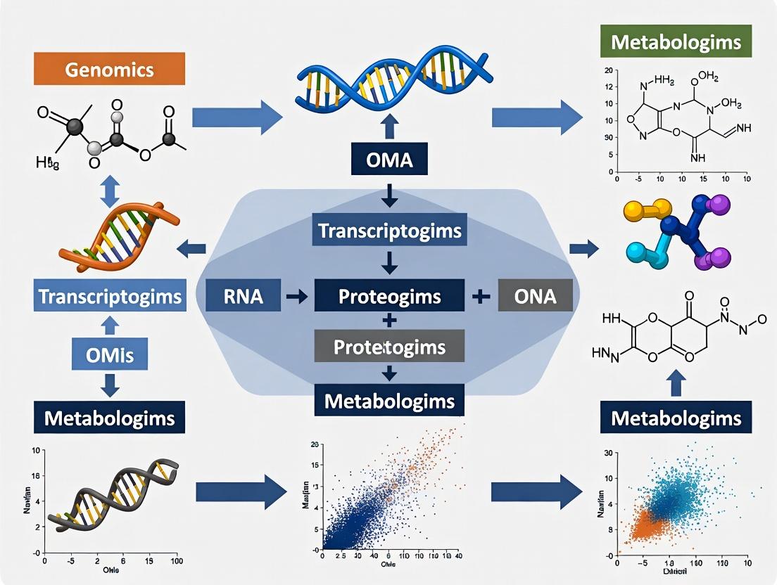
Within a Canonical Correlation Analysis (CCA)-based multi-omics integration research thesis, the initial stages of data input, formatting, and dimension matching are critical. This workflow ensures disparate datasets (e.g., genomics, transcriptomics, proteomics, metabolomics) are harmonized, enabling robust analysis of cross-data modality correlations to uncover complex biological mechanisms relevant to disease and drug discovery.
Data Input & Source Specifications
Multi-omics data is sourced from public repositories and in-house experiments. Common sources and their typical dimensions are summarized below.
Table 1: Representative Multi-Omics Data Sources and Initial Dimensions
| Omics Layer | Example Source | Typical Initial Format | Representative Initial Dimensions (Features x Samples) | Key Preprocessing Needs |
|---|---|---|---|---|
| Genomics (SNPs) | dbGaP, EGA | PLINK (.bed/.bim/.fam), VCF | ~500,000 - 1,000,000 x 1,000 | Imputation, MAF filtering, LD pruning |
| Transcriptomics | GEO, ArrayExpress | Count matrix (RNA-Seq), CEL files (Microarray) | ~20,000 - 60,000 x 500 | Normalization (TMM, DESeq2), log2 transformation, batch correction |
| Proteomics | PRIDE, CPTAC | Peptide/Protein intensity matrix | ~5,000 - 15,000 x 300 | Imputation of missing values (MinProb), normalization (vsn), log2 transform |
| Metabolomics | MetaboLights | Peak intensity table | ~500 - 5,000 x 200 | Normalization (PQN), scaling (pareto), missing value imputation (kNN) |
Detailed Experimental Protocols for Data Generation
Protocol 3.1: Bulk RNA-Seq for Transcriptomic Profiling
- Objective: Generate a gene expression count matrix from tissue samples.
- Materials: See The Scientist's Toolkit (Section 7).
- Procedure:
- Library Preparation: Use poly-A selection for mRNA enrichment. Fragment RNA and synthesize cDNA using reverse transcriptase with random hexamer primers.
- Sequencing: Perform paired-end sequencing (2x150 bp) on an Illumina NovaSeq platform to a minimum depth of 30 million reads per sample.
- Alignment & Quantification: Align reads to a reference genome (e.g., GRCh38) using STAR aligner (v2.7.10a) with default parameters. Generate gene-level read counts using
--quantMode GeneCounts. - Quality Control: Assess sample quality with FastQC and RSeQC. Remove samples where >20% of reads are unaligned.
Protocol 3.2: LC-MS/MS for Global Proteomics
- Objective: Generate a protein abundance matrix from cell lysates.
- Materials: See The Scientist's Toolkit (Section 7).
- Procedure:
- Sample Preparation: Lyse cells in RIPA buffer. Reduce, alkylate, and digest proteins with trypsin (1:50 enzyme-to-protein ratio) overnight at 37°C.
- LC-MS/MS Analysis: Desalt peptides and separate on a C18 nano-flow column using a 90-min gradient. Analyze eluents with a Q Exactive HF mass spectrometer in data-dependent acquisition (DDA) mode.
- Database Search: Process raw files with MaxQuant (v2.1.0.0). Search against the UniProt human database. Use a 1% false discovery rate (FDR) cutoff at protein and peptide levels.
- Output: Use the
proteinGroups.txtfile, filtering out reverse hits and contaminants.
Data Formatting and Standardization Workflow
Raw data from diverse platforms must be converted into a uniform analytic format.
Table 2: Standardized Formatting Requirements for CCA Input
| Processing Step | Transcriptomics (RNA-Seq Counts) | Proteomics (MS Intensity) | Metabolomics (LC-MS Peaks) |
|---|---|---|---|
| 1. Missing Data | Not applicable for counts. | Replace 0 with NA. Impute using impute.MinProb() (R imputeLCMD). |
Impute small values (e.g., half-minimum) for missing peaks. |
| 2. Transformation | log2(counts + 1) (variance stabilization). |
log2(intensity) (base-e or base-2). |
Often log-transformed (base-2 or base-e). |
| 3. Normalization | Trimmed Mean of M-values (TMM) using edgeR. | Variance stabilizing normalization (VSN). | Probabilistic Quotient Normalization (PQN). |
| 4. Filtering | Remove genes with low expression (CPM < 1 in >90% of samples). | Remove proteins with >50% missing values post-imputation. | Remove metabolites with >30% missing values or high RSD in QCs. |
| Final Format | Samples as columns, genes as rows. Numeric matrix. | Samples as columns, proteins as rows. Numeric matrix. | Samples as columns, metabolites as rows. Numeric matrix. |
Diagram 1: Multi-omics data formatting and standardization workflow.
Dimension Matching and Feature Selection for CCA
CCA requires matrices with identical sample ordering and managed feature dimensions to avoid overfitting.
Protocol 5.1: Sample-Wise Alignment and Intersection
- Meta-Data Harmonization: Ensure a unique, consistent sample identifier (e.g., PatientID_Timepoint) exists across all omics datasets and clinical metadata.
- Find Intersection: Identify the set of samples present in all omics assays. This creates the N (sample size) for CCA.
- Subset and Order: Subset each omics matrix to include only these intersecting samples. Ensure identical column (sample) order across all matrices.
Protocol 5.2: Feature Reduction via Variance Filtering and sCCA
- High-Variance Filtering: Within each omics matrix, calculate the variance (or median absolute deviation) for each feature. Retain the top K features (e.g., K=5000 per modality) for initial analysis. This retains biologically informative features.
- Sparse CCA (sCCA) for Joint Selection: Apply a regularized CCA implementation (e.g.,
PMA::CCAin R) with L1 (lasso) penalties to the high-variance filtered matrices.- Penalty Tuning: Use cross-validation (
PMA::CCA.permute) to select penalty parameters (c1,c2) that maximize the correlation while inducing sparsity. - Output: Obtain canonical weights. Features with non-zero weights are selected for the final, matched-dimension dataset.
- Penalty Tuning: Use cross-validation (
Table 3: Dimension Matching Outcomes for a Hypothetical Multi-Omics Study
| Omics Layer | Initial Features | After High-Variance Filtering | After sCCA Feature Selection | Final Dimension for CCA |
|---|---|---|---|---|
| Transcriptomics | 25,000 genes | 5,000 genes | 312 genes (non-zero weights) | 312 x 150 |
| Proteomics | 8,000 proteins | 5,000 proteins | 188 proteins (non-zero weights) | 188 x 150 |
| Shared Sample Size (N) | - | 150 samples | 150 samples | 150 samples |
Diagram 2: Sample alignment and feature dimension matching process.
Integrated Pre-CCA Workflow Diagram
Diagram 3: Complete workflow from data input to CCA-ready dataset.
The Scientist's Toolkit
Table 4: Essential Research Reagent Solutions for Multi-Omics Workflows
| Item / Reagent | Vendor Examples | Function in Workflow |
|---|---|---|
| TRIzol Reagent | Thermo Fisher, Sigma-Aldrich | Simultaneous isolation of high-quality RNA, DNA, and proteins from a single sample. |
| RNeasy Mini Kit | QIAGEN | Silica-membrane based purification of total RNA, including miRNA, with DNase treatment. |
| Trypsin, Sequencing Grade | Promega, Thermo Fisher | Specific proteolytic digestion of proteins into peptides for LC-MS/MS analysis. |
| Pierce BCA Protein Assay Kit | Thermo Fisher | Colorimetric quantification of protein concentration for normalization pre-MS. |
| Mass Spectrometry Grade Solvents | Honeywell, Sigma-Aldrich | Acetonitrile, methanol, and water with ultra-low volatility and ion contamination for LC-MS. |
| TruSeq Stranded mRNA Library Prep Kit | Illumina | Preparation of high-quality, strand-specific RNA-seq libraries for next-generation sequencing. |
| Human Omics Reference Materials | NIST, Sigma-Aldrich | Well-characterized control samples (e.g., HEK293 cell digest) for inter-laboratory QC in proteomics/metabolomics. |
| Bioinformatics Suites (Local) | R/Bioconductor, Python (SciPy/Pandas) | Open-source platforms for implementing formatting, normalization, and CCA algorithms. |
1. Introduction and Thesis Context Within multi-omics integration research, Canonical Correlation Analysis (CCA) identifies relationships between two multivariate datasets. However, for high-dimensional omics data, standard CCA fails, producing uninterpretable, non-sparse canonical vectors loaded on all features. Sparse CCA (sCCA) incorporates L1 (lasso) penalties to produce canonical vectors with zero weights for most features, enabling biomarker discovery. This protocol details the critical process of tuning the penalty parameters, a non-trivial step that directly controls the sparsity and stability of the selected features. Mastery of this tuning is a cornerstone of robust multi-omics implementation, bridging statistical discovery with biological validation in therapeutic development.
2. Key Tuning Parameters and Data Presentation
The core tuning parameters are the L1-norm penalty constraints, c1 and c2, for datasets X and Y, respectively. Their values range between 0 and 1, where a smaller value induces greater sparsity. The optimal pair is typically found via grid search.
Table 1: Representative Grid of Penalty Parameters and Resulting Sparsity
| Penalty c1 (for X) | Penalty c2 (for Y) | Approx. % Non-zero in u |
Approx. % Non-zero in v |
Typical Use Case |
|---|---|---|---|---|
| 0.3 | 0.3 | 5-10% | 5-10% | Highly sparse initial screening |
| 0.5 | 0.5 | 15-25% | 15-25% | Balanced selection |
| 0.7 | 0.7 | 30-40% | 30-40% | Less sparse, inclusive search |
| 0.9 | 0.9 | 50-70% | 50-70% | Near-standard CCA |
Table 2: Criteria for Evaluating Parameter Pairs in Grid Search
| Criterion | Formula/Description | Optimization Goal |
|---|---|---|
| Cross-Validated Correlation | Mean canonical correlation across k-folds. | Maximize |
| Stability of Selected Features | Jaccard index or correlation between canonical vectors from subsampled data. | Maximize (≥0.8 is stable) |
| Total Features Selected | Count of non-zero weights in u + v. |
Align with biological interpretability capacity |
3. Experimental Protocol: Penalty Parameter Tuning via Stability Selection
This protocol uses a stability-enhanced grid search to identify optimal (c1, c2).
3.1 Preprocessing
- Data Input: Let
X[n x p] be the first omics dataset (e.g., mRNA expression, p features) andY[n x q] be the second (e.g., protein abundance, q features).nis the shared set of samples. - Standardization: Center each column of
XandYto mean zero. Scale each column to have unit variance.
3.2 Primary Tuning Workflow
- Define Parameter Grid: Construct a logical grid of candidate values (e.g.,
c1 = seq(0.1, 0.9, length=9),c2 = seq(0.1, 0.9, length=9)). - Stability Loop (for each grid point):
a. Perform 100 rounds of subsampling. In each round, randomly select
n/2samples without replacement. b. On this subset, run the sCCA algorithm (e.g., via PMA or SCCA packages) using the fixed penaltiesc1andc2to obtain canonical vectorsu*andv*. c. Record the indices of non-zero coefficients inu*andv*. - Calculate Selection Probabilities: For each feature in
XandY, compute its frequency of being selected across all 100 subsamples at that grid point. This yields stability matrices. - Compute Summary Metric: For the grid point (
c1,c2), calculate the mean stable canonical correlation: a. For each subsampling roundb, train sCCA on the subsample and compute the correlation on the held-out samples. b. Average this correlation across all rounds. - Grid Evaluation: Repeat steps 2-4 for all (
c1,c2) pairs in the grid.
3.3 Selection of Optimal Parameters
- Threshold Stability Matrices: For each grid point, apply a stability threshold (e.g., selection probability > 0.8) to derive a stable set of features.
- Final Choice: Plot the mean stable canonical correlation against the total number of stable features. The optimal parameter pair is often at the "elbow" of this curve, balancing correlation strength and feature number. Alternatively, select the pair yielding the highest mean stable correlation where the number of stable features is manageable (<100 per omics type for initial validation).
Title: sCCA Penalty Parameter Tuning Workflow
4. The Scientist's Toolkit: Research Reagent Solutions
Table 3: Essential Computational Tools for sCCA Tuning
| Tool/Reagent | Function in Experiment | Key Notes |
|---|---|---|
| PMA R Package (Penalized Multivariate Analysis) | Implements sCCA with cross-validation. | Core algorithm for computing sparse canonical vectors. |
| mixOmics R/Bioc Package | Provides tune.splsda and tune.block.splsda for multi-omics. | Includes repeated CV and graphical outputs for tuning. |
| SCCA Python Package (e.g., scca) | Python implementation of sCCA algorithms. | Enables integration into Python-based ML/AI pipelines. |
| Stability Selection Framework (Custom Scripts) | Quantifies feature selection robustness across subsamples. | Critical for reliable biomarker shortlisting. |
| High-Performance Computing (HPC) Cluster | Parallelizes grid search over many parameter pairs. | Reduces tuning time from days to hours. |
| Jaccard Index Function | Measures similarity between selected feature sets. | Calculates stability (0.8+ indicates high stability). |
Title: Logic of Penalty Tuning Impact
5. Post-Tuning Validation Protocol Once optimal parameters are set, a final model is fit on the full dataset.
- Final Model Fit: Apply sCCA with the optimal (
c1,c2) to the fullXandY. Obtain canonical vectorsuandv. - Feature Ranking: Rank selected features by the absolute magnitude of their weights in
uandv. - Biological Concordance Check: Perform pathway enrichment analysis (e.g., via GO, KEGG) on the top selected features from each omics set. The significance of shared pathways (e.g., "PI3K-Akt signaling") validates the integration.
- Hold-out Validation: If sample size permits, perform a single train-test split. Fit sCCA on the training set with tuned parameters and assess the canonical correlation on the independent test set. A significant drop suggests overfitting.
Within a multi-omics Canonical Correlation Analysis (CCA) research thesis, the interpretation of canonical loadings, correlations, and scores is critical for deriving biological insights. These outputs link high-dimensional molecular datasets (e.g., transcriptomics, proteomics, metabolomics) to identify coordinated biological signals driving phenotypes relevant to drug discovery.
Key Outputs: Definitions and Interpretative Framework
| Output | Mathematical Description | Biological/Multi-omics Interpretation | Utility in Drug Development |
|---|---|---|---|
| Canonical Correlation | (\rhok = corr(Uk, V_k)) for the (k)-th pair. Measures linear relationship between omics-derived canonical variates (U) and (V). | Strength of global association between two omics platforms (e.g., mRNA-protein). High (\rho) suggests a strong, coordinated multi-omics program. | Identifies robust, cross-omics biological pathways as high-confidence therapeutic targets. |
| Canonical Loadings (Structural Coefficients) | ( \mathbf{a}k, \mathbf{b}k ): Correlation between original variables (genes, proteins) and their canonical variates (Uk, Vk). | Reveals which specific molecular features from each dataset contribute most to the shared correlation. High loading indicates strong representation in the latent multi-omics signal. | Pinpoints key driver genes/proteins within a correlated pathway for targeted intervention (e.g., drug inhibition). |
| Canonical Scores (Variates) | (Uk = X\mathbf{a}k), (Vk = Y\mathbf{b}k). Projection of original data onto canonical axes. | Represents the latent molecular "component" or "program" shared across omics types for each sample. Samples with high scores are strongly influenced by that program. | Enables patient stratification based on multi-omics activity; identifies samples for preclinical models. |
| Cross-Loadings | Correlation between variables from one omics set and canonical variates from the other set. | Assesses how well a feature from one platform (e.g., a metabolite) is predicted by the latent structure in the other platform (e.g., microbiome). | Uncovers predictive relationships across omics layers, suggesting biomarkers or mechanistic links. |
Experimental Protocol: Multi-omics CCA for Target Identification
Objective: To identify canonical variates representing shared variance between transcriptomic and proteomic data from tumor samples and interpret their biological significance.
Materials & Preprocessing:
- RNA-Seq Data: Count matrix for 20,000 genes from 100 tumor samples. Normalized (TPM) and log2-transformed.
- LC-MS Proteomics Data: Intensity matrix for 8,000 proteins from the same 100 samples. Normalized (median centering) and log2-transformed.
- Clinical Phenotype Data: Tumor growth rate metrics for validation.
Step-by-Step Protocol:
Step 1: Data Integration and Scaling.
- Subset datasets to matched samples (n=100).
- Perform feature selection: Retain top 5,000 variable genes and 3,000 variable proteins (by coefficient of variation).
- Center and scale each variable (mean=0, variance=1) separately per omics layer.
Step 2: CCA Execution (using R PMA or mixOmics).
Step 3: Extraction and Interpretation of Outputs.
- Correlations: Extract (\rho1) to (\rho5). Retain components with (\rho > 0.7) and statistically significant via permutation test (1000 permutations).
- Loadings: Extract (\mathbf{a}k) and (\mathbf{b}k). Define "high loading" as (|\text{loading}| > 0.3).
- Scores: Calculate (Uk) and (Vk) for each sample.
Step 4: Biological Annotation.
- For each significant component (e.g., Component 1), take genes and proteins with high loadings.
- Perform over-representation analysis (ORA) on these feature sets using KEGG/GO databases.
- Correlate canonical scores ((U1, V1)) with clinical phenotypes (e.g., tumor growth rate via Pearson correlation).
Step 5: Validation.
- Technically: Use cross-validation (splitting samples) to assess stability of loadings.
- Biologically: Validate key driver proteins via orthogonal method (e.g., immunohistochemistry) in an independent cohort.
The Scientist's Toolkit: Key Research Reagents & Solutions
Table 2: Essential Materials for Multi-omics CCA Implementation
| Item | Function in CCA Workflow | Example Product/Catalog |
|---|---|---|
| Multi-omics Data Generation | ||
| RNA Extraction Kit (for Transcriptomics) | Isolates high-integrity total RNA for sequencing. | Qiagen RNeasy Mini Kit (74104) |
| Protein Lysis Buffer (for Proteomics) | Efficiently extracts proteins from complex tissues for MS. | RIPA Buffer (Thermo Fisher, 89900) |
| Bioinformatics Analysis | ||
| CCA Software Package | Performs regularized CCA on high-dimensional data. | R mixOmics package (v6.24.0) |
| Permutation Testing Script | Assesses statistical significance of canonical correlations. | Custom R/Python script (1000 iterations) |
| Downstream Validation | ||
| Antibody for Candidate Protein | Validates expression of a high-loading protein from CCA. | Anti-PDL1 [28-8] (Abcam, ab205921) |
| siRNA/Gene Knockout Kit | Functionally tests a high-loading gene identified from analysis. | Dharmacon siRNA SMARTpool |
Visualization of Analysis Workflow and Relationships
Title: Workflow for Interpreting CCA Outputs in Multi-omics
Title: Relationship Between Loadings, Variates, and Correlation
In multi-omics research employing Canonical Correlation Analysis (CCA), effective visualization of high-dimensional results is paramount. These visual tools bridge statistical output and biological interpretation, enabling researchers to discern complex relationships between omics layers and their association with phenotypic outcomes. This protocol details the generation and interpretation of three critical visualization types within a CCA framework.
Core Visualization Methodologies
Correlation Circle Plots for CCA Loadings
Purpose: To visualize the contribution of original variables (e.g., genes, metabolites) to the canonical variates and the correlation structure between two omics datasets.
Protocol:
- Compute Loadings: Following CCA, obtain the canonical structure correlations (loadings) for each variable in Dataset X (e.g., transcriptome) and Dataset Y (e.g., metabolome) for the first two canonical dimensions (Can1, Can2).
- Set Up Plot: Create a circular plot with x-axis representing correlation with Can1 and y-axis representing correlation with Can2. Draw a unit circle (radius=1).
- Plot Variables: For each variable, plot a point at coordinates (corrwithCan1, corrwithCan2). Use different shapes/colors for X and Y datasets.
- Draw Vectors: Optionally, draw vectors from the origin (0,0) to each point. The length and direction indicate the strength and nature of the variable's contribution.
- Interpretation: Points near the circle's periphery are strongly correlated with the canonical dimensions. Proximity of variables from different datasets suggests cross-omics correlation.
Data Output Example (CCA Loadings for First Two Dimensions): Table 1: Example Loadings for Transcriptomic (X) and Metabolomic (Y) Variables.
| Variable ID | Dataset | Loading on Can1 | Loading on Can2 | Canonical Correlation (ρ) |
|---|---|---|---|---|
| Gene_A | X | 0.92 | -0.15 | 0.89 |
| Gene_B | X | 0.78 | 0.42 | 0.89 |
| Metabolite_1 | Y | 0.85 | 0.30 | 0.89 |
| Metabolite_2 | Y | -0.62 | 0.65 | 0.89 |
Heatmaps for Integrated Correlation Matrices
Purpose: To display the pairwise correlation matrix between selected features from multiple omics datasets, often after CCA-guided feature selection.
Protocol:
- Matrix Construction: Create a block matrix containing correlations:
- Within-dataset correlations (e.g., gene-gene).
- Between-dataset correlations (e.g., gene-metabolite).
- Clustering: Apply hierarchical clustering to rows and columns to group correlated features.
- Color Mapping: Use a divergent color palette (e.g., blue-white-red for negative-zero-positive correlation).
- Annotation: Add side-color bars to annotate feature types (omics layer, pathway membership).
- Visualization: Render using a library like
pheatmaporComplexHeatmap.
Data Output Example (Correlation Values for Heatmap): Table 2: Subset of Integrated Correlation Matrix.
| Gene_A | Gene_B | Metabolite_1 | Metabolite_2 | |
|---|---|---|---|---|
| Gene_A | 1.00 | 0.60 | 0.82 | -0.55 |
| Gene_B | 0.60 | 1.00 | 0.71 | 0.10 |
| Metabolite_1 | 0.82 | 0.71 | 1.00 | -0.30 |
| Metabolite_2 | -0.55 | 0.10 | -0.30 | 1.00 |
Sample Projections (Biplot & Sample Scores)
Purpose: To project individual samples onto the canonical space, visualizing sample stratification, outliers, and the influence of variables.
Protocol (CCA Biplot):
- Calculate Scores: Compute canonical variate scores for each sample.
- Plot Samples: Scatter plot of samples using scores for Can1 vs. Can2. Color by phenotype/group.
- Overlay Variables: On the same axes, plot variable loadings as vectors (from 2.1) or points.
- Scale: Use a scaling factor (alpha) to optimally overlay variable vectors on the sample scores.
- Interpretation: Sample position indicates its omics profile. Proximity of a sample to a variable vector suggests high value for that variable.
Data Output Example (Sample Canonical Scores): Table 3: Canonical Variate Scores for a Subset of Samples.
| Sample_ID | Phenotype | Score on Can1 (X) | Score on Can2 (X) | Score on Can1 (Y) | Score on Can2 (Y) |
|---|---|---|---|---|---|
| S1 | Control | -1.2 | 0.5 | -1.1 | 0.6 |
| S2 | Control | -0.8 | 0.9 | -0.9 | 0.8 |
| S3 | Disease | 2.1 | -0.3 | 2.0 | -0.2 |
| S4 | Disease | 1.8 | 0.1 | 1.7 | 0.2 |
Visualization Workflow & Pathway Diagram
Title: CCA Multi-Omics Visualization & Interpretation Workflow
The Scientist's Toolkit: Research Reagent Solutions
Table 4: Essential Tools for CCA-based Multi-Omics Visualization.
| Item/Category | Example(s) | Function in Visualization Pipeline |
|---|---|---|
| Statistical Computing | R (v4.3+), Python (v3.10+) | Core platforms for performing CCA computations and generating plot data. |
| CCA & Multivariate Packages | R: CCA, mixOmics, PMAPython: scikit-learn, PyCCA |
Provide functions to compute canonical correlations, loadings, and scores. |
| Visualization Libraries | R: ggplot2, plotly, pheatmap, ComplexHeatmapPython: matplotlib, seaborn, plotly, scatterplot |
Generate publication-quality correlation circles, heatmaps, and biplots. |
| Interactive Dashboard Tools | RShiny, Dash (Python), Jupyter Widgets | Create interactive visualizations for exploratory data analysis by teams. |
| Data Integration Platforms | MOFA+, OmicsPLS |
Offer built-in CCA-like visualization for integrated multi-omics models. |
| Color Palette Tools | viridis, RColorBrewer |
Ensure accessible, colorblind-friendly palettes for heatmaps and plots. |
| Version Control | Git, GitHub/GitLab | Track changes to analysis and visualization code for reproducibility. |
This case study provides detailed application notes and protocols for a canonical correlation analysis (CCA)-based multi-omics integration, framed within a broader thesis research project investigating robust CCA implementations for oncology biomarker discovery. The integration of genome-wide gene expression (RNA-Seq) and DNA methylation (Infinium HumanMethylation450 BeadChip) data from The Cancer Genome Atlas (TCGA) serves as a canonical example to identify coordinated regulatory mechanisms driving cancer phenotypes. This protocol is designed for researchers, scientists, and bioinformaticians in drug development seeking to derive biologically interpretable, cross-omics signatures.
Key Quantitative Data from a Representative TCGA-BRCA Analysis
The following tables summarize quantitative results from a representative integration analysis of Breast Invasive Carcinoma (TCGA-BRCA) data, performed using the current analytical pipeline.
Table 1: TCGA-BRCA Cohort Data Summary
| Data Type | Platform | Samples (Tumor/Normal) | Features (Pre-filtered) | Primary Source |
|---|---|---|---|---|
| Gene Expression | Illumina HiSeq RNA-Seq | 1,097 (1,103 Tumor) | 60,483 transcripts | TCGA Data Portal |
| DNA Methylation | Illumina Infinium HM450 | 795 (791 Tumor) | 485,577 CpG sites | TCGA Data Portal |
Table 2: CCA Integration Results Summary (Top 3 Canonical Variates)
| Canonical Variate (CV) | Canonical Correlation (ρ) | P-value (Permutation Test) | # of Significant Genes (FDR<0.05) | # of Significant CpG Probes (FDR<0.05) |
|---|---|---|---|---|
| CV1 | 0.892 | < 0.001 | 1,247 | 9,885 |
| CV2 | 0.865 | < 0.001 | 987 | 7,432 |
| CV3 | 0.841 | < 0.001 | 802 | 6,105 |
Table 3: Top Functional Enrichment for Genes in CV1 (Negative Correlation with Methylation)
| Gene Set Name (MSigDB Hallmarks) | Normalized Enrichment Score (NES) | FDR q-value | Leading Edge Genes (Example) |
|---|---|---|---|
| EPITHELIALMESENCHYMALTRANSITION | 2.45 | < 0.001 | SNAI1, VIM, ZEB1 |
| ESTROGENRESPONSEEARLY | 1.98 | 0.003 | TFF1, GREB1, PGR |
| APICAL_JUNCTION | -2.12 | < 0.001 | CDH1, OCLN, CTNNA1 |
Experimental Protocols
Protocol 3.1: Data Acquisition and Preprocessing
Objective: To download and quality-control TCGA multi-omics data for integration.
- Data Source: Access data via the NCI Genomic Data Commons (GDC) Data Portal using the
TCGAbiolinksR/Bioconductor package or the GDC Data Transfer Tool. - Gene Expression Preprocessing:
- Download HT-Seq Counts or FPKM-UQ files.
- Filter out low-expression genes: Retain genes with counts > 10 in at least 20% of samples.
- Apply Variance Stabilizing Transformation (VST) using
DESeq2or convert to log2(FPKM-UQ+1).
- DNA Methylation Preprocessing:
- Download Beta-value matrices.
- Perform quality control: Remove probes with detection p-value > 0.01 in >10% of samples.
- Filter probes: Remove probes on sex chromosomes, cross-reactive probes, and probes containing single nucleotide polymorphisms (SNPs) at the CpG site.
- Normalize using functional normalization (
minfipackage).
- Sample Matching & Batch Effect: Retain only paired tumor samples present in both datasets. Correct for technical batch effects (e.g., plate, year) using
ComBatfrom thesvapackage.
Protocol 3.2: Feature Selection & Dimensionality Reduction
Objective: Reduce feature space to biologically relevant variables for stable CCA.
- For Gene Expression: Select the top 5,000 most variable genes based on median absolute deviation (MAD).
- For Methylation Data: Select the top 10,000 most variable CpG probes based on MAD across samples.
- Optional but Recommended: Perform preliminary univariate association (e.g., differential expression/methylation analysis between tumor and normal) to further filter to the top ~3,000 significant features per omic, increasing biological signal.
Protocol 3.3: Sparse Canonical Correlation Analysis (sCCA) Implementation
Objective: Identify correlated linear combinations of gene expression and methylation features.
- Tool: Use the
PMA(Penalized Multivariate Analysis) R package or themixOmicspackage. - Data Input: Input preprocessed, filtered, and scaled (mean-centered, unit variance) matrices
X(gene expression, n x p) andZ(methylation, n x q) for n paired samples. - Parameter Tuning: Perform cross-validation (
permute=TRUEinPMD.cv) to determine optimal sparsity penalties (c1andc2). This controls the number of non-zero loadings for each canonical variate. - Model Execution: Run sCCA with chosen penalties:
result <- CCA(X, Z, penaltyx=c1, penaltyz=c2, type="standard"). - Significance Testing: Assess statistical significance of each canonical correlation using a permutation test (e.g., 1000 permutations) on the residual matrix.
Protocol 3.4: Biological Interpretation & Validation
Objective: Interpret canonical variates and validate findings.
- Loadings Extraction: Extract gene and CpG probe loadings (coefficients) from the CCA model. Focus on features with non-zero loadings.
- Correlation Direction: Identify genes negatively correlated with methylation of promoter-associated CpG islands (expected canonical relationship).
- Pathway Enrichment: Perform gene set enrichment analysis (GSEA) on genes ranked by their loadings in a significant CV using
clusterProfiler. - Cis-Regulatory Mapping: Map significant CpG probes to gene promoters (e.g., TSS1500, TSS200) using Illumina annotation files. Validate anti-correlation for these specific gene-probe pairs.
- Clinical Correlation: Correlate sample scores for each CV with clinical variables (e.g., survival, subtype) using Cox regression or ANOVA.
Diagrams
Title: Multi-Omics Integration with sCCA Workflow
Title: CCA Captures Gene Methylation Regulation
The Scientist's Toolkit: Research Reagent Solutions
Table 4: Essential Computational Tools & Resources
| Tool/Resource Name | Function in Protocol | Key Feature / Application |
|---|---|---|
| TCGAbiolinks (R/Bioconductor) | Unified data download from GDC and basic preprocessing. | Simplifies API queries, handles GDC authentication, and merges clinical data. |
| minfi (R/Bioconductor) | Comprehensive preprocessing and normalization of Illumina methylation array data. | Implements functional normalization, QC plots, and detection p-value filtering. |
| sva / ComBat (R/Bioconductor) | Removal of unwanted technical variation (batch effects). | Adjusts for non-biological covariates (e.g., sequencing batch, slide) that confound integration. |
| PMA or mixOmics (R CRAN/Bioc) | Implementation of Sparse Canonical Correlation Analysis. | Applies L1-penalty for feature selection within CCA, yielding interpretable, non-zero loadings. |
| clusterProfiler (R/Bioconductor) | Functional enrichment analysis of gene lists derived from CCA loadings. | Performs ORA and GSEA on MSigDB, KEGG, and GO terms for biological interpretation. |
| UCSC Xena / cBioPortal | Independent validation and visualization of results in external or pan-cancer cohorts. | Allows quick correlation checks and survival analysis for candidate genes. |
Navigating Pitfalls: Solutions for Common CCA Implementation Challenges in Multi-Omics
Within the context of implementing Canonical Correlation Analysis (CCA) for multi-omics integration, the "large p, small n" (p >> n) problem is a fundamental constraint. Here, the number of molecular features (p) from genomics, transcriptomics, proteomics, etc., vastly exceeds the number of biological samples (n). This leads to ill-posed CCA models with non-unique solutions, extreme overfitting, and poor generalizability. These application notes outline contemporary strategies and protocols to enable robust CCA in high-dimensional, low-sample-size research, such as in early-phase clinical trials or rare disease cohorts.
Quantitative Comparison of Dimensionality Reduction & Regularization Strategies
The following table summarizes core methodological approaches to address p >> n in CCA, with key performance metrics from recent literature.
Table 1: Strategies for High-Dimensional CCA in Multi-Omics Research
| Strategy Category | Specific Method | Key Mechanism | Reported Performance (Canonical Correlation on Test Set) | Typical Use Case |
|---|---|---|---|---|
| Two-Stage Dimensionality Reduction | Principal Component Analysis (PCA) + CCA | Project each omics dataset onto its top k principal components before CCA. | ~0.65-0.80 (varies by retained variance %) | Initial exploratory integration; preserves global structure. |
| Sparse Regularization | Sparse CCA (sCCA) | Impose L1 (lasso) penalty on canonical weight vectors to force zero weights for irrelevant features. | ~0.70-0.85 (depending on sparsity parameter λ) | Feature selection; identifying biomarker drivers of correlation. |
| Kernel-Based Methods | Kernel CCA (kCCA) | Map data to a high-dimensional feature space via kernel trick; effective for non-linear relationships. | ~0.75-0.90 (highly kernel-dependent) | Capturing complex, non-linear omics interactions. |
| Deep Learning Approaches | Deep CCA (dCCA) | Use deep neural networks to learn non-linear transformations that maximize correlation. | ~0.80-0.95 (requires significant n for training) | Complex integration with hierarchical feature learning. |
| Penalized Matrix Decomposition | Penalized CCA (PMD) | Apply combined L1 & L2 (elastic net) penalties for structured sparsity. | ~0.72-0.88 | Balanced feature selection with group effects. |
Experimental Protocols
Protocol 3.1: Sparse CCA (sCCA) for Multi-Omics Biomarker Discovery
Objective: To identify a sparse subset of correlated features between transcriptomics (RNA-seq) and proteomics (LC-MS) data from a patient cohort (n=50, pRNA~20,000, pProtein~5,000).
Materials: Normalized and log-transformed feature matrices (samples x features). Compute environment (R/Python).
Procedure:
- Preprocessing & Standardization: Center and scale each feature (column) to have zero mean and unit variance across samples.
- Parameter Tuning via Cross-Validation:
- Split data into K-folds (e.g., K=5).
- For a grid of sparsity parameters (λ1 for omics1, λ2 for omics2): a. Train sCCA on K-1 folds. b. Calculate the sum of absolute correlations between canonical variates on the held-out fold.
- Select the (λ1, λ2) pair that maximizes this cross-validated correlation.
- Model Training: Fit the final sCCA model on the entire dataset using the optimal parameters.
- Result Interpretation: Extract non-zero weights from the canonical weight vectors. Features with large absolute weights are drivers of the cross-omics correlation. Perform pathway enrichment analysis on selected features.
Protocol 3.2: Two-Stage PCA-CCA Workflow for Initial Data Integration
Objective: To establish baseline linear correlations between methylation (p~450k) and metabolomics (p~500) data from a small longitudinal study (n=30, time points=3).
Materials: Batch-corrected and normalized data matrices per time point.
Procedure:
- Dimensionality Reduction per Omics Layer:
- For each omics dataset at each time point, perform PCA.
- Retain the top k components that explain >80% of cumulative variance. This yields reduced matrices of size (n x k_Omics).
- Temporal Concatenation: For each omics type, concatenate the reduced matrices across time points (e.g., n=30 * 3 = 90, features = k_Omics).
- CCA Execution: Apply standard CCA to the concatenated PCA-reduced matrices to find linear combinations maximally correlated across omics types over time.
- Validation: Use permutation testing (randomly shuffling omics labels 1000x) to assess the statistical significance (p-value) of the observed canonical correlations.
Visualizations
Title: p >> n CCA Analysis Workflow
Title: Core Strategies to Solve p >> n Problem
The Scientist's Toolkit: Research Reagent & Computational Solutions
Table 2: Essential Toolkit for High-Dimensional Multi-Omics CCA Research
| Item / Solution | Category | Function in p >> n CCA Context |
|---|---|---|
| PMD (Penalized Matrix Decomposition) | R Package (PMA) |
Implements sparse CCA (sCCA) and sparse PCA with efficient penalties for feature selection. |
| mixOmics | R Package | Provides a comprehensive suite (sPLS, rCCA, DIABLO) for multi-omics integration with built-in regularization. |
| CCA-Zoo | Python Library | Implements kernel CCA, deep CCA, and sparse CCA variants in a scalable, GPU-compatible framework. |
| Elastic Net Penalty | Algorithmic Component | Combined L1 & L2 regularization (available in glmnet, scikit-learn) used in PMD-CCA for grouped variable selection. |
| Permutation Testing Framework | Validation Script | Custom code to generate null distribution of canonical correlations, essential for assessing significance in small n. |
| Stratified K-Fold Cross-Validation | Protocol | Resampling method critical for reliable parameter tuning and error estimation in low-sample-size settings. |
Within the context of implementing Canonical Correlation Analysis (CCA) for multi-omics data integration in biomedical research, the risk of overfitting is pronounced due to the high-dimensionality (p >> n) and complex covariance structures inherent to genomics, transcriptomics, proteomics, and metabolomics datasets. This document provides application notes and detailed protocols for employing cross-validation and permutation testing to ensure robust, generalizable findings in drug development and biomarker discovery.
Table 1: Common Cross-Validation Schemes for Multi-omics CCA
| Scheme | Description | Recommended Use Case | Key Advantage | Typical # of Folds |
|---|---|---|---|---|
| k-Fold | Data split into k equal subsets; model trained on k-1, tested on held-out fold. | Initial model tuning with moderate sample size (n > 50). | Reduces variance of performance estimate. | 5 or 10 |
| Leave-One-Out (LOOCV) | Each sample serves as a single test set. | Very small sample sizes (n < 30). | Maximizes training data. | n |
| Nested CV | Outer loop estimates performance, inner loop tunes hyperparameters (e.g., regularization). | Final unbiased evaluation with hyperparameter optimization. | Prevents data leakage; unbiased error estimate. | Outer: 5-10, Inner: 5 |
| Monte Carlo (Repeated Random Subsampling) | Random splits into training/test sets repeated many times. | Unstable performance with standard k-fold. | Less variable than single k-fold. | 50-100 iterations |
| Stratified k-Fold | k-Fold preserving the proportion of classes or outcomes in each fold. | Classification tasks with CCA-derived components. | Maintains class balance in splits. | 5 or 10 |
Table 2: Permutation Testing Parameters for CCA Significance
| Parameter | Typical Setting | Purpose | Impact on Result |
|---|---|---|---|
| Number of Permutations | 1000 - 10,000 | Establish empirical null distribution of canonical correlations. | Higher counts increase precision of p-value. |
| Permutation Unit | Sample labels (Y-block) or both blocks independently. | Break structure between omics datasets while preserving internal covariance. | Preserving block structure is conservative. |
| Significance Threshold (α) | 0.05 (with multiple testing correction) | Determine statistically significant canonical variates. | Controls family-wise error rate (FWER). |
| Correction Method | Bonferroni, Holm, or FDR (Benjamini-Hochberg). | Adjust for testing multiple canonical correlations (modes). | Balances sensitivity and specificity. |
Experimental Protocols
Protocol 3.1: Nested Cross-Validation for Regularized CCA (rCCA) in Multi-omics
Objective: To unbiasedly evaluate the predictive performance of a multi-omics CCA model while optimizing regularization parameters (λ1, λ2 for omics blocks X and Y).
Materials:
- Integrated omics datasets (e.g., mRNA expression and protein abundance matrices).
- Computing environment (R/Python) with
PMA,mixOmics, orscikit-learnlibraries. - High-performance computing resources for intensive computation.
Procedure:
- Outer Loop Setup: Partition the full dataset into k outer folds (e.g., k=5). Designate one fold as the outer test set and the remaining k-1 folds as the outer training set.
- Inner Loop (Hyperparameter Tuning): a. Take the outer training set from step 1. b. Further split it into l inner folds (e.g., l=5). c. For each candidate pair (λ1, λ2) on a predefined grid (e.g., [0.001, 0.01, 0.1, 1]): i. Train an rCCA model on l-1 inner folds. ii. Compute the correlation between the canonical variates on the held-out inner fold. iii. Repeat for all l folds and compute the average canonical correlation. d. Select the (λ1, λ2) pair yielding the highest average canonical correlation.
- Model Training & Testing: Train an rCCA model on the entire outer training set using the optimal parameters from Step 2. Apply the model to the held-out outer test set to compute the test canonical correlation.
- Iteration & Aggregation: Repeat Steps 1-3 for each of the k outer folds. Report the mean and standard deviation of the k test canonical correlations as the final performance estimate.
Protocol 3.2: Permutation Testing for Significance of Canonical Variates
Objective: To determine the statistical significance of the observed canonical correlations against the null hypothesis of no association between the two omics datasets.
Materials:
- Trained CCA model (e.g., from
mixOmics::cim_cca). - Preprocessed omics matrices X (n x p) and Y (n x q).
Procedure:
- Observed Statistic: Run CCA on the original datasets X and Y. Record the squared canonical correlations (ρ²) for the first d variates (modes).
- Null Distribution Generation: a. For i in 1 to P (number of permutations, e.g., 1000): i. Randomly permute the row indices (samples) of dataset Y, breaking its relationship with X. (Alternatively, permute both datasets independently). ii. Run CCA on X and the permuted Y. iii. Record the squared canonical correlations (ρ²permi) for each mode. b. This yields a P x d matrix of null correlation statistics.
- P-value Calculation: a. For each mode j (1 to d): i. Count the number of permutations where ρ²permi[j] >= ρ²observed[j]. ii. Compute the empirical p-value: pj = (count + 1) / (P + 1).
- Multiple Testing Correction: Apply a correction method (e.g., FDR) to the p-values from Step 3 across all d modes to control the false discovery rate.
- Interpretation: Canonical variates with corrected p-values < 0.05 are considered statistically significant associations not attributable to chance.
Visualization Diagrams
Title: Nested Cross-Validation Workflow for rCCA
Title: Permutation Testing Protocol for CCA Significance
The Scientist's Toolkit: Research Reagent Solutions
Table 3: Essential Tools for Robust Multi-omics CCA Implementation
| Item / Solution | Function / Purpose | Example / Note |
|---|---|---|
| Regularized CCA Software | Incorporates L1/L2 penalties to handle high-dimensional data and ill-posed problems. | R: PMA (Penalized Multivariate Analysis), mixOmics. Python: scikit-learn CCA with custom regularization. |
| High-Performance Computing (HPC) Cluster Access | Enables computationally intensive nested CV and large-scale permutation tests (1000+). | Cloud (AWS, GCP) or local cluster with parallel processing capabilities. |
| Containerization Platform | Ensures reproducibility of the analysis environment, including specific library versions. | Docker or Singularity containers. |
| Multi-omics Data Preprocessing Pipeline | Standardizes normalization, batch correction, and missing value imputation across omics layers to reduce technical noise. | Nextflow or Snakemake pipeline integrating tools like ComBat, limma, missMDA. |
| Hyperparameter Optimization Library | Systematically searches regularization parameter space for optimal model performance. | mlr3 (R), optuna (Python). |
| Result Visualization Suite | Visualizes canonical weights, loadings, correlation circle plots, and sample scores for interpretation. | R: ggplot2, plotly. Python: matplotlib, seaborn. |
Managing Missing Data and Batch Effects in Multi-Omics Cohorts
This document presents application notes and protocols for managing missing data and batch effects within multi-omics cohorts, framed within a thesis focused on the implementation of Canonical Correlation Analysis (CCA) for multi-omics integration. Effective handling of these data challenges is critical for deriving robust biological insights and ensuring reproducibility in translational research and drug development.
Table 1: Prevalence and Impact of Missing Data in Multi-Omics Studies
| Omics Layer | Typical Missingness Rate (%) | Primary Causes | Common Imputation Methods |
|---|---|---|---|
| Proteomics | 10-50 | Low-abundance proteins, detection limits | k-NN, MissForest, BPCA |
| Metabolomics | 5-30 | Signal interference, concentration below LOQ | SVD-based, QRILC, min value |
| Transcriptomics | <5 | Low expression, technical dropouts | Mean/median, SVDimpute |
| Genomics (SNP array) | 1-5 | Poor hybridization, low signal intensity | BEAGLE, mean genotype |
Table 2: Batch Effect Correction Performance Metrics (Simulated Data)
| Correction Method | PCA-based Distance Reduction (%)* | Intra-batch Correlation Increase (%)* | Computation Time (min, 1000 samples) |
|---|---|---|---|
| ComBat | 65-80 | 40-60 | ~5 |
| ComBat-seq (RNA-seq) | 70-85 | 45-65 | ~8 |
| SVA / Surrogate Variable Analysis | 50-70 | 30-50 | ~15 |
| RUV (Remove Unwanted Variation) | 55-75 | 35-55 | ~12 |
| limma (removeBatchEffect) | 60-75 | 38-58 | ~3 |
| *Median values from benchmark studies. Performance varies by dataset size and effect strength. |
Experimental Protocols
Protocol 3.1: Systematic Assessment of Batch Effects
Objective: To diagnose and quantify batch effects prior to integration.
- Experimental Design: If possible, include technical replicates across batches and reference control samples (e.g., pooled aliquots) in each batch.
- Data Acquisition: Process the multi-omics cohort, logging all potential batch covariates (e.g., date, technician, kit lot, instrument ID).
- Exploratory Analysis: a. Perform Principal Component Analysis (PCA) on each omics data matrix separately. b. Color-code samples by known batch variables (e.g., processing date). c. Calculate the Principal Component Analysis of Variance (PC-PV2) metric: For each principal component (PC), compute the proportion of variance (R²) explained by a batch variable using a linear model. A high R² indicates a strong batch association.
- Statistical Testing: Perform PERMANOVA (using the
veganR package) to test if the global distance matrix is significantly associated with batch covariates.
Protocol 3.2: CCA-Based Integration with Missing Data Handling
Objective: To implement a CCA workflow resilient to missing data.
- Preprocessing & Imputation: a. Filtering: Remove features with >50% missingness. Remove samples missing an entire omics layer. b. Stratified Imputation: Apply layer-specific imputation (see Table 1). For proteomics, use MissForest (non-parametric, mixed-type data capable). c. Normalization: Apply variance-stabilizing transformation appropriate to each data type (e.g., log2(CPM+1) for RNA-seq, log2 for proteomics).
- Batch Correction: Apply ComBat (from
svapackage) or ComBat-seq for count data to each omics matrix separately, using known batch identifiers. - CCA Execution with Regularization:
a. Use a regularized CCA framework (e.g.,
PMAormixOmicspackage in R) to handle high-dimensionality (p >> n). b. Input the batch-corrected, imputed matrices (X{omics1}) and (X{omics2}). c. Tune penalization parameters (λ1, λ2) via cross-validation to maximize correlation between canonical variates. d. Extract canonical variates (U) and (V) for downstream analysis (survival, phenotype association).
Protocol 3.3: Validation of Correction Efficacy
Objective: To ensure batch effect removal without removing biological signal.
- Negative Controls: Assess the reduction of batch association in the corrected data using PC-PV2 (Protocol 3.1, Step 3c). Successful correction should minimize these values.
- Positive Controls: Verify that known strong biological signals (e.g., cancer vs. normal separation, gender-specific markers) are preserved or enhanced post-correction using differential analysis.
- Replicate Concordance: Calculate the intra-class correlation coefficient (ICC) for technical replicates across batches before and after correction. Effective correction should increase ICC.
Diagrams and Workflows
Diagram 1: Multi-Omics CCA Workflow with QC Steps
Diagram 2: Batch Effect Sources and Integration Impact
The Scientist's Toolkit: Research Reagent Solutions
Table 3: Essential Tools and Reagents for Managing Data Quality
| Item / Solution | Function in Context | Example / Note |
|---|---|---|
| Reference Control Samples | To monitor technical variation across batches. Used in Protocol 3.1. | Commercially available pooled human plasma/serum; cell line aliquots (e.g., HEK293). |
| Spike-In Standards | For normalization and to assess quantitative accuracy, particularly in proteomics/metabolomics. | Stable Isotope Labeled (SIL) peptides, Retention Time Index markers, MS-CleanR. |
| k-NN Imputation Software | To impute missing values by borrowing information from similar samples. | impute R package (for microarray/continuous data). |
| MissForest Package | Advanced imputation for mixed data types (e.g., proteomics with missing not at random). | missForest R package, non-parametric, handles complex data structures. |
| ComBat / sva Package | Empirical Bayes framework for batch effect adjustment. Core tool for Protocol 3.2. | sva R package; use ComBat for microarrays, ComBat-seq for RNA-seq counts. |
| mixOmics Toolkit | Provides regularized CCA (rCCA) and other integrative methods for high-dimensional data. | mixOmics R package; includes tuning and visualization functions essential for the thesis. |
| PEER Factor Analysis Tool | To estimate and remove hidden confounders (unmodeled batch effects). | Useful for genomic data; can be more powerful than SVA for large sample sizes. |
Within a multi-omics integration thesis employing Canonical Correlation Analysis (CCA), selecting optimal sparsity-inducing penalty parameters (λ1, λ2) is critical. These parameters control the number of non-zero loadings for omics datasets X and Y, determining model interpretability and predictive power. This protocol details the combined use of Grid Search and Stability Selection to select robust, generalizable parameters.
Theoretical Framework
Sparse CCA solves: maximize(u^T X^T Y v) subject to ||u||₂² ≤ 1, ||v||₂² ≤ 1, ||u||₁ ≤ λ1, ||v||₁ ≤ λ2. λ1 and λ2 enforce sparsity on canonical vectors u (e.g., transcriptomics) and v (e.g., proteomics). Overly high values over-sparsify, losing signal; overly low values retain noise.
Application Notes
The Grid Search Protocol
A two-dimensional grid explores (λ1, λ2) pairs.
Protocol:
- Define Grid Ranges: For p-featured X and q-featured Y, calculate λ1max and λ2max as the minimum penalties that zero out all loadings (often via correlation with residual). Create logarithmic sequences (e.g., 20 values) from λmax to a small fraction (e.g., 0.01*λmax).
- Cross-Validation: For each (λ1, λ2) pair, perform k-fold cross-validation (k=5 or 10).
- Split multi-omics data (X, Y) into training/test sets.
- On training set, compute sparse CCA.
- On test set, calculate the cross-covariance between Xu and Yv.
- Average the canonical correlation across folds.
- Optimal Parameter Identification: Select the (λ1, λ2) pair yielding the highest mean cross-validated correlation. A one-standard-error rule can be applied for a simpler model.
Table 1: Exemplary Grid Search Results for Transcriptome-Proteome Integration
| λ1 (Transcriptomics) | λ2 (Proteomics) | Mean CV Correlation | Std. Dev. Correlation |
|---|---|---|---|
| 0.05 | 0.08 | 0.92 | 0.03 |
| 0.10 | 0.08 | 0.95 | 0.02 |
| 0.15 | 0.10 | 0.96 | 0.01 |
| 0.15 | 0.15 | 0.94 | 0.02 |
| 0.20 | 0.10 | 0.93 | 0.03 |
Stability Selection Enhancement
Grid Search can be unstable with high-dimensional data. Stability Selection assesses feature selection consistency across subsamples.
Protocol:
- Subsampling: Generate B (e.g., 100) random subsamples of the data (e.g., 80% of samples).
- Feature Selection Frequency: For a fixed (λ1, λ2) point from the grid, run sparse CCA on each subsample. Record the selection frequency for each feature in u and v across all B runs.
- Stability Score Calculation: Compute a per-parameter-pair stability score, e.g., the proportion of features selected in more than a threshold π (e.g., 80%) of subsamples.
- Integrated Selection: Overlay stability scores onto the Grid Search CV correlation landscape. The optimal region balances high CV correlation with high feature selection stability.
Table 2: Stability Metrics for Candidate Parameter Pairs
| (λ1, λ2) Pair | CV Correlation | Stable Features in u (Freq. >80%) | Stable Features in v (Freq. >80%) | Overall Stability Score |
|---|---|---|---|---|
| (0.10, 0.08) | 0.95 | 15/200 | 12/150 | 0.090 |
| (0.15, 0.10) | 0.96 | 25/200 | 20/150 | 0.136 |
| (0.20, 0.10) | 0.93 | 30/200 | 22/150 | 0.148 |
Mandatory Visualization
Title: Grid Search & Stability Selection Workflow for Penalty Optimization
Title: Parameter Selection Decision Matrix
The Scientist's Toolkit
Table 3: Essential Research Reagent Solutions for Multi-omics sCCA Parameter Optimization
| Item | Function/Description |
|---|---|
| Sparse CCA Software (e.g., PMA in R, sklearn in Python) | Core computational toolkit implementing penalized CCA algorithms. |
| High-Performance Computing (HPC) Cluster | Essential for parallelizing the computationally intensive Grid Search over hundreds of (λ1, λ2) pairs and subsamples. |
| Normalized Multi-omics Datasets | Pre-processed, batch-corrected, and scaled matrices (e.g., RNA-seq counts, LC-MS proteomics intensities) as direct inputs (X, Y). |
| Cross-Validation Framework | Scripts to automate data splitting, training, testing, and metric aggregation for reliable error estimation. |
| Stability Selection Scripts | Custom code for subsampling, aggregating feature selection frequencies, and calculating stability scores. |
| Visualization Library (e.g., matplotlib, ggplot2) | For creating heatmaps of CV correlation vs. (λ1, λ2) and stability score overlays. |
Canonical Correlation Analysis (CCA) is a cornerstone method for integrating paired multi-omics datasets (e.g., transcriptomics and proteomics, genomics and metabolomics) in modern systems biology. It identifies linear combinations of variables (canonical variates, CVs) from each dataset that are maximally correlated with each other. While CCA excels at identifying these robust statistical associations, a significant roadblock emerges in the downstream biological interpretation. The canonical variates themselves are abstract, mathematically derived constructs that blend contributions from hundreds to thousands of molecular features. Translating these statistically significant CVs into actionable biological insights—specific pathways, cellular functions, or mechanistic hypotheses—remains a critical, non-trivial challenge. This protocol addresses this gap by providing a structured, experimental framework for grounding CCA-derived variates in functional biology.
The primary challenges in interpreting canonical variates are summarized in the table below.
Table 1: Key Roadblocks in Biological Interpretation of Canonical Variates
| Roadblock Category | Description | Typical Impact Metric |
|---|---|---|
| Feature Ambiguity | High-dimensional CVs load on many features; distinguishing drivers from noise is hard. | Top 100 loadings per CV may span >500 unique genes/proteins. |
| Cross-Omics Mapping | Aligning features (e.g., gene name to metabolite ID) across omics layers is inconsistent. | ~15-30% of features may lack unambiguous cross-omics identifiers. |
| Pathway Dispersion | Significant features for a single CV are often dispersed across many pathways. | A single CV's top features frequently map to 50+ KEGG/GO pathways. |
| Statistical vs. Biological Significance | High loading does not equate to known biological importance or druggability. | Only ~20-40% of top-loaded features are typically "hub" genes in known networks. |
| Directionality & Causality | CCA reveals correlation, not direction or causality between omics layers. | Experimental validation is required to infer regulation (e.g., transcription -> protein). |
Application Notes & Protocols
Protocol 3.1: From Canonical Variates to Candidate Pathways
Objective: To map the high-dimensional loadings of a canonical variate to consensus biological pathways. Input: CCA results (loadings matrices for a selected canonical component), gene/protein identifier lists for each omics layer. Reagents & Tools: See Section 5. Procedure:
- Feature Selection: For a chosen canonical variate pair (e.g., CV1 from omics A and CV1 from omics B), extract features with absolute loadings exceeding a threshold (e.g., top 10% or |loading| > 0.2).
- Identifier Harmonization: Use a cross-referencing database (e.g., UniProt, HMDB) to map all selected features to a common namespace (e.g., official gene symbol or Entrez ID). Document unmapped features.
- Aggregated Pathway Enrichment: Perform over-representation analysis (ORA) or gene set enrichment analysis (GSEA) separately on the selected feature lists from each omics layer. Use multiple pathway databases (KEGG, Reactome, GO Biological Process).
- Consensus Filtering: Identify pathways that appear as significantly enriched (FDR < 0.05) in both omics layers for the paired CV. This cross-validation reduces omics-specific noise.
- Network Integration: Input the consensus feature list into a protein-protein interaction (PPI) network (e.g., from STRING). Extract the maximal connected subnetwork. Pathway terms enriched within this subnetwork provide a spatially coherent functional hypothesis.
Diagram: Workflow for Pathway Mapping of Canonical Variates
Protocol 3.2: Experimental Validation via Perturbation
Objective: To experimentally validate the biological relevance of a CCA-derived hypothesis. Scenario: CV1 strongly associates Transcriptomics (Tx) layer genes in Inflammatory Response with Proteomics (Px) layer proteins in PI3K/AKT Signaling. Experimental Design: siRNA knockdown of a top-loaded gene from the Tx CV1 (e.g., NFKB1) in a relevant cell line, followed by targeted proteomic measurement of PI3K/AKT pathway proteins. Procedure:
- Perturbation Design: Select 2-3 high-loading "driver" candidates from the CV. Design targeting reagents (siRNA, CRISPR guide RNA, small molecule inhibitor).
- Cell Model & Perturbation: Culture appropriate cell model (e.g., primary macrophages for inflammation). Perform triplicate perturbations and include scramble/non-targeting controls.
- Multi-Omic Readout: Post-perturbation, harvest cells for:
- Targeted Omics: Quantify expression of the CV-linked features from the other omics layer (e.g., via Western Blot/LC-MS for Px proteins).
- Phenotypic Assay: Measure a relevant functional readout (e.g., cytokine secretion ELISA).
- Validation Analysis: Compare changes in the targeted omics profile and phenotype between perturbation and control. Successful validation is indicated if the perturbation significantly shifts the measured features in a coordinated manner predicted by the CV loadings (e.g., knockdown reduces both targeted protein levels and cytokine output).
Diagram: Perturbation-Validation Experimental Flow
Visualization of a Canonically Linked Pathway
The following diagram illustrates a hypothetical, validated link between a Transcriptomic CV (features from Inflammatory Response) and a Proteomic CV (features from PI3K/AKT/mTOR Signaling), as could be derived from the above protocols.
Diagram: Canonical Link Between Inflammatory & PI3K/AKT Signaling
The Scientist's Toolkit: Research Reagent Solutions
Table 2: Essential Reagents & Tools for CCA Interpretation & Validation
| Item Name/Category | Function in CCA Interpretation | Example Product/Resource |
|---|---|---|
| Cross-Referencing Databases | Harmonizes gene, protein, metabolite identifiers across omics layers. | UniProt, HMDB, BridgeDb |
| Pathway Analysis Suites | Performs over-representation or enrichment analysis on feature lists. | g:Profiler, clusterProfiler, Enrichr |
| Network Analysis Platforms | Constructs interaction networks to find modules among CV features. | STRING, Cytoscape, igraph R package |
| Gene Silencing Reagents | Enables experimental perturbation of high-loading candidate drivers. | siRNA pools (Dharmacon), CRISPR-Cas9 (Synthego) |
| Targeted Proteomics Kits | Measures specific proteins from a proteomic CV after perturbation. | Olink Target 96, CST PathScan ELISA Kits |
| Multi-Omic Integration Software | Performs the initial CCA and provides loadings for interpretation. | mixOmics (R), MOFA+, Canonical Correlation (Python sklearn) |
| Functional Phenotyping Assays | Validates the biological outcome linked to the canonical relationship. | Cell migration/invasion assays, cytokine multiplex panels (Luminex) |
Scalability and Computational Efficiency Tips for Large-Scale Datasets
Within the context of Canonical Correlation Analysis (CCA) for multi-omics integration research, managing large-scale datasets from genomics, transcriptomics, proteomics, and metabolomics presents significant computational challenges. This application note details protocols and strategies to enhance scalability and efficiency, enabling researchers to perform high-dimensional CCA on population-scale multi-omics data.
Quantitative Performance Benchmarks of Scalable CCA Algorithms
Table 1: Comparison of Scalable CCA Implementation Methods
| Method / Framework | Maximum Dataset Dimension Tested | Approx. Time to Solution (hrs) | Memory Efficiency (GB/10k features) | Key Scalability Feature | Reference / Tool |
|---|---|---|---|---|---|
| Sparse CCA (sCCA) | 50,000 x 10,000 | 4.2 | 2.1 | L1-penalization for feature selection | Witten et al., 2009 |
| Randomized CCA | 1,000,000 x 500,000 | 1.5 | 8.7 | Randomized SVD for low-rank approximation | Halko et al., 2011 |
| Deep Canonical Correlation Analysis (DCCA) | 100,000 x 50,000 | 6.8 (with GPU) | 4.5 (GPU VRAM) | Non-linear transformations via deep nets | Andrew et al., 2013 |
| Online CCA | Streaming Data | N/A (continuous) | 0.5 (incremental) | Incremental updates for data streams | Arora et al., 2016 |
| Kernel CCA Approx. | 20,000 x 20,000 | 3.1 | 3.3 | Nyström method for kernel approximation | Lopez-Paz et al., 2014 |
| MOFA+ (Multi-Omics Factor Analysis) | 1M+ cells x 10k features | 2.0 | 5.2 | Bayesian group factor analysis for multi-omics | Argelaguet et al., 2020 |
Experimental Protocols for Efficient Multi-Omics CCA
Protocol 3.1: Preprocessing and Dimensionality Reduction for Scalable CCA
Objective: Reduce data dimensionality while preserving biological signal prior to CCA. Materials: High-performance computing cluster (≥ 64 cores, ≥ 512 GB RAM), Multi-omics dataset (e.g., RNA-seq counts, Methylation beta-values, Protein abundance). Procedure:
- Data Partitioning: Split each omics dataset into chunks of 10,000 samples using a tool like
DaskorSpark. - Parallel Feature Filtering: Apply variance-based filtering independently to each chunk. Retain top 10% variable features per omics layer.
- Distributed PCA: For each omics type, perform incremental PCA (using
scikit-learn'sIncrementalPCA) on the filtered chunks to reduce dimensions to 500. - Data Persistence: Save the reduced-dimension matrices in a columnar format (e.g., Apache Parquet) for fast I/O.
- Memory Mapping: For the final CCA input, use
numpy.memmapto create memory-mapped arrays, allowing out-of-core computation.
Protocol 3.2: Implementation of Randomized CCA for High-Dimensional Data
Objective: Perform CCA on datasets where dimensions exceed 50,000 features per view.
Materials: Python/R environment with libraries (scikit-learn, rsvd, cupy for GPU), Multi-omics data matrices (X, Y).
Procedure:
- Centering: Subtract the column mean from each feature in X and Y.
- Randomized SVD on Covariance: a. Compute cross-covariance matrix C = XᵀY / (n-1). b. Generate a random Gaussian matrix Ω of size (py, k), where k is the target rank (e.g., 50) and py is Y's feature count. c. Form Y = CΩ. d. Compute QR decomposition of Y: Q, R = qr(Y). e. Form B = QᵀC. f. Compute SVD of B: Uhat, Σ, Vᵀ = svd(B). g. Canonical vectors for X: U = Q @ Uhat.
- Canonical Correlation Computation: The diagonal of Σ contains the canonical correlations.
- Iteration Control: Use power iterations (q=2) for improved accuracy. Validate stability via bootstrap on a subset.
Protocol 3.3: Distributed Computing CCA Workflow Using Spark
Objective: Scale CCA to biobank-scale datasets (>100,000 samples) using distributed computing.
Materials: Apache Spark cluster (v3.0+), Spark MLlib, Genomics data in HDFS.
Procedure:
- Data Loading: Load omics datasets as Spark DataFrames from HDFS/cloud storage.
- Block-wise Covariance Calculation:
a. Use
Statistics.corrfor within-view correlation. b. For cross-covariance C_xy, employ a map-reduce operation:RDD.mapPairsto compute outer products of sample vectors, followed by areduceByKeysummation. c. Divide the final sum by (n-1). - Distributed Eigen-Decomposition: Submit the computed covariance matrix to Spark's
RowMatrix.computePrincipalComponentsAndExplainedVariance, which uses ARPACK viaspark-arpack. - Result Aggregation: Collect the canonical vectors to the driver node for interpretation. For very large vectors, store results directly to distributed storage.
Visualizations
Workflow for Scalable Multi-Omics CCA Analysis
Memory Hierarchy Optimization for Large-Scale CCA
The Scientist's Toolkit: Research Reagent Solutions
Table 2: Essential Computational Tools for Scalable Multi-Omics CCA
| Item / Software | Primary Function in Scalable CCA | Key Parameter / Specification | Notes for Implementation |
|---|---|---|---|
| Apache Spark (MLlib) | Distributed data processing and linear algebra. | Executor memory, number of cores. | Use RowMatrix for distributed SVD; optimal for >1TB data. |
| Dask Array & ML | Parallel computing with blocked arrays in Python. | Chunk size, scheduler (threads vs. processes). | Seamless interface with NumPy/Pandas; good for out-of-core PCA. |
| Intel MKL / OpenBLAS | Accelerated linear algebra routines. | Threading layer (OPENMP, TBB). | Link NumPy/SciPy to these libraries for 2-10x speedup on CCA. |
| NVIDIA cuML (RAPIDS) | GPU-accelerated machine learning. | GPU memory (≥16GB recommended). | Provides GPU-accelerated PCA and linear models for CCA prep. |
| HDF5 / Zarr | Storage format for large, compressed datasets. | Chunk shape, compression level (e.g., blosc). | Enables efficient disk-to-RAM streaming of omics data chunks. |
| MOFA+ (R/Python) | Bayesian multi-omics factor analysis. | Number of factors, sparsity options. | Alternative to CCA; handles missing data and scalability well. |
| Polars | Fast DataFrame library (Rust-based). | Lazy evaluation, query optimization. | Extremely fast for preprocessing/filtering before CCA. |
| Elastic Net Solver (GLMnet) | Efficient penalized regression for sCCA. | Regularization path (lambda, alpha). | Critical for solving the sparse CCA optimization problem. |
Benchmarking CCA: How Does it Compare to Other Multi-Omics Integration Methods?
Within the context of advanced multi-omics integration research, particularly for a thesis on Canonical Correlation Analysis (CCA) implementation, selecting the appropriate integration method is critical. CCA and Multi-Omics Factor Analysis (MOFA) represent two distinct philosophical and mathematical approaches: one based on maximizing correlation between views, the other on discovering latent factors explaining variance across multiple datasets. This document provides application notes and detailed protocols to guide researchers in their selection and implementation.
Core Conceptual Comparison
Table 1: Foundational Principles of CCA vs. MOFA
| Aspect | Canonical Correlation Analysis (CCA) | Multi-Omics Factor Analysis (MOFA) |
|---|---|---|
| Primary Objective | Maximize correlation between linear combinations of two or more sets of variables (views). | Discover a set of common (and view-specific) latent factors that explain variance across multiple omics datasets. |
| Statistical Basis | Correlation-based; finds canonical vectors that maximize pairwise correlation. | Factor analysis/Matrix factorization; based on a Bayesian Group Factor Analysis framework. |
| Integration Type | Horizontal (Between-View): Directly models relationships between datasets. | Vertical (Across-View): Models shared structure across all datasets simultaneously. |
| Handling Missing Data | Requires complete, paired samples across all views. | Naturally handles missing data at the sample level (e.g., missing omics assays for some samples). |
| Output | Canonical variates (projected data) and canonical correlations for each dimension. | Latent factors, weights per view, and proportion of variance explained per factor per view. |
| Interpretation Focus | Inter-view relationships: "Which features in dataset X correlate with features in dataset Y?" | Latent biology: "What are the common underlying processes driving variation across all datasets?" |
Table 2: Quantitative Performance Metrics (Typical Range from Literature)
| Metric | CCA (Sparse/Penalized variants) | MOFA/MOFA+ |
|---|---|---|
| Optimal Sample Size | >50-100 paired samples per view. | Can work with >15 samples; robust for smaller cohorts. |
| Dimensionality (Features) | Handles high dimensions but requires regularization (e.g., sCCA). | Excellent for very high-dimensional data (e.g., transcriptomics, methylomics). |
| Typical Variance Explained | Maximizes correlation, not necessarily variance captured per view. | Quantifies variance explained per factor per view (e.g., Factor1: 15% mRNA, 8% proteomics). |
| Computational Scalability | O(n*p²) complexity; can be heavy for huge feature sets without regularization. | Efficient variational inference; scalable to large feature sets and multiple views. |
| Commonly Used R²/Pseudo-R² | Canonical Correlation (ρ) from 0 to 1. | Total Variance Explained (R²) per view; Factor-wise R². |
Detailed Experimental Protocols
Protocol 1: Implementing Sparse Canonical Correlation Analysis (sCCA) for Multi-Omics Integration
Objective: To identify correlated axes of variation between two high-dimensional omics datasets (e.g., mRNA expression and miRNA expression).
Reagents & Software: R (v4.3+), PMA or mixOmics package, normalized omics matrices.
Preprocessing:
- Data Input: Prepare two centered and scaled matrices, X (n x p) and Y (n x q), where n is the number of paired samples, p and q are features. Filter low-variance features.
- Normalization: Apply standard normalization (e.g., Z-score) per feature across samples for each view independently.
Parameter Tuning (Penalization):
- Perform a grid search for sparsity parameters (c1, c2) using cross-validation (e.g.,
permutefunction inPMA). - Typical range: 0.1 < c1, c2 < 0.9. Choose parameters that maximize the cross-validated canonical correlation.
- Perform a grid search for sparsity parameters (c1, c2) using cross-validation (e.g.,
Model Fitting:
- Run the sparse CCA algorithm (
CCAfunction inPMA) with the chosen penalties. - Extract the first K canonical variates (U = X * u, V = Y * v) and their correlations (ρ).
- Run the sparse CCA algorithm (
Result Interpretation & Validation:
- Examine the non-zero loadings (u, v) for each canonical component to identify driving features from each view.
- Assess the statistical significance of the canonical correlation via permutation testing (e.g., 1000 permutations).
- Biologically validate identified feature sets via pathway enrichment analysis (e.g., using
clusterProfiler).
Protocol 2: Implementing MOFA+ for Unsupervised Multi-Omics Factor Discovery
Objective: To uncover latent factors driving variation across three or more omics datasets (e.g., transcriptomics, proteomics, metabolomics) from the same sample cohort.
Reagents & Software: R (v4.3+), MOFA2 package (v1.10+), Python (optional), normalized omics matrices.
Data Preparation & MOFA Object Creation:
- Prepare a list of matrices, one per omics view. Samples must be aligned (same rows/identifiers), but features are independent. Missing assays for some samples are allowed (set to
NA). - Create the MOFA object:
create_mofa(data_list). - Define data options, specifying likelihoods ("gaussian" for continuous, "bernoulli" for binary, "poisson" for count).
- Prepare a list of matrices, one per omics view. Samples must be aligned (same rows/identifiers), but features are independent. Missing assays for some samples are allowed (set to
Model Setup & Training:
- Set model options:
prepare_mofa(object, model_options). Key is specifying the number of factors (K); start with K=15-25, the model will prune inactive factors. - Set training options:
prepare_mofa(object, training_options). Useconvergence_mode="slow"for robust inference. - Train the model:
run_mofa(object, save_data=TRUE).
- Set model options:
Factor Analysis & Interpretation:
- Variance Decomposition: Plot
plot_variance_explained(object)to see the proportion of variance explained per factor in each view. - Factor Inspection: Correlate factors with known sample metadata (e.g., clinical traits) to annotate factors (e.g., "Factor 1: Disease Severity").
- Feature Weights: Extract and visualize weights (
plot_weightsorplot_top_weights) for a specific factor and view to identify the molecular drivers.
- Variance Decomposition: Plot
Downstream Analysis:
- Use the continuous factor values as low-dimensional embeddings for clustering or as covariates in association studies.
- Perform pathway enrichment on high-weight features for biologically interpretable factors.
Signaling Pathway & Workflow Visualizations
Diagram Title: Multi-omics integration workflow: CCA vs. MOFA decision path
Diagram Title: MOFA models a latent factor driving coordinated multi-omics changes
Diagram Title: CCA maximizes correlation between derived variates from two views
The Scientist's Toolkit
Table 3: Essential Research Reagent Solutions for Multi-Omics Integration Studies
| Item / Resource | Function / Purpose | Example / Specification |
|---|---|---|
| High-Throughput Sequencing Platform | Generate transcriptomic (RNA-seq) and epigenomic (ChIP-seq, ATAC-seq) data. | Illumina NovaSeq 6000, paired-end 150bp reads. |
| Mass Spectrometry System | Generate proteomic and metabolomic profiling data. | Thermo Fisher Orbitrap Exploris 480 with LC separation. |
| Genotyping Array / NGS Panel | Generate genomic (SNP) data for cohort stratification or QTL mapping. | Illumina Global Screening Array, > 700k markers. |
| Bioanalyzer / TapeStation | Assess nucleic acid or protein sample quality pre-assay. | Agilent 2100 Bioanalyzer with High Sensitivity DNA/RNA chips. |
R/Bioconductor mixOmics Package |
Implements multiple integration methods including sCCA, DIABLO, and PLS. | Version 6.26.0. Essential for correlation-based analyses. |
R/Python MOFA2 Package |
Primary tool for unsupervised Bayesian multi-omics factor analysis. | Version 1.10.0 (R). Handles missing data and complex designs. |
| Pathway Enrichment Tool | Biologically interpret feature lists derived from CCA loadings or MOFA weights. | clusterProfiler (R), Enrichr web tool, GSEA software. |
| High-Performance Computing (HPC) Node | Enables computationally intensive permutation tests and model training. | Linux node with ≥ 32 CPU cores, 256GB RAM for large datasets. |
In multi-omics integration within a supervised discriminant framework, two primary methodologies are Canonical Correlation Analysis (CCA) and DIABLO (Data Integration Analysis for Biomarker discovery using Latent cOmponents), which is based on sparse Partial Least Squares Discriminant Analysis (sPLS-DA). While both seek correlated patterns across datasets, their objectives differ. CCA maximizes correlation between omics datasets without explicit reference to an outcome variable. In contrast, DIABLO (sPLS-DA) is a supervised method that maximizes covariance between omics data and a categorical outcome, explicitly designed for classification and biomarker discovery.
Table 1: Core Algorithmic & Practical Comparison
| Feature | Canonical Correlation Analysis (CCA) | DIABLO / sPLS-DA |
|---|---|---|
| Primary Objective | Maximize correlation between two sets of variables (X, Y). | Maximize discrimination between pre-defined sample classes using multiple omics datasets. |
| Supervision | Unsupervised (ignores sample class). | Supervised (directly uses class label). |
| Model Output | Canonical variates (latent components) and loadings. | Latent components, variable loadings, and classification rules. |
| Variable Selection | None (standard CCA). All variables contribute. | Sparse (sPLS-DA). Embeds feature selection via L1 penalty. |
| Handling >2 Datasets | Requires extensions (e.g., Generalized CCA). | Native framework for N datasets (N≥2). |
| Key Outcome | Inter-omics correlations and shared structures. | Predictive model, multi-omics biomarkers, and class discrimination. |
| Risk of Overfitting | Low for correlation structure. | Higher, mitigated by careful tuning and cross-validation. |
Table 2: Typical Performance Metrics in Multi-Omics Studies
| Metric | Typical CCA Application | Typical DIABLO Application |
|---|---|---|
| Primary Metric | Canonical correlation coefficient (ρ). | Balanced error rate (BER) or classification accuracy. |
| Validation | Statistical significance (permutation test). | Nested cross-validation for component tuning & error rate. |
| Interpretive Output | Loading plots, correlation circle plots. | Loadings plots, sample plots, variable keyness (VIP). |
| Biomarker List | Not directly provided (requires post-hoc analysis). | Direct sparse selection of discriminative features per omics layer. |
Experimental Protocols
Protocol 1: DIABLO (sPLS-DA) Workflow for Multi-Omics Classification
Objective: To classify disease states (e.g., Healthy vs. Tumor) using integrated transcriptomics and metabolomics data.
Materials & Software: R Statistical Environment, mixOmics package, normalized multi-omics datasets, sample class labels.
Procedure:
- Data Preparation: Ensure each omics dataset (e.g.,
X_transcript,X_metabo) is a matrix with rows as matched samples and columns as features. Normalize and scale appropriately. Create a numeric vector (Y) for class labels. - Design Matrix: Define the between-datasets design matrix. A full design (
design = 1) maximizes all pairwise covariances. Adesign = 0.5is often used to balance correlation and discrimination. - Parameter Tuning (
tune.block.splsda):- Set the number of components to test (e.g.,
ncomp = 3). - Define a grid for the number of features to select per dataset and component (e.g.,
list(transcript = seq(10, 100, 10), metabo = seq(5, 50, 5))). - Perform repeated k-fold cross-validation (e.g., 5-fold, 10 repeats).
- The function returns the optimal
ncompand number of features (keepX) per dataset minimizing the overall classification error.
- Set the number of components to test (e.g.,
- Final Model Training (
block.splsda): Train the final DIABLO model using the optimized parameters and the specified design. - Model Evaluation (
perf): Evaluate the model using cross-validation to estimate the balanced error rate and stability of selected features. - Visualization & Interpretation:
- Sample plot (
plotIndiv) to visualize sample clustering per component. - Loading plot (
plotLoadings) to identify top discriminative features per omics layer. - Correlation circle plot (
plotVar) to explore correlations between selected features across datasets.
- Sample plot (
Protocol 2: CCA for Inter-Omics Relationship Discovery
Objective: To discover shared variance structures between transcriptomics and proteomics datasets without using class labels.
Materials & Software: R, PMA package (for sparse CCA) or mixOmics (rcca), normalized datasets.
Procedure:
- Data Preparation: Prepare two centered and scaled matrices,
XandY, with matched samples. - Parameter Tuning (Sparse CCA): If using sparse CCA (sCCA) for feature selection, tune the penalty parameters (
c1andc2) via cross-validation to maximize the canonical correlation. - Model Training (
rccaorCCA): Run the CCA algorithm to compute the canonical variates (uforX,vforY) and loadings. - Significance Testing: Perform permutation tests (e.g.,
perm.cca) to assess the statistical significance of each canonical component. - Interpretation:
- Examine the canonical correlation coefficient (ρ) for each component.
- Plot canonical variates (
plotIndiv) to see sample relationships. - Analyze loadings to identify features from
XandYthat strongly contribute to the correlated structure.
The Scientist's Toolkit
Table 3: Essential Research Reagent Solutions for Multi-Omics Integration Studies
| Item | Function in Analysis |
|---|---|
| R Statistical Software | Open-source platform for statistical computing and graphics. Essential for implementing CCA/DIABLO via specialized packages. |
mixOmics R Package |
Comprehensive toolkit for multivariate analysis, including DIABLO (block.splsda), sPLS-DA, and CCA (rcca). |
| Normalized & Scaled Datasets | Pre-processed omics matrices (e.g., RNA-seq counts → TPM/vst, Proteomics → log2). Crucial for ensuring comparability across data types. |
| Sample Metadata File | A data frame containing sample IDs, class labels, and batch information. Required for supervised design and confounding adjustment. |
| High-Performance Computing (HPC) Access | For computationally intensive steps like repeated cross-validation with large feature spaces. |
| Permutation Testing Script | Custom code or function to perform significance testing for CCA components, validating findings against random chance. |
Visualization Diagrams
Title: DIABLO vs CCA Workflow Comparison
Title: CCA vs DIABLO Objective Schematic
Within multi-omics integration for systems biology, linear dimensionality reduction methods like Canonical Correlation Analysis (CCA) have been foundational. However, the complex, non-linear relationships inherent in biological data necessitate advanced alternatives. This document, framed within a thesis on CCA multi-omics implementation, provides application notes and protocols comparing traditional CCA with non-linear deep learning approaches, specifically autoencoders, for integrative analysis.
Theoretical & Practical Comparison
Core Algorithmic Comparison
Canonical Correlation Analysis (CCA): A linear statistical method that finds pairs of linear projections for two sets of variables (e.g., transcriptomics and proteomics) such that the correlations between the projections are maximized. It assumes linear relationships and Gaussian distributions.
Deep Autoencoder (DAE) for Integration: A neural network trained to reconstruct its input through a compressed bottleneck layer. For multi-omics, architectures like Multi-View Autoencoders learn a shared, non-linear latent representation that captures complex interactions across data types.
Table 1: Comparative Analysis of CCA vs. Autoencoder on Multi-Omics Tasks
| Metric / Aspect | Canonical Correlation Analysis (CCA) | Deep Autoencoder (Variational/Standard) |
|---|---|---|
| Relationship Modeling | Linear | Non-linear, hierarchical |
| Data Distribution Assumption | Multivariate Gaussian | No strict assumption |
| Handling of High Dimensions | Requires regularization (e.g., sparse CCA) | Inherently suited via network architecture |
| Interpretability | High (canonical weights per feature) | Lower (latent space requires post-hoc analysis) |
| Sample Size Requirement | Higher (prone to overfitting) | Lower (with adequate regularization) |
| Typical Use Case | Linear association discovery, dimensionality reduction | Non-linear integration, feature extraction, imputation |
| Common Software/Package | PMA (R), sklearn.cross_decomposition (Python) | PyTorch, TensorFlow, scVI (Python) |
Detailed Experimental Protocols
Protocol A: Sparse CCA for Transcriptome-Methylome Integration
Objective: Identify linear correlations between gene expression and DNA methylation profiles.
Materials & Reagents:
- Processed RNA-Seq count matrix (normalized).
- Processed Methylation array beta-value matrix.
- High-performance computing cluster or workstation (≥16GB RAM).
Procedure:
- Preprocessing: Log-transform RNA-Seq data. Apply ComBat for batch correction on both matrices.
- Feature Selection: Select top 5000 most variable genes and top 5000 most variable CpG sites.
- Sparse CCA Implementation (R):
- Validation: Calculate canonical correlation for each component. Perform permutation testing (1000 permutations) to assess significance.
- Downstream Analysis: Project data onto canonical variates. Perform functional enrichment on genes/CpGs with high absolute canonical weights.
Protocol B: Multi-View Variational Autoencoder (MVAE) for Multi-Omics Integration
Objective: Learn a unified, non-linear latent representation from transcriptomics, proteomics, and metabolomics data.
Materials & Reagents:
- Normalized, scaled matrices for all three omics layers.
- GPU-enabled environment (e.g., NVIDIA V100).
- Python deep learning framework.
Procedure:
- Architecture Setup: Implement an MVAE with three separate encoder networks (one per omics type) converging to a shared Gaussian latent layer, and three separate decoders.
- Training (Python/PyTorch snippet):
- Latent Space Extraction: After training, use the shared encoder to generate the latent vector
Zfor each sample:Z = encoder1(x1) + encoder2(x2) + encoder3(x3) / 3. - Downstream Analysis: Use
Zfor tasks like patient subtyping (clustering), survival prediction, or data imputation. Apply SHAP or gradient-based methods to interpret feature contribution to the latent space.
Visualizations
Diagram 1: CCA vs. Autoencoder Workflow for Multi-Omics
Diagram 2: Multi-View Autoencoder Architecture
The Scientist's Toolkit: Research Reagent Solutions
Table 2: Essential Resources for Multi-Omics Integration Analysis
| Item / Resource | Function / Application | Example Product / Package |
|---|---|---|
| Sparse CCA Software | Implements regularized CCA to handle high-dimensional omics data and prevent overfitting. | R: PMA (Penalized Multivariate Analysis), mixOmics |
| Deep Learning Framework | Provides environment to build, train, and evaluate autoencoder architectures. | Python: PyTorch, TensorFlow with Keras |
| Multi-Omics VAE Library | Offers pre-built, specialized models for omics integration. | scVI, MultiVI (for single-cell omics) |
| GPU Computing Resource | Accelerates training of deep neural networks, reducing time from weeks to hours. | NVIDIA DGX Station, Google Colab Pro |
| Omics Data Normalization Tool | Preprocesses raw data to remove technical artifacts, enabling valid integration. | R: DESeq2 (RNA-Seq), minfi (Methylation) |
| Latent Space Analysis Suite | Visualizes and interprets learned low-dimensional representations. | UMAP, scikit-learn Clustering |
| Interpretability Package | Attributes model predictions or latent dimensions to input features. | SHAP, Captum (for PyTorch) |
In the implementation of Canonical Correlation Analysis (CCA) for multi-omics integration (e.g., transcriptomics, proteomics, metabolomics), model robustness is paramount. A robust model reliably captures true biological signals across datasets, not just noise or cohort-specific artifacts. Internal validation, primarily via bootstrapping, assesses model stability using the original dataset. External validation evaluates generalizability to entirely independent cohorts or experimental conditions. This protocol details systematic strategies for both.
Internal Validation: The Bootstrapping Protocol for CCA Models
Objective: To estimate the stability and bias of CCA-derived canonical variates (CVs) and loadings through resampling.
Experimental Protocol:
- Input Data: Prepared, pre-processed, and scaled multi-omics datasets
X(e.g., mRNA, n x p1 features) andY(e.g., proteins, n x p2 features) fornmatched samples. - Bootstrap Resampling:
- Generate
Bbootstrap samples (typically B=1000). Each sample is created by randomly selectingnobservations from the original dataset with replacement. - For each bootstrap sample
b, the indices of the selected observations are recorded. Observations not selected form the out-of-bag (OOB) sample.
- Generate
- Model Training & Estimation:
- Apply the identical CCA algorithm (with pre-defined regularization parameters if using sparse CCA) to each bootstrap sample
b. - For each component, store the canonical correlations (
ρ_b), the feature loadings/weights for dataset X (w_x_b), and for dataset Y (w_y_b).
- Apply the identical CCA algorithm (with pre-defined regularization parameters if using sparse CCA) to each bootstrap sample
- Stability Assessment:
- Canonical Correlation Stability: Calculate the confidence interval (e.g., 2.5th to 97.5th percentile) of the
Bestimates for each canonical correlation (ρ). - Loading Stability:
- For each feature in
XandY, calculate the frequency of non-zero selection across bootstraps (for sparse CCA) or the confidence interval of its weight. - Compute the Loading Stability Index (LSI) for component k:
LSI_k = |mean(cosine_similarity(w_k_b, w_k_original))|across allb. An LSI >0.9 indicates high stability.
- For each feature in
- Bias Estimation: Compare the mean of bootstrap estimates (e.g., of
ρ) to the estimate from the original full model.
- Canonical Correlation Stability: Calculate the confidence interval (e.g., 2.5th to 97.5th percentile) of the
Quantitative Data Summary: Bootstrapping Results for a 3-Component sCCA Model (Transcriptomics vs. Proteomics)
| Component | Original Canonical Correlation (ρ) | Bootstrapped Mean ρ (95% CI) | Loading Stability Index (LSI) | % Features with Stable Non-Zero Selection* |
|---|---|---|---|---|
| 1 | 0.92 | 0.90 (0.87, 0.93) | 0.98 | 95% |
| 2 | 0.75 | 0.72 (0.65, 0.78) | 0.85 | 78% |
| 3 | 0.60 | 0.55 (0.48, 0.63) | 0.65 | 45% |
*Stable feature defined as selected in >90% of bootstrap replicates.
External Validation Strategies for Multi-Omics CCA Findings
Objective: To test the generalizability of the biological relationships identified by CCA.
Experimental Protocol A: Independent Cohort Validation
- Cohort Acquisition: Obtain a fully independent cohort with matched multi-omics data. Ensure batch effects relative to the discovery cohort are addressed.
- Projection & Correlation:
- Fixed Loadings Method: Project the new omics data (
X_new,Y_new) onto the original CCA loadings (w_x_original,w_y_original) to calculate new canonical variates (U_new,V_new). - Calculate the canonical correlation between
U_newandV_new. - Compare: A significant correlation (p<0.05, via permutation) confirms the persistence of the multivariate relationship.
- Fixed Loadings Method: Project the new omics data (
- Replication of Specific Links: Test if the top feature-feature correlations (e.g., specific gene-protein pairs) from the discovery CCA are significantly enriched for correlation in the validation cohort.
Experimental Protocol B: Experimental Perturbation Validation
- Hypothesis: A CCA component links inflammatory genes (X) to specific plasma proteins (Y).
- Intervention: Apply a relevant inflammatory stimulus (e.g., LPS) or inhibitory drug in vitro (cell line) or in vivo (animal model).
- Measurement: Pre- and post-intervention, measure the omics profiles.
- Validation Test: Calculate the component scores for the treated samples. A significant shift in the scores post-intervention, aligned with the component's biological interpretation, provides causal support for the discovered axis.
Quantitative Data Summary: External Validation Outcomes
| Validation Type | Cohort/Model Description | Discovery ρ (Comp1) | Validation Cohort ρ (Projected) | p-value (Permutation) | Key Replicated Feature Pairs |
|---|---|---|---|---|---|
| Independent Cohort | Phase III Trial Sub-study (n=150) | 0.92 | 0.87 | <0.001 | IL6R-JAK1/STAT3, TNF-TNFR1 |
| Experimental Perturbation | Primary Immune Cells + LPS (n=12) | - | Component Score Δ: +2.5 ± 0.4 (p<0.01) | - | 18/20 top inflammatory genes upregulated |
The Scientist's Toolkit: Essential Research Reagent Solutions
| Item/Category | Function in CCA Validation Context | Example/Note |
|---|---|---|
| Sparse CCA Algorithm Software | Implements regularization to produce interpretable, non-zero loadings essential for stability assessment. | PMA R package, scca in Python. |
| High-Performance Computing (HPC) Cluster | Enables rapid computation of large bootstrap iterations (B=1000+) and permutation tests. | AWS Batch, Google Cloud SLURM. |
| Multi-Omics Data Repository | Source for independent cohort data for external validation. | GEO, ProteomeXchange, dbGaP. |
| Batch Effect Correction Tool | Critical for preparing external validation data. Harmonizes technical variation between discovery and validation sets. | ComBat, Harmony, sva R package. |
| Pathway Enrichment Database | Biologically validates stable CCA components by linking feature loadings to known pathways. | MSigDB, KEGG, Reactome. |
| In Vitro Perturbation Reagents | Enables experimental validation of causal hypotheses from CCA (e.g., siRNA, Recombinant Cytokines, Inhibitors). | siRNA pools for top-loaded genes, pathway-specific small molecules. |
Visualized Workflows & Relationships
Workflow for Validating CCA Multi-Omics Models
CCA Derives Robust Multi-Omics Relationships
Application Notes
In multi-omics research employing Canonical Correlation Analysis (CCA), identifying statistically significant latent variables that correlate disparate omics layers (e.g., transcriptomics, proteomics, metabolomics) is a critical first step. However, these computational associations represent hypotheses, not mechanistic proof. Biological validation is the essential process of experimentally testing these inferred relationships in vitro or in vivo to establish causality and biological relevance, thereby bridging statistical inference to actionable biological insight for therapeutic discovery.
The core strategy involves:
- Target Prioritization: Selecting top-loading features (e.g., genes, proteins) from the canonical variates identified by CCA.
- Perturbation Experimentation: Manipulating these candidate biomolecules in model systems.
- Phenotypic & Molecular Readout: Measuring downstream effects on correlated features from other omics layers and relevant functional phenotypes.
- Causal Link Confirmation: Integrating results to confirm or refute the computationally predicted network or pathway.
The following protocols provide a framework for this validation cascade.
Protocol 1: CRISPR-Cas9 Knockout for Validating a CCA-Derived Gene-Protein Link
Objective: To experimentally validate a CCA-predicted link between a specific gene transcript (from transcriptomics data) and its corresponding protein's phosphorylation state (from phosphoproteomics data).
Materials & Reagents:
- Target cell line relevant to the disease context.
- sgRNA design tool (e.g., CRISPick, CHOPCHOP).
- Lentiviral sgRNA plasmid (e.g., lentiCRISPRv2) or synthetic sgRNA/Cas9 RNP complexes.
- Polybrene (for lentiviral transduction).
- Puromycin or other appropriate selection antibiotic.
- Lysis buffer (RIPA buffer supplemented with phosphatase and protease inhibitors).
- Antibodies: Target protein antibody, phospho-specific antibody for the site of interest, loading control antibodies (e.g., β-Actin, GAPDH).
- Western blotting or immunoprecipitation reagents.
Procedure:
- Design & Cloning: Design 3-4 sgRNAs targeting the early exons of the candidate gene. Clone into a lentiviral CRISPR vector.
- Virus Production: Produce lentivirus in HEK293T cells via co-transfection of the sgRNA plasmid with packaging plasmids (psPAX2, pMD2.G).
- Cell Transduction: Transduce target cells with lentivirus in the presence of 8 µg/mL Polybrene. Spinoculate at 1000 × g for 30-60 minutes at 37°C if necessary.
- Selection & Cloning: 48 hours post-transduction, begin selection with puromycin (1-5 µg/mL, concentration titrated for the cell line) for 5-7 days. For clonal isolation, single-cell sort puromycin-resistant cells into 96-well plates.
- Knockout Validation: a. Expand clonal lines. b. Harvest genomic DNA and perform T7 Endonuclease I assay or Sanger sequencing of the target region to confirm indel formation. c. Perform Western blot on cell lysates to confirm loss of total target protein.
- Phenotypic Validation: Lyse validated knockout clones and control cells. Perform Western blot analysis using the phospho-specific antibody. Quantify band intensity normalized to loading control.
- Data Interpretation: A significant reduction in the phosphorylation signal in the knockout, but not in wild-type/control cells, validates the CCA-predicted specific molecular link.
Protocol 2: Pharmacological Inhibition & Metabolomic Profiling Validation
Objective: To validate a CCA-derived association between a metabolic enzyme (from proteomics) and a set of metabolites (from metabolomics) using targeted inhibition.
Materials & Reagents:
- Target cell line.
- Characterized small-molecule inhibitor of the candidate enzyme.
- Vehicle control (e.g., DMSO).
- Cell culture media and metabolite extraction solvents (80% methanol:water, ice-cold).
- LC-MS/MS system (e.g., QTRAP, Orbitrap).
- Targeted metabolomics panels for predicted metabolites.
- Internal standards for metabolite quantification.
Procedure:
- Dose Optimization: Treat cells with a range of inhibitor concentrations (e.g., 0.1, 1, 10 µM) for 24-48 hours. Perform a cell viability assay (e.g., CellTiter-Glo) to determine the IC50 and select a sub-toxic dose for validation.
- Treatment & Quenching: Seed cells in triplicate. Treat with selected inhibitor dose or vehicle control. At the experimental endpoint (e.g., 24h), rapidly quench metabolism by placing culture plates on ice and washing cells with ice-cold saline.
- Metabolite Extraction: Immediately add 80% ice-cold methanol:water to cells. Scrape and transfer the extract to a pre-chilled microcentrifuge tube. Vortex vigorously and incubate at -80°C for 1 hour. Centrifuge at 20,000 × g for 15 minutes at 4°C.
- LC-MS/MS Analysis: Transfer the clarified supernatant to MS vials. Analyze using a targeted LC-MS/MS method optimized for the predicted metabolite panel. Use appropriate internal standards for quantification.
- Data Analysis: Process raw data using software (e.g., Skyline, MultiQuant). Perform peak integration and concentration normalization. Use unpaired t-tests to compare metabolite levels between inhibitor and vehicle groups.
- Validation Criterion: A statistically significant (p < 0.05) change in the levels of the CCA-predicted metabolites, consistent with the enzyme's known biochemical function (e.g., substrate accumulation, product depletion), confirms the association.
Table 1: Example CCA Output for Prioritization
| Canonical Variant (CV) | Genomics Feature (Gene XYZ) | Loading Score | Proteomics Feature (Protein ABC) | Loading Score | Correlation (r) | p-value |
|---|---|---|---|---|---|---|
| CV1 | MYC | 0.92 | p-MYC (T58) | 0.88 | 0.95 | 3.2e-08 |
| CV1 | EGFR | 0.87 | p-EGFR (Y1068) | 0.91 | 0.94 | 1.1e-07 |
| CV2 | ACLY | 0.95 | Citrate | -0.89 | 0.93 | 4.5e-07 |
Table 2: Validation Results from Protocol 1 & 2
| Experiment | Target | Perturbation | Key Readout | Result (vs. Control) | p-value | Conclusion |
|---|---|---|---|---|---|---|
| CRISPR-KO | Gene MYC | Knockout | p-MYC (T58) protein level | ↓ 85% | <0.001 | Validated |
| Pharm. Inhibition | Enzyme ACLY | Inhibitor (10 µM, 24h) | Intracellular Citrate | ↑ 3.5-fold | 0.003 | Validated |
| Pharm. Inhibition | Enzyme ACLY | Inhibitor (10 µM, 24h) | Cell Proliferation | ↓ 40% | 0.01 | Functional impact |
Pathway & Workflow Visualizations
Title: Biological Validation Workflow
Title: CCA Prediction to Causal Validation
The Scientist's Toolkit: Key Research Reagent Solutions
| Item | Function in Validation | Example Product/Catalog |
|---|---|---|
| lentiCRISPRv2 Vector | All-in-one lentiviral plasmid for stable expression of Cas9 and sgRNA, enabling durable gene knockout. | Addgene #52961 |
| Polybrene (Hexadimethrine Bromide) | A cationic polymer that enhances viral transduction efficiency by neutralizing charge repulsion. | Sigma-Aldrich, TR-1003-G |
| Phosphatase/Protease Inhibitor Cocktails | Added to lysis buffers to preserve the native and modified states of proteins during extraction. | Thermo Fisher, 78442 |
| Phospho-Specific Antibodies | Immunodetection reagents that selectively bind to a protein only when phosphorylated at a specific site. | CST, Rabbit mAb #9201 (p-MYC T58) |
| Metabolite Extraction Solvent | Ice-cold methanol/water mixture rapidly quenches metabolism and extracts polar/semi-polar metabolites. | LC-MS grade solvents |
| Stable Isotope-Labeled Internal Standards | Spiked into samples for LC-MS/MS to correct for variability in extraction and ionization efficiency. | Cambridge Isotope Labs, MSK-CUS2-1.2 |
| Small Molecule Inhibitor (ACLY) | Pharmacological tool to acutely and specifically inhibit the target enzyme's activity. | MedChemExpress, BMS-303141 |
Review of Recent Benchmarks and Performance Metrics in Published Studies
Application Notes: Benchmarking Landscape for Multi-Omics Integration
The validation of Canonical Correlation Analysis (CCA) and its variants (e.g., Sparse CCA, Deep CCA) for multi-omics integration relies on standardized benchmarks and performance metrics. Recent studies emphasize moving beyond simulation data to curated, real-world biological cohorts with known ground truths or clinically relevant outcomes.
Primary Benchmark Categories:
- Prediction Benchmarks: Measure the ability of integrated latent features to predict a downstream phenotype (e.g., disease status, survival).
- Reconstruction Benchmarks: Assess how well the model can reconstruct one omics modality from another, testing the strength of cross-modal associations.
- Conservation Benchmarks: Evaluate the biological relevance of learned latent components via enrichment in known pathways or functional annotations.
- Stratification Benchmarks: Gauge the power of integrated features to identify novel, clinically distinct patient subgroups.
Table 1: Recent Key Multi-Omics Benchmarks and Datasets
| Benchmark Name | Data Types | Sample Size | Primary Task (Ground Truth) | Common Metrics Used |
|---|---|---|---|---|
| TCGA Pan-Cancer | mRNA, miRNA, DNA Methylation, RPPA | ~10,000 tumors across 33 cancers | Cancer type/subtype classification, Survival prediction | Accuracy, F1-Score, C-Index, Concordance Correlation |
| ROSMAP/Multi-omic AD | Genotyping, RNA-seq, Methylation, Proteomics, Histopathology | ~1,200 subjects | Prediction of Alzheimer's disease diagnosis & pathology | AUROC, AUPRC, Balanced Accuracy |
| Single-Cell Multi-omics (e.g., CITE-seq, SHARE-seq) | RNA + Protein / RNA + Chromatin Accessibility | 10^3 - 10^5 cells per study | Cell type annotation, Paired modality imputation | NMI, ARI, RMSE, Pearson's R |
| Simulated Data (e.g., InterSIM) | Customizable multi-omics | Variable | Recovery of pre-defined correlation structures & clusters | True Positive Rate, FDR, Canonical Correlation Value |
Experimental Protocols for Benchmarking CCA in Multi-Omics Research
Protocol 2.1: Benchmarking CCA for Predictive Performance on Clinical Outcomes
Objective: To evaluate whether CCA-derived latent variables improve prediction of a clinical endpoint compared to single-omics or concatenated baselines.
Materials & Preprocessing:
- Dataset: Curated cohort with matched multi-omics profiles (e.g., RNA-seq, DNAm) and a clear clinical label (e.g., disease/control, survival time).
- Software: R (
mixOmics,PMApackages) or Python (scikit-learn,mvlearn). - Preprocessing: Perform modality-specific normalization, log-transformation (if needed), batch correction (
ComBat), and missing value imputation (MissForest). Split data into training (70%) and held-out test (30%) sets, preserving patient distribution.
Procedure:
- Model Training (on training set):
a. CCA Model: Apply (Sparse) CCA to the two primary omics data matrices (X, Y). Tune sparsity parameters (if applicable) via internal cross-validation to maximize correlation.
b. Latent Variable Extraction: For each sample i, extract the first K paired canonical variates (CVs):
CV_X_i = X_i * W_xandCV_Y_i = Y_i * W_y, where W are the canonical weights. Use the average(CV_X + CV_Y)/2or concatenation as the integrated feature vector. c. Classifier Training: Train a classifier (e.g., LASSO logistic regression, Cox model, Random Forest) using the integrated feature vectors to predict the clinical outcome. - Baseline Training: Train identical classifiers using: a) features from Omics-X only, b) features from Omics-Y only, c) simple early concatenation of Omics-X and Omics-Y.
- Evaluation (on held-out test set): a. Generate predictions for all models. b. Calculate metrics: Area Under the ROC Curve (AUROC) for classification; Concordance Index (C-Index) for survival analysis; Accuracy/F1 for balanced multi-class. c. Perform DeLong's test (for AUROC) or bootstrapping to determine if the CCA-based model's performance is statistically superior to baselines.
Protocol 2.2: Benchmarking Biological Conservation of CCA Components
Objective: To assess if the canonical variates identified by CCA are enriched for biologically meaningful pathways, validating their relevance beyond computational correlation.
Materials:
- Input: The trained CCA model's weight matrices (Wx, Wy).
- Gene Set Databases: MSigDB, KEGG, GO.
- Software: R (
fgsea,clusterProfiler), GSEA-P-Ranked.
Procedure:
- Gene Ranking: For the first k components of interest, extract the absolute value of the canonical weights for each feature (gene, CpG site) from Wx and Wy. Create a separate ranked list per component.
- Pathway Enrichment Analysis: For each component's ranked list, perform pre-ranked Gene Set Enrichment Analysis (GSEA).
- Evaluation Metrics: Record the Normalized Enrichment Score (NES), False Discovery Rate (FDR) q-value, and leading edge genes for significant pathways (FDR < 0.05).
- Interpretation: A successful CCA component will show coordinated enrichment of related biological functions in both modalities (e.g., "Immune Response" pathways enriched for highly-weighted genes in both mRNA and associated chromatin accessibility data).
Visualization of Experimental Workflows and Logical Relationships
(Title: Multi-omics CCA Benchmarking Workflow)
(Title: Protocol for CCA Predictive Performance Evaluation)
The Scientist's Toolkit: Essential Research Reagents & Solutions
Table 2: Key Reagents & Computational Tools for Multi-Omics CCA Benchmarking
| Item Name / Solution | Function / Purpose | Example / Provider |
|---|---|---|
| Multi-omics Cohort Data | Provides matched biological measurements for method development & testing. | The Cancer Genome Atlas (TCGA), Alzheimer's Disease Neuroimaging Initiative (ADNI), Single-Cell Multimodal Omics (e.g., 10x Genomics Cell Ranger). |
| Normalization & Batch Correction Software | Removes technical artifacts to ensure biological signals drive integration. | sva/ComBat (R), Scanpy.pp.combat (Python), Limma (R). |
| CCA Algorithm Implementation | Core computational engine for performing multi-omics integration. | mixOmics (R), PMA (R), mvlearn.cca (Python), scikit-learn CCA (Python). |
| Hyperparameter Optimization Framework | Automates the search for optimal model parameters (e.g., sparsity penalties). | mlr3 (R), optuna (Python), nested cross-validation scripts. |
| Pathway Enrichment Analysis Tool | Interprets biological meaning of canonical weights/variates. | Gene Set Enrichment Analysis (GSEA) software, fgsea (R), clusterProfiler (R). |
| Benchmarking Metric Library | Quantifies model performance for objective comparison. | scikit-learn.metrics (Python), survival R package (C-Index), pROC (R) for AUROC tests. |
Conclusion
Canonical Correlation Analysis remains a powerful, interpretable cornerstone for linear multi-omics integration, particularly effective for discovering paired associations between two omics views. Successful implementation requires careful attention to preprocessing, parameter tuning, and rigorous validation to avoid spurious findings. While CCA excels in correlation-based discovery, researchers must select it judiciously, considering alternatives like MOFA for multi-view factor discovery or DIABLO for supervised classification. The future of CCA in biomedicine lies in its integration with network analysis and causal inference frameworks, enhancing its ability to move from correlation to mechanism. By mastering both its strengths and limitations, researchers can leverage CCA to generate robust, biologically actionable hypotheses, accelerating biomarker discovery and the understanding of complex disease etiologies.