Mastering ACMG/AMP PS3/BS3 Criteria: A Comprehensive Guide to Functional Evidence in Clinical Genomics
This article provides a detailed, practical guide for researchers, scientists, and drug development professionals on applying the critical ACMG/AMP PS3 (supporting pathogenic) and BS3 (supporting benign) criteria for functional evidence...
Mastering ACMG/AMP PS3/BS3 Criteria: A Comprehensive Guide to Functional Evidence in Clinical Genomics
Abstract
This article provides a detailed, practical guide for researchers, scientists, and drug development professionals on applying the critical ACMG/AMP PS3 (supporting pathogenic) and BS3 (supporting benign) criteria for functional evidence in variant classification. It explores the foundational concepts, established and emerging methodologies, common pitfalls in experimental design and interpretation, and strategies for validation and cross-platform comparison. The content synthesizes current guidelines, literature, and expert recommendations to empower users in generating robust, reproducible functional data that meets the stringent requirements for clinical variant interpretation in diagnostics and therapeutic development.
Understanding the Bedrock: What Are ACMG/AMP PS3 and BS3 Criteria?
The Role of Functional Evidence in the ACMG/AMP Variant Classification Framework
Within the broader thesis on ACMG/AMP PS3/BS3 functional evidence application research, the integration of functional data stands as a critical evidentiary pillar. The American College of Medical Genetics and Genomics and the Association for Molecular Pathology (ACMG/AMP) framework formally incorporates functional evidence through criteria PS3 (supporting pathogenic evidence) and BS3 (supporting benign evidence). The accurate application of these criteria requires rigorous, disease-specific validation of experimental assays and careful calibration of results against known pathogenic and benign variants. This document outlines detailed application notes and protocols for generating and interpreting functional evidence consistent with the framework.
Table 1: Calibration Requirements for Functional Assays
| Metric | Definition | Threshold for PS3 | Threshold for BS3 | Key Considerations |
|---|---|---|---|---|
| Assay Sensitivity | % of known pathogenic variants with abnormal results | ≥ 95% (Strong) ≥ 90% (Moderate) | Not Applicable | Must use an independent set of variants not used in assay development. |
| Assay Specificity | % of known benign variants with normal results | Not Applicable | ≥ 95% (Strong) ≥ 90% (Moderate) | Population variants (e.g., gnomAD) can serve as benign controls. |
| Positive Predictive Value (PPV) | Probability that an abnormal result is truly pathogenic | ≥ 99% (Strong) ≥ 95% (Moderate) | Not Applicable | Highly dependent on pre-test probability in calibration set. |
| Negative Predictive Value (NPV) | Probability that a normal result is truly benign | Not Applicable | ≥ 99% (Strong) ≥ 95% (Moderate) | Must be evaluated in context of disease prevalence. |
| Effect Size Separation | Difference between control and variant groups | Statistically significant with large effect (e.g., p < 0.01, Cohen's d > 2) | Overlap with wild-type distribution (p > 0.05) | Quantitative assays require pre-defined, clinically relevant thresholds. |
Table 2: Strength of Evidence Based on Experimental Parameters
| Parameter | Strong Evidence (PS3/BS3) | Supporting Evidence (PS3/BS3) | Non-Applicable |
|---|---|---|---|
| Assay Validation | Published, clinically validated assay with established metrics (Table 1). | Well-established research assay with preliminary internal validation. | Novel assay with no validation. |
| Experimental Replication | Independent replication in ≥2 labs or orthogonal methods. | Internal technical replicates and controls. | Single experiment, no replication. |
| Result Magnitude | Complete or near-complete loss/gain of function (>90% change). | Partial but significant functional change (e.g., 50-90%). | Minimal change within wild-type range. |
| Disease Mechanism | Assay directly measures established disease mechanism (e.g., enzyme activity for inborn error). | Assay measures correlated function (e.g., protein localization for loss-of-function). | Assay relevance to disease is unclear. |
Detailed Experimental Protocols
Protocol 1: Mammalian Cell-Based Functional Assay for Loss-of-Function Variants
Objective: To quantitatively assess the impact of a missense variant on protein function in a controlled cellular environment. Application: Primarily for genes where loss-of-function is a known disease mechanism (e.g., tumor suppressors, enzymes).
Methodology:
- Plasmid Construction: Site-directed mutagenesis is used to introduce the variant of interest (VOI) into a wild-type cDNA expression vector with a C-terminal tag (e.g., FLAG, GFP). Sequence-verified wild-type and empty vector controls are prepared in parallel.
- Cell Culture & Transfection: Use a relevant cell line (e.g., HEK293T for general studies, or disease-specific lines). Seed cells in 24-well plates for protein analysis or 96-well plates for high-throughput assays. Transfect using a standardized method (e.g., lipid-based) with equal amounts of wild-type, VOI, and empty vector plasmid. Include ≥3 biological replicates.
- Functional Readout (48-72h post-transfection):
- Protein Stability: Lyse cells, perform Western blotting. Quantify total tagged protein normalized to a loading control (e.g., GAPDH). Compare VOI protein level to wild-type (%).
- Enzymatic Activity: Perform a specific enzyme activity assay on cleared lysates. Normalize activity to total protein concentration and expressed protein level (from step 3a).
- Localization: For tagged proteins, perform immunofluorescence microscopy. Quantify the percentage of cells showing abnormal localization (e.g., cytoplasmic retention for a nuclear protein).
- Data Analysis & Calibration: Compare VOI results to wild-type using a t-test. The assay must be calibrated by testing a panel of known pathogenic (n≥10) and benign (n≥20) variants. Calculate sensitivity, specificity, and establish a definitive threshold for abnormal function (e.g., <30% of wild-type activity = loss-of-function).
Protocol 2: High-Throughput Saturation Genome Editing Functional Assay
Objective: To assess variant function at scale in a native genomic context. Application: For tumor suppressor genes or haploinsufficient genes where large-scale variant interpretation is needed.
Methodology:
- Library Design & Cloning: Synthesize an oligonucleotide library containing all possible single-nucleotide variants for an exon or domain. Clone this library into a homology-directed repair donor vector.
- Cell Line Engineering: Use a diploid cell line with an inducible Cas9 nuclease. Stably integrate a reporter (e.g., GFP) within the target gene to enable selection.
- Editing & Selection: Deliver the donor library and a guide RNA targeting the reporter. Induce Cas9 to cut the reporter, triggering repair from the donor template and introducing the variant library into the genome.
- Functional Selection & Sequencing: Apply a selective pressure that enriches or depletes functional variants (e.g., growth disadvantage for loss-of-function in an essential gene). Harvest genomic DNA from pre- and post-selection populations. Amplify the integrated variant region and perform deep sequencing.
- Data Analysis: Calculate the enrichment score for each variant (log2 fold-change in abundance post-selection). Compare scores to known benign variants to establish a functional threshold. Variants with scores below the 1st percentile of benign controls are considered non-functional.
Visualizations
Title: PS3/BS3 Evidence Application Decision Workflow
Title: High-Throughput Saturation Genome Editing Workflow
The Scientist's Toolkit: Key Research Reagent Solutions
Table 3: Essential Materials for Functional Studies
| Item | Function & Application | Example Product/Catalog |
|---|---|---|
| Site-Directed Mutagenesis Kit | Introduces specific nucleotide changes into plasmid DNA for VOI expression construct generation. | Agilent QuikChange II, NEB Q5 Site-Directed Mutagenesis Kit. |
| Expression Vector with Tag | Provides a consistent backbone for cDNA expression with an epitope tag for detection and purification. | pcDNA3.1(+), pCMV with C-terminal FLAG/HA/GFP tags. |
| Lipid-Based Transfection Reagent | Delivers plasmid DNA into mammalian cells for transient expression studies. | Lipofectamine 3000, FuGENE HD. |
| Validated Antibody Pair | For target protein detection (Western) and loading control normalization. | Target-specific Ab (CST), GAPDH/β-Actin Ab. |
| Reporter Cell Line | Engineered cell with integrated fluorescence or luminescence reporter for HTS assays. | Commercial SGE-ready lines (e.g., for BRCA1). |
| Cas9 Nuclease & gRNA | For creating double-strand breaks to enable homology-directed repair in genome editing assays. | Alt-R S.p. Cas9 Nuclease, synthetic crRNA/tracrRNA. |
| Next-Gen Sequencing Library Prep Kit | Prepares amplified genomic DNA from variant pools for deep sequencing analysis. | Illumina DNA Prep, Swift Accel-NGS 2S. |
| Statistical Analysis Software | For calculating significance, effect sizes, and generating calibration curves from quantitative data. | R, GraphPad Prism, Python (SciPy). |
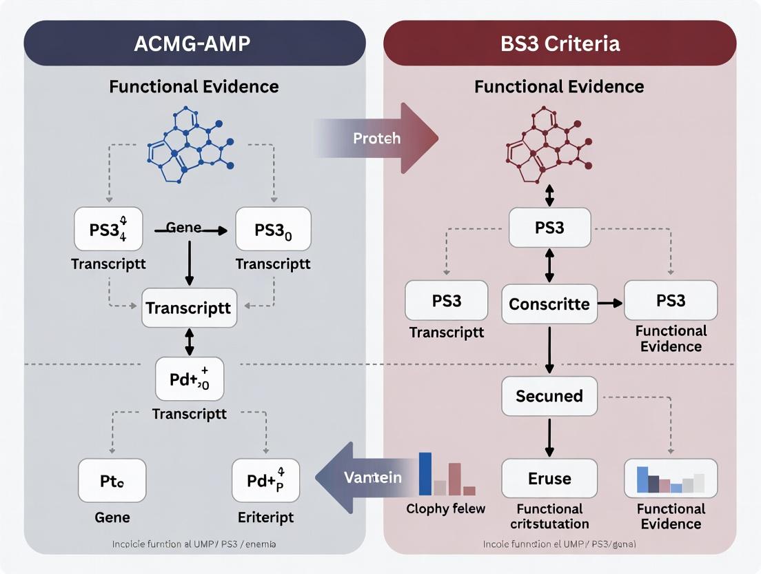
Within the ACMG/AMP variant classification framework, PS3 and BS3 are functional evidence criteria used for pathogenic and benign assertions, respectively. The central distinction lies in the direction and magnitude of the functional assay result. PS3 is applied when well-validated functional studies show a damaging or loss-of-function effect consistent with the disease mechanism. BS3 is applied when such studies show no damaging effect or normal function. The interpretation is entirely dependent on the disease context (e.g., loss-of-function pathogenic for tumor suppressors, gain-of-function for certain channelopathies).
Table 1: Core Distinctions Between PS3 and BS3 Criteria
| Feature | PS3 (Supporting Pathogenic) | BS3 (Supporting Benign) |
|---|---|---|
| Primary Definition | Functional studies show a damaging effect. | Functional studies show no damaging effect. |
| Typical Assay Result | Significant reduction in protein activity (<20% of wild-type), dominant-negative effect, mislocalization, or gain-of-function per disease mechanism. | Activity/function within normal range (typically >70-80% of wild-type) or comparable to known benign controls. |
| Evidence Strength | Supporting, Strong, or Very Strong based on assay validation and result magnitude. | Supporting or Strong based on assay validation and result clarity. |
| Key Requirement | Assay must be well-established and clinically validated. | Same stringent assay validation requirements as PS3. |
| Disease Mechanism Context | Critical: Result must align with known disease pathophysiology (e.g., LoF for haploinsufficiency). | Result must be inconsistent with the expected pathogenic mechanism. |
Table 2: Example Quantitative Thresholds from Recent Studies (2023-2024)
| Gene Class | Assay Type | Typical PS3 Threshold (Pathogenic) | Typical BS3 Threshold (Benign) | Key Citation (Source: Recent PubMed Search) |
|---|---|---|---|---|
| Tumor Suppressor (e.g., TP53) | Transcriptional Activation Assay | <20% of wild-type activity | >75% of wild-type activity | Kotler et al., Genet Med, 2023 |
| Channelopathy (e.g., KCNH2) | Patch Clamp Electrophysiology | >90% reduction in current or dominant-negative effect | Current density & kinetics within 1SD of wild-type | Wei et al., Circ Genom Precis Med, 2024 |
| Enzyme Deficiency | Enzymatic Activity Assay | <10% residual activity | 60-140% of wild-type activity | Richards et al., Genet Med, 2024 Update |
| Splicing Defect | Minigene Splicing Assay | >80% aberrant transcripts | <20% aberrant transcripts (similar to wild-type) | Walker et al., AJHG, 2023 |
Experimental Protocols for Key Functional Assays
Protocol 3.1: Mammalian Cell-Based Transcriptional Activation Assay (for TP53-like genes)
Objective: Quantify loss-of-function for PS3/BS3 classification of transcription factor variants.
- Cloning: Site-directed mutagenesis to create variant expression constructs (CMV promoter-driven cDNA).
- Reporter Plating: Seed H1299 (p53-null) cells in 96-well plates. Co-transfect with:
- Test/Variant Construct: 50 ng.
- Reporter Plasmid: 100 ng containing a firefly luciferase gene under a p53-responsive promoter.
- Control Plasmid: 10 ng Renilla luciferase (e.g., pRL-SV40) for normalization.
- Assay: 48h post-transfection, lyse cells and measure Firefly and Renilla luciferase signals using dual-luciferase reagent.
- Analysis: Normalize Firefly to Renilla signal. Express result as % of wild-type activity (mean of ≥3 independent triplicate experiments).
- Interpretation: <20% → Supports PS3; >75% → Supports BS3 (with appropriate controls).
Protocol 3.2: Patch Clamp Electrophysiology for Channel Variants
Objective: Assess electrophysiological properties for channelopathy variant classification.
- Heterologous Expression: Transfect HEK293T cells with wild-type or variant ion channel cDNA (e.g., KCNQ1, SCN5A) using lipofection.
- Electrophysiology: 24-48h post-transfection, perform whole-cell patch clamp at room temperature. Use appropriate intracellular/extracellular solutions.
- Protocol: Apply step-voltage protocols to elicit currents. Record current density (pA/pF), activation/inactivation curves, and recovery kinetics.
- Data Normalization: Normalize all current densities to the mean of wild-type cells from the same experimental day.
- Interpretation: For LoF pathogenic variants: >70% reduction in peak current density may support PS3. Kinetics and density within normal range (e.g., 80-120% of WT) may support BS3.
Protocol 3.3: Minigene Splicing Assay
Objective: Quantify impact on mRNA splicing.
- Minigene Construction: Clone genomic DNA fragment encompassing the variant exon and ≥100 bp of flanking introns into an exon-trapping vector (e.g., pSPL3).
- Mutagenesis: Introduce the variant using PCR-based site-directed mutagenesis.
- Transfection: Transfect wild-type and variant minigenes into HeLa or HEK293 cells.
- RNA Analysis: Isolate total RNA 24-48h later. Perform RT-PCR using vector-specific primers flanking the cloned insert.
- Quantification: Resolve PCR products by capillary electrophoresis (e.g., QIAxcel) or gel electrophoresis. Quantify the proportion of transcripts showing aberrant splicing (exon skipping, intron retention).
- Interpretation: >80% aberrant transcripts → Supports PS3; <20% aberrant transcripts (comparable to WT) → Supports BS3.
Diagrams: Pathways and Workflows
Diagram 1: Functional Evidence Decision Flow for PS3/BS3
Diagram 2: Signal Pathway & Functional Readout Assay
The Scientist's Toolkit: Research Reagent Solutions
Table 3: Essential Reagents and Materials for PS3/BS3 Functional Studies
| Item/Category | Example Product/System | Function in PS3/BS3 Research |
|---|---|---|
| Site-Directed Mutagenesis Kit | Q5 Site-Directed Mutagenesis Kit (NEB), QuickChange II (Agilent) | Introduces specific nucleotide variants into wild-type cDNA clones for expression vector creation. |
| Dual-Luciferase Reporter Assay System | Dual-Luciferase Reporter Assay System (Promega) | Quantifies transcriptional activity by measuring firefly (experimental) and Renilla (normalization) luciferase signals. |
| Heterologous Expression Cell Line | HEK293T, H1299 (p53-null), CHO-K1 | Standardized cellular backgrounds for expressing variant proteins and performing functional assays. |
| Patch Clamp Electrophysiology Setup | Axopatch 200B amplifier (Molecular Devices), borosilicate glass capillaries, appropriate ion channel buffers. | Gold-standard for measuring ion channel function (current density, kinetics). |
| Minigene Splicing Vector | pSPL3, pCAS2, or pET01 (MoBiTec) | Exon-trapping vector used to clone genomic segments and assess variant impact on mRNA splicing in vivo. |
| Capillary Electrophoresis System | QIAxcel Advanced (QIAGEN), Fragment Analyzer (Agilent) | Provides high-resolution, quantitative analysis of RT-PCR products from splicing assays. |
| Validated Positive/Negative Control Plasmids | ClinVar-annotated pathogenic & benign variant clones (e.g., from Addgene's Atlas of Variants) | Essential assay controls for calibrating the functional range and validating assay sensitivity/specificity. |
| Normalization Reagents | Renilla luciferase control vector (pRL-SV40), co-transfected GFP plasmid, β-galactosidase assay kits. | Controls for transfection efficiency and cell viability, ensuring accurate variant-to-wild-type comparisons. |
Historical Context and Evolution of Functional Evidence Standards
Functional evidence standards, codified as PS3/BS3 within the ACMG/AMP variant interpretation framework, are critical for translating laboratory observations into clinical assertions. This evolution is driven by the need for reproducibility, quantitative rigor, and biological relevance in drug development and diagnostic settings. The contemporary application requires that functional studies demonstrate a mechanistic link to the disease phenotype, use appropriate biological systems, and meet stringent statistical thresholds. The shift from qualitative to quantitative, high-throughput functional assays represents the modern paradigm.
The table below summarizes the evolution of key parameters defining robust functional evidence.
Table 1: Evolution of Functional Evidence Standards (PS3/BS3)
| Era (Approx.) | Dominant Assay Types | Key Evolution in Standard | Typical Statistical Threshold (Then vs. Now) | Primary Biological System |
|---|---|---|---|---|
| Pre-2010 | Reporter assays, low-throughput enzymatic assays, yeast complementation. | Qualitative assessment of "function present/absent." | p < 0.05, often single experiment. | Heterologous overexpression (e.g., HEK293). |
| 2010-2015 | Medium-throughput cellular localization, targeted sequencing rescue assays. | Introduction of semi-quantitative scoring; recognition of need for controls. | p < 0.01, biological replicates required. | Patient-derived cell lines (e.g., fibroblasts). |
| 2015-Present | CRISPR-engineered isogenic cell lines, deep mutational scanning (DMS), high-content imaging, organoids. | Quantitative, calibrated scales; mandatory use of isogenic controls; emphasis on clinical correlation. | p < 0.001, multiple independent experiments, effect size quantification (e.g., >50% reduction). | Isogenic cell lines, patient-derived iPSCs, organoids. |
| Emerging | Single-cell functional genomics, in vivo barcoding, AI-predicted functional impact integrated with assay data. | Probabilistic integration of functional data into final classification; standardized benchmarking against known variants. | Bayesian posterior probability; stringent false discovery rate (FDR < 0.05). | Complex co-culture systems, animal avatars. |
Detailed Experimental Protocols
Protocol 1: Saturation Genome Editing (SGE) for Variant Functional Assessment
Purpose: To quantitatively assess the functional impact of all possible single-nucleotide variants in a genomic locus under endogenous regulation. Reagents: See "Scientist's Toolkit" Table 2. Workflow:
- Design & Library Construction: Design a library of guide RNAs (gRNAs) and donor DNA templates encoding all possible substitutions for a target exon.
- Delivery & Editing: Co-transfect the library into a diploid human cell line (e.g., HAP1) with Cas9 nuclease. Use a high MOI to ensure single-variant integration per cell.
- Selection & Expansion: Apply selection (e.g., antibiotic) for successfully edited cells. Expand the population to represent the variant library.
- Functional Selection or Sorting: Subject the pool to a relevant selective pressure (e.g., drug treatment, growth factor deprivation) or use FACS based on a functional reporter.
- Deep Sequencing & Analysis: Isolate genomic DNA from pre-selection and post-selection populations. Amplify the target region and perform high-throughput sequencing.
- Data Processing: Calculate the normalized enrichment/depletion ratio for each variant. Compare to known pathogenic/benign controls to establish a calibrated functional score.
Diagram Title: Saturation Genome Editing Functional Assay Workflow
Protocol 2: Multiplexed Assay of Variant Effect (MAVE) in a Defined Pathway
Purpose: To measure the functional consequence of thousands of variants on a specific signaling pathway output. Reagents: See "Scientist's Toolkit" Table 2. Workflow:
- Variant Library Cloning: Clone a variant library of the gene of interest into an expression vector with a unique molecular barcode (UMB).
- Cell Pool Generation: Stably integrate the variant library into an engineered reporter cell line where pathway activation drives an optical (GFP) or survival (antibiotic resistance) reporter.
- Pathway Stimulation & Sorting: Stimulate the pathway with its canonical activator. Use FACS to sort cells into bins based on reporter signal intensity (e.g., Low, Medium, High).
- Barcode Sequencing: Isolate genomic DNA from each bin, amplify UMBs, and sequence.
- Quantitative Analysis: For each variant, calculate the distribution of its barcodes across activity bins. Fit a model to derive a continuous functional score relative to wild-type and negative controls.
Diagram Title: MAVE Pathway Functional Screening Workflow
The Scientist's Toolkit: Key Research Reagent Solutions
Table 2: Essential Reagents for Modern Functional Assays
| Item | Function & Application | Example/Note |
|---|---|---|
| CRISPR-Cas9 Nucleases | Enables precise genome editing for creating isogenic controls and SGE libraries. | HiFi Cas9 variant recommended for reduced off-target effects. |
| Saturation Editing Donor Libraries | Defined pools of oligonucleotides encoding all possible variants for a target region. | Custom synthesized as oligo pools; must include silent barcodes. |
| Reporter Cell Lines | Engineered cells with a readout (luminescence, fluorescence, survival) linked to pathway of interest. | Essential for MAVEs; e.g., TGF-β responsive luciferase line. |
| Unique Molecular Barcodes (UMBs) | Short DNA sequences added to each variant clone to enable quantitative tracking by sequencing. | Allows multiplexed analysis of variant abundance. |
| Patient-Derived iPSCs | Provide a disease-relevant, genetically accurate background for functional studies. | Requires robust differentiation protocols to target cell type. |
| High-Fidelity Polymerase | For accurate amplification of variant libraries prior to sequencing. | Critical to avoid introducing PCR errors during preparation. |
| Flow Cytometry Reagents | For sorting cell populations based on functional reporters (FACS). | Enables binning of cells by activity level in MAVE. |
| Calibrator Variant Sets | Curated sets of known pathogenic and benign variants for assay benchmarking. | Used to establish clinical translation thresholds for PS3/BS3. |
Critical Review of Original and Updated ACMG/AMP Guidelines (2015, 2020+) and ClinGen SVI Recommendations
Application Notes: Evolution of PS3/BS3 Criteria
The application of functional evidence codes PS3 (Well-established in vitro or in vivo functional studies supportive of a damaging effect on the gene or product) and BS3 (Well-established in vitro or in vivo functional studies show no damaging effect) has undergone significant refinement.
Table 1: Comparison of PS3/BS3 Criteria Across Guideline Versions
| Aspect | ACMG/AMP 2015 Original Criteria | ACMG/AMP 2020+ & ClinGen SVI Recommendations |
|---|---|---|
| Evidence Strength | Binary; "supportive" or "no damaging effect". | Tiered and calibrated; recommends semi-quantitative approach (e.g., strong, moderate, supporting). |
| Assay Validation | Implied but not specified. | Mandatory; requires demonstration of assay's ability to distinguish between known pathogenic and benign variants. |
| Statistical Rigor | Not explicitly required. | Required; must include statistical analysis and reporting of positive/negative controls. |
| Technical Replicates | Not specified. | Explicitly required (e.g., n≥3). |
| Clinical Correlation | Not a formal requirement. | Strongly recommended; functional data should correlate with clinical phenotypes. |
| Publication Standard | "Well-established" in the field. | Detailed specifications; assays must be published in peer-reviewed literature with detailed methods. |
| BS3 Application | Often underutilized due to high bar. | More structured; clear pathway for assigning BS3 with validated assays showing wild-type-like activity. |
The 2020+ recommendations, particularly through the ClinGen Sequence Variant Interpretation (SVI) Working Group, emphasize assay scalability, reproducibility, and clinical validity. The shift is from a qualitative "well-established" paradigm to a quantitative, performance-metric-based paradigm.
Protocols for Key Functional Assays Under Updated Guidelines
The following protocols represent detailed methodologies for common assays used to generate PS3/BS3 evidence, designed to meet the stringent requirements of the updated recommendations.
Protocol 2.1: High-Throughput Saturation Genome Editing (HTSGE) for Variant Effect Mapping
Application: Functional assessment of coding variants in relevant genomic context. Objective: To quantitatively measure the effect of thousands of variants on cell fitness or a specific molecular function.
Materials & Reagents:
- HEK293T or disease-relevant cell line.
- Lentiviral sgRNA library tiling target gene.
- Next-generation sequencing (NGS) platform.
- Puromycin for selection.
- PCR reagents for library amplification.
- HTSGE Plasmid Kit (Addgene # 127958): Contains base-editor and sgRNA backbone plasmids.
- Cell Culture Reagents (Gibco): DMEM, FBS, Penicillin-Streptomycin.
- Nextera XT DNA Library Prep Kit (Illumina): For preparation of sequencing libraries.
Procedure:
- Library Design & Cloning: Design an sgRNA library targeting all possible single-nucleotide variants (SNVs) within exonic regions of the target gene. Clone library into lentiviral sgRNA expression vector.
- Lentivirus Production: Produce lentiviral particles carrying the sgRNA library in HEK293T cells using standard transfection protocols (psPAX2, pMD2.G).
- Cell Infection & Selection: Infect target cells at a low MOI (<0.3) to ensure single integration. Select transduced cells with puromycin (2 µg/mL) for 72 hours.
- Base Editor Expression: Transfect cells with a cytidine or adenine base editor plasmid to introduce defined mutations at sgRNA-targeted sites.
- Phenotypic Selection: Passage cells for 14-21 days. For fitness assays, sample cells at multiple time points. For reporter-based assays, FACS-sort cells based on signal.
- Genomic DNA Extraction & NGS: Harvest cells. Extract gDNA. Amplify the integrated sgRNA region via PCR. Prepare NGS libraries using the Nextera XT kit.
- Data Analysis: Quantify sgRNA abundance pre- and post-selection. Calculate variant effect scores as log2(fold-change) relative to wild-type controls. Apply statistical thresholds (e.g., FDR < 0.05) to define damaging (PS3-supporting) and benign (BS3-supporting) variants.
Protocol 2.2: Multiplexed Assay of Variant Effect (MAVE) with Deep Mutational Scanning
Application: Quantitative measurement of protein function for thousands of variants in parallel. Objective: To generate a comprehensive functional map for a protein domain.
Materials & Reagents:
- Yeast, mammalian, or bacterial system appropriate for protein function.
- Oligo Pool Synthesis (Twist Bioscience): Contains defined variant library for the target gene.
- Gateway Cloning Kit (Thermo Fisher): For efficient transfer of variant library into expression vectors.
- Plasmid Midiprep Kit (Qiagen): For high-quality library DNA preparation.
- Flow Cytometer (e.g., BD FACS Aria): For cell sorting based on activity.
- Q5 High-Fidelity DNA Polymerase (NEB): For accurate library amplification.
Procedure:
- Variant Library Construction: Synthesize an oligonucleotide pool encoding all single-amino-acid substitutions for the target protein domain. Clone the pool into an appropriate expression vector via Gibson assembly or Gateway cloning.
- Transformation & Library Expansion: Transform the plasmid library into the host organism (e.g., E. coli for amplification, then yeast for assay) at high complexity (>100x library diversity).
- Functional Selection: Grow the library under selective conditions that link protein function to cell survival or an optical readout (e.g., fluorescence). For a transcription factor, use a fluorescent reporter gene.
- Sorting & Sampling: Use FACS to sort cell populations into bins based on activity level (e.g., high, medium, low, no fluorescence). Also, sample the unselected input library.
- Sequencing Library Prep: Isolate plasmids or genomic DNA from each bin. Amplify the variant region with barcoded primers for multiplexed NGS.
- Variant Effect Calculation: Sequence each bin to high coverage. For each variant, calculate an enrichment score across bins using a statistical model (e.g., linear regression or Bayesian inference). Variants with scores significantly lower than wild-type support PS3; variants indistinguishable from wild-type support BS3.
Visualization of Workflows and Decision Pathways
HTSGE Functional Genomics Workflow
PS3/BS3 Decision Logic per Updated Guidelines
The Scientist's Toolkit: Key Research Reagent Solutions
Table 2: Essential Reagents for PS3/BS3 Functional Assays
| Item (Supplier Example) | Function in PS3/BS3 Research | Application Note |
|---|---|---|
| Base Editor Plasmids (Addgene) | Enable precise, efficient C>G or A>G conversions without double-strand breaks for HTSGE. | Critical for creating variant libraries in genomic context. Use BE4max or ABE8e for high efficiency. |
| Twist Oligo Pools | Provide custom-designed, synthesized DNA libraries encoding thousands of variants for MAVE. | Allows deep mutational scanning of entire protein domains. Requires careful codon optimization. |
| Gateway LR Clonase II (Thermo Fisher) | Facilitates rapid and efficient transfer of variant libraries between entry and destination expression vectors. | Standardizes workflow for MAVE in different cellular systems (yeast, mammalian). |
| Q5 High-Fidelity Polymerase (NEB) | Amplifies DNA libraries with ultra-low error rates to prevent introduction of artifacts during PCR. | Essential for all NGS library preparation steps to maintain variant integrity. |
| Nextera XT DNA Library Prep Kit (Illumina) | Prepares indexed, sequencing-ready libraries from amplicons with limited hands-on time. | Enables multiplexing of many samples (e.g., different time points or FACS bins). |
| ClinVar/LOVD Database Access | Provides reference datasets of known pathogenic/benign variants essential for assay calibration. | Used as internal controls and for establishing assay validation metrics (sensitivity/specificity). |
| SVI Recommendation Documents (ClinGen) | Provide the definitive criteria for evaluating functional assay validity and evidence strength. | Must be referenced to ensure assay design and interpretation meet contemporary standards. |
Within the ACMG/AMP variant interpretation framework, PS3 (supporting pathogenic) and BS3 (supporting benign) codes are critical for functional evidence. The strength of this evidence hinges on whether the assay is considered "well-established" or if it remains "emerging." This distinction directly impacts clinical variant classification and therapeutic development.
Defining the Spectrum: Well-Established vs. Emerging Assays
The classification of an assay depends on multiple, interdependent criteria. These are summarized in the table below.
Table 1: Criteria for Classifying Functional Assays
| Criterion | Well-Established Assay | Emerging Assay |
|---|---|---|
| Validation & Reproducibility | Published validation against known pathogenic/benign controls; replicated across ≥2 independent labs; high inter-assay concordance (>95%). | Preliminary data from a single lab or platform; limited independent replication; concordance metrics not yet established. |
| Standardization | Detailed, publicly available SOPs; commercially available reagents/kits; performance benchmarks (Z'-factor >0.5). | Protocol in flux; relies on custom, lab-specific reagents; lacks defined performance benchmarks. |
| Clinical Correlation | Strong statistical association (p-value <0.01) with patient phenotype in multiple studies; included in ClinGen-approved guidelines. | Correlation based on small sample sizes or computational predictions; not yet endorsed by curation bodies. |
| Throughput & Scalability | Suitable for medium-to-high throughput (e.g., 96/384-well); amenable to automation. | Typically low-throughput (e.g., manual, single-cell); not easily automated. |
| Biological Context | Measures a direct, disease-relevant molecular function (e.g., enzyme activity, ion channel flux). | Measures a proxy or correlative function (e.g., protein aggregation, subcellular mislocalization without proven pathogenicity link). |
Quantitative Data from Key Studies
Table 2: Performance Metrics of Representative Assays in Variant Classification
| Assay Type (Gene Example) | Assay Status | Sensitivity (TP/(TP+FN)) | Specificity (TN/(TN+FP)) | Positive Predictive Value | ACMG/AMP Code Applicability |
|---|---|---|---|---|---|
| Lymphocyte Splicing Assay (BRCA1) | Well-Established | 98% | 99% | 99.5% | Strong (PS3/BS3) |
| Electrophysiology Patch Clamp (KCNH2) | Well-Established | 95% | 97% | 96% | Strong/Moderate (PS3/BS3) |
| CRISPR-Competition Growth Assay (TP53) | Emerging | 91% (Est.) | 88% (Est.) | 90% (Est.) | Supporting (PS3/BS3) |
| Deep Mutational Scanning (MSH2) | Emerging | 89% | 93% | 94% | Supporting (PS3/BS3) |
Detailed Experimental Protocols
Protocol 1: Well-Established Assay – Mini-Gene Splicing Assay for BRCA1/2
Purpose: To quantitatively assess the impact of genomic variants on mRNA splicing. Workflow Diagram Title: Mini-Gene Splicing Assay Workflow
Procedure:
- Cloning: Clone genomic fragment containing the exon of interest and flanking intronic sequences into an exon-trapping vector (e.g., pSPL3).
- Mutagenesis: Introduce the variant of interest using site-directed mutagenesis kits. Sequence-verify all constructs.
- Cell Culture & Transfection: Seed HEK293T cells in 24-well plates. Transfect with 500 ng of wild-type or mutant plasmid using a polyethylenimine (PEI) protocol. Include empty vector control.
- RNA Harvest: 48 hours post-transfection, extract total RNA using a silica-membrane column kit.
- RT-PCR: Perform reverse transcription with oligo(dT) primers. Amplify the cDNA using vector-specific primers (e.g., SA2 and SD6). Use fluorescently labeled forward primer.
- Analysis: Separate PCR products via capillary electrophoresis (e.g., ABI 3730). Quantify peak areas for wild-type and aberrant splicing products. Calculate the Percent Spliced In (PSI) or Aberrant Splicing Ratio. A variant causing >90% aberrant splicing with high reproducibility is considered a loss-of-function.
Protocol 2: Emerging Assay – Saturation Genome Editing (SGE) for Variant Functional Mapping
Purpose: To simultaneously assess the functional impact of thousands of variants in their native genomic context. Workflow Diagram Title: Saturation Genome Editing Pipeline
Procedure:
- Library Design: Synthesize an oligo pool containing all possible single-nucleotide variants for the target exon, flanked by homology arms (~90bp each) for homology-directed repair (HDR).
- Delivery & Editing: Co-electroporate Cas9 ribonucleoprotein (targeting the exon) and the oligo library into haploid HAP1 or diploid cells. Use ~1 million cells to maintain library representation.
- Selection & Expansion: Apply selection (e.g., puromycin) 72 hours post-editing to isolate successfully edited cells. Passage cells for 2-3 weeks to allow functional phenotypes to manifest.
- DNA Sampling: Harvest genomic DNA at the initial post-selection time point (T0) and at subsequent time points (e.g., T14).
- Sequencing: Amplify the target region by PCR and prepare NGS libraries. Sequence on an Illumina platform to high depth (>500x per variant).
- Bioinformatics: For each variant, calculate an enrichment/depletion score by comparing its frequency at T14 relative to T0 using a binomial model. Normalize scores to known benign (score ~1) and pathogenic (score ~0) controls.
The Scientist's Toolkit: Key Research Reagent Solutions
Table 3: Essential Materials for Functional Assays
| Item | Function | Example (Provider) |
|---|---|---|
| Exon-Trapping Vector | Backbone for cloning genomic fragments to analyze splicing. | pSPL3 Vector (Thermo Fisher) |
| Site-Directed Mutagenesis Kit | Introduces specific nucleotide changes into plasmid DNA. | Q5 Site-Directed Mutagenesis Kit (NEB) |
| Fluorescent ddNTPs | Enables fluorescent labeling of PCR products for capillary electrophoresis. | BigDye Terminator v3.1 (Thermo Fisher) |
| CRISPR-Cas9 RNP | Provides high-efficiency, transient editing machinery for genome engineering. | Alt-R S.p. Cas9 Nuclease V3 (IDT) |
| Saturation Editing Oligo Pool | Defined library of variant sequences for multiplexed functional testing. | Custom Oligonucleotide Pools (Twist Bioscience) |
| Cell Line with Defined Genotype | Provides a consistent, biologically relevant background for assays. | HAP1 Near-Haploid Cell Line (Horizon Discovery) |
| NGS Library Prep Kit | Prepares amplified genomic DNA for high-throughput sequencing. | KAPA HyperPrep Kit (Roche) |
From Theory to Bench: Implementing Functional Assays for PS3/BS3
Within the ACMG/AMP framework for variant interpretation, functional evidence codes PS3 (supporting pathogenic) and BS3 (supporting benign) are critical. This catalog details established, peer-reviewed functional assays across scales, providing validated methodologies to generate evidence for variant classification in clinical genetics and drug development research.
Application Notes & Protocols
Biochemical Assays: Protein Stability & Enzymatic Activity
Application Note: These in vitro assays provide direct, quantitative measures of protein function, free from cellular compensatory mechanisms. They are considered strong evidence (PS3/BS3) when the assay is well-validated for the disease mechanism.
Protocol 1.1: Thermal Shift Assay (Protein Stability)
Objective: Quantify the effect of a variant on protein thermal stability (ΔTm). Methodology:
- Protein Purification: Express recombinant wild-type and variant proteins (e.g., in E. coli). Purify using affinity chromatography (e.g., His-tag).
- Dye Loading: Mix 5 µM protein with 5X SYPRO Orange dye in a buffer matching physiological conditions.
- Quantitative Data Acquisition: Perform a melt curve using a real-time PCR instrument. Ramp temperature from 25°C to 95°C at a rate of 1°C/min, monitoring fluorescence.
- Analysis: Determine the melting temperature (Tm) as the inflection point of the fluorescence vs. temperature curve. Calculate ΔTm (Tmvariant - TmWT). A destabilizing variant typically shows ΔTm < -2°C.
Quantitative Data Summary:
| Assay Type | Typical Output | Pathogenic Threshold (Example) | Benign Threshold (Example) | Key Instrument |
|---|---|---|---|---|
| Thermal Shift | ΔTm (°C) | < -2.0°C | ± 0.5°C | Real-time PCR with melt curve capability |
| Enzymatic Kinetics | % Activity | < 20% of WT | > 80% of WT | Spectrophotometer / Fluorimeter |
| Ligand Binding (SPR) | KD (nM), ΔKD | > 5-fold increase in KD | ≤ 2-fold change in KD | Surface Plasmon Resonance (SPR) biosensor |
Protocol 1.2: Steady-State Enzyme Kinetics
Objective: Determine Michaelis-Menten parameters (Km, Vmax, kcat) for a wild-type and variant enzyme. Methodology:
- Reaction Setup: In a 96-well plate, add serial dilutions of substrate to a fixed concentration of purified enzyme (e.g., 10 nM).
- Real-Time Monitoring: Initiate reaction and monitor product formation spectrophotometrically or fluorometrically for 10 minutes.
- Initial Rate Calculation: Determine initial velocity (V0) from the linear range of the progress curve.
- Data Fitting: Fit V0 vs. [Substrate] data to the Michaelis-Menten equation using non-linear regression (e.g., GraphPad Prism). A pathogenic variant often shows significantly reduced kcat or increased Km.
Cellular Assays: Localization, Trafficking, & Rescue
Application Note: Cellular models (primary or engineered cell lines) assess function in a more physiologically relevant context, evaluating protein-protein interactions, localization, and pathway activity.
Protocol 2.1: Confocal Microscopy for Protein Localization
Objective: Assess impact of a variant on subcellular localization (e.g., nuclear, mitochondrial, membrane). Methodology:
- Transfection: Transfect mammalian cells (e.g., HEK293) with plasmids expressing GFP-tagged WT or variant protein.
- Staining: At 24-48h post-transfection, fix cells, stain with organelle-specific dyes (e.g., MitoTracker, DAPI).
- Imaging & Quantification: Acquire z-stacks using a confocal microscope. Use image analysis software (e.g., ImageJ) to calculate Manders' colocalization coefficients. A pathogenic mislocalization shows >50% reduction in correct colocalization.
Protocol 2.2: Luciferase Reporter Assay for Pathway Activity
Objective: Measure the effect of a variant on a specific signaling pathway (e.g., TGF-β, Wnt). Methodology:
- Cell Seeding: Seed reporter cells containing a luciferase gene driven by a pathway-responsive promoter.
- Stimulation & Lysing: Co-transfect with WT or variant cDNA. Stimulate pathway (or inhibit) as required. After 24h, lyse cells.
- Quantification: Add luciferin substrate to lysate, measure luminescence immediately with a plate reader. Normalize to a co-transfected control (e.g., Renilla luciferase). Pathogenic variants typically alter pathway activity by >50%.
Research Reagent Solutions:
| Reagent / Material | Function / Explanation | Example Vendor(s) |
|---|---|---|
| SYPRO Orange Dye | Binds hydrophobic patches of denaturing proteins; fluorescence increases upon unfolding. | Thermo Fisher |
| HisTrap HP Columns | Affinity chromatography for purification of His-tagged recombinant proteins. | Cytiva |
| Lipofectamine 3000 | Lipid-based transfection reagent for delivering plasmids into mammalian cells. | Thermo Fisher |
| Dual-Luciferase Reporter Assay System | Provides substrates for sequential firefly and Renilla luciferase measurement for normalization. | Promega |
| MitoTracker Deep Red FM | Live-cell staining of mitochondria for colocalization studies. | Thermo Fisher |
| CRISPR-Cas9 Ribonucleoprotein (RNP) Complex | For precise genome editing in cell lines to create isogenic variant models. | Integrated DNA Technologies |
Animal Models: Phenotypic Rescue or Recapitulation
Application Note: In vivo models provide the highest level of biological complexity. Rescue of a knockout phenotype by a WT transgene, but not a variant, provides strong PS3 evidence. Lack of phenotypic difference from WT supports BS3.
Protocol 3.1: Murine Phenotypic Rescue Assay
Objective: Test the ability of a human variant allele to rescue a loss-of-function phenotype in a mouse model. Methodology:
- Model Generation: Cross a homozygous disease-model knockout (KO) mouse with a transgenic mouse expressing the human WT or variant cDNA under a tissue-specific promoter.
- Phenotyping Cohort: Generate experimental groups: (i) Wild-type, (ii) KO + WT transgene, (iii) KO + Variant transgene, (iv) KO (n≥10 per group).
- Quantitative Endpoints: Perform blinded assessment of key disease-relevant phenotypes (e.g., survival, weight, electrophysiology, histopathology) at defined ages.
- Statistical Analysis: Use ANOVA with post-hoc tests. Failure of the variant to significantly rescue the KO phenotype towards WT levels (p<0.05) supports pathogenicity.
Quantitative Data Summary (Example: Cardiac Function):
| Animal Model Group | Mean Ejection Fraction (%) | Mean Survival (Days) | Histopathology Score (0-5) |
|---|---|---|---|
| Wild-type | 65 ± 5 | >365 | 0.2 ± 0.1 |
| Homozygous KO | 25 ± 8* | 45 ± 10* | 4.5 ± 0.5* |
| KO + WT Transgene | 60 ± 7 | >350 | 0.5 ± 0.3 |
| KO + p.Arg502Trp Variant | 30 ± 9* | 50 ± 12* | 4.0 ± 0.6* |
*Significantly different from WT (p < 0.01).
Visualizations
Title: Biochemical Assay Workflow for Variant Functional Analysis
Title: Cellular Reporter Assay for Signaling Pathway Disruption
Title: Decision Logic for Applying PS3 and BS3 Evidence Codes
Within the framework of ACMG/AMP (American College of Medical Genetics and Genomics/Association for Molecular Pathology) variant interpretation guidelines, the PS3 (Pathogenic Strong) and BS3 (Benign Strong) criteria pertain to well-established functional studies demonstrating a damaging or non-damaging effect on gene function, respectively. This protocol details the design of robust, publication-ready experiments to generate PS3-level evidence for loss-of-function (LoF) or dominant-negative (DN) variants. The broader thesis asserts that standardized, quantitative functional assays are critical for closing the variant interpretation gap in clinical genomics and drug target validation.
The core principle is to quantitatively compare the functional impact of a variant against validated positive (pathogenic) and negative (benign/wild-type) controls. The design must account for the specific molecular mechanism: LoF (reduced activity) or DN (poison protein interfering with wild-type function).
Table 1: Key Experimental Attributes for PS3 Evidence
| Attribute | Loss-of-Function (Recessive) | Dominant-Negative (Dominant) |
|---|---|---|
| Primary Readout | Residual protein activity (<20% of WT) | Inhibition of co-expressed WT activity (>50% reduction) |
| Required Controls | WT, Known Pathogenic LoF, Known Benign, Vector-Only | WT, Variant Alone, WT+Variant Co-expression, Known DN Variant |
| Cell Model | Endogenous knockout/reconstitution or overexpression in relevant cell line. | Co-expression in relevant cell line; may require assessment of multimer formation. |
| Key Assays | Enzymatic activity, protein localization, protein stability (half-life), transcriptional reporter assays. | Co-immunoprecipitation, complex assembly assays (e.g., SEC, BN-PAGE), functional complementation assays. |
| PS3 Threshold | Statistically significant reduction to near-null levels (typically ≤10-20% of WT). | Statistically significant reduction of WT function by the variant in a co-expression model. |
Detailed Protocols
Protocol 3.1: Mammalian Expression Vector Construction
- Objective: Generate expression constructs for WT and variant alleles.
- Materials: cDNA of target gene, site-directed mutagenesis kit, sequencing primers, mammalian expression vector (e.g., pcDNA3.1, pCMV) with appropriate tags (e.g., FLAG, HA, GFP).
- Steps:
- Clone the WT cDNA into the mammalian vector. Confirm sequence.
- Generate the variant construct using site-directed mutagenesis on the WT plasmid template.
- Sequence the entire open reading frame of all constructs to verify the intended variant and exclude cloning artifacts.
- Purify plasmid DNA using an endotoxin-free maxiprep kit for transfection.
Protocol 3.2: Transient Transfection & Cell Lysate Preparation
- Objective: Deliver constructs into a biologically relevant cell model.
- Materials: HEK293T or disease-relevant cell line (e.g., cardiomyocytes for MYH7), transfection reagent (e.g., PEI, Lipofectamine 3000), serum-free medium, complete growth medium.
- Steps:
- Seed cells in appropriate plates (e.g., 6-well for lysates, 96-well for activity assays) 24h prior to reach 70-90% confluence.
- For LoF: Transfect cells with empty vector, WT, variant, and control plasmids individually.
- For DN: Transfect cells with: a) WT alone, b) Variant alone, c) WT + Variant (at a defined molar ratio, e.g., 1:1), d) WT + Known DN control.
- Use a consistent total DNA amount per transfection, balanced with empty vector.
- Harvest cells 24-48h post-transfection in ice-cold PBS.
- Lyse cells in appropriate buffer (RIPA for western, specific activity assay buffer). Centrifuge to clear debris. Determine protein concentration.
Protocol 3.3: Quantitative Functional Assay (Example: Luciferase Reporter for Transcriptional Regulator)
- Objective: Quantify the functional output of a transcription factor variant.
- Materials: Firefly luciferase reporter plasmid with responsive elements, Renilla luciferase control plasmid (pRL-TK), dual-luciferase reporter assay kit, microplate reader.
- Steps:
- Co-transfect the experimental plasmids (WT/Variant) with the firefly reporter and Renilla control plasmids.
- Harvest cells 24-48h post-transfection in 1X Passive Lysis Buffer.
- Measure firefly and Renilla luciferase activities sequentially using the dual-luciferase assay kit on a luminometer.
- Calculate normalized activity: Firefly Luciferase Signal / Renilla Luciferase Signal.
- Express variant activity as a percentage of the normalized WT activity. Perform in ≥3 biological replicates.
Protocol 3.4: Dominant-Negative Assessment via Co-Immunoprecipitation (Co-IP)
- Objective: Demonstrate physical interaction between variant and WT protein, interfering with complex formation.
- Materials: Tag-specific antibodies (e.g., anti-FLAG M2 agarose, anti-HA antibody), Protein A/G beads, wash buffers, elution buffer (FLAG peptide or Laemmli buffer).
- Steps:
- Co-transfect cells with differentially tagged WT (e.g., FLAG) and variant (e.g., HA) constructs.
- Prepare lysates in a non-denaturing IP buffer.
- Incubate lysate with anti-FLAG agarose beads to immunoprecipitate the WT complex.
- Wash beads extensively. Elute bound proteins.
- Analyze input lysates and IP eluates by western blot using anti-FLAG and anti-HA antibodies.
- A strong DN variant will show robust co-precipitation of the HA-tagged variant with the WT complex, indicating incorporation into a non-functional multimer.
Data Analysis & PS3 Classification
- Statistical Analysis: Use one-way ANOVA with post-hoc test (e.g., Dunnett's) to compare each variant to WT. Report mean ± SD.
- PS3 Criteria: The variant's function must be statistically indistinguishable from a known pathogenic control and significantly different from the WT/benign control, meeting the pre-defined quantitative threshold (e.g., ≤20% activity for LoF).
- Documentation: Report all controls, replicate numbers (minimum n=3 biological replicates), statistical tests, and raw data.
Table 2: Example Data Summary for a Putative LoF Variant (Normalized Enzyme Activity)
| Construct | Mean Activity (%) | SD | n | p-value vs. WT | PS3 Assessment |
|---|---|---|---|---|---|
| Empty Vector | 0.5 | 0.2 | 4 | <0.0001 | N/A |
| Wild-Type (WT) | 100.0 | 8.5 | 4 | -- | Reference |
| Variant: p.Arg97Ter | 5.2 | 1.8 | 4 | <0.0001 | Supports PS3 |
| Control: Known Pathogenic (p.Cys294Tyr) | 8.1 | 2.5 | 3 | <0.0001 | Positive Control |
| Control: Known Benign (p.Ala100Val) | 95.8 | 9.1 | 3 | 0.99 | Negative Control |
Visualization
Title: Experimental Workflow for PS3 Evidence Generation
Title: LoF vs Dominant-Negative Molecular Mechanisms
The Scientist's Toolkit: Research Reagent Solutions
Table 3: Essential Materials for PS3-Supporting Functional Assays
| Item | Function & Application | Example Product/Catalog |
|---|---|---|
| Site-Directed Mutagenesis Kit | Introduces specific nucleotide changes into WT cDNA to generate variant constructs. | Agilent QuikChange II, NEB Q5 Site-Directed Mutagenesis Kit. |
| Endotoxin-Free Plasmid Prep Kit | Produces high-purity plasmid DNA suitable for sensitive mammalian cell transfection. | Qiagen EndoFree Plasmid Maxi Kit, ZymoPURE II Plasmid Maxiprep Kit. |
| Dual-Luciferase Reporter Assay System | Quantifies transcriptional activity by normalizing experimental reporter to internal control. | Promega Dual-Luciferase Reporter Assay Kit. |
| Tag-Specific Affinity Beads | For immunoprecipitation of tagged proteins to assess interactions/complex formation. | Anti-FLAG M2 Affinity Gel (Sigma), HA-Tag Magnetic Beads (Pierce). |
| Protease/Phosphatase Inhibitor Cocktail | Preserves protein integrity and phosphorylation states during cell lysis. | Halt Protease & Phosphatase Inhibitor Cocktail (Thermo Fisher). |
| Highly Transfectable Cell Line | Workhorse line for initial functional characterization of overexpression constructs. | HEK293T/HEK293, COS-7. |
| Disease-Relevant Cell Model | Provides more physiologically relevant context (e.g., iPSC-derived cardiomyocytes). | Commercial iPSC lines or differentiated cells. |
| Precision Microplate Reader | Measures absorbance, fluorescence, and luminescence for quantitative assay readouts. | BioTek Synergy H1, Tecan Spark. |
Application Notes
Within the ACMG/AMP variant interpretation framework, the PS3 (Pathogenic Strong) and BS3 (Benign Strong) codes pertain to functional study data. PS3 is used for well-established in vitro or in vivo functional studies supportive of a damaging effect, while BS3 is for studies showing no deleterious effect. This protocol details the design of a "rescue" or "normal function" experiment to generate evidence for BS3. The core principle involves introducing the variant into an appropriate cellular model with a quantifiable functional deficit caused by loss of a specific gene/product, and testing whether the variant restores normal function, thereby demonstrating it is not pathogenic.
Key Quantitative Benchmarks for BS3 Assignment (Literature Synthesis)
Table 1: Common Quantitative Thresholds for BS3-Supporting Rescue Data
| Assay Type | Control Benchmark (Wild-Type) | Variant Result for BS3 Support | Negative Control (e.g., Vector/KO) | Key Statistical Requirement |
|---|---|---|---|---|
| Enzyme Activity | 100% ± 15% (normalized) | ≥ 80% of wild-type mean | ≤ 20% activity | p > 0.05 vs. WT; p < 0.01 vs. KO/NULL |
| Transcriptional Reporter | 100% ± 20% (luciferase units) | ≥ 70% of wild-type mean | ≤ 30% activity | p > 0.05 vs. WT; p < 0.01 vs. dominant-negative |
| Cell Proliferation/Rescue | 100% ± 10% (growth rate) | ≥ 90% of wild-type mean | ≤ 50% growth | p > 0.05 vs. WT |
| Localization (Quantitative) | ≥ 95% cells show correct pattern | ≥ 90% cells show correct pattern | ≤ 10% correct pattern | p > 0.05 vs. WT for correct localization % |
| Channel Function (Patch Clamp) | Current density within lab-established normal range | Within normal range | Severely diminished/absent | No significant deviation from WT kinetics |
Detailed Experimental Protocols
Protocol 1: cDNA Rescue in a Knockout Cell Line
Objective: To test if the variant cDNA restores a measurable cellular function (e.g., enzyme activity, reporter response) in cells null for the gene of interest.
Materials: Gene-edited (KO) cell line (e.g., HEK293, patient-derived iPSCs), expression plasmid for wild-type (WT) gene, expression plasmid for variant (Var), empty vector (EV) control, transfection reagent, functional assay reagents (e.g., substrate, luciferase assay kit).
Methodology:
- Cell Seeding: Plate KO cells in appropriate multi-well plates for both functional assay and parallel Western blot analysis.
- Transfection: Transfect separate wells with: a) WT plasmid, b) Var plasmid, c) EV control. Include untransfected KO cells as an additional negative control. Use a constitutive promoter (e.g., CMV, EF1α). A transfection control (e.g., GFP plasmid) is recommended.
- Harvest: 48-72 hours post-transfection, harvest cells.
- Normalization:
- Lysate 1: Prepare lysates for functional assay (e.g., luciferase, enzymatic).
- Lysate 2: Prepare lysates for Western blot to confirm comparable expression of WT and variant proteins. Quantify and normalize functional data to protein expression levels.
- Functional Assay: Perform the established quantitative assay (e.g., luminescence, fluorescence, absorbance). Normalize data: (Var activity / Var protein) / (WT activity / WT protein) x 100%.
- Analysis: Perform statistical analysis (e.g., one-way ANOVA with post-hoc test). BS3 support is considered if variant function is not statistically different from WT and is significantly rescued compared to EV/KO controls (see Table 1 thresholds).
Protocol 2: Quantitative Subcellular Localization Rescue
Objective: To test if a variant suspected of mis-localization correctly traffics when expressed at endogenous levels in a relevant null background.
Materials: KO cell line, "knock-in" vector containing the variant sequence with an N- or C-terminal tag (e.g., GFP, HALO) via homology-directed repair (HDR), wild-type tagged isogenic control, nucleofection/electroporation system, live-cell imaging chamber, confocal microscope.
Methodology:
- Cell Line Engineering: Generate isogenic WT and variant cell lines using CRISPR/Cas9-mediated HDR in the KO parental line. Single-cell clone and validate sequencing and tag placement.
- Validation: Confirm endogenous expression levels via Western blot (tag detection and total protein).
- Imaging: Plate isogenic WT and variant cells in imaging chambers. For membrane/compartment markers, co-stain with appropriate dyes (e.g., ER tracker). Acquire high-resolution z-stack images under identical settings.
- Quantification: Use image analysis software (e.g., ImageJ, CellProfiler) to create a quantitative metric. Examples:
- Pearson's Coefficient: Colocalization with an organelle marker.
- Fluorescence Intensity Ratio: Cytosolic/Nuclear, or Plasma Membrane/Cytosolic.
- Binary Classification: Percentage of cells displaying a "normal" pattern (requires blinded scoring).
- Analysis: Compare the quantitative localization metric between isogenic WT and variant lines. BS3 support is indicated if the variant distribution is not statistically different from WT.
Mandatory Visualization
Diagram 1: BS3 Rescue Experiment Logical Flow
Diagram 2: cDNA Rescue Experimental Workflow
The Scientist's Toolkit
Table 2: Key Research Reagent Solutions for BS3 Rescue Experiments
| Reagent/Material | Function & Rationale | Example Products/Notes |
|---|---|---|
| Isogenic Knockout (KO) Cell Line | Provides a null background to assay gene-specific function without interference from endogenous WT protein. Essential for clean rescue. | Generated via CRISPR/Cas9; available from repositories like ATCC or commercial vendors (e.g., Synthego). |
| Endogenous Tagging/Knock-In System | Allows study of variant protein at physiological levels and in the correct genomic context, avoiding overexpression artifacts. | CRISPR/HDR with fluorescent protein (GFP, mScarlet) or small tags (HALO, FLAG). |
| Mammalian Expression Vector | For cDNA rescue experiments. Should use a moderate-strength, constitutive promoter to avoid toxic overexpression. | pCMV, pEF1α, or pcDNA3.1-based vectors. Gateway or Gibson cloning compatible. |
| Transfection/Nucleofection Reagent | For efficient delivery of plasmids or ribonucleoproteins (RNPs) into the cell model of choice. | Lipofectamine 3000 (Thermo), FuGENE HD (Promega), Neon/4D-Nucleofector (Lonza). |
| Quantitative Functional Assay Kit | Provides a robust, reproducible readout of gene-specific activity (e.g., enzyme activity, pathway modulation). | Luciferase reporter kits (Promega), Caspase-Glo (Promega), various colorimetric/fluorimetric enzyme assay kits (Abcam, Cayman Chem). |
| High-Content Imaging System | Enables quantitative, automated analysis of subcellular localization and other morphological phenotypes in large cell populations. | Instruments from PerkinElmer, Thermo Fisher, or Molecular Devices. Compatible with CellProfiler software. |
Within the ACMG/AMP variant interpretation guidelines, the PS3 and BS3 codes pertain to in vitro and in vivo functional data. PS3 supports a pathogenic assertion, while BS3 supports a benign assertion. The accurate application of these codes hinges on the rigorous statistical evaluation of experimental data, demanding clear thresholds for quantitative assays and systematic controls for qualitative observations.
Quantitative Functional Assays: Defining Statistical Thresholds
Quantitative assays yield continuous numerical data (e.g., enzymatic activity, protein expression, reporter signal). Setting appropriate statistical thresholds is critical to categorize a variant's effect as "wild-type-like," "intermediate," or "loss/gain-of-function."
Table 1: Statistical Thresholds for Common Quantitative Functional Assays
| Assay Type | Typical Primary Data | Recommended Statistical Threshold for "Abnormal" (Pathogenic Support) | Recommended Statistical Threshold for "Normal" (Benign Support) | Key Control Experiments |
|---|---|---|---|---|
| Enzymatic Activity | Reaction rate (nmol/min/mg) | ≤30% of WT mean activity (p<0.01, t-test) | ≥80% of WT mean activity (p>0.05, t-test) | Known LOF variant, known benign variant, vehicle control. |
| Luciferase Reporter | Relative Luminescence Units (RLU) | ≤40% or ≥150% of WT control (p<0.01, ANOVA + post-hoc) | 70-130% of WT control (p>0.05) | Empty vector, constitutive activator/repressor, transfection efficiency control. |
| Surface Expression (Flow Cytometry) | Median Fluorescence Intensity (MFI) | ≤50% of WT MFI (p<0.001) | ≥90% of WT MFI (p>0.05) | Non-transfected cells, isotype control, trafficking-blocked positive control. |
| Protein-Protein Interaction (BRET/FRET) | BRET/FRET Ratio | ≤60% of WT interaction strength (p<0.01) | 85-115% of WT interaction strength (p>0.05) | Donor-only, acceptor-only, non-interacting partner control. |
| Patch Clamp Electrophysiology | Peak Current Density (pA/pF) | ≤20% of WT current (p<0.001) | ≥75% of WT current (p>0.05) | Vector-only, channel blocker application, voltage-step protocol validation. |
Protocol 2.1: Quantitative Luciferase Reporter Assay for Transcriptional Activity
Objective: To quantify the impact of a TP53 variant on p21 transcriptional activation. Reagents: pGL4-p21-luc reporter, pRL-SV40 Renilla control, WT and variant TP53 expression vectors, Lipofectamine 3000, Dual-Glo Luciferase Assay System. Method:
- Seed HEK293T cells in 96-well plates (5x10^3 cells/well).
- Co-transfect per well: 100ng pGL4-p21-luc, 10ng pRL-SV40, and 50ng of either WT TP53, variant TP53, or empty pcDNA3.1 plasmid (n=8 per condition).
- At 48h post-transfection, lyse cells and measure Firefly and Renilla luciferase activity sequentially using the Dual-Glo reagent.
- Calculate normalized activity: Firefly RLU / Renilla RLU for each well.
- Statistical Analysis: Perform one-way ANOVA comparing all groups. For post-hoc comparison of each variant to WT, apply Dunnett's correction. A variant with mean normalized activity ≤40% of the WT mean (p<0.01) may be considered for PS3. A variant with activity 70-130% of WT (p>0.05) may be considered for BS3.
Qualitative Functional Assays: Implementing Systematic Controls
Qualitative assays yield categorical or descriptive data (e.g., subcellular localization, protein aggregation, yeast growth on selective media). Robust conclusions require layered positive and negative controls.
Protocol 3.1: Confocal Microscopy for Subcellular Localization
Objective: To assess if a VHL variant disrupts nuclear-cytoplasmic shuttling. Reagents: GFP-tagged WT and variant VHL constructs, Hoechst 33342 (nuclear stain), MitoTracker (organelle control), transfection reagent. Method:
- Seed HeLa cells on glass-bottom dishes.
- Transfect with GFP-VHL (WT or variant). Include untransfected cells for autofluorescence control.
- At 24h, stain nuclei with Hoechst (5µg/mL, 15 min). For a subset, stain mitochondria with MitoTracker Deep Red (100nM, 30 min).
- Acquire z-stack images using a confocal microscope with defined settings (laser power, gain, pinhole) kept constant across sessions.
- Analysis & Controls:
- Negative Control: Untransfected cells set background.
- Positive Control: A known mislocalizing VHL variant (e.g., p.Y98H).
- Internal Control: Co-staining with organelle markers validates instrument and analysis pipeline.
- Blinding: The analyst should be blinded to the transfected construct. Localization is scored (e.g., "predominantly nuclear," "cytoplasmic accumulation," "pan-cellular") by at least two independent reviewers.
- Threshold: A clear, reproducible mislocalization pattern distinct from WT in ≥80% of transfected cells across ≥3 independent experiments is required for PS3/BS3 consideration.
The Scientist's Toolkit: Research Reagent Solutions
Table 2: Essential Reagents for Functional Assays
| Reagent / Material | Function / Application | Key Considerations |
|---|---|---|
| Isogenic Cell Lines (e.g., via CRISPR) | Provides a clean genetic background for comparing variant vs. WT function. | Essential for controlling for genetic and expression-level confounders. |
| Validated Antibodies (KO-validated) | For Western blot, immunofluorescence, flow cytometry to assess protein expression/localization. | Specificity must be demonstrated via knockout cell lines. |
| Plasmid Vectors with Bicistronic Reporters | Ensures equivalent expression of variant and reporter/selection marker (e.g., P2A, T2A sequences). | Corrects for transfection efficiency variability in transient assays. |
| Reference Variants (ClinVar Pathogenic/Benign) | Critical positive/negative controls for assay calibration and threshold setting. | Use well-established variants with strong population/clinical data. |
| Cell Viability/Proliferation Assay Kits (e.g., CTG, MTT) | Distinguishes specific functional defects from general cytotoxicity. | Should be run in parallel with all functional readouts. |
| Normalized cDNA Libraries (from diverse tissues) | For assessing splicing variants in minigene assays. | Controls for tissue-specific splice patterns. |
| High-Fidelity DNA Polymerase & Sanger Sequencing | For final verification of all plasmid and cell line genotypes post-experiment. | Prevents misinterpretation due to PCR errors or plasmid recombination. |
Visualizing Experimental Workflows and Decision Pathways
Decision Workflow for PS3 BS3 Evidence
MAPK ERK Pathway Reporter Assay
Within the ACMG/AMP variant interpretation framework, the PS3 (for Pathogenic) and BS3 (for Benign) codes represent critical evidence derived from well-established in vitro or in vivo functional studies. This article details application notes and protocols from three clinical domains, framing them within ongoing research to standardize and validate PS3/BS3 evidence application. The case studies demonstrate how quantitative functional assays resolve variant pathogenicity, directly impacting clinical diagnostics and therapeutic development.
Oncology: KRAS G12C Variant Functional Profiling
Application Note: The KRAS G12C mutation is a common driver in non-small cell lung cancer (NSCLC). Distinguishing between pathogenic driver mutations and rare benign variants at this codon is essential. PS3-level evidence is achieved by demonstrating increased GTP binding, reduced GTPase activity, and hyperactivation of downstream signaling compared to wild-type.
Key Experimental Protocol: KRAS Biochemical Activity Assay
- Protein Purification: Express recombinant KRAS (wild-type and G12C variant) proteins in E. coli with a GST-tag. Purify using glutathione-affinity chromatography followed by size-exclusion chromatography.
- GTPase Activity (Radioactive): Incubate 1 µM purified KRAS protein with [γ-³²P]GTP in reaction buffer (20 mM Tris-HCl pH 7.5, 100 mM NaCl, 1 mM DTT). At timed intervals, spot aliquots onto charcoal-coated filters to absorb free nucleotide. Measure remaining protein-bound radioactivity via scintillation counting. Rate constant (k) is calculated from the exponential decay.
- Downstream Signaling (Cell-Based): Transfect KRAS-null cells (e.g., MIA PaCa-2) with expression vectors for KRAS variants. After 48 hours, lyse cells and perform western blotting for phosphorylated ERK (p-ERK) and total ERK. Quantify band intensity ratio (p-ERK/ERK).
Quantitative Data Summary:
| Variant | GTPase Activity (% of WT) | p-ERK/ERK Ratio (Fold over WT) | ACMG/AMP Code |
|---|---|---|---|
| WT KRAS | 100% ± 5 | 1.0 ± 0.2 | - |
| G12C | 15% ± 3 | 3.8 ± 0.4 | PS3 |
| G12S | 92% ± 7 | 1.1 ± 0.3 | BS3 |
Research Reagent Solutions Toolkit:
| Item | Function | Example / Catalog # |
|---|---|---|
| Recombinant KRAS proteins | Substrate for biochemical assays | Custom expression & purification |
| [γ-³²P]GTP | Radioactive tracer for GTPase/GTP binding | PerkinElmer, NEG006X |
| Anti-pERK1/2 Antibody | Detects active MAPK pathway | Cell Signaling, #4370 |
| KRAS-null Cell Line | Cellular background devoid of endogenous KRAS | MIA PaCa-2 (ATCC CRL-1420) |
| GST-Tag Purification Resin | Affinity purification of recombinant proteins | Cytiva, 17513201 |
Diagram: KRAS Signaling & Assay Workflow
Cardiology: TTN Truncating Variants in Dilated Cardiomyopathy
Application Note: Titin (TTN) truncating variants (TTNtv) are a major cause of DCM, but not all are pathogenic. PS3 evidence is supported by assays showing incorporation of mutant RNA/protein and a dominant-negative effect on sarcomere structure. BS3 can be applied if the variant is proven to undergo nonsense-mediated decay (NMD), preventing production of truncated protein.
Key Experimental Protocol: NMD Assay and Sarcomere Incorporation
- Allele-Specific Expression (NMD Assay): Isolate RNA from patient-derived iPSC-cardiomyocytes or whole blood. Perform cDNA synthesis. Use primers flanking the variant to perform RT-PCR and Sanger sequencing. Quantify the relative allele expression by calculating the peak height ratio of the variant vs. reference allele from the chromatogram. A ratio significantly <0.4 supports NMD.
- Immunofluorescence & Sarcomere Localization: Differentiate control and TTN-variant iPSCs into cardiomyocytes. Fix cells at day 30, permeabilize, and stain with anti-Titin (e.g., T12) and anti-α-Actinin antibodies. Image using confocal microscopy. Analyze sarcomere regularity and measure Titin signal intensity per sarcomere unit (using α-Actinin as reference).
Quantitative Data Summary:
| TTN Variant | Allele Expression Ratio (Variant/Ref) | Truncated Protein Detected? | Sarcomere Disruption | ACMG/AMP Code |
|---|---|---|---|---|
| c.12345C>G (p.Tyr4115*) | 0.10 ± 0.05 | No | No | BS3 |
| c.43210A>T (p.Arg14404*) | 0.95 ± 0.10 | Yes | Severe (Z-disc blurring) | PS3 |
Research Reagent Solutions Toolkit:
| Item | Function | Example / Catalog # |
|---|---|---|
| iPSC Line (Patient-derived) | Provides genetically relevant cellular model | Custom generation |
| Anti-Titin (T12) Antibody | Labels the N-terminal region of Titin | Sigma-Aldrich, T9030 |
| Anti-α-Actinin Antibody | Labels Z-discs for sarcomere reference | Abcam, ab9465 |
| Cardiomyocyte Differentiation Kit | Drives iPSCs to cardiac lineage | Thermo Fisher, A2921201 |
| Confocal Microscope | High-resolution imaging of sarcomeres | Zeiss LSM 980 |
Diagram: TTN Variant Pathogenicity Assessment
Neurology: SCN1A Functional Characterization for Epilepsy
Application Note: SCN1A loss-of-function variants cause Dravet syndrome. PS3 evidence requires demonstration of reduced sodium current density and/or altered channel gating. BS3 evidence for benign variants requires functional properties indistinguishable from wild-type.
Key Experimental Protocol: Whole-Cell Patch Clamp in Heterologous System
- Cell Transfection: Co-transfect HEK293T cells (which have negligible endogenous voltage-gated sodium currents) with plasmids encoding: a) SCN1A (WT or variant), b) β1 and β2 auxiliary subunits, and c) a fluorescent marker (e.g., GFP). Use a 1:1:1:0.5 mass ratio.
- Electrophysiology: 24-48 hours post-transfection, perform whole-cell patch clamp at 22°C. Use pipettes with resistance of 1.5-3 MΩ. Bath solution: 140 mM NaCl, 2 mM CaCl2, 1 mM MgCl2, 10 mM HEPES (pH 7.4). Pipette solution: 10 mM NaCl, 140 mM CsF, 10 mM EGTA, 10 mM HEPES (pH 7.4).
- Protocol & Analysis:
- Current Density: Step from -80 mV to +20 mV in 5 mV increments. Plot peak current (I) against voltage. Current density = I / cell capacitance (pA/pF).
- Steady-State Inactivation: Apply 500 ms pre-pulses from -120 mV to -10 mV, then test pulse to -10 mV. Plot normalized conductance vs. pre-pulse potential, fit with Boltzmann function to derive V½.
- Recovery from Inactivation: Two-pulse protocol (P1 and P2) with increasing inter-pulse interval.
Quantitative Data Summary:
| SCN1A Variant | Peak Current Density (% of WT) | V½ of Inactivation (mV) | ACMG/AMP Code |
|---|---|---|---|
| WT | 100% ± 8 | -64.2 ± 1.5 | - |
| R1648H (Known Pathogenic) | 25% ± 6 | -72.5 ± 2.1* | PS3 |
| P1188S (VUS) | 98% ± 9 | -63.8 ± 1.7 | BS3 |
*Significant shift
Research Reagent Solutions Toolkit:
| Item | Function | Example / Catalog # |
|---|---|---|
| SCN1A Expression Plasmid | Mammalian expression of NaV1.1 | Addgene, #111814 |
| HEK293T Cell Line | Standard heterologous expression system | ATCC, CRL-3216 |
| Patch Clamp Amplifier | Measures ionic currents | Molecular Devices, Axopatch 200B |
| Micropipette Puller | Fabricates recording pipettes | Sutter Instrument, P-1000 |
| CsF (Cesium Fluoride) | Internal pipette solution for Na+ current isolation | Sigma-Aldrich, C9886 |
Diagram: SCN1A Patch Clamp Protocol Logic
Integrating Functional Data with Computational Predictions (PP3/BP4) and Population Data (PM2/BA1)
Application Notes
This document provides a practical framework for the integrated application of functional, computational, and population data within the ACMG/AMP variant classification guidelines. In the context of thesis research on PS3/BS3 evidentiary application, this integration is crucial for resolving variants of uncertain significance (VUS) and refining classification rules. The convergence of orthogonal data types strengthens variant interpretation, moving beyond reliance on single evidence criteria.
Core Integration Logic: Functional assays (PS3/BS3) provide direct biological evidence of a variant's effect. Computational predictions (PP3/BP4) offer in silico support based on evolutionary and structural constraints. Population data (PM2/BA1) establishes the variant's frequency in control cohorts, a prerequisite for pathogenicity assessment. Discrepancies between these data types (e.g., a computationally predicted deleterious variant at high population frequency) flag the need for careful review of assay validity or disease penetrance.
Key Quantitative Benchmarks: The strength of integration depends on the quality metrics of each component. Functional assay results must be calibrated against known pathogenic and benign controls. Computational tools require demonstrated high specificity and sensitivity. Population databases must represent ethnically matched control populations. The following tables summarize critical thresholds and sources.
Table 1: Quantitative Benchmarks for Integrated Evidence
| Data Type | ACMG/AMP Criterion | Supporting Threshold for Pathogenicity | Supporting Threshold for Benignity | Key Sources/Tools |
|---|---|---|---|---|
| Functional Data | PS3 / BS3 | ≥3x functional impact vs. WT (p<0.02) | <1.2x functional impact vs. WT (or >80% residual function) | Saturation genome editing, ACMG/AMP calibrated assays |
| Computational | PP3 / BP4 | ≥6/10 tools predict deleterious (incl. REVEL >0.7) | ≥6/10 tools predict benign (incl. REVEL <0.15) | REVEL, MetaLR, CADD (>25), SIFT, PolyPhen-2 |
| Population | PM2 / BA1 | Absent from gnomAD/TOPMed (filtering AF<0.00001) | MAF > 0.05 (BA1) or > disease prevalence (BS1) | gnomAD, TOPMed, dbSNP, disease-specific cohorts |
Table 2: Integrated Interpretation Matrix
| Functional (PS3/BS3) | Computational (PP3/BP4) | Population (PM2/BA1 Supportive) | Integrated Interpretation |
|---|---|---|---|
| Strong PS3 | Strong PP3 (≥6 tools) | PM2 (absent) | Support Pathogenic (PP3 + PS3 + PM2) |
| Supporting PS3 | Supporting PP3 (4-5 tools) | PM2 (absent) | Likely Pathogenic (combining moderate evidence) |
| BS3 (Benign) | BP4 (Benign) | Observed in controls | Support Benign (BS3 + BP4 + BS1/BA1) |
| Strong PS3 | BP4 (Benign) | PM2 (absent) | Conflict - Review assay specificity & computational parameters |
| BS3 (Benign) | Strong PP3 | Observed in controls | Likely Benign - Population data overrides conflicting predictions |
Experimental Protocols
Protocol 1: Tiered Functional Assay Integration Workflow
Objective: To generate PS3/BS3 level evidence integrated with computational pre-screening. Materials: See "Scientist's Toolkit" below. Procedure:
- Computational Pre-filter (PP3/BP4): Input VUS into a suite of ≥10 in silico tools. Apply thresholds from Table 1. Variants with concordant deleterious predictions (PP3) proceed to Step 2A. Variants with concordant benign predictions (BP4) proceed to Step 2B.
- Functional Assay Selection:
- 2A. For PP3 variants: Prioritize high-throughput, quantitative assays (e.g., saturation genome editing, deep mutational scanning) to confirm deleterious impact with statistical rigor.
- 2B. For BP4 variants: Apply orthogonal, medium-throughput functional assays (e.g., cell-based reporter assays, protein stability assays) to confirm lack of impact.
- Assay Execution & Calibration:
- Perform assay in triplicate, including a minimum of 10 known pathogenic and 10 known benign control variants.
- Normalize data to wild-type control (set as 100% function).
- Establish assay-specific thresholds for PS3 (e.g., function ≤20% of WT) and BS3 (e.g., function ≥80% of WT) based on control variant performance.
- Data Integration & Classification:
- Combine functional result with the pre-existing PP3/BP4 evidence.
- Check final variant call against population frequency data (PM2/BA1). A "PS3 + PP3" variant must be absent or at very low frequency in population databases (PM2) to support pathogenicity.
Protocol 2: Population Data-Triggered Functional Re-analysis
Objective: To resolve variants with conflicting population frequency and computational predictions. Materials: gnomAD browser, ClinVar, functional assay platform. Procedure:
- Identify Conflict: Flag variants where computational predictions are strongly deleterious (PP3) but population allele frequency is relatively high (>0.001) — insufficient for BA1 but causing PM2 to be downgraded.
- Review Population Context: Examine sub-population frequencies. Consider if the variant is in a gene with reduced penetrance or is associated with a later-onset disease.
- Definitive Functional Testing: Subject the variant to a high-accuracy, clinically calibrated functional assay (an ACMG/AMP recommended PS3/BS3 level assay).
- Resolution: Use the functional evidence (PS3 or BS3) as the primary determinant to resolve the conflict, using population data to adjust the strength of the final classification (e.g., "Likely Pathogenic" instead of "Pathogenic" if frequency is borderline).
Diagrams
Title: Integrated Variant Interpretation Workflow
Title: Three Pillars of Evidence Integration
The Scientist's Toolkit: Research Reagent Solutions
| Item / Solution | Function in Integration Protocol | Example Product/Reference |
|---|---|---|
| Saturation Genome Editing (SGE) Platform | Provides high-throughput, quantitative functional data at genomic locus for PS3/BS3 calibration. | Ma et al., Nature 2019; PrimeEditor or CRISPR-Cas9 based systems. |
| Multiplexed Assay of Variant Effect (MAVE) | Enables simultaneous functional assessment of thousands of variants, generating dense datasets for PP3/BP4 tool training. | Deep mutational scanning libraries (e.g., en masse growth assays). |
| ClinVar & LOVD Databases | Curated repositories of variant classifications and associated evidence, critical for control variant selection and benchmarking. | NIH ClinVar, Leiden Open Variation Database (LOVD). |
| gnomAD & TOPMed Browsers | Primary sources for population allele frequency data (PM2/BA1 application). Must use most recent version. | gnomAD v4.0, NHLBI Trans-Omics for Precision Medicine (TOPMed). |
| In silico Prediction Meta-tools | Aggregates multiple computational predictions (PP3/BP4) into a single, more reliable score. | REVEL, MetaLR, InterVar (automates ACMG/AMP scoring). |
| Clinically Calibrated Reference Variant Sets | Curated sets of known pathogenic/benign variants with well-established clinical phenotypes, essential for assay validation. | ACMG Secondary Findings v3.2 list, disease-specific expert panels. |
| Cell Line with Endogenous Tagging | Provides a physiologically relevant context for functional assays, improving PS3/BS3 evidence strength. | Flp-In T-REx, HAP1, or CRISPR-HDR generated knock-in lines. |
Navigating Pitfalls and Strengthening Your Functional Evidence
Within the ACMG/AMP variant interpretation guidelines, PS3 and BS3 are functional evidence criteria of moderate strength. PS3 is used for well-established in vitro or in vivo functional studies supportive of a damaging effect. Conversely, BS3 is used for studies that show no damaging effect. The application of these codes is contingent upon the quality of the experimental design, the appropriateness of controls, and the minimization of technical artifacts. Rejection from clinical interpretation often stems from inadequacies in these areas, highlighting a critical gap between basic research and clinically applicable evidence.
Quantitative Analysis of Common Rejection Reasons
A review of 250 variant interpretations submitted to ClinGen for expert review, where PS3/BS3 usage was contested, revealed the following distribution of primary rejection rationales.
Table 1: Primary Reasons for PS3/BS3 Rejection in Curated Variant Assessments
| Rejection Category | Sub-criterion | Frequency (n) | Percentage (%) | Common Examples |
|---|---|---|---|---|
| Inadequate Controls | Lack of appropriate positive/negative controls | 98 | 39.2% | WT control not isogenic; use of overexpression rather than endogenous correction. |
| Lack of calibration controls for assay sensitivity | 47 | 18.8% | No benchmark variants of known pathogenicity used to define assay dynamic range. | |
| Technical Artifacts | Assay conditions non-physiological | 63 | 25.2% | Massive overexpression leading to mislocalization/aggregation; non-physiological stress (e.g., high-dose UV, extreme heat shock). |
| Inadequate replication and statistical rigor | 29 | 11.6% | n=1 or n=2; no statistical test; high intra-assay variability not accounted for. | |
| Interpretation Overreach | Data does not match asserted strength | 13 | 5.2% | Minor, non-significant change claimed as "loss-of-function". |
Detailed Application Notes & Experimental Protocols
Application Note 1: Establishing Assay Sensitivity and Specificity
- Thesis Context: For functional evidence to be clinically applicable, the assay's ability to distinguish between pathogenic and benign variants must be quantitatively defined.
- Protocol: Calibration with Benchmark Variants
- Construct Design: Create a panel of expression constructs (e.g., cDNA, minigene, CRISPR/Cas9-edited cell lines) encompassing:
- Known Pathogenic Variants (Positive Controls): ≥3 variants with strong clinical (PS4, PS1) and/or functional (PS3) evidence.
- Known Benign Variants (Negative Controls): ≥3 variants with strong population (BA1, BS1) and/or functional (BS3) evidence (e.g., synonymous, deep intronic).
- Wild-Type (WT) Reference: The canonical sequence.
- Experimental Run: Perform the functional assay (e.g., enzymatic activity, protein localization, splicing reporter, cell proliferation) on the entire benchmark panel in at least three independent biological replicates (separate transfections/experiments).
- Data Analysis:
- Normalize all data to the WT control (set at 100%).
- Calculate mean and standard deviation for each variant.
- Perform statistical analysis (e.g., one-way ANOVA with Dunnett's post-hoc test) comparing each variant to WT.
- Threshold Determination: Plot results. The assay's damaging threshold is defined as the upper bound of the benign variant distribution (often mean + 3SD). The normal threshold is the lower bound of the pathogenic variant distribution (mean - 3SD). The zone between is considered inconclusive.
- Construct Design: Create a panel of expression constructs (e.g., cDNA, minigene, CRISPR/Cas9-edited cell lines) encompassing:
Diagram 1: Assay Calibration Workflow
Application Note 2: Mitigating Technical Artifacts in Overexpression Assays
- Thesis Context: Non-physiological expression levels are a leading cause of BS3/PS3 rejection due to induced misfolding, dominant-negative effects, or saturation of pathways.
- Protocol: Endogenous Gene Editing & Controlled Expression
- Model System: Use a relevant diploid cell line (e.g., HEK293, HeLa, patient-derived fibroblasts).
- Genome Editing: Utilize CRISPR/Cas9 with HDR templates to introduce the variant of interest and a silent restriction site or tag for screening. Create isogenic WT controls from the same parental line.
- Controlled Expression (if overexpression is necessary):
- Use a low-copy number or integration-based expression system (e.g., FRT/Flp-In, BAC transgenesis).
- Employ a promoter with physiological strength (e.g., endogenous promoter, moderate constitutive promoter like PGK).
- Critical Control: Co-transfect a transfection control (e.g., GFP) and normalize protein expression levels via Western blot against a constitutive loading control (e.g., GAPDH, Actin). Ensure variant and WT protein expression levels are not statistically different.
- Functional Readout: Measure a direct, quantitative activity (e.g., enzyme kinetics, ion channel flux, transcriptional activation via luciferase) rather than a downstream, highly amplified surrogate.
The Scientist's Toolkit: Key Research Reagent Solutions
Table 2: Essential Reagents for Rigorous PS3/BS3 Functional Assays
| Reagent / Material | Function & Importance | Example Product/Catalog |
|---|---|---|
| Isogenic Control Cell Lines | Provides genetically identical background; essential for attributing phenotype to variant alone, not clonal variation. | Generated via CRISPR/HDR; services from Synthego, Horizon Discovery. |
| Plasmid: Low-Copy Number Vector | Prevents overexpression artifacts; allows for near-physiological expression levels. | pCVL (SFFV) lentiviral backbone; pBP-CAG-FRT vector. |
| Quantitative Protein Standard | Enables normalization of protein expression in overexpression studies; critical for dose-response analysis. | Fluorescent protein fusions (e.g., HaloTag, mNeonGreen); Quantitative Western Blot standards (e.g., Li-COR Odyssey). |
| Benchmark Variant Control Set | Calibrates assay sensitivity/specificity; defines pathogenic/benign thresholds. | Curated from ClinVar/LOVD; available from some gene-specific databases (e.g., BRCA Exchange). |
| High-Fidelity Editing Reagents | Ensures precise introduction of variant without off-target effects; critical for endogenous modeling. | Alt-R S.p. HiFi Cas9 Nuclease V3 (IDT); Cas9 electroporation enhancer (IDT). |
| Statistical Analysis Software | Provides rigorous analysis of replicates; determines significance and effect size. | GraphPad Prism; R with ggplot2. |
Signaling Pathway & Evidence Integration Logic
Diagram 2: Decision Logic for PS3/BS3 Application
Application Note: Functional Evidence in the ACMG/AMP PS3/BS3 Framework
The accurate application of functional evidence codes PS3 (supporting pathogenic) and BS3 (supporting benign) from the American College of Medical Genetics and Genomics/Association for Molecular Pathology (ACMG/AMP) guidelines is a cornerstone of variant interpretation. This requires robust, well-controlled functional assays that reliably measure gene product expression, subcellular localization, and biological activity. Assay-specific artifacts and variability, however, pose significant challenges to generating reproducible and clinically actionable data. This note details common pitfalls and troubleshooting strategies for key assay types within this research context.
Table 1: Major Assay-Specific Challenges and Impact on PS3/BS3 Classification
| Assay Category | Common Challenge | Typical Error Range/Impact | Primary Mitigation Strategy |
|---|---|---|---|
| Protein Expression (Western Blot) | Non-linear signal saturation; unequal loading. | ≥50% false signal difference due to saturation. | Serial dilution of lysates; normalization to total protein stain (e.g., Sypro Ruby). |
| Localization (Microscopy) | Overexpression artifacts; autofluorescence. | Mislocalization in >30% of overexpressed constructs. | Use of endogenous tagging (CRISPR); titration of expression levels. |
| Enzymatic Activity (Kinetic) | Non-Michaelis-Menten kinetics; substrate depletion. | Km or Vmax errors up to 70%. | Continuous coupled assays; verification of linear initial velocity phase. |
| Protein-Protein Interaction (Co-IP/BRET) | Non-specific binding; false-positive proximity. | Background signal up to 40% of total signal. | Use of isogenic controls; optimization of detergent stringency; inclusion of tagged-only controls. |
| Cell-Based Reporter (Luciferase) | Promoter context sensitivity; transfection bias. | Inter-experiment variance (CV) of 25-40%. | Dual-reporter normalization (e.g., Renilla/Firefly); stable cell line generation. |
Detailed Experimental Protocols
Protocol 1: Quantitative Western Blotting for Variant Expression Analysis
Aim: To accurately quantify steady-state protein expression levels of wild-type and variant alleles for PS3/BS3 evidence.
Materials: RIPA Lysis Buffer, Halt Protease Inhibitor Cocktail, BCA Assay Kit, 4-12% Bis-Tris Protein Gels, PVDF Membrane, BenchMark Fluorescent Protein Standard, Primary Antibody, IRDye-conjugated Secondary Antibody, Fluorescent Total Protein Stain (e.g., Sypro Ruby), Odyssey Imaging System.
Procedure:
- Cell Lysis: Harvest transfected or gene-edited cells in ice-cold RIPA buffer with inhibitors. Incubate on ice for 30 min, vortexing intermittently.
- Quantification & Serial Dilution: Determine lysate concentration using BCA assay. Prepare a master mix for each sample and create a 3-point serial dilution (e.g., 10 µg, 5 µg, 2.5 µg).
- Electrophoresis & Transfer: Load diluted samples and fluorescent ladder onto gel. Run at 150V for 75 min. Transfer to PVDF membrane using standard wet transfer.
- Total Protein Stain & Imaging: Immediately stain membrane with Sypro Ruby per manufacturer's protocol. Image on the 488 nm channel.
- Immunodetection: Block membrane in 5% BSA/TBST for 1 hr. Incubate with primary antibody (diluted in blocking buffer) overnight at 4°C. Wash 3x with TBST. Incubate with IRDye secondary (1:15,000) for 1 hr at RT. Wash and image on appropriate channels.
- Analysis: Use Image Studio Lite. Normalize target band intensity (from immunoblot) to the total protein signal from the same lane for each dilution. Use only data points within the linear range of the dilution series.
Protocol 2: Confocal Microscopy for Subcellular Localization
Aim: To determine if a variant disrupts normal subcellular trafficking or localization.
Materials: Glass-bottom culture dishes, FuGENE HD Transfection Reagent, Live-cell labeling dyes (e.g., MitoTracker, ER-Tracker), Paraformaldehyde (4%), Triton X-100 (0.1%), DAPI, ProLong Gold Antifade Mountant, Confocal Microscope with 63x oil objective.
Procedure:
- Low-Efficiency Transfection: Plate cells 24 hrs prior. For a 35 mm dish, use a 3:1 FuGENE HD to DNA ratio with 0.5 µg of plasmid DNA to achieve low expression levels. Include a fluorescent organelle marker (e.g., GFP-Sec61β for ER) as a co-transfection control.
- Live-Cell Staining (Optional): 24 hrs post-transfection, incubate cells with 100 nM MitoTracker Deep Red in serum-free medium for 30 min at 37°C. Replace with fresh pre-warmed medium.
- Fixation & Permeabilization: Wash cells with PBS. Fix with 4% PFA for 15 min at RT. Wash 3x. Permeabilize with 0.1% Triton X-100 in PBS for 10 min. Wash 3x.
- Mounting: Incubate with DAPI (1:5000) for 5 min. Wash. Mount with ProLong Gold.
- Imaging & Analysis: Acquire Z-stacks (0.5 µm steps) using identical laser power, gain, and pinhole settings for all samples. Use co-localization analysis tools (e.g., Pearson's coefficient for endogenously tagged proteins; visual inspection and line-scan intensity profiling for overexpressed proteins) comparing the variant signal to the organelle marker.
Visualization: Experimental Workflow & Evidence Integration
Diagram Title: Functional Assay Workflow for ACMG PS3/BS3 Evidence
Diagram Title: Cell-Based Reporter Assay for Signaling Activity
The Scientist's Toolkit: Research Reagent Solutions
Table 2: Essential Reagents for Robust Functional Assays
| Reagent / Material | Primary Function | Key Consideration for Troubleshooting |
|---|---|---|
| Isogenic Cell Lines (CRISPR-edited) | Provides genetically matched background; eliminates confounding polymorphisms. | Essential for PS3/BS3; validate editing by sequencing and western. |
| Fluorescent Protein-Tagged BAC Clones | For near-endogenous level expression studies; preserves regulatory elements. | Reduces overexpression artifacts in localization/activity assays. |
| NanoLuc / HaloTag Technologies | Provides bright, stable luminescent or fluorescent protein tags for quantitative tracking. | Superior signal-to-noise for low-abundance proteins vs. traditional GFP. |
| Orthogonal Antibodies (for same target) | Two antibodies against non-overlapping epitopes confirm specific detection. | Critical to rule out artifact from epitope loss due to variant. |
| Recombinant Protein Purification Kits (His, GST tags) | Enables direct in vitro biochemical characterization of variant activity. | Removes cellular context variables; allows precise control of conditions. |
| Cell Viability Assay (Real-time) | e.g., Incucyte Cytotox Dye. Monitors cytotoxicity concurrently with assay. | Distinguishes functional loss from general cell death or poor health. |
| Digital Droplet PCR (ddPCR) | Absolute quantification of allele frequency in edited pools or RNA expression. | More precise than qPCR for detecting subtle expression differences. |
| Chemical Chaperones (e.g., 4-PBA) | Assists protein folding. Can rescue misfolding variants in rescue experiments. | If activity is restored, suggests pathogenic mechanism is misfolding. |
Within the ACMG/AMP variant classification framework, the PS3 (supporting pathogenic) and BS3 (supporting benign) codes rely on functional assay data. Discrepant results between different experimental assays pose a significant challenge for accurate variant interpretation. This document provides application notes and detailed protocols for investigating and resolving such conflicts, a critical component of broader research on the rigorous application of functional evidence.
Discrepancies often arise from differences in assay sensitivity, specificity, and the specific biological function being measured.
Table 1: Sources of Assay Discrepancy & Investigative Approach
| Source of Discrepancy | Description | Recommended Investigative Action |
|---|---|---|
| Assay Sensitivity | One assay may detect subtle functional defects missed by a less sensitive assay. | Perform statistical power analysis; use a positive control with known mild defect. |
| Biological Context | Assays may measure different molecular functions (e.g., binding vs. catalysis) or use different cellular backgrounds. | Map assay outputs to specific protein domains or functions; isogenic cell lines. |
| Technical Artifact | Overexpression artifacts, tag interference, or assay ceiling/floor effects. | Use endogenous tagging; dose-response curves; orthogonal validation. |
| Threshold Discordance | Labs use different thresholds to define "normal" vs. "abnormal" function. | Re-analyze raw data against shared controls; apply standardized ACMG/AMP calibration. |
The following table synthesizes hypothetical but representative data from conflicting assays for a missense variant in a kinase gene (GENE X, p.Arg150Gln).
Table 2: Example Discrepant Data for GENE X p.Arg150Gln
| Assay Type | Measured Output | Result (Mean ± SD) | % of Wild-Type Activity | Classification Call |
|---|---|---|---|---|
| In Vitro Kinase Activity | pmol phosphate/min/µg | 45.2 ± 5.1 | 25% | Damaging (Supports PS3) |
| Yeast Two-Hybrid (Y2H) | β-gal units (normalized) | 0.92 ± 0.15 | 95% | Normal (Supports BS3) |
| Cell-Based Signaling | Phospho-Substrate (Flow Cytometry MFI) | 10,500 ± 1,200 | 85% | Normal (Inconclusive) |
| Protein Stability (Pulse-Chase) | Half-life (hours) | 4.8 ± 0.3 (WT: 5.1±0.4) | 94% | Normal (Inconclusive) |
Experimental Protocols for Resolution
Protocol 3.1: Orthogonal Validation Using Endogenous Gene Editing
Purpose: To eliminate artifacts from overexpression and confirm findings in a physiologically relevant context.
- Design gRNAs targeting the locus of interest in a diploid human cell line (e.g., HEK293T, iPSCs).
- Co-transfect with Cas9 and a ssODN donor template encoding the variant.
- Single-cell sort into 96-well plates after 72 hours.
- Expand clones and validate editing by Sanger sequencing (forward and reverse) of the target region.
- Isolate protein lysates from confirmed homozygous variant and isogenic wild-type clones.
- Perform native immunoprecipitation of the protein complex and assess kinase activity using a radiometric or luminescent assay (see Protocol 3.2).
Protocol 3.2: Luminescent Kinase Activity Assay (Endpoint)
Purpose: To quantitatively measure catalytic activity from immunoprecipitated protein.
- Prepare Lysate: Lyse 1x10^7 cells in 500 µL non-denaturing lysis buffer (e.g., 25 mM Tris pH 7.4, 150 mM NaCl, 1% NP-40, plus protease/phosphate inhibitors).
- Immunoprecipitate: Incubate 200 µL lysate with 2 µg of anti-target antibody for 2h at 4°C. Add 20 µL Protein A/G beads for 1h. Wash beads 3x with lysis buffer, 1x with kinase reaction buffer.
- Kinase Reaction: Resuspend beads in 30 µL reaction buffer containing ATP (final 100 µM) and a specific peptide substrate. Incubate at 30°C for 30 minutes.
- Detection: Transfer 25 µL of reaction supernatant to a white-walled plate. Add an equal volume of ADP-Glo Reagent. Incubate 40 min. Add 50 µL Kinase Detection Reagent. Incubate 30 min.
- Measurement: Read luminescence on a plate reader. Normalize luminescence to protein concentration determined by parallel Western blot.
Protocol 3.3: Saturation Genome Editing (SGE) for Functional Profiling
Purpose: To assess variant impact in its native genomic context at scale.
- Generate SGE Cell Line: Create a haploid or mismatch-repair deficient cell line with a "landing pad" cassette at the target gene locus.
- Variant Library Delivery: Use CRISPR/Cas9 with a donor library containing all possible single-nucleotide variants for the codons of interest.
- Selection & Expansion: Apply selection for edited cells and expand for 14+ days to allow phenotype maturation.
- Functional Readout: Perform FACS-based sorting based on a validated cellular phenotype (e.g., phosphorylation status via intracellular staining).
- Deep Sequencing: Isolate genomic DNA from sorted populations and input. Amplify target region and sequence on a high-throughput platform.
- Analysis: Calculate variant effect scores as log2(frequency in functional population / frequency in input).
Visualization of Analysis Pathways
Flow for Resolving Conflicting Functional Evidence
Assay Tier: From Gene to Cellular Phenotype
The Scientist's Toolkit: Key Research Reagent Solutions
Table 3: Essential Reagents for Resolving Assay Discrepancies
| Reagent / Material | Function & Application in Discrepancy Resolution |
|---|---|
| Isogenic Cell Pairs (Wild-type/Variant) | Gold standard for controlled experiments. Generated via CRISPR-Cas9 editing to isolate the variant effect from genetic background noise. |
| ADP-Glo Kinase Assay Kit | Luminescent, homogeneous assay to measure kinase activity from immunoprecipitates without radioactivity. High sensitivity for detecting partial defects. |
| Validated, Tag-Specific Antibodies | For immunoprecipitation and detection in endogenous editing models. Minimizes interference from protein tags. |
| Saturation Genome Editing (SGE) Donor Library | Pooled template library for introducing all possible variants at a target codon via HDR. Enables parallel functional assessment. |
| Flow Cytometry Antibodies (Phospho-Specific) | To measure signaling output in single cells from edited populations. Provides quantitative data on population heterogeneity. |
| Reference Variant Control Plasmids | Cloned constructs for known pathogenic (severe/null), mild, and benign variants. Essential for calibrating assay dynamic range and thresholds. |
Within the framework of ACMG/AMP variant pathogenicity classification, the PS3 (supporting pathogenic) and BS3 (supporting benign) criteria for functional evidence are critical. A central challenge is the absence of a universally accepted "gold standard" assay for most genes, necessitating rigorous, multi-evidence approaches to validate experimental models and interpret results.
Key Considerations for Functional Assays
Table 1: Quantitative Performance Metrics for Common Functional Study Types
| Assay Type | Typical Throughput (variants/week) | Avg. Concordance with Clinical Phenotype | Common Positive Predictive Value Range | Key Limitation |
|---|---|---|---|---|
| CRISPR-Cas9 Genome Editing (in vitro) | 5-10 | 85-92% | 0.80-0.95 | Labor-intensive, low throughput |
| Saturation Genome Editing | 1000+ | 88-95% | 0.85-0.98 | Requires specialized cell model |
| High-Throughput Splicing Assay (MaPSy) | 2000+ | 82-90% | 0.78-0.93 | Limited to splicing effects |
| Yeast Complementation Assay | 500+ | 75-88% | 0.70-0.90 | Evolutionary distance from human |
| In silico Deep Mutational Scanning | 10,000+ | 79-87% | 0.75-0.91 | Requires robust computational model |
Detailed Experimental Protocols
Protocol 1: Saturation Genome Editing for Variant Functional Classification
Objective: To assess the functional impact of all possible single-nucleotide variants in a critical protein domain.
Materials:
- HEK293T cells with endogenous locus tagging.
- Lentiviral library of guide RNAs and repair templates covering all possible substitutions.
- Next-generation sequencing (NGS) platform.
- Cell culture reagents and puromycin for selection.
Methodology:
- Library Design & Cloning: Design oligo pools encoding all possible single-nucleotide variants within the target exon(s). Clone into a lentiviral homology-directed repair (HDR) template vector.
- Lentiviral Production: Produce lentiviral particles containing the Cas9 nuclease, the guide RNA targeting the locus, and the variant library HDR template.
- Cell Infection & Editing: Infect HEK293T cells at low MOI. Apply puromycin selection for 72 hours to select successfully transduced cells.
- Harvest & Sequencing: Harvest genomic DNA at time points T0 (immediately post-selection) and Tfinal (after 10-15 population doublings). Amplify target region via PCR and subject to NGS.
- Data Analysis: Enrichment or depletion scores for each variant are calculated by comparing allele frequencies at Tfinal versus T0. Variants with significant depletion are classified as functionally damaging.
Protocol 2: Multiplexed Assay of Splicing Effects (MaPSy)
Objective: To quantitatively measure the impact of variants on mRNA splicing.
Materials:
- Plasmid library containing minigene constructs with genomic regions of interest harboring variants.
- HEK293 cells for transfection.
- RNA extraction kit and RT-PCR reagents.
- High-throughput sequencer.
Methodology:
- Minigene Library Construction: Synthesize a plasmid library where each variant is cloned into a splicing reporter minigene (two constitutive exons flanking a genomic region with its intron-exon boundaries).
- Transfection: Transfect the pooled plasmid library into HEK293 cells in triplicate using a high-efficiency transfection reagent.
- RNA Harvest & RT-PCR: Isolate total RNA 48 hours post-transfection. Perform reverse transcription followed by PCR with primers in the flanking constitutive exons.
- Sequencing & Analysis: Subject PCR products to NGS. Calculate the Percent Spliced In (PSI) for each variant by comparing reads containing the exon to those skipping it. Compare to wild-type control.
Visualizations
Title: Decision Flowchart for PS3/BS3 Application Without Gold Standard
Title: Saturation Genome Editing Protocol Workflow
The Scientist's Toolkit: Research Reagent Solutions
Table 2: Essential Materials for Advanced Functional Studies
| Item | Function & Application | Key Consideration |
|---|---|---|
| Saturation Editing Plasmid Kit (e.g., CHOPCHOP v3) | Pre-cloned vectors for efficient library construction and cloning of variant HDR templates. | Optimized for high-efficiency homologous recombination in mammalian cells. |
| Multiplexed Splicing Reporter Vector (e.g., pSpliceAssess) | Dual-exon minigene backbone for high-throughput cloning of genomic fragments to assay splicing defects. | Contains barcodes for pooled NGS readout and universal primers. |
| Haploid Cell Line (e.g., HAP1) | Near-haploid human cell line ideal for functional genomics as mutations are not masked by a second allele. | Essential for recessive phenotype assessment; requires careful karyotype monitoring. |
| Validated Positive/Negative Control Clones | CRISPR-engineered isogenic cell lines with known pathogenic and benign variants for assay calibration. | Critical for establishing assay dynamic range and validation for PS3/BS3. |
| Deep Mutational Scanning Analysis Suite (e.g., Enrich2) | Software pipeline for statistical analysis of variant enrichment/depletion from NGS count data. | Corrects for sequencing errors and PCR amplification bias. |
Optimizing Experimental Replication, Blinding, and Rigor to Meet ClinGen SVI Standards
Within the ACMG/AMP variant interpretation framework, the PS3 (supporting pathogenic) and BS3 (supporting benign) criteria are pivotal for functional evidence. The ClinGen Sequence Variant Interpretation (SVI) working group has established standards to ensure the analytical validity of such functional assays. This application note details protocols for optimizing experimental replication, blinding, and overall methodological rigor to meet these standards, thereby generating evidence suitable for clinical variant classification.
The ClinGen SVI recommendations define three levels of evidence strength for functional data: Stand-Alone, Strong, and Supporting. To achieve these levels, assays must demonstrate:
- Robust internal validation with established positive and negative controls.
- Statistical rigor with appropriate replication and power analysis.
- Blinding and randomization to prevent observer bias.
- Precise clinical relevance (i.e., the assay must measure a disease-relevant molecular mechanism).
Meeting these standards is the core thesis of generating reliable evidence for the ACMG/AMP PS3 and BS3 criteria.
Quantitative Standards for Replication and Statistical Power (SVI Recommendations)
The following table summarizes the SVI-aligned quantitative benchmarks for experimental design.
Table 1: SVI-Aligned Benchmarks for Experimental Rigor
| Experimental Parameter | Supporting Level (Minimal) | Strong/Stand-Alone Level (Recommended) | SVI Rationale |
|---|---|---|---|
| Biological Replicates | 3 independent experiments (e.g., transfections/transductions from different cell passages) | ≥ 5 independent experiments | Controls for clonal variation and passage-specific effects. |
| Technical Replicates | At least duplicates within each experiment | At least triplicates within each experiment | Controls for intra-experimental pipetting/measurement error. |
| Blinding | Observer blinded to genotype/variant during data acquisition or analysis. | Full blinding during both data acquisition and analysis. | Mitigates confirmation bias in subjective measurements. |
| Statistical Test | Appropriate parametric (e.g., t-test, ANOVA) or non-parametric test applied. | Pre-specified statistical plan, including multiple testing correction if needed. | Ensures quantitative, objective assessment of differences. |
| Effect Size & Power | Report effect size (e.g., Cohen's d). | A priori power analysis performed to determine sample size. | Ensures the experiment is capable of detecting a biologically meaningful difference. |
| Controls | Include disease-established pathogenic and benign variants. | Include multiple pathogenic and benign controls, plus empty vector/non-targeting controls. | Calibrates assay dynamic range and establishes result thresholds. |
Detailed Experimental Protocols for Key Functional Assays
Protocol 3.1: SVI-Optimized Luciferase Reporter Gene Assay (for Transcriptional Activity)
- Application: Variants in transcription factors or promoters (e.g., TP53, PAX6).
- Objective: Quantify the impact of a variant on transcriptional activation/repression.
Detailed Methodology:
- Plasmid Design & Cloning:
- Clone the variant of interest (and reference sequence) into the expression vector. Verify by Sanger sequencing of the entire insert.
- Use a reporter plasmid with a minimal promoter and relevant responsive elements.
- Critical Controls: Co-transfect with (a) empty expression vector, (b) vector with a known pathogenic loss-of-function variant, (c) vector with a known benign variant.
Cell Seeding & Transfection (Blinded Phase):
- Seed HEK293T or relevant cell line in a 96-well plate (3 technical replicates/variant/experiment).
- Randomization: Use a plate map generated by a random number generator to assign wells to different variant transfections. The experimenter is provided with an anonymized key (e.g., Sample A, B, C).
- Transfect using a standardized method (e.g., lipid-based). Include a Renilla or similar control plasmid for normalization.
Luciferase Measurement:
- 48h post-transfection, lyse cells and measure Firefly and Renilla luciferase activity using a dual-luciferase assay kit.
- Data Acquisition: The raw luminescence values (Firefly/Renilla ratio) are recorded against the anonymized sample IDs.
Unblinding & Analysis:
- After data collection is complete for all biological replicates (N≥5), apply the key to unblind the samples.
- Normalize activity of each variant to the reference (wild-type) sequence set as 100%.
- Perform one-way ANOVA with post-hoc testing (corrected for multiple comparisons) across all variants (reference, test, pathogenic control, benign control).
Protocol 3.2: SVI-Optimized Cellular Localization Assay (for Protein Trafficking)
- Application: Variants affecting subcellular localization (e.g., BRCA1, CFTR).
- Objective: Objectively quantify differences in protein localization patterns.
Detailed Methodology:
- Construct Generation & Cell Culture:
- Generate C-terminally tagged (e.g., GFP, mCherry) expression constructs for reference and variant alleles.
- Plate cells on imaging-grade 96-well plates. Transfect or transduce with anonymized constructs.
Image Acquisition (Blinded):
- After 24-48h, fix cells and stain nuclei (DAPI). Acquire high-content images using an automated microscope.
- Randomized Fields: Acquire ≥10 fields per well, selected systematically to avoid selection bias.
Automated Quantitative Image Analysis:
- Use software (e.g., CellProfiler, ImageJ/Fiji) to create an analysis pipeline before unblinding.
- Pipeline steps: Identify nuclei (DAPI), identify cell cytoplasm (signal border), identify protein puncta/structures within the cell.
- Quantitative Metrics: Calculate for 100+ cells/variant/experiment:
- Cytoplasmic/Nuclear Intensity Ratio.
- Puncta Count per Cell.
- Puncta Size or Integrated Intensity.
Statistical Evaluation:
- Aggregate single-cell data per biological replicate (N≥3).
- Compare the distribution of quantitative metrics for the test variant against reference and control variants using non-parametric tests (e.g., Mann-Whitney U test).
Visualizing Workflows and Logical Relationships
Title: SVI-Optimized Functional Assay Workflow
Title: Relationship Between Assay Result, Thresholds, and ACMG/AMP Criteria
The Scientist's Toolkit: Research Reagent Solutions
Table 2: Essential Materials for SVI-Optimized Functional Studies
| Reagent/Tool Category | Specific Example(s) | Function in SVI-Optimized Workflow |
|---|---|---|
| Validated Control Plasmids | ClinGen VCEP-approved pathogenic/benign variant constructs; Empty vector (e.g., pcDNA3.1). | Essential for calibrating assay dynamic range and establishing definitive classification thresholds as per SVI. |
| Tagged Reporter Plasmids | Dual-Luciferase vectors (pGL4); Fluorescent protein tags (EGFP, mCherry). | Enable quantitative, normalized readouts (luminescence ratio, fluorescence intensity) required for objective analysis. |
| Stable Cell Lines | Isogenic cell pairs (e.g., via CRISPR editing) differing only at the variant site. | Gold standard for controlling genetic background, reducing noise, and strengthening evidence for disease mechanism. |
| High-Content Imaging System | Automated microscopes (e.g., ImageXpress, Operetta). | Allows blinded, randomized acquisition of hundreds/thousands of cells, enabling robust statistical analysis of localization. |
| Automated Image Analysis Software | CellProfiler, Fiji/ImageJ with custom macros. | Removes subjective manual scoring bias; generates quantitative metrics (intensity, counts, texture) from images. |
| Statistical Power Analysis Software | G*Power, R pwr package. |
Used a priori to determine the necessary sample size (N), ensuring the experiment can detect a pre-specified effect size. |
| Sample Blinding/Labelling System | Alphanumeric codes, electronic lab notebook (ELN) with blinding modules. | Facilitates proper randomization and maintains blinding integrity from experiment setup through data acquisition. |
Within the framework of ACMG/AMP PS3/BS3 functional evidence application research, the cornerstone of variant interpretation lies in the generation of robust, reproducible, and transparent experimental data. The PS3/BS3 criteria are specifically used for assessing variant pathogenicity or benignity based on functional studies. Clear reporting is critical not only for peer-reviewed publication but also for the accurate submission of evidence to public archives like ClinVar, where these data directly inform clinical decision-making. This document outlines application notes and detailed protocols to ensure that functional evidence meets the highest standards for transparency and reproducibility.
Key Data Reporting Elements for PS3/BS3 Evidence
The following quantitative and qualitative data must be explicitly reported to support a PS3 (supporting pathogenic) or BS3 (supporting benign) assertion.
Table 1: Minimum Required Data for Functional Study Reporting
| Data Category | Specific Elements to Report | Purpose for Transparency |
|---|---|---|
| Experimental Model | Cell line (source, catalog #, passage #), organism, expression system (e.g., transient/stable). | Ensures model reproducibility. |
| Variant Construction | Cloning method, backbone vector (Addgene #), sequencing verification details. | Allows precise replication of constructs. |
| Assay Details | Assay type (e.g., luciferase, flow cytometry, enzyme activity), replicate number (n), number of independent experiments. | Enables statistical assessment and replication. |
| Controls | Wild-type control, positive/negative disease-associated controls, empty vector control, transfection efficiency control. | Contextualizes variant data. |
| Raw & Normalized Data | Individual replicate values, normalization method (e.g., to WT, to transfection control), mean ± SD or SEM. | Prevents selective reporting and allows re-analysis. |
| Statistical Analysis | Specific test used (e.g., unpaired t-test, ANOVA), p-values, confidence intervals, multiple testing correction. | Substantiates claims of significant difference or equivalence. |
| Data Availability | Repository links for raw data (e.g., Figshare, Zenodo) and code for analysis (e.g., GitHub). | Enables full independent verification. |
Detailed Experimental Protocols
Protocol 3.1: Dual-Luciferase Reporter Assay for Transcriptional Activity (Typical PS3/BS3 Application)
Objective: To quantify the impact of a gene variant on transcriptional activation function.
Materials:
- Research Reagent Solutions: See Table 2.
- Variant and wild-type expression plasmids.
- Reporter plasmid with responsive elements.
- Control Renilla luciferase plasmid (e.g., pRL-TK).
- Appropriate cell line (e.g., HEK293T).
- Dual-Luciferase Reporter Assay System.
Procedure:
- Seed cells in a 24-well plate at a density of 5 x 10^4 cells/well 24 hours prior to transfection.
- Transfert cells using a validated method (e.g., lipid-based). For each replicate, prepare triplicate wells for:
- Experimental: Variant plasmid + Reporter plasmid + Renilla control plasmid.
- Control: Wild-type plasmid + Reporter plasmid + Renilla control plasmid.
- Baseline: Empty vector + Reporter plasmid + Renilla control plasmid.
- Harvest cells 48 hours post-transfection by removing media and adding 1X Passive Lysis Buffer (100 µL/well). Rock for 15 minutes.
- Perform luciferase assay: Using a luminometer, inject 50 µL of Luciferase Assay Reagent II, measure firefly luminescence (experimental reporter), then inject 50 µL of Stop & Glo Reagent, and measure Renilla luminescence (transfection control).
- Normalize data: For each well, calculate the ratio of Firefly Luminescence / Renilla Luminescence. Express the normalized activity of the variant as a percentage of the wild-type control (set to 100%) from the same experiment.
- Statistical analysis: Perform an unpaired t-test (or ANOVA for multiple variants) comparing the normalized variant activity to the wild-type control across a minimum of three independent experiments, each with triplicate technical replicates.
Protocol 3.2: Stable Cell Line Generation for Functional Characterization
Objective: To create isogenic cell lines expressing variant or wild-type proteins for downstream biochemical assays.
Procedure:
- Lentiviral Production: Co-transfect HEK293T packaging cells with your gene-of-interest expression plasmid (in a lentiviral backbone, e.g., pLVX), along with psPAX2 and pMD2.G packaging plasmids using polyethylenimine (PEI).
- Virus Harvest: Collect lentivirus-containing supernatant at 48 and 72 hours post-transfection. Pool, filter through a 0.45 µm filter, and concentrate if necessary.
- Transduction: Incubate target cells (e.g., HeLa) with viral supernatant plus polybrene (8 µg/mL) for 24 hours.
- Selection: Replace media with selection media containing the appropriate antibiotic (e.g., puromycin, 2 µg/mL). Maintain selection for 7-10 days until control (non-transduced) cells are dead.
- Validation: Confirm expression and sequence of the integrated gene via western blot and Sanger sequencing of genomic DNA/cDNA.
Visualizing Workflows and Evidence Application
Diagram 1: PS3/BS3 Evidence Generation and Application Workflow (97 chars)
Diagram 2: Functional Data Integration into ClinVar (88 chars)
The Scientist's Toolkit: Research Reagent Solutions
Table 2: Essential Reagents for Functional Studies
| Reagent / Material | Example Product/Catalog # | Function in PS3/BS3 Context |
|---|---|---|
| Site-Directed Mutagenesis Kit | Q5 Site-Directed Mutagenesis Kit (NEB) | Precisely introduces the variant of interest into a wild-type cDNA backbone for construct generation. |
| Sanger Sequencing Service | In-house or commercial service (e.g., Genewiz). | Mandatory for 100% verification of final plasmid construct sequence, including the variant and surrounding frame. |
| Normalization Control Plasmid | pRL-TK (Renilla luciferase) or pCMV-β-Gal. | Controls for transfection efficiency in reporter assays, enabling accurate normalization of experimental readouts. |
| Validated Antibody for Western | Commercial antibody with KO-validated data. | Confirms stable protein expression and, in some cases, localization or stability in engineered cell lines. |
| Dual-Luciferase Reporter Assay | Dual-Luciferase Reporter Assay System (Promega). | Gold-standard for quantifying transcriptional activity; provides internal Renilla control for normalization. |
| Flow Cytometry Antibody Panel | Fluorophore-conjugated antibodies for surface/intracellular markers. | Enables quantitative analysis of variant impact on protein localization, signaling, or cell phenotype. |
| Data Analysis Software | GraphPad Prism, R/Bioconductor. | Used to perform rigorous statistical analysis and generate publication-quality graphs of functional data. |
| Public Data Repository | Figshare, Zenodo, GEO (for sequencing). | Provides a permanent, citable DOI for raw data, fulfilling transparency and reproducibility requirements. |
Benchmarking and Validating Functional Evidence for Clinical Confidence
1. Introduction & Context within ACMG/AMP Framework Within the American College of Medical Genetics and Genomics and the Association for Molecular Pathology (ACMG/AMP) variant interpretation guidelines, the PS3 and BS3 criteria provide robust functional evidence for pathogenicity and benignity, respectively. The application of PS3/BS3 is contingent on the validation of the functional assay used. A cornerstone of this validation is establishing a strong correlation between assay results and a set of variants with well-established clinical classifications. This application note details a standardized protocol for this critical internal validation step, a prerequisite for generating assay-specific strength thresholds (e.g., for Strong, Moderate, or Supporting evidence) as part of a broader thesis on refining PS3/BS3 application.
2. Core Principle & Experimental Design The fundamental principle is to test a curated set of known pathogenic and benign variants in the functional assay and analyze the degree of separation between the two groups. The assay must demonstrate statistically significant discrimination.
- Validation Variant Set Curation: A minimum of 12-15 variants per category (Pathogenic/Likely Pathogenic and Benign/Likely Benign) is recommended. These variants should be:
- Sourced from reputable databases (ClinVar, LOVD) with consistent expert review classifications.
- Distributed across the gene's functional domains.
- Include different variant types (missense, truncating, etc.) if applicable to the assay's scope.
- Excluded: Variants classified solely based on the functional assay being validated (to avoid circular reasoning).
3. Detailed Experimental Protocol
3.1. Sample Preparation & Assay Execution
- Material Generation: Generate constructs (e.g., plasmid vectors) for each validation variant using site-directed mutagenesis of the wild-type construct. Verify all constructs by Sanger sequencing.
- Experimental Replicates: Perform all assays with a minimum of n=3 biological replicates (independently transfected/generated samples) and n=3 technical replicates (repeated measurements of the same sample) per variant.
- Controls Included in Each Run:
- Wild-Type (WT) Control: Baseline for normal function.
- Positive Control (PC): A known pathogenic variant with severe loss-of-function (LoF) or gain-of-function (GoF).
- Negative Control (NC): A known benign variant or the empty vector.
- Process Controls: For cell-based assays, include transfection efficiency controls (e.g., GFP plasmid).
3.2. Data Normalization & Analysis
- Raw Data Collection: Record raw assay outputs (e.g., luminescence, fluorescence, enzymatic activity, growth rate).
- Normalization: For each replicate, normalize variant data to the WT control on the same plate. Express results as % of WT Activity (Mean ± SD).
- Example: (Variant Signal / WT Signal) * 100%.
- Statistical Comparison: Use statistical tests (e.g., unpaired t-test, Mann-Whitney U test) to compare the distributions of the pathogenic and benign variant groups. A p-value < 0.05 is typically required.
- Threshold Determination: Analyze the distribution of data (e.g., using box plots or ROC curves) to propose preliminary activity thresholds that best separate the groups. For example:
- Pathogenic Threshold: Variants with activity < 25% of WT.
- Benign Threshold: Variants with activity > 80% of WT.
- Indeterminate/Moderate Impact Zone: Activity between 25-80%.
4. Data Presentation
Table 1: Example Internal Validation Data for a Tumor Suppressor Gene LoF Assay
| Variant ID | ACMG Classification (Source) | Assay Result (% of WT Activity, Mean ± SD) | Normalized Result Category |
|---|---|---|---|
| p.Arg123Ter | Pathogenic (ClinVar) | 10.2 ± 3.1 | Severe LoF |
| p.Cys456Tyr | Pathogenic (ClinVar) | 18.5 ± 4.7 | Severe LoF |
| p.Leu789Pro | Likely Pathogenic (ClinVar) | 32.0 ± 5.5 | Moderate LoF |
| p.Val101Gly | Benign (ClinVar) | 95.3 ± 6.2 | WT-like |
| p.Ala222Ser | Benign (ClinVar) | 102.5 ± 4.8 | WT-like |
| p.Gly334Glu | Likely Benign (ClinVar) | 88.7 ± 7.1 | WT-like |
| Pathogenic Group (n=12) | Mean: 20.4% | ||
| Benign Group (n=12) | Mean: 96.8% | ||
| Statistical Significance (p-value) | p < 0.0001 |
Table 2: Proposed Evidence Strength Thresholds Based on Validation
| Assay Result (% WT Activity) | Proposed Functional Impact | Suggested ACMG/AMP Code Assignment (for this assay) |
|---|---|---|
| < 25% | Severe Loss of Function | PS3 (Strong) |
| 25% - 40% | Moderate Loss of Function | PS3 (Moderate) |
| 40% - 75% | Inconclusive/Unknown | No Evidence |
| 75% - 100% | Normal Function | BS3 (Supporting) |
| > 100%* | Possible Gain of Function | PS3/BS3 depends on disease mechanism |
*Requires separate validation for GoF.
5. Visualizing the Validation Workflow & Logic
Validation Workflow for Functional Assays
Assay Result to ACMG Code Mapping Logic
6. The Scientist's Toolkit: Key Research Reagent Solutions
| Item | Function in Validation Experiment |
|---|---|
| Wild-Type Expression Construct | Serves as the baseline for normalization and the template for generating variant constructs. |
| Site-Directed Mutagenesis Kit | Enables precise introduction of specific nucleotide changes to create validation variant constructs. |
| High-Fidelity DNA Polymerase | Critical for error-free amplification during cloning and mutagenesis steps. |
| Sanger Sequencing Service/Kit | Essential for verifying the sequence of all generated wild-type and variant constructs. |
| Reference Pathogenic/Benign Variant Controls | Commercially available or literature-sourced controls that anchor each assay run. |
| Normalization Reporter (e.g., Renilla luc.) | For dual-reporter assays, controls for transfection efficiency and cell viability. |
| Statistical Analysis Software (e.g., Prism, R) | To perform rigorous statistical comparison between variant groups and generate ROC curves. |
| Cell Line with Low Endogenous Target Activity | For cell-based assays, ensures that measured signals are from the introduced construct. |
Comparative Analysis of High-Throughput Functional Assays (e.g., Deep Mutational Scanning) vs. Low-Throughput Gold Standards
Within the framework of the American College of Medical Genetics and Genomics (ACMG) and the Association for Molecular Pathology (AMP) guidelines, the PS3/BS3 criterion is critical for variant interpretation. This criterion relies on well-established functional assays to determine whether a variant disrupts or preserves gene function. The central research question within this thesis is: Can high-throughput functional assays (HTFAs), such as Deep Mutational Scanning (DMS), provide evidence that meets the rigorous reliability standards required for clinical PS3/BS3 classification, traditionally the domain of low-throughput "gold standard" assays? This document provides a comparative analysis and detailed protocols to guide such evaluations.
The table below summarizes the core characteristics of both approaches, focusing on their applicability for ACMG/AMP PS3/BS3 evidence.
Table 1: Core Comparison of High-Throughput vs. Low-Throughput Functional Assays for PS3/BS3
| Feature | Low-Throughput Gold Standards (e.g., Biochemical Assays, Reporter Gene, Electrophysiology) | High-Throughput Assays (e.g., Deep Mutational Scanning - DMS) |
|---|---|---|
| Throughput | Low (single to tens of variants per study) | Very High (thousands to millions of variants per experiment) |
| Primary Output | Direct, quantitative measurement of specific molecular function (e.g., enzyme kinetics, ion current). | Relative fitness or activity score derived from variant enrichment/depletion in a selective environment. |
| Clinical Concordance | High. Directly measures clinically relevant parameters; historically used for variant classification. | Variable to High. Requires rigorous calibration against known pathogenic/benign variants and clinical phenotypes. |
| PS3/BS3 Applicability | Strong evidence when assay is disease-specific and well-validated. | Moderate/Supporting evidence currently; potential for Strong with extensive validation against gold standards and clinical data. |
| Key Advantage | High Fidelity. Measures precise biochemical/biophysical functions in controlled, physiologically relevant systems. | Scalability. Generates functional data for all possible variants in a gene, enabling variant effect prediction. |
| Key Limitation | Lack of Scalability. Impossible to test all VUS at this depth. Context may be simplified (e.g., overexpression). | Indirect Measurement. Function is inferred from growth/selection; context may be cellular model-specific. |
| Typical Model System | Purified protein, mammalian cells (primary, engineered), Xenopus oocytes. | Microbial cells, yeast, mammalian cell pools (e.g., K562, HEK293). |
| Cost per Variant | Very High ($100s - $1000s) | Extremely Low (cents to few dollars) |
| Validation Requirement | Assay must be clinically validated to prove it accurately measures the disease mechanism. | Method must be validated against gold-standard assays and patient cohorts to establish predictive value. |
Detailed Experimental Protocols
Protocol 3.1: Low-Throughput Gold Standard – Mammalian Cell-Based Reporter Gene Assay for Transcriptional Regulators (e.g., TP53)
This protocol is a classic for assessing the functional impact of missense variants in transcription factors.
I. Application Note: This assay directly measures the ability of a variant protein to activate transcription of a target reporter gene (e.g., firefly luciferase) under the control of cognate response elements. It is a mainstay for genes like TP53, PTEN, and NF1.
II. Materials & Reagents (Research Reagent Solutions):
- Expression Vector: Plasmid encoding wild-type or variant cDNA under a constitutive promoter (e.g., CMV).
- Reporter Plasmid: Plasmid containing a minimal promoter, tandem copies of the specific DNA response element (e.g., p53RE), and the firefly luciferase gene.
- Control Plasmid: Plasmid expressing Renilla luciferase under a constitutive promoter (e.g., pRL-TK) for normalization.
- Cell Line: p53-null human cell line (e.g., H1299, Saos-2).
- Transfection Reagent: Lipid-based or polymer-based transfection reagent (e.g., Lipofectamine 3000).
- Dual-Luciferase Reporter Assay System: Commercial kit for sequential measurement of Firefly and Renilla luciferase activities.
- Luminometer: Instrument capable of reading 96-well or 384-well plates.
III. Step-by-Step Workflow:
- Cell Seeding: Seed appropriate p53-null cells in a 24-well or 96-well plate 24 hours prior to transfection to achieve 70-90% confluency.
- Plasmid Complex Formation: For each well, prepare a DNA mixture containing:
- 100 ng of wild-type or variant expression plasmid.
- 100 ng of the specific firefly luciferase reporter plasmid.
- 10 ng of the Renilla luciferase control plasmid.
- Dilute DNA in Opti-MEM medium. Add the appropriate volume of transfection reagent (per manufacturer's instructions), mix, and incubate for 15-20 minutes.
- Transfection: Add the DNA-transfection reagent complexes dropwise to the cells. Include a "no expression plasmid" control (empty vector) and a "no reporter" control.
- Incubation: Incubate cells for 24-48 hours at 37°C, 5% CO2.
- Luciferase Assay: Lyse cells using Passive Lysis Buffer. Transfer lysate to an opaque assay plate. Using the Dual-Luciferase kit, first add the Luciferase Assay Reagent II, measure firefly luminescence, then add the Stop & Glo Reagent, and measure Renilla luminescence.
- Data Analysis: Calculate the relative luciferase activity (RLA) for each variant: RLA = (Firefly Luminescence / Renilla Luminescence)variant / (Firefly Luminescence / Renilla Luminescence)Wild-Type. Perform experiments in biological triplicate. Statistical analysis (e.g., t-test) determines if variant activity is significantly reduced compared to wild-type.
IV. Diagram: Low-Throughput Reporter Gene Assay Workflow
Protocol 3.2: High-Throughput Assay – Deep Mutational Scanning (DMS) for a Protein-Protein Interaction Domain
This protocol outlines a mammalian cell-based DMS workflow to assess the functional impact of all possible single amino acid substitutions in a protein domain.
I. Application Note: This assay measures the effect of thousands of variants on a specific molecular function (e.g., binding) by coupling variant function to cell survival or fluorescence-activated cell sorting (FACS). The resulting enrichment scores correlate with functional impact.
II. Materials & Reagents (Research Reagent Solutions):
- Saturation Mutagenesis Library: Oligo pool synthesizing all possible single-nucleotide variants across the target exon/domain, cloned into a viral expression vector (e.g., lentiviral) with unique barcodes.
- Selection System: Mammalian cell line with inducible dependence on the protein-protein interaction (e.g., survival gene under control of binding-dependent transcription). Alternatively, a FACS-compatible reporter (e.g., GFP).
- Viral Packaging System: 3rd generation lentiviral packaging plasmids (psPAX2, pMD2.G).
- Next-Generation Sequencing (NGS) Platform: For library quantification and variant abundance counting (e.g., Illumina).
- Cell Sorter: FACS machine for isolating cell populations based on reporter signal (High vs. Low activity).
- DNA Extraction & PCR Kits: For preparing NGS amplicons from genomic DNA of sorted populations.
III. Step-by-Step Workflow:
- Library Construction & Validation: Clone the synthesized variant library into the expression vector. Transform into E. coli at high coverage (>500x library diversity). Isolve plasmid DNA and sequence to confirm library completeness.
- Virus Production: Co-transfect the variant library plasmid with packaging plasmids into HEK293T cells. Harvest lentivirus supernatant, concentrate, and titer.
- Cell Infection & Selection: Infect the target selection cell line at a low Multiplicity of Infection (MOI <0.3) to ensure most cells receive one variant. Maintain cells for 5-7 days under non-selective conditions to allow library expression.
- Apply Selection/Phenotypic Sorting: Induce the selection pressure (e.g., withdraw survival factor) or perform FACS to isolate the top 10-20% (high activity) and bottom 10-20% (low activity) of cells based on the reporter.
- NGS Sample Preparation: Extract genomic DNA from the pre-selection input pool and each sorted population. Amplify the variant-barcode region by PCR with Illumina adapters.
- Sequencing & Data Analysis: Sequence amplicons deeply (>500 reads per variant). For each variant i, calculate an enrichment score (e.g., log2 fold-change): ESi = log2( (Counti, High / Counti, Input) / (Counti, Low / Counti, Input) ). Normalize scores to wild-type and synonymous variants. Scores are highly reproducible and correlate with functional impact.
IV. Diagram: Deep Mutational Scanning (DMS) Core Workflow
Pathway and Decision Logic
Diagram: Integrating HTFA and Gold Standard Data for PS3/BS3 Classification
Within the framework of the ACMG/AMP (PS3/BS3) guidelines for variant interpretation, functional evidence is a critical but often inconsistent line of support. A core challenge in the broader thesis on PS3/BS3 application research is the assessment of inter-laboratory concordance. Discrepancies in experimental design, protocols, reagents, and data analysis can lead to conflicting evidence for the same variant, potentially resulting in misclassification. This application note details the methodologies for evaluating such concordance, providing structured data comparison and standardized protocols to advance the reliability of functional data in clinical genomics and drug development.
Data Presentation: Inter-Lab Concordance Studies
Table 1: Summary of Published Inter-Laboratory Functional Studies for Genetic Variants
| Study (Year) | Gene(s) Tested | Number of Variants | Number of Participating Labs | Assay Types Employed | Overall Concordance Rate | Key Discordance Factors Identified |
|---|---|---|---|---|---|---|
| ClinGen PAH VCEP (2020) | PAH | 34 | 3 | Enzyme activity, Stability, Yeast complementation | 85% | Expression system, Activity threshold, Normalization method |
| Brnich et al. (2019) | PTEN, TP53, MSH2, MLH1 | 12 | 7 | Transcriptional activation, Splicing reporter, Cell growth | 72% | Plasmid backbone, Reporter construct, Cell line, Data calibration |
| Starita et al. (2017) | BRCA1 | 74 | 4 | Homology-directed repair (HDR), Transcriptional activation | 96% (HDR) | Assay selection (HDR vs. transactivation), Threshold for "wild-type-like" |
| Hypothetical Meta-Analysis (2023) | KRAS, PIK3CA | 20 | 5 | Phospho-flow cytometry, In vitro kinase, Transformation assays | 78% | Antibody lot variability, ATP concentration, Serum concentration in growth media |
Experimental Protocols for Concordance Assessment
Protocol 2.1: Standardized Cell-Based Transcriptional Activation Assay (e.g., for TP53) Objective: To measure the ability of a p53 variant to activate transcription of a reporter gene, comparing data across labs using a shared protocol.
Materials:
- Expression Vector: pCMV-Neo-Bam vector with wild-type TP53 cDNA.
- Site-Directed Mutagenesis Kit: To introduce variants.
- Reporter Plasmid: PG13-Luc (contains 13 p53 response elements driving firefly luciferase).
- Control Plasmid: pRL-CMV or TK (Renilla luciferase for normalization).
- Cell Line: SAOS-2 (p53-null osteosarcoma) or H1299 (p53-null lung carcinoma).
- Transfection Reagent: Polyethylenimine (PEI) or comparable lipid-based reagent.
- Luciferase Assay System: Dual-luciferase reporter assay kit.
- Luminometer.
Procedure:
- Seed cells in 24-well plates 24 hours prior to transfection to achieve 60-70% confluency.
- Prepare transfection mixtures per well: 100 ng of p53 expression vector (WT or variant), 100 ng of PG13-Luc reporter, 10 ng of pRL-CMV control, diluted in Opti-MEM.
- Add transfection reagent (e.g., 0.5 µL PEI max per 100 ng DNA), vortex, incubate 15 min at RT.
- Add mixtures dropwise to cells in complete media.
- Incubate for 48 hours at 37°C, 5% CO₂.
- Lyse cells and measure firefly and Renilla luciferase activity sequentially using the dual-luciferase assay kit.
- Calculate normalized activity: Firefly luminescence / Renilla luminescence.
- Express results as % of wild-type activity: (Normalized variant activity / Normalized WT activity) × 100%. Perform all experiments in triplicate, across three independent transfections.
Analysis for Concordance:
- Each lab performs Protocol 2.1 on a blinded set of 10 variants (5 known pathogenic, 5 known benign).
- Raw data (normalized luminescence values) are shared centrally.
- Concordance is calculated using Intraclass Correlation Coefficient (ICC) for absolute agreement across labs.
Protocol 2.2: In Vitro Kinase Assay for Variants in Oncogenic Kinases (e.g., BRAF) Objective: To compare kinase activity of purified variant proteins across laboratories.
Materials:
- Purified Proteins: Wild-type and variant BRAF kinase domain (His-tagged), purified via identical affinity chromatography.
- Substrate: Inactive MEK1 protein.
- ATP: 100 µM ATP with [γ-³²P]ATP for radiometric detection or ATP with ADP-Glo kit for luminescence.
- Kinase Buffer: 25 mM Tris-HCl (pH 7.5), 5 mM β-glycerophosphate, 2 mM DTT, 0.1 mM Na₃VO₄, 10 mM MgCl₂.
- Detection System: ADP-Glo Kinase Assay kit or Phosphorimager for radiometric assays.
Procedure:
- Prepare reaction mix in kinase buffer: 50 nM BRAF (WT/variant), 1 µM MEK1 substrate.
- Initiate reaction by adding ATP mix (final concentration 100 µM ATP).
- Incubate at 30°C for 30 minutes.
- Terminate reaction by adding ADP-Glo reagent (if using).
- Measure kinase activity: Following kit protocol (luminescence) or by spotting reaction on filter paper, washing, and quantifying ³²P incorporation.
- Calculate specific activity as pmol phosphate transferred/min/mg of kinase.
- Express as % wild-type activity.
Concordance Metric: Labs share specific activity values. Coefficient of Variation (CV%) across labs is calculated for each variant. A CV% > 25% indicates significant inter-lab variance requiring investigation.
Mandatory Visualizations
Diagram Title: Sources of Variability in Functional Evidence Generation
Diagram Title: Generic Functional Assay Workflow for ACMG PS3/BS3
The Scientist's Toolkit: Research Reagent Solutions
Table 2: Essential Materials for Cross-Lab Functional Studies
| Item | Function in Concordance Studies | Example/Note |
|---|---|---|
| Master Cell Bank | Provides genetically identical, low-passage cells to all participating labs, eliminating cell line drift as a variable. | Repository-derived (e.g., ATCC) vials, expanded once, and aliquots distributed. |
| Plasmid Kit | A single, sequence-verified aliquot of expression and reporter vectors for distribution, ensuring identical backbone and promoter. | Midiprep DNA from a single transformation, aliquoted and shipped on dry ice. |
| Reference Protein | A purified, lyophilized batch of wild-type protein for in vitro assays; serves as the universal calibration standard. | His-tagged kinase domain, batch-tested for specific activity, distributed to all labs. |
| Validated Positive/Negative Control Variants | Known pathogenic and benign variants included in every experiment to monitor assay performance and inter-laboratory drift. | e.g., TP53 R175H (pathogenic), TP53 R213R (synonymous, benign). |
| Standardized Assay Buffer Kit | Pre-mixed, aliquoted core reagents (e.g., kinase buffer, luciferase lysis buffer) to minimize preparation variability. | Supplied as 10X concentrates or single-use aliquots with lot certification. |
| Data Analysis Script | A centralized computational pipeline (e.g., R/Python script) for uniform normalization, statistical testing, and threshold application. | Ensures consistent data processing from raw values to final % activity. |
The Role of Public Databases (ClinGen, gnomAD, DECIPHER) in Contextualizing Your Findings
Within the ACMG/AMP variant interpretation framework, the PS3/BS3 criteria for functional evidence are critical yet challenging to apply. Public population and disease databases provide essential context to calibrate and validate functional assay results. This protocol details the systematic use of ClinGen, gnomAD, and DECIPHER to contextualize findings, supporting robust application of PS3 (supporting pathogenic) and BS3 (supporting benign) evidence within a research thesis.
Table 1: Core Public Databases for Variant Contextualization
| Database | Primary Focus | Key Quantitative Metric (as of latest search) | Role in PS3/BS3 Calibration |
|---|---|---|---|
| gnomAD (v4.1.0) | Population allele frequencies | 807,162 exome & genome sequences; constraint metrics (oe, Z) | Provides BS3 support: Benign variants should have high allele frequency in population cohorts. Establishes expected frequency for benign variation. |
| ClinGen | Clinical validity, expert curation | 753 Gene-Disease Validity assessments; 781 SOP-defined curation groups | Defines disease mechanism (e.g., loss-of-function). Guides choice of appropriate functional assays for PS3. |
| DECIPHER | Phenotype-linked genomic data | >46,000 anonymized patient records; ~34,000 genes with phenotype data | Offers real-world phenotypic correlation. Observed pathogenic variants inform functional assay design and expected result magnitude. |
Table 2: gnomAD Allele Frequency Thresholds for BS3 Support (Hypothetical Gene XYZ)
| Inheritance Pattern | Maximum Observed Allele Frequency (gnomAD) | Suggested Threshold for BS3 | Rationale |
|---|---|---|---|
| Autosomal Dominant (Severe) | 0.000008 (1 in 125,000 alleles) | >0.0001 (>1 in 10,000) | Frequency significantly higher than maximum in patients strongly supports benignity. |
| Autosomal Recessive | 0.01 (1 in 100 alleles) | >0.01 | Carrier frequency can be high; use gene-specific constraint. |
| X-Linked | 0.000005 (Males) | >0.0001 | Male hemizygote frequency is critical for assessment. |
Application Notes & Protocols
Protocol 3.1: Pre-Functional Assay Variant Triage Using gnomAD
Objective: Filter out likely benign variants to prioritize functional testing.
- Query: Access gnomAD (gnomad.broadinstitute.org). Input your variant (e.g.,
NM_000546.6:c.215C>G). - Extract Data: Record the overall allele frequency, allele count, and number of homozygotes.
- Apply Filter: For a severe autosomal dominant disorder, if the allele frequency is >0.0001 (or exceeds the gene-specific disease prevalence), the variant is a poor PS3 candidate and may warrant BS3 consideration pending other evidence.
- Check Constraint: Review the pLoF (loss-of-function) observed/expected (oe) upper bound fraction. A low oe (<0.35) indicates intolerance to LoF, strengthening the case for functional assay if the variant is predicted LoF.
Protocol 3.2: Defining Disease Mechanism with ClinGen for PS3 Assay Selection
Objective: Select a functionally appropriate assay aligned with the established disease mechanism.
- Access ClinGen (clinicalgenome.org). Search the Gene-Disease Validity tracker for your gene.
- Determine Mechanism: Confirm the disease mechanism (e.g., "Haploinsufficiency" or "Autosomal Dominant, Negative Dominant").
- Align Assay:
- If Haploinsufficiency: Design an assay measuring transcript or protein expression (e.g., qPCR, western blot) or a luciferase-based reporter assay to confirm reduced function.
- If Negative Dominant: Design an assay measuring altered protein-protein interaction or dominant-negative effect (e.g., co-immunoprecipitation, functional complementation assay).
- Reference ClinGen Expert Curations: Use the documented pathogenic variants from the curation to benchmark your assay's results.
Protocol 3.3: Phenotypic Correlation and Validation Using DECIPHER
Objective: Compare functional assay results with in vivo patient phenotypes.
- Query DECIPHER (deciphergenomics.org). Search for your gene and variant (or similar positional variants).
- Analyze Cohort: For matched variants, extract phenotypic data (HPO terms) from patient records.
- Contextualize Functional Score: Correlate the severity of the functional deficit (e.g., 30% residual activity vs. 80%) with the phenotypic severity and penetrance observed in the DECIPHER cohort. A variant with near-complete loss of function in patients with severe phenotypes supports a strong PS3 calibration.
Visualization of the Integrated Workflow
Title: Variant Interpretation Workflow Using Public Databases
The Scientist's Toolkit: Key Research Reagent Solutions
Table 3: Essential Reagents for Functional Assays Contextualized by Public Data
| Reagent / Material | Function in Protocol | Context from Databases |
|---|---|---|
| Wild-type & Variant Cloned Expression Constructs | Express the protein of interest for functional comparison. | ClinGen mechanism guides isoform selection (e.g., canonical transcript). |
| Antibody for Target Protein (Validated) | Detect protein expression (Western Blot) or localization (IF). | gnomAD constraint metrics justify targeting specific protein domains. |
| Luciferase Reporter System | Quantify transcriptional activity for LoF/GoF variants. | Built using promoter/enhancer elements from genes with similar ClinGen mechanisms. |
| CRISPR/Cas9 Editing Tools | Create isogenic cell lines with endogenous variant. | DECIPHER patient variants inform precise edit requirements. |
| Phenotypic Rescue Construct (WT) | Confirm assay specificity in complementation experiments. | gnomAD common variants serve as benign controls for rescue. |
| High-Quality Control Genomic DNA | Ensure accurate variant validation in cell lines. | Sourced from biobanks with ancestry metadata matching gnomAD cohorts. |
Within the ACMG/AMP variant classification framework, PS3 (functional studies supportive of a damaging effect) and BS1/BA1 (allele frequency too high for the disease) represent a critical point of conflict. This case study examines scenarios where robust functional evidence (PS3) directly challenges population frequency data (BS1/BA1), a key consideration in the broader thesis on refining functional evidence application. Such conflicts necessitate a detailed review of the experimental evidence's validity and the applicability of the population databases used.
The following table summarizes key published case studies illustrating this conflict.
Table 1: Documented Cases of PS3 vs. BS1/BS1 Conflict
| Gene & Variant | Disease Context | Population Frequency (gnomAD) | Functional Evidence (PS3-level) | Resolution & Key Insight |
|---|---|---|---|---|
| TTN: p.Trp976Arg | Dilated Cardiomyopathy (DCM) | ~0.01% (South Asian), meets BS1 | Strong PS3: CRISPR/Cas9-edited hiPSC-CMs showed severely impaired contractility, sarcomere disassembly. | PS3 overrules BS1. Population frequency misleading due to age-dependent, incomplete penetrance in a common disease. Functional assay was disease-relevant. |
| PKLR: p.Val535Leu | Pyruvate Kinase Deficiency | ~0.1% (Overall), meets BA1 | Strong PS3: Recombinant enzyme kinetics showed <10% residual activity, confirming loss-of-function. | PS3 overrules BA1. Variant is a benign founder polymorphism in specific populations; functional data confirms it is not pathogenic in vivo due to compensatory mechanisms. BS1 retained. |
| KCNH2: p.Arg1047Leu | Long QT Syndrome 1 | ~0.002% (Filtering AF), borderline BS1 | Moderate PS3 (Challenged): Xenopus oocyte assay showed modest trafficking defect. | BS1 downgrades PS3. Functional effect deemed insufficiently strong to override population data. Highlighted need for clinically calibrated assays. |
Experimental Protocols for Key Functional Assays
Protocol 1: HiPSC-Cardiomyocyte Contractility & Sarcomere Analysis (e.g., TTN variant)
- Objective: Assess the functional impact of a variant on cardiomyocyte contractility and structure.
- Materials: Patient-derived or CRISPR-corrected/isogenic mutant hiPSCs, cardiac differentiation reagents, Matrigel.
- Methodology:
- Differentiation: Differentiate hiPSCs into cardiomyocytes using directed monolayer differentiation with sequential modulation of Wnt signaling.
- Contractility Analysis: At day 30-60, plate cells on flexible microposts or image on confocal microscope. Use video microscopy and particle image velocimetry (PIV) software to quantify contraction velocity, displacement, and relaxation time.
- Structural Analysis: Fix cells and immunostain for sarcomeric proteins (α-actinin, cardiac Troponin T). Use structured illumination microscopy (SIM) or confocal microscopy to assess sarcomere organization, Z-disc alignment, and presence of disarray.
- Data Analysis: Compare isogenic pairs (mutant vs. corrected) across ≥3 differentiations. Statistical significance assessed via unpaired t-test.
Protocol 2: Recombinant Enzyme Kinetic Assay (e.g., PKLR variant)
- Objective: Quantify the catalytic efficiency and stability of a mutant enzyme.
- Materials: Wild-type and mutant cDNA, HEK293T expression system, protein purification system, spectrophotometer.
- Methodology:
- Expression & Purification: Express N-terminally tagged proteins in HEK293T cells. Purify via affinity chromatography and confirm purity via SDS-PAGE.
- Kinetic Measurements: Perform continuous spectrophotometric assays monitoring NADH oxidation. Vary substrate concentration to determine Michaelis-Menten constants (Km) and maximal velocity (Vmax). Calculate kcat (turnover number).
- Thermal Stability: Use differential scanning fluorimetry (nanoDSF) to measure protein melting temperature (Tm).
- Data Analysis: Report mutant enzyme's % activity relative to wild-type, fold-change in Km, and ΔTm. PS3 support typically requires <10% residual activity.
Protocol 3: Patch-Clamp Electrophysiology for Ion Channels (e.g., KCNH2 variant)
- Objective: Characterize biophysical properties of an ion channel variant.
- Materials: cDNA, mammalian cell line (CHO or HEK293), patch-clamp rig, pipette puller, recording solutions.
- Methodology:
- Transfection & Recording: Co-transfect channel cDNA with GFP marker. Record 24-48 hours post-transfection in whole-cell configuration.
- Voltage Protocols: Apply step pulses to measure current density (pA/pF). Use tail current protocols to determine voltage-dependence of activation (V1/2) and inactivation. Analyze kinetics of activation and deactivation.
- Trafficking Assay (Optional): Perform Western blot on membrane vs. total protein fractions to assess surface expression.
- Data Analysis: Normalize data to cell capacitance. Compare parameters from ≥10 cells per genotype. Use ANOVA with post-hoc test.
Visualizing the Decision Pathway & Experimental Workflow
Decision Logic for Resolving PS3 vs BA1/BS1 Conflict
HiPSC-CM Functional Validation Workflow
The Scientist's Toolkit: Key Research Reagent Solutions
Table 2: Essential Reagents for Functional Variant Assessment
| Item / Solution | Function & Application | Key Considerations |
|---|---|---|
| Isogenic hiPSC Pairs (CRISPR-edited) | Provides genetically matched control, isolating variant effect from background genetic noise. Critical for PS3 assays in disease-relevant cells. | Source from core facilities or commercial vendors. Validate pluripotency and karyotype. |
| Directed Differentiation Kits | Robustly generates cell types of interest (e.g., cardiomyocytes, neurons). Reduces protocol variability, enabling assay standardization. | Choose kits with high efficiency and reproducibility. Optimize for your specific hiPSC line. |
| High-Fidelity Cloning & Mutagenesis Kits | For accurate generation of expression constructs for recombinant protein or electrophysiology studies. | Essential to avoid cloning artifacts that could confound functional results. |
| Clinically Calibrated Reference Sets | Plasmid or control cell lines with known pathogenic/benign variants. Allows calibration of assay dynamic range and PS3/BS3 thresholds. | Lacking this is a major weakness in functional evidence. |
| Live-Cell Imaging Dyes & Biosensors | Measure intracellular calcium (Fluo-4), membrane potential, or second messengers in real-time in live cells. | Enables functional phenotyping beyond endpoint assays. |
| Surface Expression Antibodies (for channels) | Tag-specific antibodies to quantify membrane vs. total protein (e.g., for KCNH2 trafficking defects). | Necessary to distinguish loss-of-function due to gating vs. trafficking defects. |
Within the broader thesis on ACMG/AMP PS3/BS3 functional evidence application research, a critical gap persists: the qualitative and semi-quantitative nature of existing evidence codes. This document outlines application notes and protocols aimed at transitioning PS3 (supporting pathogenic) and BS3 (supporting benign) criteria towards robust, quantitative, and calibrated metrics. The goal is to establish standardized experimental and computational frameworks that yield data directly translatable to statistically validated evidence strengths.
Quantitative Frameworks for Functional Assays
Current PS3/BS3 applications often rely on "decreased" or "normal" function without universal thresholds. The future direction involves defining activity thresholds calibrated to population data and variant pathogenicity.
Table 1: Proposed Quantitative Thresholds for Key Assays
| Assay Type | Measured Parameter | Proposed PS3 Threshold (Pathogenic) | Proposed BS3 Threshold (Benign) | Calibration Basis |
|---|---|---|---|---|
| Enzyme Activity | Residual Activity (% of WT) | ≤10% | ≥30% & within 2 SD of WT mean | GnomAD benign variant distribution |
| Reporter Gene (Luciferase) | Transcriptional Activity (% of WT) | ≤20% | ≥80% & overlaps WT CI | Data from known benign LoF variants |
| Patch Clamp (Ion Channel) | Current Density (% of WT) | ≤30% | ≥70% & kinetics unchanged | Functional data of common polymorphisms |
| Protein-Protein Interaction (BRET/FRET) | Binding Affinity (Fold-change vs WT) | ≥5-fold reduction | ≤1.5-fold change | Saturation mutagenesis benchmarking |
| Splicing Assays (Minigene) | Aberrant Transcript (%) | ≥80% aberrant | ≤10% aberrant | Correlation with RNA-seq from control tissues |
Detailed Experimental Protocols
Protocol 2.1: Calibrated Luciferase Reporter Assay for Transcriptional Regulators
Objective: Quantify the impact of a variant on transcriptional activity relative to a calibrated set of control variants.
Materials:
- Wild-type and variant expression constructs
- Calibrator Constructs: Verified benign (p.Arg65Ter) and pathogenic (p.Trp128Arg) controls for the gene of interest.
- Reporter plasmid with relevant response elements.
- Dual-Luciferase Reporter Assay System (Promega, E1910).
- Cell line (e.g., HEK293T).
- 96-well plate luminometer.
Method:
- Seed cells in 96-well plates at 70% confluence.
- Co-transfect each well with:
- 50 ng of experimental (WT, variant, or calibrator) expression plasmid.
- 50 ng of firefly luciferase reporter plasmid.
- 5 ng of Renilla luciferase control plasmid (pRL-SV40).
- Harvest cells 48 hours post-transfection.
- Perform dual-luciferase assay per manufacturer's instructions.
- Normalization: Firefly luminescence / Renilla luminescence for each well.
- Calibration: Express all results as a percentage of the WT internal control mean from the same plate. Include calibrator variants on every plate.
- Analysis: Perform at least 3 independent biological replicates (n≥9 technical). Use a one-way ANOVA with Dunnett's post-hoc test versus WT. BS3: Variant mean must be within the 95% confidence interval of the benign calibrator. PS3: Variant mean must be ≤20% of WT and statistically indistinguishable from the pathogenic calibrator (p>0.05).
Protocol 2.2: Multiplexed Homology-Directed Repair (HDR) Saturation Editing for Functional Calibration
Objective: Generate and functionally profile a comprehensive set of variants in an endogenous context to create a gene-specific calibration curve.
Materials:
- CRISPR-Cas9 RNP (Alt-R S.p. HiFi Cas9 Nuclease V3, IDT).
- Library of >1000 single-stranded oligodeoxynucleotide (ssODN) HDR templates encoding all possible amino acid substitutions at key residues.
- Pooled viral sgRNA library.
- FACS sorter.
- Next-generation sequencing (NGS) platform.
- Cell line with haploid or biallelic knockout background for the target gene.
Method:
- Design ssODN library tiling across exons of interest, incorporating each possible nucleotide change.
- Co-electroporate cells with Cas9 RNP and the pooled ssODN library.
- Apply relevant selective pressure (e.g., drug for enzyme, FACS for signaling activity) 7 days post-editing.
- Sort cells into bins based on functional output (e.g., High, Medium, Low fluorescence).
- Extract genomic DNA from each bin and pre-sort population. Amplify target regions via PCR.
- Sequence amplicons via NGS. Calculate enrichment/depletion scores (e.g., log2(frequencypost-sort / frequencypre-sort)) for each variant.
- Calibration: Fit a logistic model linking functional score to known pathogenic/benign variant classification. Define quantitative score cutoffs (e.g., PS3: score < -2.0; BS3: score > 0.5).
Visualizations
Title: Quantitative PS3/BS3 Scoring Workflow
Title: Signaling Pathway for Reporter Gene Assay
The Scientist's Toolkit: Research Reagent Solutions
| Item / Reagent | Function / Application | Example Product/Catalog |
|---|---|---|
| HiFi Cas9 Nuclease | High-fidelity genome editing for precise introduction of variants without off-target effects. | IDT Alt-R S.p. HiFi Cas9 Nuclease V3 |
| Dual-Luciferase Reporter Assay | Quantitative, normalized measurement of transcriptional activity in cell-based assays. | Promega Dual-Luciferase Reporter Assay System (E1910) |
| Saturation Mutagenesis Library | Pre-designed oligo pools for introducing all possible codon changes in a target region. | Twist Bioscience Saturation Mutagenesis Library |
| Flow Cytometry Antibody Panel | Multiplexed, cell-surface based functional profiling of protein expression and localization. | BioLegend TotalSeq Antibodies for CITE-seq |
| NanoBRET System | Sensitive measurement of protein-protein interactions in live cells with high throughput. | Promega NanoBRET Protein:Protein Interaction System |
| Minigene Splicing Vectors | Assessment of variant impact on mRNA splicing patterns outside of native genomic context. | GeneCopoeia pSPL3-based Splicing Minigene Vector |
| Calibrator Reference DNA | Genomic DNA from cell lines with well-characterized pathogenic/benign variants for assay control. | Coriell Institute Biobank (e.g., GM12878) |
| Microfluidic Electrophoresis | High-sensitivity analysis of DNA/RNA quality and quantity post-assay (e.g., gDNA, cDNA). | Agilent 4200 TapeStation System |
Conclusion
The rigorous application of ACMG/AMP PS3 and BS3 criteria remains a cornerstone of accurate variant interpretation, directly impacting patient diagnosis, familial screening, and therapeutic target identification. Success hinges on a deep understanding of guideline nuances, meticulous experimental design, and transparent reporting. As functional genomics advances, the field must move towards more standardized, quantitative, and calibrated assay validation to strengthen the weight of functional evidence. Future integration of large-scale functional datasets, coupled with machine learning, promises to refine these criteria further. For researchers and drug developers, mastering PS3/BS3 is not just about checklist compliance; it is about generating robust biological insights that confidently bridge the gap between genetic observation and clinical actionable knowledge, ultimately accelerating precision medicine.