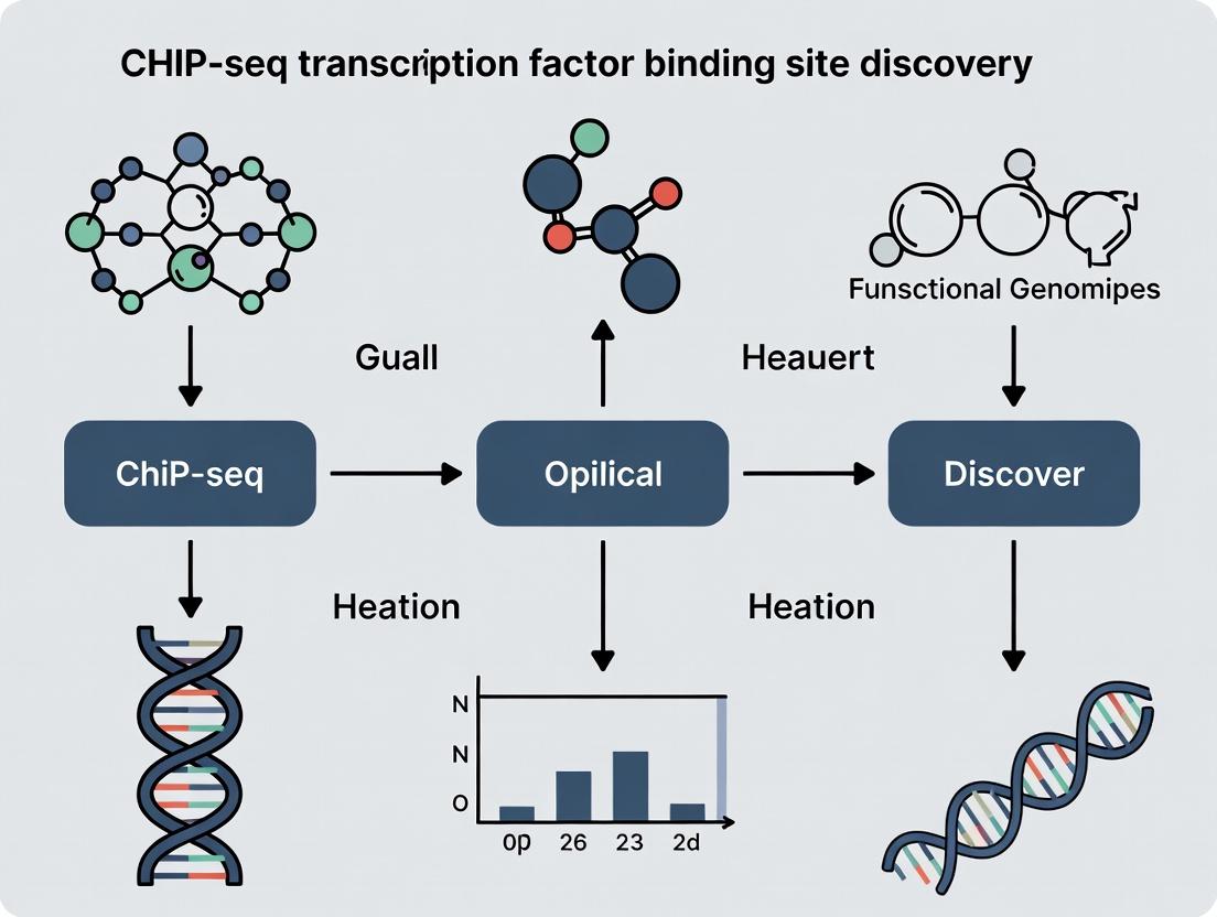
Mastering ChIP-seq: A Comprehensive Guide to Transcription Factor Binding Site Discovery in Biomedical Research
This definitive guide provides researchers, scientists, and drug development professionals with a complete workflow for ChIP-seq transcription factor binding site discovery.
Mastering ChIP-seq: A Comprehensive Guide to Transcription Factor Binding Site Discovery in Biomedical Research
Abstract
This definitive guide provides researchers, scientists, and drug development professionals with a complete workflow for ChIP-seq transcription factor binding site discovery. We cover fundamental chromatin biology principles and the role of TFs in gene regulation, then progress through detailed experimental protocols and NGS library preparation. The article addresses common troubleshooting scenarios, quality control metrics, and peak-calling optimization strategies. Finally, we explore rigorous validation methods and comparative analyses against techniques like CUT&RUN and ATAC-seq. This resource equips you to design, execute, and interpret robust ChIP-seq experiments for mechanistic insights and therapeutic target identification.
Decoding the Genome's Blueprint: Chromatin, Transcription Factors, and the Rationale for ChIP-seq
Transcription Factors as Master Regulators of Gene Expression and Cellular Identity
1. Introduction Within the nucleus, transcription factors (TFs) function as molecular interpreters and executors, binding to specific DNA sequences to activate or repress gene transcription. This article, framed within the context of Chromatin Immunoprecipitation followed by sequencing (ChIP-seq) research for TF binding site discovery, posits that the combinatorial logic of TF binding and interaction with chromatin modifiers constitutes the primary algorithm defining cellular identity, plasticity, and disease states. Precise mapping of these interactions is therefore foundational for mechanistic biology and targeted drug development.
2. Core Mechanisms of TF Action TFs exert control through modular domains: DNA-binding domains (DBDs) confer sequence specificity, while transactivation/repression domains recruit co-regulators and the basal transcriptional machinery. Master regulator TFs, such as OCT4, SOX2, and NANOG in pluripotency, often operate within dense, autoregulatory networks, binding to their own promoters and to each other's, creating stable transcriptional circuits.
Table 1: Key Master Transcription Factor Families and Their Roles
| TF Family (Example DBD) | Prototypical Members | Primary Role | Associated Disease Link |
|---|---|---|---|
| Homeodomain | OCT4 (POU5F1), HOX genes | Embryonic development, cell fate | Cancer, congenital disorders |
| Basic Helix-Loop-Helix (bHLH) | MYC, MYOD, NEUROD1 | Cell cycle, differentiation, neurogenesis | Ubiquitous in cancer |
| Zinc Finger (C2H2) | ZNFs, KLF4, SP1 | Ubiquitous regulation, pluripotency | Various cancers, immunological |
| Nuclear Receptor | Estrogen Receptor (ER), Androgen Receptor (AR) | Steroid hormone response | Breast & prostate cancer |
| Winged Helix / Forkhead | FOXO1, FOXP3 | Metabolism, immune regulation | Diabetes, autoimmunity, cancer |
A critical pathway demonstrating TF hierarchy is the pluripotency network, which maintains embryonic stem cell identity.
Figure 1: Core Pluripotency Transcription Factor Network
3. ChIP-seq: The Definitive Methodology for TF Binding Site Discovery ChIP-seq remains the gold standard for genome-wide mapping of TF occupancy. Its resolution and accuracy are critical for deconvoluting regulatory networks.
3.1 Detailed ChIP-seq Experimental Protocol
- Crosslinking: Treat cells with 1% formaldehyde for 10 min at room temperature to covalently link TFs to DNA.
- Cell Lysis & Chromatin Shearing: Lyse cells and sonicate chromatin to 200-500 bp fragments using a focused ultrasonicator (e.g., Covaris). Validate fragment size by agarose gel electrophoresis.
- Immunoprecipitation: Incubate sheared chromatin with 2-5 µg of validated, target-specific antibody pre-bound to magnetic Protein A/G beads overnight at 4°C. Include an isotype control IgG IP.
- Washing & Elution: Wash beads sequentially with Low Salt, High Salt, LiCl, and TE buffers. Elute complexes with 1% SDS, 0.1M NaHCO3.
- Reverse Crosslinking & Purification: Incubate eluates at 65°C overnight with 200 mM NaCl to reverse crosslinks. Treat with RNase A and Proteinase K, then purify DNA with SPRI beads.
- Library Preparation & Sequencing: Prepare sequencing libraries from ChIP and Input DNA using a commercial kit (e.g., NEB Next Ultra II). Perform 50-75 bp single-end sequencing on an Illumina platform to a depth of 20-40 million reads per sample.
3.2 Primary Data Analysis Workflow
Figure 2: ChIP-seq Primary Data Analysis Pipeline
Table 2: Key ChIP-seq Quality Control Metrics and Benchmarks
| Metric | Description | Optimal Target Value |
|---|---|---|
| FRiP (Fraction of Reads in Peaks) | Proportion of mapped reads falling under called peaks. Signal-to-noise measure. | > 1-5% (TF ChIP-seq) |
| NSC (Normalized Strand Cross-correlation coefficient) | Ratio of cross-correlation at the read-length shift vs. background. Measures signal strength. | > 1.05 (≥1.1 is good) |
| RSC (Relative Strand Cross-correlation) | Ratio of fragment-length shift vs. read-length shift. Corrects for poorly shifted libraries. | > 0.8 (≥1 is good) |
| Peak Number | Total reproducible peaks identified. | Varies by TF; 10,000-80,000 is typical |
| PCR Bottlenecking Coefficient (PBC) | Measures library complexity based on read duplication. | > 0.8 (0.9 is excellent) |
4. The Scientist's Toolkit: Research Reagent Solutions
Table 3: Essential Reagents for TF ChIP-seq Research
| Item | Function & Critical Notes |
|---|---|
| High-Affinity, ChIP-Validated Antibody | Specificity is paramount. Must be validated for immunoprecipitation using knockout cell controls. Sources: Cell Signaling, Active Motif, Abcam. |
| Magnetic Protein A/G Beads | Provide efficient capture of antibody-antigen complexes with low non-specific binding. |
| Formaldehyde (Electrophoresis Grade) | For efficient and consistent crosslinking. Freshness and purity affect efficiency. |
| Protease & Phosphatase Inhibitor Cocktails | Preserve post-translational modification states and prevent protein degradation during lysis. |
| Covaris AFA Focused-Ultrasonicator | Provides consistent, reproducible chromatin shearing with minimal heat-induced damage. |
| SPRI (Solid Phase Reversible Immobilization) Beads | For consistent size selection and purification of DNA after elution. |
| High-Sensitivity DNA Assay Kit (e.g., Qubit) | Accurate quantification of low-concentration ChIP DNA is critical for library prep success. |
| Commercial Library Prep Kit for Low Input | Optimized for sub-nanogram DNA input to construct sequencing libraries with minimal bias. |
5. Advanced Applications & Drug Development Context Integrating ChIP-seq with other modalities (ATAC-seq, RNA-seq) defines transcriptional regulatory networks (TRNs). In oncology, mapping TF dependencies (e.g., MYC, AR, ER) reveals direct target genes and vulnerabilities. Emerging therapeutic strategies aim to disrupt pathogenic TF activity via:
- Small Molecule Inhibitors: Targeting TF-cofactor interfaces (e.g., p300/CBP bromodomain inhibitors).
- PROTACs: Specifically degrading oncogenic TFs.
- Gene Regulation: CRISPR-based gene activation/repression to reprogram TF networks.
Table 4: Example Drug Development Targets Based on TF Dysregulation
| TF Target | Cancer Context | Therapeutic Approach (Example) | Development Stage |
|---|---|---|---|
| Androgen Receptor (AR) | Prostate Cancer | AR antagonists (Enzalutamide), PROTACs (ARV-110) | Approved / Clinical |
| MYC | Multiple Cancers | Indirect targeting via BET bromodomain inhibitors (e.g., JQ1) | Preclinical/Clinical |
| STAT3 | Inflammatory, Solid Tumors | Phosphorylation inhibitors, Decoy oligonucleotides | Clinical |
| p53 Mutants | TP53-mutant Cancers | Reactivators (e.g., APR-246/Eprenetapopt) | Clinical |
6. Conclusion Transcription factors are the central processors of genomic information, translating cellular signals into precise transcriptional programs. Rigorous ChIP-seq methodology provides the indispensable map of their genomic binding landscape, forming the basis for decoding the logic of cellular identity and its dysregulation in disease. This map is the starting point for the rational design of interventions aimed at reprogramming or correcting pathological transcriptional states, a frontier in precision medicine.
Chromatin Architecture and the Challenge of Mapping Protein-DNA Interactions In Vivo
The quest to define the complete cis-regulatory code of eukaryotic genomes hinges on the accurate mapping of transcription factor (TF) binding events within their native chromatin context. Chromatin architecture—the dynamic, three-dimensional organization of DNA, histones, and associated proteins—presents a formidable barrier to in vivo protein-DNA interaction mapping. Techniques like Chromatin Immunoprecipitation followed by sequencing (ChIP-seq) have become the cornerstone of TF binding site discovery research. However, the interplay between nucleosomal occupancy, chromatin accessibility, and higher-order folding introduces significant noise and bias, challenging the distinction between functional binding events and non-functional or indirect interactions. This technical guide examines the core challenges posed by chromatin architecture in ChIP-seq experiments and outlines current methodologies to overcome them, framing the discussion within the broader thesis of achieving a physiologically complete regulome map for therapeutic target identification.
The Core Challenge: Chromatin as a Dynamic Filter
Chromatin does not merely package DNA; it actively regulates the protein-DNA interactome. The primary challenges include:
- Nucleosomal Blockage: The canonical nucleosome core particle obscures ~147 bp of DNA, sterically hindering TF access to cognate motifs.
- Transient and Low-Abundance Binding: Many TFs bind dynamically with short residence times, and lowly expressed TFs yield limited ChIP-seq material.
- Indirect Recruitment: TFs can be recruited via protein-protein interactions without direct DNA contact, leading to ChIP-seq peaks lacking the canonical motif.
- Architectural Confounding: Long-range interactions mediated by cohesin, CTCF, or looping can bring a TF precipitated at one locus into physical proximity with a non-bound DNA fragment, creating artifactual "shadow peaks."
Recent genome-wide studies quantify this challenge. For example, a 2023 benchmark analysis of public ChIP-seq datasets estimated that ~15-30% of peaks for a typical TF may represent indirect binding or technical artifacts, a figure that escalates for factors with strong co-activator interactions.
Table 1: Quantifying Key Challenges in In Vivo TF Mapping
| Challenge | Typical Impact Metric | Experimental Manifestation | Common in TFs with |
|---|---|---|---|
| Nucleosomal Occlusion | >70% of motifs are nucleosome-occupied in inactive cells | Low signal-to-noise at repressed loci; motif enrichment in flanking regions | Pioneer capability |
| Indirect Recruitment | 15-30% of ChIP-seq peaks lack canonical motif | Peaks enriched for motifs of co-bound partners, not the immunoprecipitated TF | Strong activation domains |
| Low-Abundance Binding | Signal can be near background levels | High fraction of irreproducible discoveries (IDR) | Low expression, transient binders |
| Architectural Artifacts | Accounts for ~5% of long-range (>10kb) interactions in HiChIP | Peaks coinciding with anchor points of chromatin loops (e.g., CTCF sites) | Involvement in enhancer-promoter looping |
Advanced Methodologies: From ChIP-seq to Nuanced Solutions
To address these challenges, the field has evolved beyond standard ChIP-seq. Below are detailed protocols for key methodologies.
Protocol: Cleavage Under Targets & Release Using Nuclease (CUT&RUN)
CUT&RUN uses a targeted nuclease (pAG-MNase) to cleave DNA adjacent to the antibody-bound protein in situ, offering high resolution and low background.
Detailed Protocol:
- Cell Preparation: Permeabilize intact nuclei from ~500k cells using Digitonin buffer (0.01% Digitonin, 150mM NaCl, 20mM HEPES pH 7.5, 0.5mM Spermidine, protease inhibitors).
- Antibody Binding: Incubate nuclei with a primary antibody against the target protein (e.g., 1:50-1:100 dilution) in 200µL Digitonin buffer for 2 hours at 4°C with rotation.
- pAG-MNase Binding: Wash unbound antibody and incubate with pAG-MNase fusion protein (1:100 dilution) for 1 hour at 4°C.
- Targeted Cleavage: Wash and place tubes on ice. Add 2mM CaCl₂ to activate MNase. Incubate for exactly 30 minutes on ice.
- Reaction Stop & DNA Release: Add an equal volume of 2X Stop Buffer (340mM NaCl, 20mM EDTA, 4mM EGTA, 0.02% Digitonin, 50µg/mL RNase A, 50µg/mL Glycogen). Incubate at 37°C for 10 min to release fragments.
- DNA Purification: Centrifuge, transfer supernatant, and purify DNA using Phenol-Chloroform extraction or SPRI beads.
- Library Prep & Sequencing: Construct sequencing libraries from the eluted DNA (typically low-input protocols) and sequence on an Illumina platform (2x50bp recommended).
Protocol: Simultaneous Mapping of Protein-DNA Interactions and Chromatin Accessibility (CoBATCH)
CoBATCH integrates TF profiling with accessibility in the same assay using a Tn5 transposase-based approach.
Detailed Protocol:
- Cell & Nuclear Preparation: As per CUT&RUN (Step 1).
- Antibody Conjugation: Pre-conjugate a primary antibody specific to the target TF with Protein A-Tn5 fusion complexes loaded with sequencing adaptors. Use a 1:2 molar ratio, incubate for 1 hour at room temperature.
- In-Situ Binding & Tagmentation: Incubate permeabilized nuclei with the antibody-Protein A-Tn5 complex for 2 hours at 4°C. Directly add MgCl₂ to a final concentration of 10mM and incubate at 37°C for 1 hour to initiate tagmentation.
- DNA Extraction & Purification: Halt reaction with EDTA, add SDS and Proteinase K, and incubate at 55°C overnight. Purify DNA via Phenol-Chloroform.
- PCR Amplification: Amplify purified DNA with indexed primers for 12-15 cycles.
- Sequencing & Data Separation: Sequence on an Illumina platform. Bioinformatically separate reads based on the adaptor signature: reads with adapter1-adapter2 represent TF-bound fragments, while adapter1-adapter1 represent accessible regions.
Diagram 1: CoBATCH Experimental Workflow (79 characters)
Visualizing Cross-Method Relationships
The choice of method depends on the specific chromatin challenge being addressed.
Diagram 2: Decision Logic for In Vivo TF Mapping Methods (71 characters)
The Scientist's Toolkit: Research Reagent Solutions
Table 2: Essential Reagents for Advanced In Vivo Mapping
| Reagent / Material | Supplier Examples | Critical Function & Role |
|---|---|---|
| pAG-MNase Fusion Protein | Cell Signaling Tech, homemade | The core enzyme for CUT&RUN/CUT&Tag. Protein A/G binds antibody, MNase performs targeted cleavage. |
| Protein A-Tn5 Transposase | Epicypher, homemade | Engineered transposase for tagmentation-based methods (CUT&Tag, CoBATCH). Conjugates antibody binding to DNA cutting/adapter insertion. |
| Digitonin (High-Purity) | MilliporeSigma, Thermo Fisher | A mild detergent used at precise concentrations to permeabilize nuclear membranes without destroying chromatin structure. |
| Magnetic Concanavalin A Beads | Bangs Laboratories | Used to immobilize nuclei or cells in CUT&RUN/Tag protocols for efficient buffer exchange and washing. |
| Validated ChIP-seq Grade Antibodies | Diagenode, Abcam, CST | Antibodies with proven specificity and efficiency in immunoprecipitation under fixed or native conditions. Crucial for signal-to-noise. |
| Dual-Indexed Sequencing Adapters | Illumina, IDT | For multiplexed library preparation, especially critical for low-input methods where library complexity is a concern. |
| SPRI (Solid Phase Reversible Immobilization) Beads | Beckman Coulter, Thermo Fisher | Magnetic beads for size-selective purification and cleanup of DNA fragments during library prep. |
| Protease & Phosphatase Inhibitor Cocktails | Roche, Thermo Fisher | Preserve the endogenous protein-DNA interaction landscape by inhibiting post-lysis degradation and modification. |
The intricate architecture of chromatin is not merely an obstacle to be overcome in TF binding site discovery; it is the very medium through which regulatory logic is encoded. The evolution from ChIP-seq to more nuanced techniques like CUT&RUN and CoBATCH represents a paradigm shift towards methods that work in harmony with, rather than against, native chromatin structure. By carefully selecting methodologies based on the biological question and chromatin context, and by employing rigorous reagents from the toolkit, researchers can generate more accurate maps of the protein-DNA interactome. This progress is fundamental to the broader thesis of decoding transcriptional regulation for identifying disease-associated cis-regulatory elements and developing novel epigenetic therapeutics. Future directions will likely involve even more integrated multi-omics approaches, capturing TF binding, chromatin states, and 3D conformation simultaneously within single cells.
Within the broader thesis of ChIP-seq for transcription factor (TF) binding site discovery, Chromatin Immunoprecipitation (ChIP) stands as the indispensable foundational technique. It is the critical biochemical step that isolates protein-DNA complexes from living cells, enabling the precise mapping of in vivo TF occupancy across the genome. This whitepaper details the core principle, protocols, and reagents essential for successful ChIP experiments.
Core Biochemical Principle
The principle of ChIP is to "freeze" transient TF-DNA interactions in situ, shear the chromatin, and immunoprecipitate the protein of interest along with its bound DNA fragments. The specificity of the antibody determines the selectivity of the capture. The recovered DNA fragments, once purified, represent a library of genomic sequences bound by the TF at the time of crosslinking.
Detailed Experimental Protocol for Crosslinking ChIP
Key Steps:
- In Vivo Crosslinking: Treat cells with 1% formaldehyde for 8-12 minutes at room temperature to covalently link TFs to DNA. Quench with 125mM glycine.
- Cell Lysis and Chromatin Preparation: Harvest cells. Lyse with a series of buffers (e.g., containing SDS, Triton X-100) to isolate nuclei.
- Chromatin Shearing: Fragment crosslinked chromatin to 200-1000 bp fragments, typically via sonication. Optimization is required for each cell type.
- Immunoprecipitation: Incubate sheared chromatin with a target-specific antibody (e.g., anti-TF antibody) pre-bound to Protein A/G magnetic beads or agarose. Include controls (IgG, input DNA).
- Washing and Elution: Wash beads stringently (e.g., with low-salt, high-salt, LiCl, and TE buffers) to remove non-specific binding. Elute complexes with elution buffer (e.g., 1% SDS, 100mM NaHCO3).
- Reverse Crosslinking & DNA Purification: Incubate eluate and Input at 65°C overnight with high salt to reverse formaldehyde links. Treat with RNase A and Proteinase K. Purify DNA via column or phenol-chloroform extraction.
Table 1: Typical ChIP Experimental Parameters and Yields
| Parameter | Typical Value/Range | Notes |
|---|---|---|
| Formaldehyde Concentration | 1% | Balance between crosslinking efficiency and epitope masking. |
| Crosslinking Time | 8-12 min | Cell-type dependent; over-crosslinking impedes shearing. |
| Sonication Fragment Size | 200-500 bp | Ideal for high-resolution mapping; verified by gel electrophoresis. |
| Antibody Amount per IP | 1-10 µg | Must be validated for ChIP; high titer is critical. |
| Input DNA Percentage | 1-10% of total chromatin | Used for normalization in downstream qPCR/seq. |
| DNA Yield per ChIP | 1-100 ng | Highly variable based on target abundance and antibody quality. |
| Enrichment (qPCR Validation) | 10- to 1000-fold over IgG | Measured at positive control genomic sites. |
Table 2: Common ChIP-Seq QC Metrics
| Metric | Target Value | Purpose |
|---|---|---|
| Library Fragment Size | ~200-300 bp post-adapter ligation | Confirms proper size selection. |
| PCR Duplication Rate | <20-30% | Indicates library complexity; high rates suggest low input. |
| Fraction of Reads in Peaks (FRiP) | >1% for TFs, >5% for histones | Measures signal-to-noise; assay-specific. |
| Peak Number (Mammalian TF) | 10,000 - 50,000 | Varies by TF, cell type, and statistical threshold. |
The Scientist's Toolkit: Key Research Reagent Solutions
Table 3: Essential Materials for ChIP Experiments
| Item | Function & Critical Notes |
|---|---|
| High-Purity Formaldehyde (37%) | Creates protein-DNA crosslinks. Must be fresh, methanol-free. |
| ChIP-Validated Antibody | The single most critical reagent. Must be validated for specificity and efficacy in ChIP. |
| Protein A/G Magnetic Beads | Facilitate antibody capture and easy washing. Reduce background vs. agarose. |
| Sonicator (Cup-horn or Probe) | Shears crosslinked chromatin. Consistent power and cooling are vital for reproducibility. |
| Protease Inhibitor Cocktail | Prevents proteolytic degradation of TFs and chromatin during isolation. |
| RNase A & Proteinase K | Enzymatic treatments post-IP to remove RNA and proteins prior to DNA purification. |
| DNA Purification Columns/Reagents | For clean isolation of low-abundance ChIP DNA, critical for sequencing. |
| qPCR Primers for Positive/Negative Genomic Loci | Essential for validating enrichment. Positive control: known binding site. Negative control: gene desert/IgG. |
| High-Sensitivity DNA Assay Kits (e.g., Qubit) | Accurately quantify low-concentration ChIP DNA for library preparation. |
Workflow and Pathway Visualizations
Title: ChIP Experimental Workflow
Title: Core ChIP Capture Principle
Within the context of advancing transcription factor (TF) binding site discovery research, the evolution from microarray-based chromatin immunoprecipitation (ChIP-chip) to next-generation sequencing (NGS) based ChIP-seq represents a paradigm shift. This whitepaper details the technical superiority of ChIP-seq, establishing it as the uncontested gold standard for genome-wide binding profiling in drug development and basic research.
The Technological Evolution: Quantitative Comparison
Table 1: ChIP-chip vs. ChIP-seq Core Performance Metrics
| Feature | ChIP-chip (Microarray) | ChIP-seq (NGS) |
|---|---|---|
| Genomic Coverage | Limited to predefined probe regions (~2-3% of genome). | Comprehensive, unbiased whole-genome coverage. |
| Resolution | 100-500 bp, constrained by probe density. | Single-base-pair resolution for precise binding site mapping. |
| Dynamic Range | Limited by fluorescence saturation, ~2-3 orders of magnitude. | Vast, limited only by sequencing depth (5+ orders of magnitude). |
| Input DNA Required | High (micrograms). | Low (nanograms). |
| Cost per Sample (Typical) | ~$500-$1,000 (array dependent). | ~$100-$500 (sequencing depth dependent). |
| Signal-to-Noise Ratio | Lower, susceptible to cross-hybridization artifacts. | Higher, with precise background modeling. |
| Data Output | Fluorescence intensity ratios. | Digital read counts directly proportional to protein-DNA complex abundance. |
Detailed ChIP-seq Experimental Protocol
Core Protocol for Transcription Factor ChIP-seq:
- Crosslinking: Treat cells with 1% formaldehyde for 8-12 minutes at room temperature to covalently link proteins to DNA.
- Cell Lysis & Chromatin Shearing: Lyse cells and sonicate chromatin to shear DNA into fragments of 150-500 bp using a focused ultrasonicator. Validate fragment size by agarose gel electrophoresis.
- Immunoprecipitation (IP): Incubate sheared chromatin with a validated, high-specificity antibody against the target transcription factor (5-10 µg antibody per 10^6 cells). Use Protein A/G magnetic beads to capture antibody-protein-DNA complexes. Wash stringently to reduce non-specific binding.
- Reverse Crosslinking & Purification: Elute complexes, reverse crosslinks at 65°C with high salt, and digest RNA and protein with RNase A and Proteinase K.
- Library Preparation:
- End-repair and A-tailing of immunoprecipitated DNA fragments.
- Ligation of platform-specific sequencing adapters.
- Size selection (e.g., 200-400 bp) via SPRI bead cleanup.
- Limited-cycle PCR amplification (typically 12-18 cycles) to enrich adapter-ligated fragments.
- Quantify library using qPCR or bioanalyzer.
- Sequencing & Data Analysis: Perform high-throughput sequencing (typically 20-50 million reads per sample on Illumina platforms). Process data through a pipeline: alignment (e.g., BWA, Bowtie2), peak calling (e.g., MACS2), and downstream annotation/analysis.
Visualizing the ChIP-seq Workflow
Diagram Title: ChIP-seq Core Experimental Workflow
The Scientist's Toolkit: Essential Research Reagent Solutions
Table 2: Key Reagents and Materials for ChIP-seq Experiments
| Item | Function | Critical Consideration |
|---|---|---|
| High-Quality Antibody | Specific recognition and pulldown of the target TF. | Must be validated for ChIP (ChIP-grade). Specificity is paramount. |
| Protein A/G Magnetic Beads | Efficient capture of antibody-TF-DNA complexes. | Consistency in size and binding capacity reduces noise. |
| Formaldehyde (1%) | Reversible protein-DNA crosslinking. | Fresh preparation required for consistent efficiency. |
| Protease/Phosphatase Inhibitors | Preserve protein epitopes and modification states during lysis. | Essential cocktail for studying phospho-TFs. |
| Sonication Device (Covaris, Bioruptor) | Shears chromatin to optimal fragment size. | Reproducible shearing is critical for resolution. |
| DNA Size Selection Beads (e.g., SPRI) | Cleanup and selection of DNA fragments after library prep. | Determines final insert size for sequencing. |
| NGS Library Prep Kit (Illumina, NEB) | Prepares DNA for sequencing with adapters and barcodes. | Kit efficiency impacts required input material. |
| High-Fidelity DNA Polymerase | Amplifies library fragments with minimal bias. | Reduces PCR duplicates and artifacts. |
| qPCR Quantification Kit | Accurately quantifies final library yield. | Prevents under/overloading of sequencer. |
Bioinformatics & Data Analysis Pathway
Diagram Title: ChIP-seq Bioinformatics Analysis Pipeline
For transcription factor binding site discovery research, ChIP-seq has decisively superseded microarray-based approaches. Its unparalleled resolution, dynamic range, genome-wide coverage, and digital quantitative output provide researchers and drug developers with a definitive tool for mapping regulatory landscapes, identifying novel therapeutic targets, and understanding disease mechanisms at an elemental level.
Chromatin Immunoprecipitation followed by sequencing (ChIP-seq) is the cornerstone technology for mapping in vivo transcription factor (TF) binding sites and histone modifications genome-wide. Within the broader thesis of ChIP-seq-driven discovery, the trajectory from identifying a binding event to defining a therapeutic target is a multi-stage process. This guide details the key applications along this continuum: starting with the basic mechanistic elucidation of gene regulatory networks and culminating in the pinpointing of druggable regulatory nodes for therapeutic intervention.
Application I: Basic Mechanism Elucidation
The primary application of ChIP-seq is the foundational dissection of transcriptional mechanisms. This involves identifying where a TF binds and inferring its functional consequences.
Core Experimental Protocol: ChIP-seq
Detailed Methodology:
- Cross-linking: Cells are treated with formaldehyde (1% final concentration, 10 min at room temperature) to covalently link TFs to DNA.
- Cell Lysis & Chromatin Shearing: Cells are lysed, and chromatin is fragmented to 200-600 bp fragments via sonication (e.g., Covaris S220, 20% duty cycle, 200 cycles per burst, 5 min) or enzymatic digestion (e.g., MNase).
- Immunoprecipitation: Sheared chromatin is incubated with a protein-specific antibody (e.g., 1-5 µg of anti-STAT3) bound to magnetic beads overnight at 4°C. An isotype control IgG is used in parallel.
- Washing & Elution: Beads are washed with low-salt, high-salt, LiCl, and TE buffers. Cross-links are reversed (65°C overnight with 200 mM NaCl), and proteins are digested with Proteinase K.
- DNA Purification: Immunoprecipitated DNA is purified using silica membrane columns.
- Library Preparation & Sequencing: DNA is end-repaired, A-tailed, adapter-ligated, PCR-amplified (12-15 cycles), and size-selected for sequencing on platforms like Illumina NovaSeq.
Table 1: Quantitative Metrics for ChIP-seq Data Quality Assessment
| Metric | Optimal Range/Value | Interpretation |
|---|---|---|
| Peak Number | Experiment-dependent | Too few may indicate poor IP; too many may suggest background noise. |
| FRiP Score | >1% (TF), >10% (Histone) | Fraction of Reads in Peaks; primary measure of signal-to-noise. |
| NSC (Normalized Strand Coefficient) | ≥1.05 | Measures enrichment relative to background; >1.1 is good. |
| RSC (Relative Strand Coefficient) | ≥1 | Corrects for low-quality profiles; >1 is good. |
| Library Complexity (NRF) | >0.8 | Non-Redundant Fraction; indicates PCR over-amplification if low. |
Identifying Direct Targets & Binding Motifs
Peak calling algorithms (MACS2, HOMER) identify statistically significant binding sites. De novo motif discovery within peaks reveals the bound TF's consensus sequence and can infer co-binding partners.
Diagram 1: From ChIP-seq reads to target gene annotation.
Integrating with Transcriptomics (RNA-seq)
Correlating TF binding with gene expression changes (upon TF knockdown/overexpression) distinguishes active regulators from silent binders.
Table 2: Integrative Analysis of ChIP-seq & RNA-seq Data
| Binding Context | Gene Expression Change | Interpretation | Potential Functional Role |
|---|---|---|---|
| Promoter/Enhancer | Up-regulated | Direct Activation | Transcriptional Activator |
| Promoter/Enhancer | Down-regulated | Direct Repression | Transcriptional Repressor |
| Promoter/Enhancer | Unchanged | Poised/Inactive | Pioneer Factor, Bookmarking |
| No Binding | Up/Down-regulated | Indirect Effect | Secondary Target |
Application II: Mapping Regulatory Networks & Crosstalk
Advanced ChIP-seq applications map complex interactions between multiple TFs and chromatin states.
Multi-TF ChIP-seq & Co-occupancy Analysis
Sequential or parallel ChIP-seq for multiple TFs reveals hierarchical or cooperative regulation.
Diagram 2: Hierarchical TF cooperation in enhancer activation.
Protocol: ChIP-reChIP (Sequential ChIP)
To confirm direct TF co-occupancy on the same DNA molecule:
- Perform first ChIP with antibody for TF A.
- Elute bound complexes with 10 mM DTT at 37°C for 30 min.
- Dilute eluate 1:50 and perform a second ChIP with antibody for TF B.
- Process and sequence the final DNA. Peaks represent genomic sites bound by both TFs.
Application III: Identifying Druggable Regulatory Nodes
The ultimate translational application is to dissect disease-driving regulatory circuits and pinpoint vulnerable, pharmacologically targetable nodes.
Defining Oncogenic Transcription Factors in Disease
Differential binding analysis (using tools like diffBind) compares ChIP-seq profiles between disease (e.g., cancer) and normal cells, identifying gained/lost regulatory elements.
Table 3: Characteristics of a "Druggable" Regulatory Node
| Characteristic | Description | Assessment Method |
|---|---|---|
| Disease-Specific Activity | Hyper-bound or mutated in disease vs. normal. | Differential ChIP-seq, Mutation analysis. |
| Essentiality | Required for cell survival/proliferation. | CRISPR Knockout Screen (e.g., DepMap). |
| "Ligandability" | Possesses a domain amenable to small-molecule inhibition. | Structural analysis (kinase, bromodomain, etc.). |
| Clear Phenotypic Output | Regulates a critical, therapeutically relevant gene set. | Integrated ChIP-seq/RNA-seq. |
Targeting TF Complexes or Cofactors
Directly inhibiting a DNA-binding TF is often challenging. Strategies shift to targeting its essential cofactors (e.g., kinases, epigenetic readers/writers).
Diagram 3: Targeting upstream kinases or coactivators of an oncogenic TF.
Protocol: Functional Validation via CRISPRi/a
To validate node druggability:
- Design: Design sgRNAs targeting the regulatory element (enhancer) of a key target gene, or the gene encoding the TF/cofactor itself.
- Delivery: Lentivirally deliver dCas9-KRAB (for CRISPR interference/CRISPRi) or dCas9-VP64 (for CRISPR activation/CRISPRa) and sgRNAs into disease-relevant cells.
- Phenotyping: Measure proliferation (CellTiter-Glo), apoptosis (Caspase-3/7 assay), or transcriptomic changes (RNA-seq) to confirm node essentiality.
The Scientist's Toolkit: Research Reagent Solutions
Table 4: Essential Reagents for ChIP-seq & Translational Follow-up
| Reagent/Material | Function | Key Considerations |
|---|---|---|
| High-Quality Antibodies | Target-specific immunoprecipitation. | Validate for ChIP-seq grade (low cross-reactivity). Cite publications. |
| Magnetic Protein A/G Beads | Capture antibody-target complexes. | Superior recovery and lower background vs. agarose. |
| Crosslinking Reagents | Fix protein-DNA interactions. | Formaldehyde is standard. For distal loops, consider dual crosslinkers (e.g., DSG + formaldehyde). |
| Library Prep Kits | Prepare sequencing-ready DNA libraries. | Select kits optimized for low-input/ChIP DNA (e.g., NEBNext Ultra II). |
| CRISPR/dCas9 Systems | Functional perturbation of regulatory nodes. | Choose appropriate effector (KRAB for repression, VP64/p300 for activation). |
| Small Molecule Inhibitors | Pharmacological validation of druggable nodes. | Use tool compounds with established target specificity and potency (e.g., BETi: JQ1, OTX015). |
| Viable Disease Models | In vivo validation of targets. | Patient-derived organoids, xenografts, or genetically engineered mouse models (GEMMs). |
From Cells to Data: A Step-by-Step ChIP-seq Protocol and Experimental Design Framework
1. Introduction: A Thesis Framework for Robust TF Discovery
In ChIP-seq research aimed at discovering transcription factor (TF) binding sites, the integrity of the final dataset and the validity of subsequent biological conclusions are irrevocably established in the initial experimental phases. This guide details the three critical, interdependent pillars of this foundation—antibody validation, cell fixation, and chromatin shearing—framed within the broader thesis that rigorous, optimized upstream protocols are non-negotiable for generating high-specificity, low-noise maps of the protein-DNA interactome. Failures at these stages propagate irrecoverably, leading to false positives, obscured true signals, and unreliable data for downstream drug target identification.
2. Pillar I: Antibody Validation – The Specificity Imperative
The antibody is the primary determinant of specificity in ChIP-seq. Using an unvalidated reagent risks mapping irrelevant genomic regions.
Key Validation Strategies:
- Genetic Knockdown/Knockout (Gold Standard): Perform ChIP-qPCR on wild-type vs. TF-deficient cells. Signal loss at positive control sites confirms specificity.
- Peptide Blocking: Pre-incubate antibody with its immunizing peptide before ChIP. A significant reduction in enrichment indicates on-target activity.
- Comparative Western Blot: The antibody should recognize a single band of the correct molecular weight in a whole-cell lysate.
- Use of Validated Public Resources: Consult databases like ENCODE, CISTROME, or vendor-provided validation data.
Quantitative Metrics for Validation: Signal-to-Noise Ratio (SNR) and Enrichment over IgG are critical metrics. Data from a typical validation experiment might yield:
Table 1: Example ChIP-qPCR Validation Data for a Hypothetical TF 'X'
Sample Positive Locus 1 (Ct) Negative Locus (Ct) ΔCt (Neg-Pos) Fold Enrichment (2^ΔΔCt) Anti-TF (WT Cells) 24.5 33.2 8.7 ~420 IgG (WT Cells) 32.1 33.0 0.9 ~1.9 Anti-TF (KO Cells) 31.8 33.1 1.3 ~2.5 Protocol: Genetic Knockdown Validation by ChIP-qPCR
- Cell Preparation: Culture isogenic wild-type and TF-specific knockout cell lines.
- Crosslinking: Fix cells with 1% formaldehyde for 10 min at room temperature.
- Cell Lysis: Lyse cells in SDS lysis buffer (1% SDS, 10 mM EDTA, 50 mM Tris-HCl, pH 8.1) with protease inhibitors.
- Chromatin Shearing: Sonicate to an average fragment size of 200-500 bp.
- Immunoprecipitation: Incubate pre-cleared chromatin with 2-5 µg of target antibody or species-matched IgG overnight at 4°C. Capture with protein A/G beads.
- Wash & Elution: Wash sequentially with Low Salt, High Salt, LiCl, and TE buffers. Elute complexes in freshly prepared elution buffer (1% SDS, 0.1M NaHCO3).
- Reverse Crosslinks & Purification: Add NaCl to 200 mM and incubate at 65°C overnight. Treat with RNase A and Proteinase K. Purify DNA with silica-membrane columns.
- qPCR Analysis: Perform qPCR on known positive control genomic regions and known negative (gene desert) regions. Calculate % Input and Fold Enrichment over IgG.
3. Pillar II: Cell Fixation – Capturing Transient Interactions
Formaldehyde crosslinking creates covalent protein-protein and protein-DNA bonds, "freezing" transient TF-DNA interactions.
- Optimization Variables: Formaldehyde concentration (0.5%-2%) and crosslinking time (5-30 min) must be titrated. Over-crosslinking impedes chromatin shearing and epitope recognition; under-crosslinking loses weak interactions.
Dual Crosslinking: For recalcitrant TFs or complexes, a combination of DSG (a protein-protein crosslinker) followed by formaldehyde can improve capture.
Protocol: Titration of Formaldehyde Crosslinking Conditions
- Aliquot identical samples of cultured cells (e.g., 1 x 10^6 cells per condition).
- Prepare formaldehyde solutions to final concentrations of 0.5%, 1%, and 1.5% in growth medium.
- Incubate each aliquot for 5, 10, and 15 minutes at room temperature with gentle agitation.
- Quench with 125 mM glycine for 5 min.
- Proceed with lysis and sonication. Analyze shearing efficiency via agarose gel electrophoresis. The optimal condition yields the most DNA in the 200-500 bp range post-sonication.
4. Pillar III: Chromatin Shearing – Balancing Yield and Resolution
Optimal shearing generates fragments small enough for precise mapping (~200-300 bp) while preserving protein-DNA complexes.
- Shearing Methods: Sonicators (tip-probe or focused ultrasonicator) are standard. Enzymatic shearing (e.g., MNase) offers an alternative for fragile complexes.
Critical Optimization Parameters: The following table summarizes key variables and their impact:
Table 2: Optimization Parameters for Sonicator-Based Chromatin Shearing
Parameter Typical Range Effect of Increasing Parameter Optimal Goal Peak Power 50-75% (probe) Increased fragmentation efficiency; more heat. Efficient shearing without overheating. Duration/Cycle Time 5-15 cycles (30s ON/30s OFF) Smaller fragment size. Majority of fragments between 200-500 bp. Sample Volume 0.5-1 mL Reduced shearing efficiency if too high. Consistent volume across runs. Cell Count 0.5-2 x 10^6 per mL Higher density requires more energy/sonication. Avoid overloading. Buffer Composition Varies (e.g., RIPA, SDS) Lower SDS may reduce efficiency but preserve epitopes. Compatible with antibody and fixation. Quality Control: Always run an aliquot of sheared, reverse-crosslinked DNA on a 1.5% agarose gel or Bioanalyzer to verify fragment size distribution before proceeding to immunoprecipitation.
5. The Scientist's Toolkit: Essential Research Reagents
Table 3: Key Reagent Solutions for Initial ChIP-seq Steps
| Reagent/Material | Function & Critical Notes |
|---|---|
| Validated ChIP-grade Antibody | High-affinity, high-specificity antibody against the target TF or histone mark. Check for citations in ChIP-seq literature. |
| Formaldehyde (37%) | Primary crosslinking agent. Use high-purity, freshly opened aliquots if possible. |
| Glycine (2.5M Stock) | Quenches formaldehyde to stop crosslinking. |
| Protease Inhibitor Cocktail (PIC) | Prevents proteolytic degradation of TFs/complexes during lysis. Add fresh to all buffers. |
| SDS Lysis Buffer | Efficiently lyses nuclei and denatures proteins to expose chromatin for shearing. |
| Protein A/G Magnetic Beads | For efficient capture of antibody-antigen complexes. Choice depends on antibody species/isotype. |
| Sonication Device | Tip-probe or focused ultrasonicator. Consistent, clean shearing is vital. |
| RNase A & Proteinase K | Enzymes used post-IP to digest RNA and proteins prior to DNA purification. |
| DNA Clean-up Columns | Silica-membrane columns for efficient purification of low-concentration ChIP DNA. |
| qPCR Reagents & Primers | For validation of shearing efficiency (size distribution) and antibody specificity (positive/negative loci). |
6. Visualizing the Workflow and Logical Dependencies
Diagram 1: ChIP-seq Foundational Workflow & QC Gates
Diagram 2: Crosslinking Captures TF Complexes on DNA
Conclusion
The path to credible transcription factor binding site discovery is paved with meticulous attention to these initial technical steps. Systematic antibody validation, empirical fixation optimization, and rigorous shearing control collectively form the non-negotiable foundation. By investing in these critical first steps, researchers ensure that their subsequent ChIP-seq data accurately reflects the in vivo binding landscape, providing a solid basis for mechanistic insights and target identification in drug development.
In the context of Chromatin Immunoprecipitation followed by sequencing (ChIP-seq) for transcription factor binding site discovery, the immunoprecipitation (IP) step is the critical enrichment phase. This workflow determines the specificity and signal-to-noise ratio of the entire experiment. Efficient capture of protein-DNA complexes, rigorous removal of non-specifically bound material, and gentle yet complete elution are paramount for generating high-quality, interpretable sequencing data. This guide details the core IP protocol, focusing on bead selection, wash stringency optimization, and elution strategies to maximize target enrichment for downstream NGS library preparation.
Core Components: Beads, Buffers, and Their Functions
Research Reagent Solutions Toolkit
| Reagent / Material | Function in ChIP-seq IP |
|---|---|
| Protein A/G Magnetic Beads | Solid-phase support for antibody immobilization. Magnetic properties enable rapid buffer changes and minimal mechanical loss of chromatin. |
| ChIP-Validated Primary Antibody | Binds specifically to the target transcription factor or histone modification. Must be validated for IP and specificity. |
| Sonication Sheared Chromatin | Crosslinked and fragmented DNA-protein complexes (200–500 bp average size) ready for immunoenrichment. |
| Low-SDS Lysis Buffer | Maintains integrity of protein-DNA complexes while solubilizing chromatin and providing initial washing conditions. |
| High-Salt Wash Buffer | Removes non-specifically bound chromatin through ionic disruption of weak electrostatic interactions. |
| LiCl Wash Buffer | Removes contaminating RNA and protein aggregates via chaotropic effects. |
| TE Buffer (Low EDTA) | Final wash to prepare complexes for elution in a low-ion, nuclease-inhibiting environment. |
| Elution Buffer (1% SDS, 0.1M NaHCO3) | Disrupts antibody-antigen binding and releases captured chromatin complexes into solution for crosslink reversal. |
| Proteinase K | Digests proteins post-elution to facilitate DNA purification and library preparation. |
| RNase A | Optional post-elution treatment to remove residual RNA that may interfere with library prep. |
Detailed Immunoprecipitation Protocol for ChIP-seq
Day 1: Pre-clearing and Binding
- Bead Preparation: Resuspend protein A/G magnetic beads thoroughly. For each IP, aliquot 50 µL of bead slurry (approx. 25 µL bead volume) into a low-retention microfuge tube.
- Wash Beads: Place tube on a magnetic separator for 30 seconds. Discard supernatant. Wash beads twice with 1 mL of cold IP Dilution Buffer (or Lysis Buffer).
- Antibody Coupling: Resuspend washed beads in 500 µL of Dilution Buffer containing 1–5 µg of the target-specific antibody. Incubate with rotation for 2 hours at 4°C to overnight.
- Chromatin Pre-clearing: While antibodies couple, add 50 µL of untreated washed beads to the total volume of sonicated chromatin (input from 1-10 million cells). Incubate with rotation for 1 hour at 4°C. Place on magnet and transfer the pre-cleared supernatant to a new tube.
- Immunoprecipitation: Wash the antibody-coupled beads twice with 1 mL of cold Dilution Buffer. Resuspend the beads in the pre-cleared chromatin supernatant. Incubate with rotation for 4–6 hours (or overnight) at 4°C.
Day 2: Washes and Elution
- Capture Complexes: Place the IP tube on a magnetic separator for 2 minutes. Carefully remove and save the supernatant (this is the "unbound" fraction).
- Stringent Washes: Perform sequential washes on the magnet with the following cold buffers, incubating with rotation for 5 minutes per wash:
- Wash 1: Low-SDS Lysis Buffer (1 mL)
- Wash 2: High-Salt Buffer (1 mL)
- Wash 3: LiCl Buffer (1 mL)
- Wash 4: TE Buffer (1 mL, twice)
- Elution: After the final TE wash, fully remove supernatant. Resuspend beads in 150 µL of freshly prepared Elution Buffer. Vortex briefly. Incubate at 65°C for 30 minutes with intermittent vortexing (every 5-10 minutes).
- Collect Eluate: Place tube on magnet and carefully transfer the eluate (containing enriched chromatin) to a new tube.
- Reverse Crosslinks & Purify: Add 6 µL of 5M NaCl and 2 µL of Proteinase K (20 mg/mL) to the eluate. Incubate at 65°C for 2 hours (or overnight). Proceed to DNA purification using SPRI beads or phenol-chloroform extraction.
Table 1: Bead Type Selection Guide
| Bead Type | Binding Specificity | Best For | Recommended Amount per IP (Slurry) |
|---|---|---|---|
| Protein A | IgG of most mammals (strong for rabbit, human, mouse) | Rabbit polyclonal antibodies | 25–50 µL |
| Protein G | Broad mammalian IgG (strong for mouse, rat, human) | Mouse monoclonal antibodies | 25–50 µL |
| Protein A/G | Combined A and G affinities | Polyclonal/monoclonal mixes or unknown species | 25–50 µL |
| Antigen-Specific Beads | Covalently coupled target-specific antibody | High-throughput or standardized assays; reduces antibody contamination in eluate | As per manufacturer |
Table 2: Wash Buffer Stringency Impact
| Buffer | Key Components | Purpose | Effect on Stringency | Typical Volume/Time |
|---|---|---|---|---|
| Low-SDS Lysis | 1% Triton, 0.1% SDS, 150mM NaCl | Removes soluble proteins & lipids | Low | 1 mL, 5 min |
| High-Salt | 1% Triton, 0.1% SDS, 500mM NaCl | Disrupts non-specific ionic interactions | High | 1 mL, 5 min |
| LiCl | 250mM LiCl, 1% NP-40, 1% Deoxycholate | Removes RNA & protein aggregates | Medium | 1 mL, 5 min |
| TE | 10mM Tris, 1mM EDTA | Removes detergents/salts; prepares for elution | Very Low | 1 mL x 2, 2 min |
Table 3: Elution Method Comparison
| Method | Conditions | Efficiency | Pros | Cons |
|---|---|---|---|---|
| SDS/Heat | 1% SDS, 0.1M NaHCO₃, 65°C, 30 min | High (>90%) | Simple, effective, standard for ChIP | Harsh, may co-elute contaminants |
| Low pH Glycine | 0.2M Glycine, pH 2.5-3.0 | Moderate-High | Gentle on protein epitopes | May require immediate neutralization |
| Competitive Peptide | HA or FLAG peptide excess | Variable | Gentle, epitope-specific | Expensive, requires epitope tag |
Visualizing the ChIP-seq IP Workflow and Key Pathways
Title: ChIP-seq Immunoprecipitation and Wash Workflow
Title: Molecular Interactions on IP Bead Surface
Title: Elution Strategy Decision Tree for ChIP-seq
Within the framework of ChIP-seq for transcription factor (TF) binding site discovery, the construction of high-quality sequencing libraries is a critical determinant of success. Following chromatin immunoprecipitation, the isolated DNA fragments must be converted into a format compatible with high-throughput sequencing platforms. This technical guide details the three pivotal wet-lab steps—End-Repair, Adapter Ligation, and Size Selection—that transform ChIP-enriched DNA into a sequencer-ready library. The fidelity of these steps directly impacts mapping accuracy, data complexity, and the ultimate sensitivity in identifying bona fide TF binding events.
End-Repair (or End Polishing)
Purpose: ChIP-derived DNA fragments possess heterogeneous ends, including 5' overhangs, 3' overhangs, and nicks. The end-repair reaction converts all DNA termini to blunt-ended, 5'-phosphorylated molecules, which is a mandatory substrate for subsequent adapter ligation.
Detailed Protocol:
- Assemble the reaction on ice in a thin-walled PCR tube:
- ChIP DNA (in ≤ 50 µL): Variable volume (e.g., 1-100 ng typical for TF ChIP-seq).
- 10X End Repair Buffer: 7 µL (provides Mg2+, ATP, and dNTPs).
- T4 DNA Polymerase: 3 µL. Possesses 5'→3' polymerase activity (fills in 5' overhangs) and strong 3'→5' exonuclease activity (removes 3' overhangs).
- Klenow Fragment: 1 µL. Possesses 5'→3' polymerase activity with lower exonuclease activity, assisting in blunt-end formation.
- T4 Polynucleotide Kinase (PNK): 3 µL. Phosphorylates 5' hydroxyl termini, essential for ligation.
- Nuclease-free water to a final volume of 70 µL.
- Mix thoroughly by pipetting and incubate in a thermal cycler at 20°C for 30 minutes.
- Purification: Immediately clean up the reaction using a spin-column-based purification kit (e.g., AMPure XP beads). Elute in 20-25 µL of nuclease-free water or low-EDTA TE buffer.
Key Considerations: Reaction temperature is critical; T4 DNA Polymerase is most active at 20°C for blunt-end formation. Over-incubation can lead to excessive exonuclease activity and DNA loss.
Adapter Ligation
Purpose: To ligate platform-specific oligonucleotide adapters to both ends of the blunt-ended, phosphorylated DNA. These adapters contain the primer binding sites for cluster amplification and sequencing on instruments like Illumina platforms.
Detailed Protocol:
- Prepare the Ligation Mix on ice:
- End-Repaired DNA: 20 µL.
- 2X Rapid Ligation Buffer: 25 µL (contains ATP and PEG for enhanced efficiency).
- Indexed Adapter Oligo Mix: 2.5-5 µL (use a concentration appropriate for low-input ChIP DNA to minimize adapter-dimer formation).
- T4 DNA Ligase: 3 µL.
- Nuclease-free water to 50 µL.
- Mix gently and incubate at 20°C for 15 minutes.
- Purification: Purify immediately using AMPure XP beads. Perform a double-sided size selection during bead cleanup: use a bead-to-sample ratio of 0.8X to remove large adapter concatenates, then add beads to the supernatant at a ratio of 1.2X to capture the desired library fragments. Elute in 20 µL.
Key Considerations: Adapter concentration must be titrated based on input DNA mass to maximize yield of desired product while minimizing adapter-dimer artifacts, which are particularly detrimental in low-input ChIP-seq libraries.
Size Selection
Purpose: To isolate library fragments within an optimal size range (typically 200-500 bp for TF ChIP-seq). This removes unligated adapters, adapter-dimers (~120 bp), and excessively large fragments, ensuring uniform amplification and sequencing.
Detailed Protocol (Dual-Sided Solid-Phase Reversible Immobilization - SPRI): This is the most common method using AMPure XP beads.
- Remove Large Fragments: To the ligated library (20 µL), add AMPure XP beads at a 0.5X ratio (10 µL). Mix thoroughly and incubate for 5 minutes. Pellet beads on a magnet and transfer the supernatant (containing DNA <~700 bp) to a new tube. Discard beads.
- Recover Desired Fragments: To the supernatant, add AMPure XP beads at a 0.8X ratio of the original library volume (16 µL). Incubate for 5 minutes. Place on magnet. Wash beads twice with 80% ethanol while on the magnet.
- Elute: Air-dry beads for 2-3 minutes, then elute DNA in 20 µL of nuclease-free water or resuspension buffer.
Quantitative Data Summary of Key Reagents:
Table 1: Key Enzymes for End-Repair
| Reagent | Core Function | Optimal Temperature | Critical Note for ChIP-seq |
|---|---|---|---|
| T4 DNA Polymerase | 5'→3' pol / 3'→5' exo | 20°C | Primary enzyme for blunt-end generation. |
| Klenow Fragment | 5'→3' polymerase | 37°C | Assists in filling 5' overhangs. |
| T4 PNK | 5' phosphorylation | 37°C | Essential for ligation competency. |
Table 2: Size Selection Parameters (Using AMPure XP Beads)
| Target to Remove | Bead:Sample Ratio | Approximate Size Cutoff | Fraction Kept |
|---|---|---|---|
| Large fragments / Concatenates | 0.5X - 0.7X | > 700-500 bp | Supernatant |
| Small fragments / Adapter-dimers | 0.8X (of original) | < 150-200 bp | Bead Pellet |
| Final Library Recovery | 1.0X - 1.2X | Broad range | Bead Pellet |
The Scientist's Toolkit: Research Reagent Solutions
Table 3: Essential Materials for ChIP-seq Library Construction
| Item | Function/Description | Example Vendor/Kit |
|---|---|---|
| T4 DNA Polymerase | Creates blunt ends via polymerase/exonuclease activity. | NEB, Thermo Fisher |
| T4 Polynucleotide Kinase (PNK) | Adds 5' phosphate groups for ligation. | NEB |
| T4 DNA Ligase | Catalyzes the attachment of adapters to DNA inserts. | NEB Rapid Ligase |
| Platform-Specific Adapters | Double-stranded oligos with indexing and sequencing primer sites. | Illumina TruSeq, IDT for Illumina |
| SPRI Magnetic Beads | For purification and size-based selection of DNA fragments. | Beckman Coulter AMPure XP |
| High-Sensitivity DNA Assay | Fluorometric quantification of low-concentration libraries. | Agilent Bioanalyzer, Qubit |
| Library Amplification Mix | High-fidelity PCR mix for final library enrichment. | KAPA HiFi, NEB Next Ultra II |
Visualizing the ChIP-seq Library Construction Workflow
Diagram 1: ChIP-seq library prep workflow from end-repair to final lib.
Diagram 2: Molecular steps of end-repair and adapter ligation.
In ChIP-seq transcription factor (TF) binding site discovery research, the statistical power to detect true binding events is fundamentally governed by two interrelated experimental design factors: sequencing depth (total reads per sample) and biological replicate number. This technical guide provides a framework for conducting power analysis to optimize resource allocation, ensuring robust and reproducible discoveries in both basic research and drug development contexts where TF dysregulation is a target.
Foundational Concepts: Power Analysis in ChIP-seq
Statistical power is the probability of correctly rejecting the null hypothesis (i.e., detecting a true TF binding peak) when a true effect exists. In ChIP-seq, effect size relates to the fold-enrichment of reads at a binding site over background. Key parameters are:
- α (Significance Threshold): The false positive rate (e.g., 0.05).
- 1-β (Power): The desired probability of detecting true peaks (typically 0.8-0.9).
- Effect Size: The minimum fold-change in read density at a peak deemed biologically significant.
- Variability: Biological and technical variance between replicates.
Power analysis helps determine the required N (replicates) and depth (reads) to achieve a given power for an expected effect size, given the natural variability of the system.
Quantitative Framework: The Impact of Depth and Replicates
Table 1: Recommended Minimum Sequencing Depth for Common ChIP-seq Targets
| Target Type | Recommended Minimum Depth (Mapped Reads) | Rationale |
|---|---|---|
| Point-source TFs | 20-30 million | Sharp, localized peaks require sufficient coverage for precise summit calling. |
| Broad Histone Marks | 40-60 million | Wide enrichment regions require more reads to distinguish signal from background over large genomic intervals. |
| Input/Control | Matched or greater than IP depth | Essential for accurate background modeling and peak calling, especially in complex genomes. |
Table 2: Simulated Power Analysis for Detecting a 2-fold Enrichment Peak (α=0.05)
| Biological Replicates (N) | Sequencing Depth per Sample (Millions) | Estimated Statistical Power (1-β) | Relative Cost Factor |
|---|---|---|---|
| 2 | 20 | ~0.65 | 1.0x (Baseline) |
| 3 | 20 | ~0.82 | 1.5x |
| 2 | 40 | ~0.78 | 2.0x |
| 3 | 30 | ~0.90 | 2.25x |
| 4 | 20 | ~0.92 | 2.0x |
Note: Power estimates are simulated for a typical mammalian TF with moderate variability. Actual values depend on antibody quality, cell type homogeneity, and genomic background.
Experimental Protocols for Power Assessment
Protocol A: In Silico Power Analysis Using Downsampling
Purpose: To determine if existing data has sufficient depth.
- Start with a deeply sequenced ChIP-seq sample (e.g., 50 million reads).
- Use bioinformatics tools (e.g.,
samtoolsview -s) to randomly subsample the aligned BAM file at descending depths (e.g., 40M, 30M, 20M, 10M reads). - Call peaks at each depth level using your standard pipeline (e.g., MACS2).
- Plot the number of high-confidence peaks (e.g., -log10(p-value) > 5) against sequencing depth. The point where the curve plateaus indicates sufficient depth for that sample.
Protocol B: Empirical Power and Reproducibility Assessment
Purpose: To determine the optimal number of biological replicates.
- Perform ChIP-seq with at least 3-4 true biological replicates (independently cultured and cross-linked samples).
- Process each replicate identically with matched sequencing depth.
- Perform peak calling on all possible combinations of replicates (e.g., using
idrorDESeq2for count-based overlap). - Calculate the consistency rate (e.g., Irreproducible Discovery Rate - IDR) as a function of replicate number. Plot the number of high-confidence peaks (IDR < 0.05) against N. The gain from adding another replicate diminishes at the optimal N.
Visualizing the Experimental Design Logic
Power Analysis Design Workflow
The Scientist's Toolkit: Research Reagent Solutions
Table 3: Essential Materials for Robust ChIP-seq Experimental Design
| Item | Function & Importance for Power |
|---|---|
| High-Specificity Antibody | Primary determinant of signal-to-noise ratio. Validated for ChIP-seq (ChIP-grade) is critical. Poor antibody efficiency directly lowers effect size, requiring more depth/replicates. |
| Cell Line Authentication Kit | Ensures biological replicate consistency. Misidentified or contaminated cells introduce uncontrollable variability, undermining power calculations. |
| Cross-linking Reagent (e.g., formaldehyde) | Standardizes fixation time/concentration. Inconsistent cross-linking creates technical variance, increasing required N. |
| Magnetic Protein A/G Beads | For consistent chromatin-antibody complex pulldown. Bead lot variability is a major technical confounder; using a single, large lot for a project is recommended. |
| High-Fidelity Library Prep Kit | Minimizes PCR duplicates and bias. Kits with low duplicate rates maximize usable reads per input amount, optimizing depth. |
| Unique Molecular Identifiers (UMI) Adapters | Allows precise deduplication at the molecular level. Critical for accurately assessing true sequencing depth and removing PCR artifacts. |
| Spike-in Control Chromatin | Provides an external reference for normalization, especially crucial for experiments comparing conditions where global binding changes are suspected. |
| Validated qPCR Primers | For positive & negative genomic loci. Essential for quality control of each IP reaction prior to sequencing, ensuring failed replicates do not waste resources. |
Advanced Considerations: Multi-Factor Experimental Designs
In drug development, experiments often compare multiple conditions (e.g., drug vs. vehicle, time course). Power analysis must account for multiple comparisons and increased design complexity. Tools like DESeq2 or edgeR for count-based differential binding analysis require careful estimation of dispersion from pilot data to accurately model power.
A principled approach to sequencing depth and replicate design, grounded in power analysis, is non-negotiable for robust statistical discovery in ChIP-seq research. Investing in pilot studies and in silico simulations to define these parameters prevents underpowered, irreproducible studies and overexpenditure of resources, thereby accelerating the translation of TF biology into actionable drug discovery insights.
In the pursuit of mapping transcription factor (TF) binding landscapes via ChIP-seq, data interpretation hinges on distinguishing genuine biological signal from pervasive technical and biological noise. This whitepaper asserts that a robust triad of control experiments—Input DNA, IgG, and TF-specific knockout/depletion—forms the non-negotiable foundation for credible TF binding site discovery, directly impacting target validation in drug development.
The Control Triad: Function and Quantitative Impact
The following table summarizes the purpose and typical data outcome for each mandatory control.
| Control Type | Primary Function | Key Metric in Analysis | Expected Outcome for a True Peak |
|---|---|---|---|
| Input DNA | Controls for genomic DNA shearing efficiency, sequencing bias, and open chromatin artifacts. | Used as background for peak calling statistical models (e.g., in MACS2). | Significant enrichment over local input background. |
| IgG (or non-specific antibody) | Controls for non-specific antibody binding and magnetic bead/protein A/G interactions. | Fold-enrichment over IgG. Typically shows low, uniform signal. | High, localized enrichment compared to IgG genome-wide. |
| Knockout/Depletion | Provides biological specificity control; confirms signal depends on the target TF's presence. | Loss of >70-90% of peaks in knockout vs. wild-type. | Peak disappears or is drastically reduced in knockout condition. |
Detailed Experimental Protocols
Input DNA Control Preparation
- Protocol: Process an aliquot of the same cross-linked cell sonicate used for ChIP, but omit the immunoprecipitation step.
- Steps: After sonication and centrifugation (as per your ChIP protocol), take 50 µL of lysate. Reverse cross-links (65°C overnight with 200 mM NaCl), treat with RNase A and Proteinase K, and purify DNA via phenol-chloroform extraction or spin columns.
- Key Detail: The input should represent 1-10% of the total chromatin used per ChIP reaction. It is sequenced to a depth comparable to or greater than the ChIP sample (often 1.5-2x deeper) to model background accurately.
IgG Control ChIP-seq
- Protocol: Perform a parallel immunoprecipitation using a non-specific antibody from the same host species (e.g., rabbit IgG) as the specific TF antibody.
- Steps: Use the same cell lysate, antibody amount (µg), and all subsequent wash/elution steps as the specific ChIP. This controls for non-specific binding to beads or chromatin.
- Key Detail: The IgG control is essential for identifying artifacts from highly accessible genomic regions. Its low-complexity library often requires higher PCR cycle numbers during library prep, but over-amplification should be minimized.
TF Knockout/Depletion Control Experiment
- Method A (Genetic Knockout): Use CRISPR-Cas9 to generate a clonal cell line with a frameshift mutation in the gene encoding the TF of interest. Perform ChIP-seq in parallel on knockout and isogenic wild-type cells.
- Method B (Acute Depletion): For essential TFs, use an auxin-inducible degron system or siRNA/shRNA-mediated knockdown. Perform ChIP-seq at the time of maximal protein depletion (confirmed by western blot).
- Key Detail: The knockout control is the most stringent test for antibody specificity. Peaks persisting in the knockout are false positives, likely resulting from cross-reactivity or open chromatin artifacts.
Visualizing the Control Strategy
Diagram 1: ChIP-seq Control Experimental Workflow
Diagram 2: Data Analysis & Peak Validation Logic
The Scientist's Toolkit: Essential Research Reagents & Materials
| Item | Function & Rationale |
|---|---|
| Validated ChIP-grade α-TF Antibody | Must be validated for specificity in ChIP applications, ideally by knockout control. The critical reagent defining the experiment's success. |
| Species-Matched IgG | Isotype control for non-specific binding. Must be from the same host species and immunoglobulin class as the primary antibody. |
| Protein A/G Magnetic Beads | For efficient antibody-chromatin complex pulldown. Choice of A, G, or A/G depends on the antibody species and isotype. |
| CRISPR-Cas9 KO Cell Line | Gold-standard biological control. Provides definitive proof of antibody specificity and peak authenticity. |
| Ultrasonic Shearing Device | To fragment cross-linked chromatin to optimal size (200-500 bp). Consistent shearing is vital for resolution and background. |
| Crosslinking Agent (Formaldehyde) | Reversible protein-DNA cross-linker to "freeze" TF-DNA interactions in living cells. |
| High-Fidelity DNA Polymerase | For minimal-bias amplification of low-input ChIP and control DNA libraries during NGS preparation. |
| SPRI Beads | For size selection and clean-up of DNA fragments post-sonication and post-library preparation. |
| Dual-Indexed NGS Adapters | Enable multiplexed sequencing of multiple controls and replicates in a single run, reducing batch effects. |
| Peak Calling Software (MACS2, etc.) | Statistical tool to identify enriched regions by comparing ChIP signal against Input DNA background model. |
Solving the Puzzle: Troubleshooting Poor Signal, Background, and Peak-Calling Challenges
In ChIP-seq for transcription factor (TF) binding site discovery, low enrichment of target regions is the primary technical failure mode, leading to poor signal-to-noise ratios and compromised data. This directly undermines the core thesis of such research: to accurately map the cis-regulatory landscape governing gene expression programs. This guide provides a systematic, technical framework for diagnosing the three most critical bottlenecks—antibody specificity, crosslinking efficiency, and chromatin shearing—to ensure robust and reproducible TF binding data.
Core Diagnostic Pillars: Quantitative Benchmarks
Successful ChIP-seq experiments operate within defined quantitative windows. Deviations from these benchmarks indicate specific failure points.
Table 1: Quantitative Benchmarks for Key ChIP-seq QC Metrics
| QC Metric | Target Range (TF ChIP-seq) | Indication of Problem |
|---|---|---|
| Crosslinking Efficiency | >95% bound DNA (Indirect) | Incomplete fixation leads to loss of transient interactions. |
| Fragment Size Distribution (Post-sonication) | Majority between 100-500 bp, peak ~200-300 bp | Over-shearing (<100 bp) damages epitopes; under-shearing (>1000 bp) reduces resolution. |
| DNA Yield Post-IP | 1-50 ng (varies by target abundance) | Yields <1 ng suggest poor IP efficiency. |
| % Input DNA Recovery | 0.1% - 5% (Target-dependent) | Consistently <0.1% suggests global enrichment failure. |
| PCR Duplication Rate | <20% for high-complexity libraries | High rates (>50%) indicate low starting DNA material. |
| FRiP Score | >1% (≥5% for strong TFs, ≥0.3% for pioneers) | FRiP < 1% indicates poor signal enrichment over background. |
Pillar I: Antibody Issues
The antibody is the most variable reagent. A non-specific or low-affinity antibody cannot be compensated for downstream.
Experimental Protocol: Antibody Validation Pre-ChIP
- Western Blot: Perform on whole-cell lysate and nuclear extract. A single band at the correct molecular weight confirms specificity.
- Immunofluorescence: Confirm expected sub-nuclear localization.
- Knockdown/Knockout Control: The gold standard. Perform ChIP-qPCR on known positive control regions using cells with the TF genetically ablated. Enrichment should drop to background levels.
- Comparison to Publicly Available Datasets: For established TFs, the enrichment profile on a positive control locus should match published data.
The Scientist's Toolkit: Research Reagent Solutions
| Item | Function & Diagnostic Role |
|---|---|
| Validated ChIP-grade Antibody | Primary driver of specificity. Use datasets from ENCODE or literature as reference. |
| Isoform-Specific Antibody | Critical for TFs with multiple isoforms that may have distinct functions. |
| Phospho-Specific Antibody | Essential for mapping activation-dependent TF binding events. |
| Competing Peptide/Protein | Control for antibody specificity by pre-incubating antibody with antigen. |
| Species-Matched IgG | Standard negative control for non-specific binding. |
| Anti-RNA Polymerase II Antibody | Universal positive control for successful ChIP workflow. |
Title: Antibody Validation Diagnostic Workflow
Pillar II: Crosslinking Efficiency
Incomplete crosslinking fails to capture transient TF-DNA interactions, while over-crosslinking masks epitopes and impedes shearing.
Experimental Protocol: Reversible Crosslinking & qPCR Assessment
- Treat Cells with formaldehyde (typically 1% for 8-10 min). Quench with glycine.
- Harvest and Lyse cells. Split lysate: one portion is reversed (heat/NaCl), the other is not.
- Purify DNA from both samples.
- Design qPCR Primers for 2-3 known strong binding sites and 2 negative control regions.
- Calculate % Bound DNA: Using the crosslinked sample as "Input" and the reversed sample as "Bound". The formula:
% Bound = 2^(Ct(Reversed) - Ct(Crosslinked)) * 100. Target >95% bound DNA.
Title: Crosslinking Efficiency QC Protocol
Pillar III: Shearing Problems
Optimal shearing balances epitope preservation with fragment resolution. The goal is a tight distribution centered at ~200-300 bp.
Experimental Protocol: Sonication Optimization & Analysis
- Crosslink 1x10^6 cells per condition.
- Lyse cells and isolate nuclei.
- Shear chromatin using a focused ultrasonicator. Test a gradient (e.g., 3, 6, 9, 12 cycles of 30 sec ON/30 sec OFF at 4°C).
- Reverse crosslinks for each condition, purify DNA.
- Run DNA on a high-sensitivity Bioanalyzer or TapeStation.
- Analyze profile. Select the lowest sonication condition yielding a peak between 200-300 bp with minimal fragments >600 bp.
- Proceed to IP with the optimized condition.
Table 2: Shearing Problem Diagnosis & Solutions
| Observed Fragment Profile | Primary Diagnosis | Corrective Action |
|---|---|---|
| Majority > 1000 bp | Under-shearing | Increase sonication time/cycles; ensure sample is kept cold; check sonicator tip alignment/condition. |
| Smear < 100 bp | Over-shearing | Reduce sonication time/cycles; increase cell number per sample. |
| Bimodal distribution | Incomplete cell/nuclear lysis | Optimize lysis buffer (SDS concentration); ensure sufficient mechanical disruption. |
| No DNA post-reversal | Crosslinking too harsh | Reduce formaldehyde concentration or incubation time. |
Title: Chromatin Shearing Problem Diagnosis
Integrated Diagnostic Workflow
A systematic approach is required to isolate the root cause.
Table 3: Sequential Diagnostic Checkpoints
| Checkpoint | Method | Pass Criteria | If Fail, Next Step |
|---|---|---|---|
| 1. Input Material | Bioanalyzer post-shearing | Peak at 200-300 bp | Re-optimize shearing (Pillar III). |
| 2. IP Efficiency | qPCR on positive control vs IgG, post-IP | Enrichment >10x over IgG | Suspect antibody (Pillar I) or crosslinking (Pillar II). |
| 3. Library Complexity | Sequencing metrics (PCR duplicates) | Duplication rate <20% | Low IP DNA yield; revisit all three pillars. |
| 4. Final Enrichment | FRiP Score from sequencing | FRiP > 1% (TF-dependent) | If previous steps passed, may indicate weakly bound/transient TF requiring protocol intensification. |
By rigorously applying this diagnostic framework to antibody validation, crosslinking QC, and shearing optimization, researchers can systematically overcome low enrichment, thereby generating high-fidelity data to robustly test hypotheses in transcription factor binding site discovery.
In chromatin immunoprecipitation followed by sequencing (ChIP-seq), the accurate discovery of transcription factor (TF) binding sites is paramount for elucidating gene regulatory networks in health, disease, and drug response. A pervasive challenge confounding this accuracy is high background noise, which often manifests as an abundance of false-positive peaks. This technical whitepaper dissects two principal, interlinked contributors to this noise: non-specific antibody binding and insufficient washing stringency. Within the broader thesis of robust TF binding site discovery, managing these factors is not merely a procedural step but a foundational requirement for data integrity and biological interpretation.
Core Mechanisms of Background Noise Generation
Non-Specific Binding (NSB)
NSB occurs when the immunoprecipitating antibody interacts with epitopes or protein surfaces other than its intended target antigen. In ChIP-seq, this leads to the spurious pull-down of genomic regions not bound by the TF of interest.
Primary Causes:
- Antibody Cross-Reactivity: Binding to other proteins with similar epitopes or modified states (e.g., other post-translationally modified histones).
- Protein-Protein Interactions: Non-specific ionic or hydrophobic interactions with chromatin-associated proteins or the solid-phase matrix (beads).
- Non-Bioinformatic "Sticky" Genomic Regions: Certain chromatin contexts (e.g., open chromatin, high GC-content) are prone to artefactual enrichment across experiments, often mistaken for specific signal.
Insufficient Washing Stringency
The washing steps after immunoprecipitation are designed to remove NSB complexes. Insufficient stringency—defined by suboptimal ionic strength, detergent concentration, or wash duration—fails to disrupt these weak interactions, leaving them to co-purify with truly bound fragments.
Key Washing Parameters:
- Salt Concentration (NaCl): Moderates ionic strength. Too low preserves non-ionic interactions; too high can disrupt specific antibody-antigen bonds.
- Detergent Type & Concentration: Agents like SDS (ionic) and Triton X-100 (non-ionic) solubilize membranes and disrupt hydrophobic interactions.
- Lithium Chloride (LiCl): Often used in later washes to disrupt protein-protein interactions without denaturing antibodies.
- Temperature and Duration: Influence the kinetics of dissociation for non-specific complexes.
Quantitative Impact on ChIP-seq Data Quality
The following table summarizes key metrics affected by NSB and poor washing, based on recent methodological studies and benchmarking papers.
Table 1: Impact of Noise Contributors on ChIP-seq Quality Metrics
| Quality Metric | Definition | Impact of High NSB/Weak Washes | Typical Target Range (TF ChIP-seq) |
|---|---|---|---|
| FRiP (Fraction of Reads in Peaks) | Proportion of sequenced reads falling under called peaks. | Artificially inflated due to widespread, low-signal background peaks. | >1% (TF), >5-10% (Histone) |
| Signal-to-Noise Ratio | Enrichment of reads at true binding sites vs. background genomic regions. | Severely decreased. | High, as measured by peak enrichment scores. |
| Peak Count | Total number of binding sites called. | Exaggerated, with many low-confidence, broad peaks. | Variable, but should be biologically plausible. |
| Irreproducible Discovery Rate (IDR) | Measure of consistency between replicates. | Increases dramatically, indicating poor replicate concordance. | <5% (for top peaks between replicates) |
| Peak Shape/Profile | Sharpness and symmetry of read pileup at binding sites. | Peaks become diffuse, broad, and poorly defined. | Sharp, narrow summits for most TFs. |
Experimental Protocols for Mitigation
Protocol 4.1: Pre-clearing to Reduce NSB
Objective: Remove chromatin fragments that bind non-specifically to the bead matrix or IgG before adding the specific antibody.
- After chromatin sonication and centrifugation, take the soluble chromatin supernatant.
- Incubate with Protein A/G beads (or equivalent) without antibody for 1-2 hours at 4°C with rotation.
- Pellet beads and carefully transfer the pre-cleared chromatin supernatant to a new tube for the IP step.
Protocol 4.2: Optimized High-Stringency Wash Buffer Series
Objective: Employ a stepwise increase in stringency to remove weakly bound material while preserving specific complexes.
- Low Salt Wash Buffer (2x): 20 mM Tris-HCl (pH 8.0), 150 mM NaCl, 2 mM EDTA, 1% Triton X-100, 0.1% SDS.
- High Salt Wash Buffer (1x): 20 mM Tris-HCl (pH 8.0), 500 mM NaCl, 2 mM EDTA, 1% Triton X-100, 0.1% SDS.
- LiCl Wash Buffer (1x): 10 mM Tris-HCl (pH 8.0), 250 mM LiCl, 1 mM EDTA, 1% NP-40, 1% Sodium Deoxycholate.
- TE Wash Buffer (1x, 2x): 10 mM Tris-HCl (pH 8.0), 1 mM EDTA. Workflow: Perform one wash with Low Salt, one with High Salt, one with LiCl, and two final washes with TE. Perform all washes for 5 minutes at 4°C with rotation.
Protocol 4.3: Validation with Negative Controls
Objective: Empirically define background using control experiments.
- IgG Control: Use a non-specific immunoglobulin from the same host species as the ChIP antibody.
- Input DNA: Sequence sheared, non-immunoprecipitated chromatin (critical for peak calling algorithms).
- Knockout/Knockdown Control: Perform ChIP-seq in a cell line or tissue where the target TF is genetically absent or depleted.
Visualizing the Workflow and Decision Process
Diagram 1: ChIP-seq Noise Diagnosis & Mitigation Workflow (100 chars)
Diagram 2: Specific vs. Non-Specific Binding in ChIP Washes (99 chars)
The Scientist's Toolkit: Research Reagent Solutions
Table 2: Essential Reagents for Managing ChIP-seq Background
| Reagent Category | Specific Example(s) | Function in Noise Reduction |
|---|---|---|
| Validated Antibodies | CRISPR/Cas9-validated monoclonal antibodies; ChIP-seq grade polyclonals. | Minimizes cross-reactivity and non-specific epitope recognition at the source. |
| Bead Blocking Agents | BSA (0.5-1.0%), Sheared Salmon Sperm DNA, Yeast tRNA. | Saturates non-specific binding sites on Protein A/G magnetic beads before IP. |
| High-Stringency Wash Buffers | Commercial ChIP wash buffer kits with LiCl buffers; lab-prepared series with SDS/Triton. | Disrupts weak ionic and hydrophobic interactions during post-IP clean-up. |
| Nuclease-Free Molecular Biology Reagents | Ultra-pure Tris, EDTA, Salts, Detergents. | Prevents exogenous DNase/RNase contamination that can degrade samples and create artefacts. |
| Negative Control Antibodies | Species-matched Normal IgG (Rabbit, Mouse). | Provides an essential experimental baseline to distinguish specific signal from genome-wide background. |
| Protease/Phosphatase Inhibitor Cocktails | Broad-spectrum cocktails (e.g., PMSF, Aprotinin, Sodium Orthovanadate). | Maintains chromatin and TF integrity during extraction, preventing degradation-related artefacts. |
| Magnetic Bead Separation System | Low-binding magnetic stands and tubes. | Enables efficient, complete buffer exchange during washes to carry away dissociated contaminants. |
Introduction Within ChIP-seq transcription factor binding site (TFBS) discovery research, the computational step of "peak calling" is critical. It transforms aligned sequencing data into a list of genomic regions enriched for protein-DNA interactions. Two of the most widely used peak callers are MACS2 and HOMER. A core thesis of modern ChIP-seq analysis is that default parameters are rarely optimal for all experimental designs, and inappropriate tuning is a significant source of false positives and false negatives, ultimately jeopardizing downstream biological interpretation and drug target validation.
Core Algorithmic Parameters and Their Impact The accuracy of peak detection hinges on how algorithms model background signal and distinguish true enrichment. Misconfiguration leads directly to analytical pitfalls.
MACS2 (Model-based Analysis of ChIP-seq) MACS2 employs a dynamic Poisson distribution to model the tag distribution, shifting reads to predict fragment centers and building a local lambda for each potential peak.
- --qvalue (or -q): The minimum false discovery rate (FDR) cutoff for peak reporting. A stringent value (e.g., 0.01) reduces false positives but may miss weaker, biologically relevant sites.
- --broad: Used for broad histone marks (e.g., H3K27me3). Disables the default sharp peak model.
- --broad-cutoff: The cutoff for broad peak reporting. Less stringent than the default q-value.
- --shift & --extsize: Manually control the shift/extension size. Critical for factors with unusual fragment lengths.
- --keep-dup: Controls handling of PCR duplicates. "auto" (default) or a specific integer value can drastically alter sensitivity in high-depth experiments.
HOMER (Hypergeometric Optimization of Motif EnRichment) HOMER uses a binomial distribution to compare tag counts in a putative peak region versus a local background region, often factoring in GC content.
- -style: The core parameter defining the peak finding style (
factorfor sharp peaks,histonefor broad marks,groseqfor precision nuclear run-on). - -size: The fixed length for peak analysis (e.g., 200 for factors, 1000 for histones). Must match the expected binding event footprint.
- -minDist: The minimum distance between significant peaks. Prevents fragment pileup from being called as multiple peaks.
- -F (fold enrichment): Minimum fold-enrichment over background.
- -P (Poisson p-value): The p-value cutoff. Combined with
-Fto define stringency.
Quantitative Comparison of Parameter Effects The following tables summarize the impact of key parameters on output characteristics.
Table 1: Impact of Key MACS2 Parameters on Peak Calling Output
| Parameter | Default Value | Increased Value Effect | Decreased Value Effect | Primary Pitfall if Mis-set |
|---|---|---|---|---|
-q (FDR) |
0.05 | Fewer, high-confidence peaks. Risk of false negatives. | More peaks, lower confidence. Risk of false positives. | Over/under-estimation of TF binding landscape. |
--broad-cutoff |
0.1 | Fewer broad regions. | More broad regions. | Misclassification of broad domains as noise or sharp peaks. |
--keep-dup |
auto |
all retains all duplicates, inflating coverage. 1 keeps only one. |
N/A | Artifactual peaks from PCR over-amplification or underestimation of signal. |
--extsize |
Predicted | Over-extended peaks merge distinct binding events. | Under-extended peaks split true binding sites. | Incorrect peak width and summit location. |
Table 2: Impact of Key HOMER Parameters on Peak Calling Output
| Parameter | Default (factor style) | Increased Value Effect | Decreased Value Effect | Primary Pitfall if Mis-set |
|---|---|---|---|---|
-F (Fold) |
10 | Fewer, highly enriched peaks. | More, lower-fold peaks. | Missing lower-avidity binding sites or capturing noise. |
-P (p-value) |
0.0001 | Fewer, significant peaks. | More peaks, less stringent. | Similar to -F; conflating statistical and biological significance. |
-size |
200 | Larger, less precise peaks. | Smaller, narrowly defined peaks. | Poor resolution of binding site or incomplete region capture. |
-minDist |
Auto | Forces merging of nearby peaks. | Allows closely spaced peaks. | Artificially merging distinct binding events or over-splitting. |
Experimental Protocol for Systematic Parameter Optimization A robust tuning strategy is essential for thesis-level research.
Protocol: Empirical Parameter Calibration for a Novel TF ChIP-seq Dataset
- Data Preparation: Align reads using Bowtie2 or BWA. Generate sorted BAM files for IP and Input control.
- Baseline Calling: Run MACS2 and HOMER with default, literature-standard parameters for your factor type (sharp vs. broad).
- Parameter Grid Design: Create a matrix of key parameters (e.g., for MACS2:
-q[0.001, 0.01, 0.05, 0.1]; for HOMER:-F[5, 10, 20] and-P[1e-4, 1e-5, 1e-6]). - Parallel Peak Calling: Execute peak calling across all parameter combinations.
- Benchmarking Against Orthogonal Data:
- Positive Control: Overlap peaks with known binding sites from validated databases (e.g., ENCODE) using
bedtools intersect. Calculate % recovery (sensitivity). - Negative Control: Assess overlap with genomic "blacklist" regions (e.g., DAC Exclusion List). Calculate % of peaks in blacklist (specificity indicator).
- Motif Analysis: Use HOMER's
findMotifsGenome.plon the peak sets. Track the enrichment (p-value, % of targets) of the expected TF motif as a function of parameters.
- Positive Control: Overlap peaks with known binding sites from validated databases (e.g., ENCODE) using
- Decision Metrics: Plot the number of peaks, motif enrichment, and orthogonal validation rates against parameter values. Select the parameter set that optimizes specificity and sensitivity for your biological context.
Visualization of the Optimization Workflow
Title: Workflow for Empirical Peak Caller Parameter Optimization
The Scientist's Toolkit: Essential Research Reagents & Solutions
Table 3: Key Reagents and Tools for ChIP-seq Peak Calling Analysis
| Item | Function in Analysis |
|---|---|
| High-Quality Antibody (IP-grade) | Specific immunoprecipitation of the target protein is the foundational step; antibody specificity dictates signal-to-noise. |
| PCR-free or Low-PCR Library Prep Kit | Minimizes duplicate reads, preventing algorithmic confusion and overestimation of enrichment. |
| Standardized Input Control DNA | Serves as the essential background model for peak callers; its quality controls for open chromatin and technical biases. |
| Genomic DNA Spike-Ins (e.g., S. cerevisiae) | Enables normalization across experiments, critical for comparing peak counts and intensities between conditions. |
| Benchmark Positive Control Regions | Validated binding sites from orthogonal assays (e.g., CRISPR validation) used to calibrate sensitivity. |
| Genomic Blacklist (e.g., ENCODE DAC) | A BED file of problematic regions to assess and filter out false-positive calls from repetitive sequences. |
| Motif Database (e.g., JASPAR, CIS-BP) | Reference TF binding motifs required to validate that called peaks are enriched for the expected sequence pattern. |
Conclusion Accurate TFBS discovery in ChIP-seq research is not a push-button operation. It requires a thesis-driven, empirical approach to parameter tuning in peak callers like MACS2 and HOMER. By understanding the algorithmic models, systematically testing parameters against orthogonal benchmarks, and leveraging appropriate controls, researchers can avoid critical pitfalls. This rigor ensures that subsequent analyses—such as differential binding assessment, motif discovery, and target gene linkage—are built upon a foundation of reliable genomic annotations, a necessity for robust biological inference and drug development.
Within the framework of a thesis investigating ChIP-seq for transcription factor (TF) binding site discovery, rigorous data quality assessment is the foundational step that determines the validity of all downstream biological conclusions. Poor data quality can lead to false positives, obscured true binding events, and ultimately, flawed scientific inferences. This technical guide details three critical, hierarchical quality control (QC) tiers used in contemporary ChIP-seq analysis: initial sequence quality (FASTQC), assay-specific enrichment (Cross-Correlation), and peak-calling reliability (FRiP scores).
Tier 1: Raw Sequence Quality with FASTQC
FASTQC provides a comprehensive initial assessment of raw sequencing reads, highlighting potential issues arising from the sequencing process itself.
Key Metrics and Interpretation: Table 1: Core FASTQC Metrics for ChIP-seq QC
| Metric | Ideal Outcome | Failure Indicator | Impact on ChIP-seq |
|---|---|---|---|
| Per Base Sequence Quality | Phred scores >28 across all cycles. | Scores dropping below 20. | Poor base calls can misalign, reducing mappability and peak resolution. |
| Per Sequence Quality Scores | Tight distribution with high median (>30). | Low median scores or broad distribution. | Indicates a subset of low-quality reads that contribute noise. |
| Sequence Duplication Levels | Low duplication for diverse, complex samples. | High duplication (>50% in marked duplicates). | Can indicate PCR over-amplification or low library complexity, inflating enrichment metrics. |
| Adapter Content | Negligible adapter sequence detected. | Adapters present in >5% of reads. | Adapter contamination leads to truncated, unalignable reads, reducing usable data. |
| K-mer Content | No significant overrepresented k-mers. | Significant k-mer enrichment. | Suggests contamination or specific sequence bias. |
Experimental Protocol (FASTQC Execution):
- Input: Raw FASTQ files (R1, and R2 if paired-end).
- Tool Execution: Run FASTQC via command line:
fastqc sample.fastq.gz -o ./qc_output/. - Batch Processing: Use parallelization or tools like MultiQC to aggregate results from multiple samples.
- Interpretation: Visually inspect the HTML report, focusing on the metrics in Table 1. Sequence quality and adapter content are critical pass/fail checks.
FASTQC Workflow and Decision Path
Tier 2: ChIP-seq Specific Enrichment with Cross-Correlation Analysis
Cross-correlation analysis assesses the fragmentation and strand-shift characteristics of a ChIP-seq library, distinguishing true punctate TF binding from noise.
Key Metrics:
- NSC (Normalized Strand Coefficient): Ratio of the cross-correlation peak at the fragment length versus the background. NSC ≥ 1.05 is typical; ≥1.1 indicates strong enrichment.
- RSC (Relative Strand Correlation): Ratio of the fragment-length peak versus the read-length peak. RSC ≥ 0.8 is acceptable; ≥1.0 indicates high quality.
- Fragment Length Estimate: The predicted average length of sequenced fragments, inferred from the peak of cross-correlation.
Table 2: Cross-Correlation Metric Benchmarks
| Assay Type | Ideal NSC | Ideal RSC | Fragment Length (TF) | Primary Quality Concern |
|---|---|---|---|---|
| Transcription Factor | > 1.1 | > 1.0 | 150-300 bp | Low signal-to-noise, weak enrichment. |
| Histone Mark (Broad) | > 1.05 | > 0.8 | Variable | Diffuse signal, but clear strand shift should be present. |
Experimental Protocol (SPP/DeepTools for Cross-Correlation):
- Input: Aligned BAM file (coordinate-sorted, duplicate-marked).
- Compute Cross-Correlation: Using SPP (PhantomPeakQualTools) or
plotFingerprintfrom DeepTools.- SPP Command (R):
run_spp(tagAlign_file, spp_version="spp_1.0") - DeepTools Command:
plotFingerprint -b sample.bam --plotFile fingerprint.pdf
- SPP Command (R):
- Output: Metrics (NSC, RSC) and a plot. The plot should show a clear phantom peak at the fragment length shift, taller than the peak at the read-length shift.
Cross-Correlation Analysis Logic
Tier 3: Peak-Calling Reliability with FRiP Score
The Fraction of Reads in Peaks (FRiP) score quantifies the fraction of all mapped reads that fall within called peak regions. It is a direct measure of signal-to-noise and antibody enrichment efficiency.
Interpretation: A higher FRiP indicates more successful enrichment. Benchmarks are experiment-dependent. Table 3: Typical FRiP Score Benchmarks
| Experiment Type | Minimum FRiP | Good FRiP | Excellent FRiP |
|---|---|---|---|
| Transcription Factor | 1% | 3% - 5% | > 5% |
| Broad Histone Mark | 10% | 20% - 30% | > 30% |
Experimental Protocol (FRiP Calculation with MACS2 & BEDTools):
- Input: Aligned BAM file and called peaks (BED or narrowBroad file).
- Call Peaks: Use MACS2 for TF ChIP-seq:
macs2 callpeak -t chip.bam -c control.bam -f BAM -g hs -n sample --outdir peaks - Count Reads in Peaks: Use
bedtools intersectorfeatureCounts.bedtools intersect -a sample_sorted.bam -b sample_peaks.narrowPeak -wa -c | awk '{sum+=$NF} END {print sum}'to get reads in peaks.
- Calculate FRiP: (Reads in Peaks) / (Total Mapped Reads). Tools like
plotEnrichmentin DeepTools automate this.
FRiP Score Calculation Workflow
The Scientist's Toolkit: Essential Research Reagents & Materials
Table 4: Key Reagent Solutions for ChIP-seq Quality Control
| Item | Function in ChIP-seq QC | Example/Note |
|---|---|---|
| High-Quality, Specific Antibody | Immunoprecipitates the target protein. Critical for high NSC/RSC and FRiP. | Validated for ChIP; check publications. |
| Proteinase K | Digests proteins post-IP to release cross-linked DNA. Affects DNA purity. | Molecular biology grade. |
| DNA Clean-up Beads/Columns | Purifies ChIP-enriched DNA for library prep. Impacts library complexity. | SPRI beads are standard. |
| PCR Amplification Kit (Low-Bias) | Amplifies the ChIP library for sequencing. Major driver of duplication levels. | Use kits designed for low-input, high-fidelity. |
| Size Selection Beads/ Gel | Isolates DNA fragments of desired length (~200-600 bp). Defines fragment length distribution. | Critical for sharp cross-correlation peaks. |
| High-Sensitivity DNA Assay Kit | Quantifies library DNA accurately before sequencing. Prevents under/overloading flow cell. | e.g., Qubit dsDNA HS Assay. |
| Sequencing Control Spike-ins | External standards to monitor IP efficiency and normalization. | e.g., Drosophila chromatin, S. pombe cells. |
A robust ChIP-seq QC pipeline, systematically evaluating data from FASTQC through Cross-correlation to FRiP scores, is non-negotiable for credible transcription factor binding site discovery. These metrics form a diagnostic chain: FASTQC identifies technical sequencing flaws, cross-correlation confirms successful ChIP enrichment physics, and FRiP quantifies the biological signal strength. Integrating these assessments ensures that the foundational data for a thesis is sound, thereby validating subsequent genomic localization, motif analysis, and mechanistic biological insights.
Within the broader thesis of ChIP-seq transcription factor binding site discovery, a fundamental challenge persists: mapping the epigenomic landscape of rare, low-abundance, or difficult-to-acquire cell populations. Conventional ChIP-seq protocols require millions of cells, rendering studies of rare immune subsets, tumor-initiating cells, or fine neuronal populations impractical. This whitepaper details advanced MicroChIP (μChIP) methodologies and carrier strategies that enable robust, high-resolution binding site profiling from as few as 100-10,000 cells, thereby expanding the frontiers of functional genomics in translational research and drug discovery.
Quantitative Comparison of Low-Input ChIP Strategies
The performance of low-input ChIP strategies is defined by key quantitative metrics. The table below summarizes data from current methodologies, highlighting their applicability for rare cell research.
Table 1: Performance Metrics of Low-Input ChIP-seq Strategies
| Strategy | Minimum Cell Number | Recommended Antibody | Estimated Sequencing Depth | Key Advantages | Primary Limitations |
|---|---|---|---|---|---|
| Standard ChIP-seq | 500,000 - 10⁷ | 1-10 µg polyclonal | 20-40 Million reads | Robust, established protocols | Impractical for rare populations |
| MicroChIP (μChIP) | 1,000 - 10,000 | 0.5-2 µg high-titer | 10-20 Million reads | Adapted from standard protocols, uses carrier | Background from carrier DNA |
| Carrier ChIP (e.g., Drosophila S2) | 100 - 10,000 | 1-5 µg | 15-30 Million reads | Dramatically increases yield | Carrier genome alignment critical |
| ULI-NChIP (Nucleosome) | 10,000 - 100,000 | 0.5-1 µg | 5-10 Million reads | Excellent for histone marks | Less effective for TFs |
| tagmentation-based (ChIPmentation/CUT&Tag) | 500 - 50,000 | 0.5-2 µg | 5-15 Million reads | Fast, in-situ, low background | Optimization needed for each TF |
Core Methodologies and Protocols
MicroChIP with Drosophila S2 Carrier Chromatin
This protocol is optimized for transcription factor (TF) binding site discovery from 1,000-10,000 mammalian cells.
Detailed Protocol:
- Cell Crosslinking & Lysis: Combine your target rare cell population (e.g., FACS-sorted) with 5x10⁵ Drosophila melanogaster S2 cells as carrier. Crosslink with 1% formaldehyde for 8 minutes at room temperature. Quench with 125mM glycine.
- Chromatin Preparation: Lyse cells in SDS Lysis Buffer (1% SDS, 10mM EDTA, 50mM Tris-HCl pH 8.1) with protease inhibitors. Sonicate using a focused ultrasonicator (e.g., Covaris) to shear chromatin to 200-500 bp fragments. Centrifuge to remove debris.
- Immunoprecipitation: Dilute lysate 10-fold in ChIP Dilution Buffer (0.01% SDS, 1.1% Triton X-100, 1.2mM EDTA, 16.7mM Tris-HCl pH 8.1, 167mM NaCl). Add high-quality, validated antibody against the target TF. Incubate overnight at 4°C with rotation.
- Bead Capture & Washes: Add pre-blocked Protein A/G magnetic beads for 2 hours. Wash sequentially: twice with Low Salt Wash Buffer (0.1% SDS, 1% Triton X-100, 2mM EDTA, 20mM Tris-HCl pH 8.1, 150mM NaCl), once with High Salt Wash Buffer (500mM NaCl), once with LiCl Wash Buffer (0.25M LiCl, 1% NP-40, 1% deoxycholate, 1mM EDTA, 10mM Tris-HCl pH 8.1), and twice with TE Buffer.
- Elution & Decrosslinking: Elute DNA in Fresh Elution Buffer (1% SDS, 100mM NaHCO₃). Add NaCl to 200mM and reverse crosslinks at 65°C overnight.
- DNA Purification & Library Prep: Treat with RNase A and Proteinase K. Purify DNA using silica-membrane columns (e.g., MinElute). Use a high-sensitivity library preparation kit (e.g., ThruPLEX) for amplification. Sequence with ~20 million reads per sample.
Carrier-Free tagmentation-based ChIP (ChIPmentation)
This method is suitable for 500-50,000 cells and integrates tagmentation into the ChIP workflow.
Detailed Protocol:
- Cell Preparation & Tagmentation: Crosslink cells (1% formaldehyde, 10 min). Lyse and perform a brief sonication. Incubate washed chromatin-bound beads with a custom Tn5 transposase loaded with sequencing adapters (commercially available) in Tagmentation Buffer for 5-30 minutes at 37°C. Stop with SDS.
- Immunoprecipitation: Dilute tagmented chromatin in standard ChIP Dilution Buffer and proceed with antibody incubation and bead capture as in 3.1.
- Post-IP Washes & PCR: Perform stringent washes. Elute DNA directly into a PCR tube. Use a limited-cycle PCR (e.g., 12-15 cycles) with dual-indexed primers to amplify the library.
- Size Selection & Sequencing: Clean up PCR product with SPRI beads, performing a double-sided size selection (e.g., 0.5x/1.2x ratios) to exclude primer dimers and large fragments. Sequence on an appropriate platform.
Visualizing Workflows and Strategies
MicroChIP-Carrier Strategy Workflow
Title: MicroChIP Workflow Using Carrier Cells
Carrier vs. Carrier-Free Strategy Decision Logic
Title: Choosing a Low-Input ChIP Strategy
The Scientist's Toolkit: Research Reagent Solutions
Table 2: Essential Reagents for Low-Input ChIP Experiments
| Reagent / Kit | Function / Role | Critical Application Note |
|---|---|---|
| Drosophila S2 Cells | Inert chromatin carrier. Increases precipitate mass, improving handling efficiency and yield. | Genome must be filtered out during bioinformatics analysis. Do not use if studying evolutionarily conserved sequences. |
| High-Titer, Validated ChIP-Grade Antibody | Specific immunoprecipitation of the target antigen (TF or histone mark). | The primary determinant of success. Validate for specificity and efficiency in low-complexity IPs. |
| Protein A/G Magnetic Beads | Capture of antibody-antigen complexes for easy washing and elution. | Pre-block with BSA and sheared salmon sperm DNA to reduce non-specific binding. |
| ThruPLEX or SMARTer ChIP-Seq Kits | Ultra-low input DNA library preparation. Amplify picogram amounts of ChIP DNA for sequencing. | Minimize PCR cycles to retain complexity and avoid duplicates. |
| Covaris Focused-Ultrasonicator | Reproducible, controllable shearing of chromatin to optimal fragment sizes (200-500bp). | Critical for resolution and IP efficiency. Tube type and duty cycle must be optimized. |
| Tn5 Transposase (Loaded) | For ChIPmentation. Simultaneously fragments and tags chromatin with sequencing adapters. | Commercial kits (e.g., Nextera) ensure consistent adapter loading and activity. |
| SPRIselect Beads | Size selection and purification of DNA fragments post-library amplification. | Double-sided size selection is crucial for tagmentation-based libraries to remove adapter dimers. |
| Dual-Indexed PCR Primers | Multiplexing of multiple libraries in a single sequencing lane. | Essential for cost-effective sequencing of many low-input samples. |
Beyond the Peak List: Validating Findings and Comparing ChIP-seq to Modern Alternatives
Chromatin Immunoprecipitation followed by sequencing (ChIP-seq) is a powerful, high-throughput method for identifying putative transcription factor (TF) binding sites across the genome. However, as a thesis on TF discovery will emphasize, ChIP-seq data is inherently probabilistic and can contain false-positive signals due to antibody non-specificity, peak-calling artifacts, or bioinformatic overestimation. Therefore, direct biochemical and functional validation of key candidate binding sites is a non-negotiable step to establish biological relevance. This technical guide details three cornerstone wet-lab techniques—quantitative PCR (qPCR), electrophoretic mobility shift assay (EMSA), and luciferase reporter assays—that together provide a robust, multi-layered confirmation of TF-DNA interactions, moving from in vitro binding to cellular function.
Core Validation Assays: Principles and Applications
Quantitative PCR (qPCR) for ChIP Enrichment Validation
Principle: qPCR is used to quantitatively validate the enrichment of specific genomic regions identified from ChIP-seq analysis. It confirms that the immunoprecipitation successfully pulled down a region of interest (ROI) compared to a negative control region.
Application in Thesis Work: Following bioinformatic peak calling, select top candidate peaks (e.g., highest significance, near key genes) and design primers flanking the putative binding site. Validate the ChIP efficiency by comparing the enrichment (measured as % input or fold-change) of these sites in the specific antibody ChIP versus an IgG control ChIP.
Quantitative Data Summary: Typical qPCR Validation Metrics
| Metric | Acceptable Range | Interpretation |
|---|---|---|
| Fold-Enrichment (vs. IgG) | > 5-10 fold | Strong evidence of specific enrichment. |
| % Input (Target Site) | 0.1% - 10%* | Varies by TF abundance and ChIP efficiency. |
| % Input (Negative Control Region) | ~0.01% - 0.1% | Should be near background. |
| PCR Efficiency (Standard Curve) | 90-110% | Essential for accurate ΔΔCt calculation. |
| Dependent on factor and cell type. |
Detailed Protocol: ChIP-qPCR Validation
- Primer Design: Design amplicons 80-150 bp spanning the peak summit. Include a positive control region (known binding site) and negative control region (gene desert, inactive promoter).
- Template Preparation: Use DNA eluted from the experimental ChIP (specific antibody) and control ChIP (IgG/Negative). Also prepare a dilution series of "Input" DNA (1%, 0.1%, 0.01%) for standard curves.
- qPCR Setup: Perform reactions in triplicate using a SYBR Green master mix. Standard cycling conditions: 95°C for 10 min, then 40 cycles of (95°C for 15 sec, 60°C for 1 min), followed by a melt curve.
- Data Analysis: Calculate % Input = 2^(Ct[Input] - Ct[ChIP] - log2(Input Dilution Factor)). Fold enrichment over IgG = 2^(Ct[IgG] - Ct[specific Ab]).
Electrophoretic Mobility Shift Assay (EMSA) for DirectIn VitroBinding
Principle: EMSA detects direct protein-DNA interactions by observing the reduced electrophoretic mobility of a protein-bound DNA probe compared to a free probe.
Application in Thesis Work: Confirms that the purified TF of interest can bind directly to the exact DNA sequence identified from ChIP-seq, proving the sequence is a bona fide binding element independent of chromatin context.
Detailed Protocol: Native EMSA
- Probe Preparation: Design complementary biotin-labeled oligonucleotides covering the ~25-35 bp core putative binding site. Anneal and purify the double-stranded probe.
- Protein Extract: Prepare nuclear extract from relevant cells or use purified recombinant TF protein.
- Binding Reaction: Incubate 5-20 fmol labeled probe with 2-10 µg nuclear extract or 10-100 ng purified protein in binding buffer (10 mM HEPES, 50 mM KCl, 1 mM DTT, 2.5% glycerol, 5 mM MgCl2, 0.05% NP-40, 50 ng/µL poly(dI·dC)) for 20-30 min at room temperature.
- Competition Assays: Include 50-200x molar excess of unlabeled wild-type (specific) or mutated (non-specific) competitor probes to demonstrate binding specificity.
- Supershift: Add an antibody against the TF after the initial binding reaction. A further mobility shift ("supershift") confirms the identity of the bound protein.
- Electrophoresis & Detection: Load samples onto a pre-run 6% non-denaturing polyacrylamide gel in 0.5x TBE buffer. Run at 100V for 60-90 min at 4°C. Transfer to a nylon membrane, crosslink, and detect the biotin-labeled probe using a chemiluminescent substrate.
Luciferase Reporter Assay for Functional Validation
Principle: This assay tests the functional consequence of TF binding. A DNA sequence containing the putative binding site is cloned upstream of a minimal promoter driving a luciferase gene. Co-transfection with a TF expression vector assesses the site's ability to mediate transcriptional activation or repression.
Application in Thesis Work: Moves beyond binding to demonstrate that the identified site can regulate transcription in a living cell, providing critical evidence for its biological role.
Detailed Protocol: Dual-Luciferase Reporter Assay
- Reporter Construct Cloning: Synthesize and clone the wild-type genomic sequence (200-500 bp surrounding the peak) into a reporter vector (e.g., pGL4.23[luc2/minP]). Generate a mutant control with key nucleotides in the TF binding motif scrambled or deleted.
- Cell Transfection: Seed relevant cells (e.g., HEK293, HeLa, or a pertinent cell line) in 24-well plates. Co-transfect each well with:
- 400 ng reporter plasmid (wild-type or mutant).
- 100 ng TF expression plasmid (or empty vector control).
- 10 ng Renilla luciferase control plasmid (e.g., pRL-SV40) for normalization.
- Luciferase Measurement: 24-48 hours post-transfection, lyse cells and measure firefly and Renilla luciferase activities using a dual-luciferase assay kit on a luminometer.
- Data Analysis: Normalize firefly luciferase activity to Renilla activity for each well. Calculate fold activation by dividing the normalized activity of the TF + wild-type reporter by the empty vector + wild-type reporter control. Compare wild-type to mutant reporter activity.
Quantitative Data Summary: Reporter Assay Interpretation
| Result | Typical Fold-Change | Biological Conclusion |
|---|---|---|
| Strong Activator Site | > 5-10x | TF binding significantly increases transcription. |
| Modest Activator/Repressor | 2-5x or 0.5-0.2x | TF exerts a measurable regulatory effect. |
| No Functional Effect | ~1x | Site may be non-functional, redundant, or require other co-factors not present. |
| Site-Dependent Effect | WT >> Mutant | Confirms function is specific to the tested sequence. |
The Scientist's Toolkit: Research Reagent Solutions
| Reagent / Material | Function & Application |
|---|---|
| ChIP-Grade Antibody | High-specificity antibody for the target TF, validated for chromatin immunoprecipitation. Critical for clean ChIP-seq and ChIP-qPCR. |
| Proteinase K | Digests proteins and nucleases post-ChIP, enabling clean DNA recovery for qPCR library preparation. |
| SYBR Green qPCR Master Mix | Contains hot-start Taq polymerase, dNTPs, buffer, and a fluorescent DNA-binding dye for real-time PCR quantification in ChIP-qPCR. |
| Biotin-End-Labeled DNA Oligos | Used as probes in EMSA; biotin allows for sensitive chemiluminescent detection after gel shift. |
| Poly(dI·dC) | Non-specific competitor DNA added in excess to EMSA binding reactions to minimize non-specific protein-DNA interactions. |
| Non-Denaturing PAGE Gel | Matrix for separating protein-DNA complexes from free probe in EMSA based on size/sharge, without disrupting non-covalent bonds. |
| Dual-Luciferase Reporter Assay System | Provides optimized lysis buffers and substrates for sequential measurement of firefly and Renilla luciferases, enabling robust normalization. |
| Minimal Promoter Luciferase Vector (e.g., pGL4.23) | Backbone for cloning candidate enhancers; contains a TATA-box but no enhancers, providing a low background to test regulatory elements. |
| Transfection Reagent (Lipid-based or Electroporation) | Facilitates efficient delivery of reporter and expression plasmids into mammalian cells for functional assays. |
Integrated Workflow & Pathway Diagrams
Title: Three-Stage Workflow for Validating ChIP-Seq Binding Sites
Title: Mechanism of a Luciferase Reporter Assay for TF Activity
Within the broader context of a thesis on ChIP-seq transcription factor binding site (TFBS) discovery, a critical advancement lies in moving beyond mere cataloging of binding events. Integrative analysis seeks to establish functional correlation between TF binding, transcriptional outcomes (RNA-seq), and the epigenetic landscape (other ChIP-seq marks). This technical guide details the methodologies and analytical frameworks for such multi-omics integration, a cornerstone for understanding gene regulation mechanisms in basic research and for identifying novel therapeutic targets in drug development.
Foundational Concepts and Data Types
The integrative analysis hinges on three primary high-throughput sequencing data modalities:
1. ChIP-seq for Transcription Factors: Identifies genomic loci where a protein of interest (e.g., a TF, co-activator) is bound. The primary output is a set of enriched peaks, representing putative binding sites. 2. RNA-seq: Quantifies gene expression levels (mRNA abundance) under matched experimental conditions. Differential expression analysis identifies genes up- or down-regulated upon TF perturbation or across cell states. 3. ChIP-seq for Epigenetic Marks: Maps histone modifications (e.g., H3K27ac for active enhancers, H3K4me3 for active promoters, H3K27me3 for Polycomb repression) or chromatin accessibility (via ATAC-seq or DNase-seq). These marks delineate functional genomic elements and chromatin states.
The core hypothesis is that TF binding influences gene expression, and this activity is modulated by the permissive or restrictive nature of the local chromatin environment.
Experimental Protocols for Data Generation
Chromatin Immunoprecipitation and Sequencing (ChIP-seq)
- Cell Fixation: Cross-link proteins to DNA using 1% formaldehyde for 10 minutes at room temperature. Quench with 125mM glycine.
- Cell Lysis & Chromatin Shearing: Lyse cells and sonicate chromatin to fragment DNA to 200-500 bp fragments. Validate size distribution by agarose gel electrophoresis.
- Immunoprecipitation: Incubate lysate with antibody specific to the target protein. Use Protein A/G magnetic beads to capture antibody-protein-DNA complexes.
- Washing & Elution: Wash beads stringently. Reverse cross-links at 65°C overnight.
- DNA Purification & Library Prep: Purify DNA. Prepare sequencing library using end-repair, A-tailing, and adapter ligation steps. Amplify by PCR.
- Sequencing: Sequence on an Illumina platform to achieve 20-50 million mapped reads per sample.
RNA Sequencing (RNA-seq)
- RNA Extraction: Isolate total RNA using TRIzol or column-based kits. Assess integrity (RIN > 8).
- Poly-A Selection or Ribodepletion: Enrich for mRNA or remove ribosomal RNA.
- Library Preparation: Fragment RNA, synthesize cDNA, ligate adapters, and amplify.
- Sequencing: Perform paired-end sequencing (e.g., 2x150 bp) to a depth of 25-40 million reads per sample.
Data Generation for Integration
For robust correlation, experiments must be designed with matched biological samples (same cell type, treatment, and passage) for ChIP-seq and RNA-seq. Biological replicates (n≥3) are mandatory for statistical rigor in differential analysis.
Analytical Workflow and Methodologies
The integrative analysis follows a logical pipeline from data processing to statistical correlation.
Diagram Title: Integrative Multi-Omics Analysis Workflow
Key Correlation Strategies
- Proximity Association: Link TF peaks to genes based on genomic proximity (e.g., within promoter region ±5 kb of TSS). Test for enrichment of differentially expressed genes among TF-bound genes.
- Regression Modeling: Model gene expression (RNA-seq counts) as a function of TF binding signal intensity (ChIP-seq read density) at regulatory regions, incorporating epigenetic marks as covariates.
- Co-Binding/Colocalization Analysis: Identify genomic regions where TF binding overlaps with specific epigenetic marks (e.g., H3K27ac). Use tools like
ChIPpeakAnnoorbedtools. - Machine Learning Approaches: Train classifiers (Random Forest, SVM) to predict gene expression changes or chromatin states using combined ChIP-seq signals from multiple factors/marks as features.
Table 1: Typical Sequencing Depth and Parameters for Integrative Studies
| Data Type | Recommended Depth | Key Quality Metric | Typical Aligner |
|---|---|---|---|
| TF ChIP-seq | 20-50 million mapped reads | FRiP score > 1% | Bowtie2, BWA |
| Histone ChIP-seq | 30-60 million mapped reads | FRiP score > 10% | Bowtie2, BWA |
| RNA-seq | 25-40 million paired-end reads | RIN > 8, Mapping Rate > 70% | STAR, HISAT2 |
Table 2: Common Statistical Tools for Integrative Analysis
| Tool/Package | Primary Function | Key Output |
|---|---|---|
| DiffBind | Differential peak analysis from ChIP-seq | Consensus peakset, differential binding sites |
| DESeq2 / edgeR | Differential expression analysis from RNA-seq | List of differentially expressed genes (DEGs) |
| ChIPseeker | ChIP peak annotation and visualization | Genomic annotation of peaks (promoter, intron, etc.) |
| bedtools | Genome arithmetic (intersect, merge, coverage) | Overlap files between peak sets and genomic features |
| MEME-ChIP / HOMER | De novo motif discovery in peak regions | Enriched DNA binding motifs |
The Scientist's Toolkit: Research Reagent Solutions
Table 3: Essential Reagents and Kits for Featured Experiments
| Item | Function | Example Product/Provider |
|---|---|---|
| Crosslinking Reagent | Fixes protein-DNA interactions for ChIP | Formaldehyde (Sigma-Aldrich); DSG for distant crosslinks (Thermo Fisher) |
| ChIP-Grade Antibody | Specific immunoprecipitation of target protein | Validated antibodies from Abcam, Cell Signaling Technology, Diagenode |
| Magnetic Protein A/G Beads | Capture of antibody-protein-DNA complexes | Dynabeads (Thermo Fisher), Magna ChIP beads (MilliporeSigma) |
| Chromatin Shearing Reagent | Fragment chromatin to optimal size | Covaris ultrasonicator; Micrococcal Nuclease (MNase) for nucleosome mapping |
| High-Sensitivity DNA/RNA Assay | Accurate quantification of low-concentration nucleic acids | Qubit dsDNA/RNA HS Assay (Thermo Fisher), Bioanalyzer/Tapestation (Agilent) |
| Library Prep Kit | Prepares sequencing libraries from ChIP-DNA or RNA | NEBNext Ultra II DNA/RNA Library Prep Kit (NEB), KAPA HyperPrep Kit (Roche) |
| DNA Cleanup Beads | Size selection and purification of DNA fragments | SPRIselect Beads (Beckman Coulter) |
| RNase Inhibitor | Protects RNA integrity during extraction and cDNA synthesis | Recombinant RNase Inhibitor (Takara) |
Signaling Pathway Integration
Integrative analysis often reveals TFs acting within broader signaling networks. A common pathway is the MAPK/ERK cascade leading to immediate-early gene activation.
Diagram Title: TF Signaling to Expression via Epigenetic Modification
This diagram illustrates how integrative analysis connects an extracellular signal to a transcriptional outcome: A phosphorylated TF binds DNA, recruits co-activators that deposit active histone marks (detectable by ChIP-seq), leading to changes in gene expression (measured by RNA-seq).
The systematic discovery of transcription factor (TF) binding sites is foundational to understanding gene regulatory networks. For over a decade, Chromatin Immunoprecipitation followed by sequencing (ChIP-seq) has been the cornerstone methodology for in vivo TF profiling, forming the basis of countless studies and public databases like ENCODE. However, its technical limitations regarding cell input, resolution, and signal-to-noise have driven the development of enzymatic and cleavage-based alternatives. This whitepaper, framed within a thesis on advancing TF binding site discovery, provides a head-to-head technical comparison of the established ChIP-seq paradigm against the emerging techniques CUT&RUN and CUT&Tag. We evaluate their applicability for TF studies, focusing on data quality, resource requirements, and practical implementation for modern research and drug discovery.
Core Methodologies & Protocols
2.1 Chromatin Immunoprecipitation Sequencing (ChIP-seq)
- Principle: Crosslink protein to DNA, shear chromatin, immunoprecipitate with target-specific antibody, reverse crosslinks, and sequence isolated DNA.
- Detailed Protocol:
- Crosslinking: Treat cells with 1% formaldehyde for 8-12 minutes.
- Cell Lysis & Sonication: Lyse cells and shear chromatin via sonication to ~200-500 bp fragments.
- Immunoprecipitation: Incubate sheared chromatin with protein A/G beads coated with TF-specific antibody overnight at 4°C.
- Washes & Elution: Wash beads stringently, elute bound complexes.
- Reverse Crosslinks & Purification: Heat eluate at 65°C overnight, treat with RNase and proteinase K, purify DNA.
- Library Prep & Sequencing: Prepare sequencing library from purified DNA (end repair, A-tailing, adapter ligation, PCR amplification).
2.2 Cleavage Under Targets & Release Using Nuclease (CUT&RUN)
- Principle: Permeabilize cells, bind antibody, recruit Protein A-Micrococcal Nuclease (pA-MN) fusion protein, perform targeted cleavage in situ, and release fragments.
- Detailed Protocol:
- Permeabilization: Bind cells to Concanavalin A-coated beads. Permeabilize with digitonin.
- Antibody Binding: Incubate with primary antibody against target TF.
- pA-MN Recruitment: Add pA-MN fusion protein.
- Activation & Cleavage: Add Ca²⁺ to activate MN, inducing precise cleavage (~50-900 bp) around the antibody target.
- Termination & Release: Stop reaction with EDTA, release cleaved fragments into supernatant.
- Purification & Sequencing: Purify DNA and proceed to library prep.
2.3 Cleavage Under Targets & Tagmentation (CUT&Tag)
- Principle: Permeabilize cells, bind antibody, recruit Protein A-Tn5 transposase (pA-Tn5) fusion loaded with sequencing adapters, perform targeted tagmentation in situ.
- Detailed Protocol:
- Permeabilization & Antibody Binding: Similar to CUT&RUN (bead-bound cells, digitonin permeabilization). Incubate with primary and secondary antibody.
- pA-Tn5 Recruitment: Add pA-Tn5 transposase pre-loaded with sequencing adapters.
- Tagmentation: Add Mg²⁺ to activate Tn5, which simultaneously cuts and ligates adapters to genomic DNA surrounding the target.
- DNA Extraction: Release and purify tagmented DNA via SDS/proteinase K treatment and phenol-chloroform extraction.
- PCR & Sequencing: Amplify purified DNA with PCR to add full adapters and barcodes, then sequence.
Table 1: Technical & Performance Comparison for TF Profiling
| Feature | ChIP-seq | CUT&RUN | CUT&Tag |
|---|---|---|---|
| Starting Material | 0.1-10 million cells | 10,000 - 100,000 cells | 1,000 - 100,000 cells |
| Hands-on Time | 3-4 days | 1-2 days | 1-2 days |
| Crosslinking Required | Yes (formaldehyde) | No (native) | No (native) |
| Chromatin Handling | Sonication (variable) | In-situ cleavage (pA-MN) | In-situ tagmentation (pA-Tn5) |
| Resolution | 100-300 bp | ~50-100 bp | ~50-100 bp |
| Background Noise | High (from sonication/IP) | Low | Very Low |
| Mapping Reads (%) | ~70-85% | >90% | >90% |
| FRiP Score (Typical) | 1-5% | 10-40% | 30-70% |
| Sequencing Depth | 20-40 million reads | 3-10 million reads | 1-5 million reads |
| Key Advantage | Established, vast literature | Low background, high resolution | Ultra-sensitive, fast protocol |
| Key Limitation | High input, high background | Manual buffer optimization | Adapter background if over-tagmented |
Table 2: Research Reagent Solutions Toolkit
| Reagent / Material | Function in Experiment |
|---|---|
| Protein A/G Magnetic Beads | Universal scaffold for antibody capture in ChIP-seq. |
| Concanavalin A Magnetic Beads | Binds cell membranes, immobilizing permeabilized cells for CUT&RUN/Tag. |
| Digitonin | Detergent used to permeabilize cell membranes in CUT&RUN/Tag. |
| pA-MN Fusion Protein | Key enzyme for targeted chromatin cleavage in CUT&RUN. |
| pA-Tn5 Transposase | Key enzyme for targeted cleavage and adapter ligation in CUT&Tag. |
| TF-Specific Validated Antibody | Critical for all techniques; specificity dictates success. |
| Size Selection Beads (SPRI) | For post-library DNA purification and size selection. |
| Dual-Indexed Sequencing Adapters | For multiplexing samples during NGS library preparation. |
Visualized Workflows & Pathways
ChIP-seq Experimental Workflow
CUT&RUN Experimental Workflow
CUT&Tag Experimental Workflow
Evolution from ChIP-seq to CUT&Tag
Discussion & Strategic Recommendations
For a thesis focused on TF discovery, technique selection depends on the biological question and resources. ChIP-seq remains relevant for studies requiring comparison with historical datasets or when working with robust, abundant cell types. Its principal drawbacks are its inefficiency and noise.
CUT&RUN offers superior resolution and signal-to-noise for mapping TFs with high precision, making it ideal for detailed mechanistic studies in accessible cell populations. CUT&Tag is transformative for low-input scenarios (e.g., rare cell populations, biopsies) or high-throughput profiling, offering the highest sensitivity and simplest workflow.
For drug development, where sample material is often limited and quantitative accuracy is paramount, CUT&Tag presents a compelling choice. Its ability to generate high-quality TF profiles from minimal cells accelerates target validation and pharmacodynamic biomarker assessment. The field is moving towards a hybrid thesis: leveraging CUT&Tag/CUT&RUN for novel discovery and primary research, while ChIP-seq maintains its role in contextualizing findings within the established genomic landscape.
Within the framework of ChIP-seq-based transcription factor (TF) binding site discovery, a critical initial step is identifying cis-regulatory elements (CREs) such as promoters and enhancers where TFs are likely to bind. This necessitates the precise mapping of chromatin accessibility—the physical availability of DNA for protein interactions. ATAC-seq (Assay for Transposase-Accessible Chromatin with sequencing) and DNase-seq (DNase I hypersensitive sites sequencing) are two cornerstone techniques for this purpose. This technical guide details their complementary roles in CRE annotation, providing context for their integration with downstream ChIP-seq experiments to elucidate transcriptional regulatory networks in both basic research and drug discovery.
Technical Foundations & Comparative Analysis
Core Principles
- DNase-seq: Utilizes the endonuclease DNase I to cleave DNA in accessible, nucleosome-depleted regions. The resulting fragments are size-selected, typically for short fragments indicative of transcription factor footprints, and sequenced.
- ATAC-seq: Employs a hyperactive mutant Tn5 transposase that simultaneously fragments and tags accessible DNA with sequencing adapters. Transposition occurs preferentially in open chromatin regions.
Quantitative Performance Comparison
Table 1: Key Performance Metrics of ATAC-seq vs. DNase-seq
| Metric | ATAC-seq | DNase-seq | Implication for TFBS Discovery |
|---|---|---|---|
| Input Cells | 500 - 50,000 (standard), down to 1-500 (nuclear) | 1,000,000 - 10,000,000 | ATAC-seq is superior for rare cell populations or limited clinical samples. |
| Hands-on Time | ~3-4 hours | ~2 days | ATAC-seq enables higher throughput and rapid screening. |
| Sequencing Depth | 25 - 50 million mapped reads (mammalian) | 200 - 300 million mapped reads | ATAC-seq is more cost-effective per sample for genome-wide accessibility mapping. |
| Resolution | Nucleosome-level (~200 bp peaks) | Single-base pair (footprinting) | DNase-seq excels in detecting precise TF footprint patterns within accessible regions. |
| Signal-to-Noise | High (direct tagmentation) | Moderate (requires fragment sizing) | ATAC-seq data often has clearer peak calls. |
| Multimodal Data | Can infer nucleosome positioning | Primarily accessibility only | ATAC-seq provides additional regulatory layer information. |
Detailed Experimental Protocols
ATAC-seq Protocol (Adapted from Buenrostro et al., 2015, 2023)
A. Cell Lysis and Transposition
- Cell Preparation: Harvest 50,000 - 100,000 viable cells. Pellet and wash with cold PBS.
- Lysis: Resuspend pellet in cold lysis buffer (10 mM Tris-HCl pH 7.4, 10 mM NaCl, 3 mM MgCl2, 0.1% IGEPAL CA-630). Incubate on ice for 3 minutes.
- Nuclei Wash: Pellet nuclei, carefully remove supernatant, and resuspend in cold PBS.
- Tagmentation: Prepare a 25 µL reaction containing nuclei, 1x TD Buffer, and 2.5 µL Tn5 Transposase (Illumina). Incubate at 37°C for 30 minutes in a thermomixer with shaking.
- DNA Purification: Immediately purify tagmented DNA using a MinElute PCR Purification Kit (Qiagen) or equivalent.
B. Library Amplification and Sequencing
- PCR Setup: Use 1x NPM Master Mix and custom Adapters. Determine optimal cycle number via qPCR side reaction to avoid over-amplification.
- Amplify: Run PCR: 72°C for 5 min; 98°C for 30 sec; then cycles of [98°C for 10 sec, 63°C for 30 sec, 72°C for 1 min].
- Clean-up: Purify library with double-sided SPRI bead selection (e.g., 0.5x and 1.5x ratios) to remove primer dimers and large fragments.
- QC & Sequencing: Assess library profile on a Bioanalyzer/TapeStation (expected peak ~200-600 bp). Sequence on an Illumina platform (typically paired-end).
DNase-seq Protocol (Adapted from Boyle et al., 2008; updated)
A. Nuclei Isolation and DNase I Digestion
- Nuclei Prep: Isolate nuclei from 1-10 million cells using Dounce homogenization in buffer A (15 mM Tris-HCl pH 8.0, 15 mM NaCl, 60 mM KCl, 1 mM EDTA, 0.5 mM EGTA, 0.5 mM Spermidine, 0.15 mM Spermine).
- DNase I Titration: Aliquot nuclei and digest with a range of DNase I concentrations (e.g., 0.1 to 10 units) in digestion buffer at 37°C for 3 minutes. The goal is to achieve predominantly mono- and di-nucleosome-sized fragments.
- Reaction Stop: Add equal volume of Stop Buffer (50 mM Tris-HCl pH 8.0, 100 mM NaCl, 0.1% SDS, 100 mM EDTA, 1 mM Spermidine, 0.3 mM Spermine) with Proteinase K. Incubate at 55°C overnight.
B. Fragment Size Selection and Library Construction
- DNA Extraction: Purify DNA with phenol-chloroform extraction and ethanol precipitation.
- Size Selection: Run digested DNA on a 1.8% agarose gel. Excise the region corresponding to fragments < 500 bp (primarily mononucleosomal). Gel extract and purify.
- End-Repair & Adapter Ligation: Perform standard Illumina library prep steps: end-repair, A-tailing, and adapter ligation.
- PCR Amplification & Clean-up: Amplify with 12-18 PCR cycles and purify. Sequence paired-end on an Illumina platform.
Integration with ChIP-seq for TFBS Discovery
ATAC-seq/DNase-seq data is not an endpoint but a critical guide for TF research. Open chromatin maps prioritize genomic regions for further investigation. Candidate CREs identified are used to:
- Select TFs for ChIP-seq: Focus on TFs expressed in the cell type of interest that are predicted to bind motifs within accessible regions.
- Guide Peak Calling: ChIP-seq peak callers can use accessibility data as a control to filter out false-positive signals in closed chromatin.
- Interpret TF Binding: Confirmed TF binding within an accessible region validates it as a functional site. Binding in a closed region may suggest pioneering activity or an artifact.
Title: ATAC/DNase-seq Informs ChIP-seq for TFBS Discovery
The Scientist's Toolkit: Essential Research Reagents & Materials
Table 2: Key Reagents and Solutions for Chromatin Accessibility Assays
| Reagent/Solution | Primary Function | Example/Notes |
|---|---|---|
| Hyperactive Tn5 Transposase | Simultaneously fragments and tags accessible DNA. Core of ATAC-seq. | Illumina Tagmentase TDE1; DIY purified Tn5. |
| DNase I (RNase-free) | Enzyme for digesting DNA in accessible regions. Core of DNase-seq. | Worthington, Roche, or Qiagen grade. |
| Digitonin or IGEPAL CA-630 | Detergent for cell membrane lysis while preserving nuclear integrity. | Concentration is critical (e.g., 0.01% digitonin for permeabilization). |
| SPRI (Solid Phase Reversible Immobilization) Beads | Magnetic beads for DNA size selection and clean-up. Crucial for library prep. | Beckman Coulter AMPure XP, KAPA Pure Beads. |
| TD Buffer (Tagmentation DNA Buffer) | Optimized buffer for Tn5 transposase activity in ATAC-seq. | Provided commercially or custom-made (e.g., 10 mM TAPS pH 8.5, 5 mM MgCl2). |
| Stop Buffer (for DNase-seq) | Halts DNase I activity and begins protein digestion. Contains SDS, EDTA, Proteinase K. | Must be prepared fresh or aliquoted to prevent degradation. |
| Nextera-style Adapters (i5/i7) | Double-stranded DNA adapters for library amplification and indexing. | Illumina or IDT for TruSeq. Essential for multiplexing. |
| High-Sensitivity DNA Assay Kits | Quantification and quality control of libraries prior to sequencing. | Agilent Bioanalyzer/TapeStation HS DNA kits, Qubit dsDNA HS Assay. |
In ChIP-seq transcription factor (TF) binding site discovery research, the generation of high-quality, reproducible results is paramount. The availability of vast, well-annotated public datasets has transformed the field, enabling rigorous benchmarking of novel algorithms and serving as a springboard for new biological discoveries. This whitepaper provides an in-depth technical guide on leveraging key repositories, specifically the Encyclopedia of DNA Elements (ENCODE) and the Gene Expression Omnibus (GEO), within the context of ChIP-seq TF binding research. We focus on practical methodologies for data retrieval, quality assessment, benchmarking, and integrative analysis to drive hypothesis generation and validation.
Key Public Data Repositories: ENCODE and GEO
The ENCODE Project
The ENCODE consortium systematically maps functional elements in the human and mouse genomes. For TF binding studies, it is the gold standard, providing uniformly processed ChIP-seq data for hundreds of TFs across numerous cell lines, with strict quality metrics and controls (e.g., input DNA, IgG, knockdown/knockout validation for antibodies).
The Gene Expression Omnibus (GEO)
GEO is a public functional genomics data repository that archives and freely distributes microarray, next-generation sequencing, and other high-throughput data submitted by the research community. It contains a vast, diverse, and ever-growing collection of ChIP-seq datasets, though with variable quality and metadata completeness.
Quantitative Comparison of ENCODE and GEO for ChIP-seq Research
Table 1: Core Characteristics of ENCODE and GEO for ChIP-seq Data
| Feature | ENCODE | GEO |
|---|---|---|
| Primary Purpose | Generate & disseminate a comprehensive encyclopedia of functional elements. | Archive & distribute community-submitted high-throughput data. |
| Data Curation | Rigorous, uniform pipeline; central quality control. | Variable; dependent on submitter's provided metadata and processing. |
| Standardized Metadata | Excellent (controlled vocabulary, consistent ontologies). | Inconsistent (free-text fields, varying detail). |
| Data Processing | Uniform pipeline (e.g., ENCODE4: mm10/hg38 alignment, IDR for peaks). | Highly variable; raw (FASTQ), processed (BAM, peaks), or both. |
| Key Strengths | Benchmarking gold standard; matched controls; validated antibodies; integrative data (ATAC-seq, RNA-seq). | Volume; diversity of conditions, tissues, diseases, and novel TFs; rapid access to cutting-edge data. |
| Typical Use Case | Algorithm benchmarking, establishing baseline patterns, training models. | Hypothesis generation, validation in specific contexts, meta-analysis. |
| Estimated TF ChIP-seq Datasets (Human/Mouse) | ~2,100 (as of 2023) | >20,000 (as of 2023) |
Table 2: Key Metrics for Dataset Selection & Quality Assessment
| Metric | Target/Threshold | Source in Metadata |
|---|---|---|
| Read Depth | > 10 million non-redundant reads for broad marks; > 20 million for TFs. | ENCODE: total_reads. GEO: Check SRR/SRX stats or submitted files. |
| Fraction of Reads in Peaks (FRiP) | > 1% for TFs; > 5% for histone marks. | ENCODE: Provided. GEO: Often needs calculation. |
| Peak Caller Reproducibility (IDR) | IDR threshold of 0.05 (5% irreproducible discovery rate). | ENCODE: Standard output. GEO: Rarely available. |
| Control Experiments | Matched input DNA or IgG essential. | ENCODE: Always required. GEO: Check SRA for linked samples. |
| Antibody Validation | CRISPR knockout, siRNA knockdown, or recombinant protein specificity. | ENCODE: "Characterized by" metadata. GEO: Seldom provided. |
Experimental Protocols for Utilizing Public Data
Protocol: Building a Benchmarking Dataset from ENCODE
Objective: Assemble a standardized set of TF ChIP-seq datasets to evaluate a novel peak-calling or motif discovery algorithm.
Materials:
- ENCODE Portal API or website.
- Unix/Linux computing environment with
wgetorcurl. - Reference genome (hg38/mm10).
Method:
- Define Scope: Select cell line(s) of interest (e.g., K562, GM12878 for human; CH12 for mouse).
- Query via API: Use a structured query to fetch all TF ChIP-seq experiments with
status=released,assay_title=TF ChIP-seq, andassembly=hg38.
- Filter by Quality: Parse JSON output to select experiments with:
- A matched
controlexperiment. quality_metricsincludingFRiP> 0.01.- Available
optimizedIDR thresholded peak files (.bed).
- A matched
- Download Data: Script the download of IDR peak files and corresponding control BAM files using the accessions and
@downloadURLs. - Create Gold Standard: For each TF, use the IDR-peaks as the "true" binding set for benchmarking sensitivity and specificity.
Protocol: Discovery-Driven Mining of GEO
Objective: Identify novel co-binding partners or context-specific binding of a TF of interest (e.g., NF-κB in sepsis models).
Materials:
- GEO website or
GEOfetch/SRAtools. - SRA Toolkit for
fastq-dumporfasterq-dump. - Basic ChIP-seq analysis pipeline (aligner, peak caller).
Method:
- Advanced GEO Search: Use the query
"NF-kB"[All Fields] AND "ChIP-seq"[All Fields] AND "sepsis"[All Fields]on the NCBI GEO DataSet browser. - Manual Curation: Inspect search results (
GSEseries). Prioritize series with:- Detailed protocol descriptions.
- Clearly linked
GSMsamples for IP and control. - Supplementary processed peak files.
- Retrieve Accessions: Note the
GSMIDs for IP samples and their paired controlGSMIDs from theSRAlink. - Download Raw Data: Use the
prefetchandfasterq-dumpfrom the SRA Toolkit on the correspondingSRRrun accessions. - Re-process Uniformly: Align all downloaded FASTQs to the same reference genome using
Bowtie2orBWA. Call peaks using a consistent algorithm (e.g.,MACS2) with appropriate parameters and matched controls. - Integrative Analysis: Compare peak locations, intensities, and motif enrichments across conditions to generate hypotheses about differential TF activity.
Visualizing Data Utilization Workflows
Workflow Diagram for Benchmarking Study
Diagram Title: Benchmarking Workflow Using ENCODE Data
Pathway for Discovery from GEO
Diagram Title: Discovery Pipeline Leveraging GEO
The Scientist's Toolkit: Research Reagent Solutions
Table 3: Essential Toolkit for Public Data-Driven ChIP-seq Research
| Item | Function & Relevance to Public Data | Example/Note |
|---|---|---|
| ENCODE Consortium Antibodies | Provide a vetted list of antibodies with validated ChIP-seq performance. Critical for confirming the usability of public data and planning new experiments. | Anti-CTCF (Millipore 07-729) used in ENCODE; high reproducibility. |
| SRA Toolkit | Command-line tools to download sequence data from GEO's Sequence Read Archive (SRA). Foundational for data retrieval. | prefetch, fasterq-dump. |
| Reference Genomes & Annotations | Consistent genome builds (hg38, mm10) and gene annotations (GENCODE) are essential for re-analyzing and integrating diverse datasets. | Use the same version as the target public data. |
| Uniform Processing Pipelines | Standardized software (e.g., ENCODE ChIP-seq pipeline) ensures fair comparisons when re-processing data from GEO. | bwa/bowtie2 for alignment, MACS2/SPP for peak calling. |
| Metadata Parsing Scripts | Custom scripts (Python/R) to parse JSON (ENCODE API) or SOFT files (GEO) are needed for automated, large-scale dataset collection. | Essential for reproducible workflow construction. |
| Quality Metric Calculators | Tools to compute FRiP, NSC, RSC, and cross-replicate correlation metrics to assess dataset quality post-download. | phantompeakqualtools for cross-correlation; bedtools for FRiP. |
| Integrative Analysis Suites | Software for combining ChIP-seq peaks with RNA-seq or ATAC-seq data from the same repositories. | ChIPseeker (R), HOMER, bedtools. |
Conclusion
ChIP-seq remains an indispensable tool for constructing genome-wide maps of transcription factor occupancy, providing foundational insights into gene regulatory networks. Mastering the technique requires a synergistic understanding of molecular biology, rigorous experimental design, informed bioinformatics analysis, and orthogonal validation. As we move forward, integration of ChIP-seq with single-cell methodologies, long-read sequencing, and advanced perturbation screens will further refine our understanding of transcriptional dynamics. For drug discovery, robust ChIP-seq data can pinpoint critical transcription factors driving disease pathways, revealing novel, high-value targets for therapeutic intervention. By adhering to the principles outlined—from foundational concepts through validation—researchers can generate reliable, impactful data that advances both basic science and translational medicine.