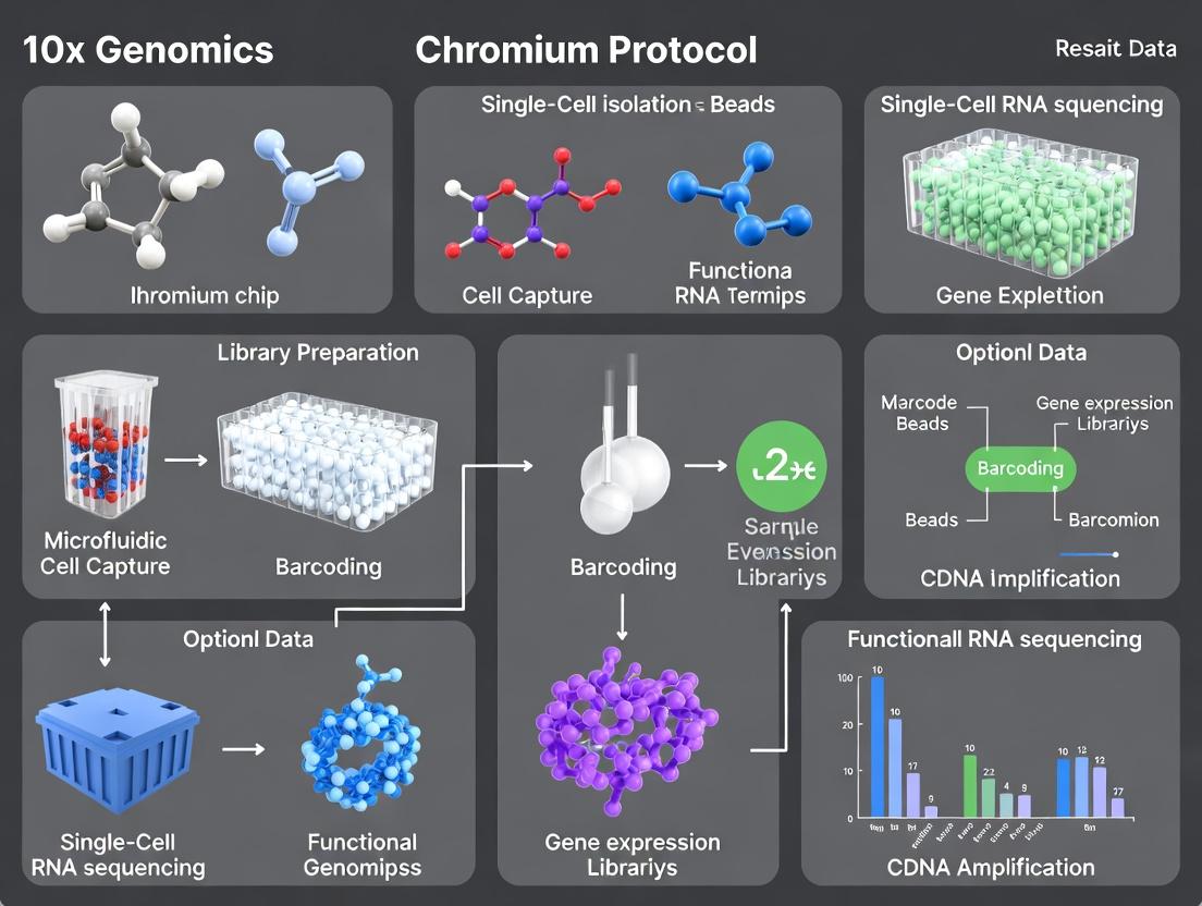
Mastering Single-Cell RNA-seq: A Complete Guide to the 10x Genomics Chromium Protocol
This comprehensive guide provides researchers and drug development professionals with an in-depth exploration of the 10x Genomics Chromium single-cell RNA sequencing (scRNA-seq) platform.
Mastering Single-Cell RNA-seq: A Complete Guide to the 10x Genomics Chromium Protocol
Abstract
This comprehensive guide provides researchers and drug development professionals with an in-depth exploration of the 10x Genomics Chromium single-cell RNA sequencing (scRNA-seq) platform. We cover foundational principles, from the core chemistry of Gel Bead-in-emulsion (GEM) generation to cellular indexing. The article details the complete workflow from sample preparation to data analysis, addresses common troubleshooting and optimization challenges, and critically validates performance metrics while comparing Chromium to alternative platforms like SMART-seq and droplet-based methods. The guide concludes with insights into translational applications in immunology, oncology, and neuroscience, offering a practical resource for experimental design and execution.
Demystifying the 10x Chromium Platform: Core Principles and Single-Cell Biology Applications
Bulk RNA-seq has been foundational in transcriptomics, measuring the average gene expression across thousands to millions of cells in a sample. However, this approach masks cellular heterogeneity, a fundamental characteristic of tissues, tumors, and immune systems. Single-cell RNA sequencing (scRNA-seq) technologies, such as the 10x Genomics Chromium platform, resolve this by profiling gene expression in individual cells, enabling the discovery of novel cell types, states, and dynamics invisible in bulk analysis.
Quantitative Comparison: Bulk RNA-seq vs. Single-Cell RNA-seq
The following table summarizes the core differences between the two approaches, highlighting the paradigm shift enabled by single-cell resolution.
Table 1: Core Comparison of Bulk RNA-seq and Single-Cell RNA-seq
| Feature | Bulk RNA-seq | Single-Cell RNA-seq (e.g., 10x Chromium) |
|---|---|---|
| Resolution | Population average | Individual cell |
| Primary Output | Aggregate gene expression profile | Gene expression matrix (Cells x Genes) |
| Key Capability | Detect differentially expressed genes between conditions | Identify cell types, states, trajectories, and rare populations |
| Information on Heterogeneity | Obscured and lost | Explicitly measured and characterized |
| Typical Cells per Sample | One measurement (pool of millions) | 500 - 10,000+ individual cell measurements |
| Cost per Cell | Very low | Higher, but continuously decreasing |
| Complexity of Data Analysis | Relatively standardized | High, requiring specialized pipelines for QC, clustering, etc. |
| Application Example | Comparing tumor vs. normal tissue expression | Deconstructing tumor microenvironment (T cells, macrophages, cancer stem cells) |
Detailed Protocol: 10x Genomics Chromium Single-Cell 3' RNA-seq
This protocol outlines the key steps for library preparation using the 10x Genomics Chromium Controller and associated kits.
Title: 10x Chromium Single-Cell 3' Reagent Kit v3.1 Workflow
Principle: Gel Bead-In-EMulsions (GEMs) are formed where each GEM contains a single cell, a single Gel Bead with barcoded oligonucleotides, and RT reaction mix. Within each GEM, cell lysis, reverse transcription, and barcoding occur, uniquely tagging all cDNA from an individual cell.
Materials & Reagents:
- Chromium Controller & Chip G
- 10x Genomics Chromium Single Cell 3' GEM, Library & Gel Bead Kit v3.1
- Single cell suspension (viability >90%, concentration optimized for target cell recovery)
- SPRISelect magnetic beads
- Thermal cycler
- Bioanalyzer/TapeStation
Procedure:
- Single-Cell Suspension Preparation: Prepare a single-cell suspension in appropriate buffer (e.g., PBS + 0.04% BSA). Filter through a flow cytometry-compatible strainer (e.g., 40 µm). Count and assess viability. Adjust concentration to the target for loading (e.g., ~1,000 cells/µl for 10,000 cell target).
- GEM Generation & RT:
- Combine Cells, Master Mix, and Gel Beads onto a Chromium Chip G.
- Load the chip into the Chromium Controller. The instrument partitions the mixture into nanoliter-scale GEMs.
- Transfer the GEMs to a PCR tube and perform reverse transcription in a thermal cycler (53°C for 45 min, 85°C for 5 min). This generates full-length, barcoded cDNA.
- Cleanup & Amplification:
- Break GEMs and recover barcoded cDNA using Recovery Agent.
- Purify cDNA with SPRISelect beads.
- Amplify the cDNA via PCR (98°C for 3 min; [98°C for 15s, 63°C for 20s, 72°C for 1 min] x 12 cycles; 72°C for 1 min).
- Purify amplified cDNA again with SPRISelect beads. Quantify and check size distribution (~1-10 kb).
- Library Construction:
- Fragment, end-repair, and A-tail the amplified cDNA.
- Ligate sample index adaptors via a second PCR (98°C for 45s; [98°C for 20s, 54°C for 30s, 72°C for 20s] x 12-16 cycles; 72°C for 1 min).
- Perform a double-sided size selection with SPRISelect beads to remove very short and very long fragments.
- QC & Sequencing:
- Assess final library quality (size, concentration) using a Bioanalyzer.
- Sequence on an Illumina platform (typical read configuration: Read 1: 28 cycles, i7 Index: 10 cycles, i5 Index: 10 cycles, Read 2: 90 cycles).
Visualizing the Single-Cell RNA-seq Revolution
The following diagrams illustrate the conceptual shift and the core workflow.
Title: From Bulk Average to Single-Cell Resolution
Title: 10x Chromium Single-Cell Partitioning & Barcoding
The Scientist's Toolkit: Key Reagents for 10x Chromium scRNA-seq
Table 2: Essential Research Reagent Solutions for 10x Chromium Experiments
| Reagent/Material | Function | Critical Note |
|---|---|---|
| Chromium Single Cell 3' Gel Bead Kit | Contains barcoded gel beads, partitioning oil, enzymes, and buffers for GEM generation and RT. | Kit version (e.g., v3.1, v4) dictates chemistry and sensitivity. Must match Chip. |
| Chromium Chip G | Microfluidic chip for generating single-cell GEMs. | Specific to cell throughput (e.g., Chip G for 10k cells). |
| SPRISSelect Beads | Solid-phase reversible immobilization (SPRI) magnetic beads for post-RT and post-PCR cleanups. | Essential for cDNA and library purification. Size selection ratios are critical for library quality. |
| Dual Index Kit Plate Sets | Provides unique combinatorial i7 and i5 indices for multiplexing samples in a single sequencing run. | Allows pooling of up to 96 libraries. Index hopping is minimized. |
| Live/Dead Cell Stain (e.g., AO/PI, DAPI) | Used to assess viability of the single-cell suspension prior to loading. | >90% viability is strongly recommended to limit background from dead cells. |
| Phosphate-Buffered Saline (PBS) with 0.04% BSA | Buffer for preparing and diluting single-cell suspensions. | BSA reduces cell adhesion and loss. Calcium/Magnesium-free PBS is often used. |
| Nuclease-Free Water | Used for resuspending and diluting various reagents. | Prevents degradation of RNA and enzymatic reactions. |
Within the context of advancing single-cell RNA sequencing (scRNA-seq) research, the 10x Genomics Chromium System has emerged as a foundational platform. It enables high-throughput, droplet-based partitioning of single cells, facilitating the detailed analysis of cellular heterogeneity. This ecosystem is pivotal for researchers and drug development professionals investigating complex biological systems, disease mechanisms, and therapeutic targets.
The Chromium System's performance is characterized by key metrics that define its utility in scalable single-cell research.
Table 1: Chromium Platform Performance Metrics (Current Generation)
| Metric | Chromium X Series | Chromium Connect | Notes |
|---|---|---|---|
| Cells Recovered per Run | 10,000 - 1,000,000+ | 1,000 - 80,000 | Scalable based on chip and reagent kit selection. |
| Cell Throughput (Cells/Hour) | Up to 80,000 | Up to 16,000 | Includes time for library preparation. |
| Cell Multiplexing Samples per Run | Up to 96 (with CellPlex) | Up to 12 (with CellPlex) | Enables sample pooling and demultiplexing. |
| Gene Detection per Cell | 1,000 - 10,000+ | 500 - 5,000+ | Varies by cell type, viability, and protocol. |
| Reads Required per Cell | 20,000 - 50,000 | 10,000 - 30,000 | For standard 3' or 5' gene expression. |
| Droplet Generation Rate | ~15,000 droplets/sec | ~6,000 droplets/sec | Ensures high cell capture efficiency. |
| Single-Cell Capture Efficiency | 40-65% | 40-65% | Percentage of cells loaded that are encapsulated. |
| Multiplet Rate | <0.9% per 1k cells | <1.5% per 1k cells | Lower at lower cell loading concentrations. |
Detailed Application Notes & Protocols
Protocol 1: Standard Single Cell 3’ Gene Expression (v3.1/v4.0)
Objective: To generate barcoded, sequencing-ready cDNA libraries from single cells for transcriptome quantification.
Key Steps:
- Cell Suspension Preparation: Create a single-cell suspension with >90% viability in PBS + 0.04% BSA. Target cell concentration is 700-1,200 cells/µL.
- Master Mix Preparation: Combine RT reagents, Gel Beads containing barcoded oligonucleotides (Illumina TruSeq Read 1, 16bp 10x Barcode, 12bp UMI, poly-dT), and partitioning oil.
- Droplet Partitioning: Load the cell suspension and master mix onto a Chromium Next GEM Chip. The Chromium Controller generates Gel Bead-In-EMulsions (GEMs), where each droplet contains a single cell, a single Gel Bead, and RT reagents.
- Reverse Transcription (In-Droplet): GEMs are transferred to a thermal cycler. Cells are lysed, and poly-adenylated mRNA is captured by the Gel Bead oligo-dT and reverse-transcribed into barcoded, full-length cDNA.
- Cleanup & Amplification: GEMs are broken, and pooled cDNA is recovered. SPRIselect cleanup is performed. cDNA is then PCR-amplified (12 cycles).
- Library Construction: Amplified cDNA is enzymatically fragmented, end-repaired, A-tailed, and ligated to a sample index adapter via Chromium i7 Multiplex Kit. Final PCR (12 cycles) adds P5, P7, and i7 sample index sequences.
- Quality Control & Sequencing: Libraries are quantified (qPCR) and sized (Bioanalyzer/TapeStation). Recommended sequencing depth is 20,000-50,000 reads per cell on Illumina NovaSeq or HiSeq platforms.
Protocol 2: Single Cell Multimodal (ATAC + Gene Expression)
Objective: To simultaneously profile chromatin accessibility (ATAC-seq) and transcriptome (RNA-seq) from the same single nucleus/cell.
Key Steps:
- Nuclei Isolation: Isolate nuclei from fresh frozen or fixed tissue using a lysis buffer. Critical to maintain nuclear integrity.
- Tagmentation & Partitioning: Combine nuclei with a transposase loaded with mosaic adapters and Gel Beads containing multimodality barcoded oligos. Load onto a Chromium Next GEM Chip for partitioning.
- In-Droplet Reactions: Within each GEM, two simultaneous reactions occur: a) Transposition of accessible chromatin, and b) Reverse transcription of poly-adenylated mRNA.
- Post-GEM Processing: GEMs are broken, and products are split into two aliquots for separate processing.
- ATAC Library Prep: The transposed DNA aliquot undergoes a primer extension and PCR (12 cycles) using a Chromium i7 Multiplex Kit to add P5, P7, and sample index.
- Gene Expression Library Prep: The cDNA aliquot undergoes cDNA amplification, fragmentation, and adapter ligation (similar to Protocol 1) to generate the GEX library.
- Sequencing: ATAC and GEX libraries are sequenced separately. Recommended: 25,000 GEX reads and 25,000 ATAC fragments per nucleus.
The Scientist's Toolkit: Key Research Reagent Solutions
Table 2: Essential Chromium Reagents & Materials
| Item | Function in Workflow | Key Notes |
|---|---|---|
| Chromium Next GEM Chip (e.g., G, K, X) | Microfluidic device for generating uniform droplets (GEMs). | Chip type determines max cell throughput. Single-use. |
| Chromium Gel Beads | Deliver barcoded oligonucleotides (cell barcode, UMI, adapter) into each droplet. | Bead type is assay-specific (3’, 5’, ATAC, Multiome, etc.). |
| Chromium Partitioning Oil | Immiscible oil phase for stable droplet formation within the chip. | Critical for consistent GEM generation. |
| Chromium i7 Multiplex Kit | Provides unique dual-index adapters (i7 & i5) for sample multiplexing. | Essential for pooling multiple libraries in one sequencing lane. |
| SPRIselect Beads | Solid-phase reversible immobilization beads for size selection and cleanup of cDNA/libraries. | Used for post-RT cleanup and post-library size selection. |
| Buffer Kit (e.g., Reverse Transcription, Lysis) | Contains enzymes and buffers for in-droplet cell lysis, RT, and cDNA amplification. | Kit-specific; optimized for performance. |
| Single-Cell Suspension Reagent (e.g., PBS/0.04% BSA) | Suspension buffer to minimize cell adhesion and maintain viability. | Must be nuclease-free. BSA is a carrier protein. |
| Viability Stain (e.g., Trypan Blue, AO/PI) | To assess cell viability and concentration pre-loading. | >90% viability is strongly recommended. |
| DNA/RNA Shield | Stabilization reagent for fixed samples or tissue storage. | Preserves nucleic acids for later analysis. |
Within the framework of 10x Genomics Chromium protocol single-cell RNA-seq research, the generation of Gel Bead-in-Emulsions (GEMs) is the foundational step that enables massively parallel, barcoded analysis of thousands of single cells. This application note details the principles, quantitative parameters, and step-by-step protocols for robust GEM formation and barcoding, critical for researchers and drug development professionals aiming to implement high-throughput single-cell genomics.
GEM formation is a microfluidic process that partitions individual cells, lysis reagents, and uniquely barcoded gel beads into nanoliter-scale aqueous droplets within an oil emulsion. Each gel bead is loaded with oligonucleotides containing a shared 10x Barcode, a Unique Molecular Identifier (UMI), and a poly-dT sequence for mRNA capture. The co-partitioning of a single cell with a single gel bead in a GEM ensures that all cDNA derived from that cell shares the same barcode, enabling pooled sequencing and computational deconvolution.
Quantitative Parameters of GEM Formation
Table 1: Key Quantitative Metrics for Chromium GEM Formation
| Parameter | Typical Value / Range | Significance |
|---|---|---|
| Target Cell Recovery Rate | 65% | Percentage of input cells successfully partitioned into single-cell GEMs. |
| Single-Cell Multiplexing Capacity | Up to 10,000 cells per channel | Maximum number of cells loaded to maintain high single-cell capture efficiency. |
| Gel Beads per GEM | ~1 bead per droplet (Poisson loading) | Ensures barcode uniqueness. |
| Partition Volume | ~1 nL | Defines reaction volume for reverse transcription. |
| Number of Barcodes | 750,000 per bead; 4 million per channel | Provides vast diversity to label each cell's transcripts uniquely. |
| Recommended Cell Viability | >90% | Minimizes ambient RNA from dead cells. |
| Doublet Rate | ~0.8% per 1,000 cells loaded | Function of cell concentration and Poisson distribution. |
Table 2: Reagent Volumes for Chromium Next GEM Chip Kits (Example: Single Cell 3')
| Reagent | Volume per Reaction (µL) | Function |
|---|---|---|
| Master Mix (Cells, Buffer, RT reagents) | 36.8 | Contains cells and reagents for reverse transcription. |
| Gel Beads | 5.2 | Source of barcoded oligonucleotides. |
| Partitioning Oil | 310 | Creates the emulsion. |
| Recovery Reagent | 165 | Breaks emulsions and recovers aqueous phase. |
Detailed Protocol: GEM Generation and Barcoding
Protocol 1: Preparing the Single Cell Suspension
- Cell Preparation: Harvest and wash cells in PBS supplemented with 0.04% BSA. Filter through a 40 µm flow cytometry strainer.
- Viability and Counting: Determine viability and exact cell concentration using an automated cell counter (e.g., Trypan Blue exclusion). Adjust concentration to the target for your chip type (e.g., 1,000 cells/µL for a target of 10,000 cells).
- Master Mix Assembly: On ice, combine in a nuclease-free tube:
- Single Cell Suspension:
X µL(for target cell count) - Nuclease-Free Water:
(32.8 - X) µL - Master Mix (from kit):
4.0 µL - Total Volume:
36.8 µL
- Single Cell Suspension:
- Mix gently by pipetting and keep on ice until loading.
Protocol 2: Microfluidic Chip Loading and GEM Generation
- Chip Preparation: Place a new Chromium Next GEM Chip into the chip holder.
- Loading Reagents: Pipette reagents into the designated wells:
- Well 1:
50 µLof Partitioning Oil. - Well 2:
36.8 µLof prepared cell Master Mix. - Well 3:
50 µLof Partitioning Oil. - Well 4:
5.2 µLof Gel Beads. - Well 5:
50 µLof Partitioning Oil. - Well 6:
50 µLof Partitioning Oil. - Well 7:
50 µLof Partitioning Oil. - Well 8:
50 µLof Partitioning Oil.
- Well 1:
- Run the Chip: Place the loaded chip into the Chromium Controller and run the "Single Cell 3' v3.1" program. The instrument uses pressure-based microfluidics to precisely combine reagents and generate up to 1 million partitions per channel.
- GEM Collection: Post-run, carefully collect the GEMs (~100 µL emulsion) from the recovery well into a clean 0.2 mL PCR tube. Proceed immediately to reverse transcription.
Protocol 3: Post-GEM Reverse Transcription and Cleanup
- Incubate for RT: Place the tube in a pre-heated thermal cycler and run:
53°Cfor 45 minutes (Reverse Transcription)85°Cfor 5 minutes (enzyme inactivation)- Hold at
4°C.
- Break Emulsions: Add
165 µLof Recovery Reagent to the GEMs. Mix by pipetting up and down 10 times. Incubate at room temperature for 2 minutes. A biphasic solution will form. - Purify cDNA: Combine the aqueous lower phase with
200 µLof DynaBeads Cleanup Mix (from kit). Follow kit instructions for bead-based purification. Elute in45 µLof Elution Buffer. - Amplify cDNA: Perform PCR amplification on the purified barcoded cDNA using recommended cycles (typically 12-14 cycles). Purify the amplified cDNA using SPRIselect beads.
Visualizing the GEM Formation and Barcoding Workflow
Diagram Title: GEM Formation and Barcoding Experimental Workflow
The Scientist's Toolkit: Key Research Reagent Solutions
Table 3: Essential Materials for GEM-based Single-Cell RNA-seq
| Item | Function | Critical Notes |
|---|---|---|
| Chromium Next GEM Chip | Microfluidic device with precisely etched channels to generate uniform partitions. | Single-use. Different chips (e.g., X, K) accommodate different cell targets. |
| Barcoded Gel Beads | Hydrogel beads containing billions of oligonucleotide constructs with unique 10x Barcodes and UMIs. | Stored at 4°C. Critical for assigning reads to single cells. |
| Partitioning Oil | Fluorinated oil with surfactants to stabilize water-in-oil emulsions. | Prevents droplet coalescence and ensures compartmentalization. |
| Master Mix | Contains reverse transcriptase, dNTPs, and reagents for cell lysis and cDNA synthesis. | Proprietary formulation optimized for performance within GEMs. |
| Recovery Reagent | Destabilizes the emulsion for aqueous phase recovery post-RT. | Contains PEG and other agents to break the oil-water interface. |
| SPRIselect Beads | Solid-phase reversible immobilization (SPRI) magnetic beads for size selection and cleanup. | Used for post-amplification cDNA and final library purification. |
| Chromium Controller | Instrument that applies pressure to drive precise microfluidic mixing and GEM generation. | Essential for consistent, high-quality partition formation. |
Within the framework of a thesis investigating tumor heterogeneity using 10x Genomics Chromium single-cell RNA sequencing (scRNA-seq), a precise understanding of core reagents is critical. These components enable the partitioning, barcoding, and reverse transcription of thousands of single cells, forming the foundation for high-throughput transcriptomic analysis in drug discovery and basic research.
Master Mix
The Master Mix is a proprietary, enzyme-based solution central to the 10x Genomics workflow. It contains reverse transcriptase, template-switching oligonucleotides, dNTPs, and necessary co-factors.
Function: Upon cell lysis within a Gel Bead-in-EMulsion (GEM), the Master Mix initiates reverse transcription. It converts poly-adenylated mRNA into full-length, barcoded cDNA, leveraging template switching to add universal primer sequences.
Key Components Table:
| Component | Function in scRNA-seq |
|---|---|
| Reverse Transcriptase | Synthesizes cDNA from mRNA template. |
| Template Switch Oligo (TSO) | Enables strand switching for universal adapter addition. |
| dNTPs | Building blocks for cDNA synthesis. |
| RNase Inhibitor | Protects RNA integrity during reaction. |
| Reducing Agent | Maintains enzyme stability in the reaction environment. |
Gel Beads
Gel Beads are micron-sized, degradable beads each impregnated with millions of copies of a unique oligonucleotide barcode.
Function: Each Gel Bead delivers a unique 10x Barcode, a Unique Molecular Identifier (UMI), and a poly(dT) primer sequence into a single partition. This ensures all cDNA from a single cell receives the same cell barcode, while each mRNA molecule receives a unique UMI for digital quantification.
Gel Bead Oligo Structure:
[10x Barcode] [UMI] [Poly(dT) Primer]
Partitioning Oil
Partitioning Oil is a surfactant-based reagent used to generate nanoliter-scale droplets in the Chromium chip.
Function: It flows alongside the aqueous stream containing cells, Master Mix, and Gel Beads to create stable, water-in-oil emulsions (GEMs). The oil's properties ensure single-cell encapsulation and prevent coalescence of droplets.
Single Cell 3' vs. 5' Kits
These kit families determine which end of the transcript is enriched and barcoded, influencing the biological questions addressable.
Comparative Table: 3' vs. 5' Gene Expression Kits
| Feature | Single Cell 3' Kit | Single Cell 5' Kit |
|---|---|---|
| Target | 3' end of poly-adenylated mRNA | 5' end of mRNA (or V(D)J transcripts) |
| Barcoding Location | 3' UTR region | 5' start of transcript |
| Primary Application | Gene expression profiling, differential expression | Paired gene expression + immune receptor profiling (TCR/BCR) |
| Compatible Add-ons | Feature Barcoding (CRISPR, Antibody) | V(D)J Enrichment, Feature Barcoding |
| Ideal For Thesis On | General tumor heterogeneity, cell type identification | Tumor immunology, immune cell clonality |
Detailed Protocol: GEM Generation & Barcoding
This protocol is integral to the 10x Genomics Chromium Controller workflow.
Materials:
- Chromium Controller & appropriate Chip
- Single Cell 3' or 5' Kit (contains Gel Beads, Master Mix, Partitioning Oil)
- Single Cell Suspension (viability >80%, concentration adjusted)
- Nuclease-free water
- Recovery Reagents (from kit)
Procedure:
- Prepare Reagents: Thaw Master Mix, Gel Beads, and Partitioning Oil. Vortex and spin down Gel Beads thoroughly.
- Load Chromium Chip: Using a single-channel pipette:
- Add Partitioning Oil to the oil wells.
- Load Master Mix and single-cell suspension into the assigned right-side wells.
- Load Gel Beads into the assigned left-side wells.
- Run on Chromium Controller: Place chip into the controller and run the "Single Cell" program. This automatically creates up to 80,000 GEMs.
- GEM-RT Incubation: Transfer the GEMs from the collection chamber to a PCR tube. Perform reverse transcription in a thermal cycler (53°C for 45 min, 85°C for 5 min, hold at 4°C).
- GEM Breakage & Cleanup: Add Recovery Reagents to break the oil emulsion. Use DynaBeads (provided) to clean up the barcoded cDNA.
- Amplification: Amplify the cDNA via PCR (98°C for 3 min; cycled: 98°C for 15s, 63°C for 20s, 72°C for 1 min; 72°C for 1 min).
- Quality Control: Check cDNA fragment size (~1000-10,000 bp) using an Agilent Bioanalyzer High Sensitivity DNA chip.
The Scientist's Toolkit: Essential Research Reagent Solutions
| Item | Function in 10x Genomics Workflow |
|---|---|
| Chromium Chip B | Microfluidic device for generating single-cell GEMs. |
| SPRIselect Reagent | Size-selective magnetic beads for post-amplification library purification. |
| Dual Index Kit TT Set A | Provides sample indexes for multiplexing libraries for sequencing. |
| Buffer EB (Elution Buffer) | Low-EDTA TE buffer for eluting and storing final libraries. |
| Agilent High Sensitivity DNA Kit | For quality control of cDNA and final libraries pre-sequencing. |
Visualized Workflows
Title: 10x Chromium Single-Cell Partitioning and Barcoding Workflow
Title: 3' vs 5' Kit cDNA Synthesis Mechanism
Within single-cell RNA sequencing (scRNA-seq) using the 10x Genomics Chromium platform, cellular indexing and molecular tagging are foundational for highly multiplexed, accurate analysis. Cellular barcodes assign a unique identifier to each cell, enabling thousands of cells to be pooled and sequenced simultaneously. Unique Molecular Identifiers (UMIs) tag individual mRNA molecules, allowing for the digital counting of transcripts and correction for amplification bias. Together, these technologies enable precise, high-throughput measurement of gene expression at single-cell resolution, crucial for research in oncology, immunology, and drug development.
Key Concepts and Quantitative Data
Table 1: Comparison of Indexing and Tagging Elements
| Element | Sequence Length (bp) | Primary Function | Key Property | Typical Count |
|---|---|---|---|---|
| Cell Barcode | 10-16 bp (10x: 16bp) | Uniquely labels all mRNA from a single cell | Enables sample/cell multiplexing | Up to 4^10 (10^6) theoretical combinations |
| Unique Molecular Identifier (UMI) | 10-12 bp (10x: 12bp) | Tags individual mRNA molecules | Enables PCR duplicate removal & absolute quantification | 4^12 (16.8M) theoretical combinations |
| Illumina i7/i5 Index | 8-10 bp | Demultiplexes pooled libraries by sample | Enables sample-level multiplexing on sequencer | Standard for Illumina platforms |
| Poly(dT) Primer | 30 bp | Binds to mRNA poly-A tail | Initiates reverse transcription | N/A |
Table 2: Impact of UMI Correction on Data Fidelity
| Metric | Without UMI Deduplication | With UMI Deduplication | Improvement |
|---|---|---|---|
| PCR Duplicate Rate | 30-60% of reads | Reduced to <5% | >6-fold reduction |
| Quantification Accuracy | Overestimates expression | Reflects true molecule count | Essential for digital counting |
| Detection of Lowly Expressed Genes | Impaired by duplicate noise | Enhanced sensitivity | Critical for rare cell populations |
Detailed Protocols
Protocol 1: 10x Genomics Chromium Single Cell 3' Reagent Kit v3.1 - Library Construction
This protocol details the key steps where cellular barcodes and UMIs are incorporated.
Materials:
- 10x Genomics Chromium Controller & Chip
- Single Cell 3' Gel Beads (v3.1)
- Reverse Transcription Master Mix
- Partitioning Oil
- DynaBeads MyOne SILANE
- SPRIselect Reagent Kit
Procedure:
- Cell Suspension Preparation: Prepare a single-cell suspension at 700-1,200 cells/µL in PBS + 0.04% BSA. Aim for >90% viability.
- Master Mix Assembly: Combine RT Master Mix, cells, and gel beads. The gel beads contain:
- Billions of unique oligonucleotides with:
- Illumina R1 sequence
- 16bp Cellular Barcode
- 12bp UMI
- 30bp Poly(dT) sequence
- Billions of unique oligonucleotides with:
- Partitioning on Chromium Chip: Load the master mix and partitioning oil onto a Chromium Chip. The Controller generates nanoliter-scale Gel Bead-In-EMulsions (GEMs). Each GEM ideally contains a single cell, a single gel bead, and reagents.
- Reverse Transcription within GEMs: Incubate at 53°C for 45 min. Within each GEM, poly-adenylated mRNA binds to the poly(dT) primer. Reverse transcription creates full-length cDNA tagged with the cell barcode and UMI.
- GEM-RT Cleanup & cDNA Amplification: Break emulsions, pool contents. Purify cDNA with DynaBeads. Amplify cDNA via PCR (12 cycles).
- Library Construction: Fragment, A-tail, and ligate sample indexes and adapters (Illumina i5/i7). Perform SPRIselect size selection (typically 200-600 bp).
- Quality Control: Assess library concentration (qPCR) and size distribution (Bioanalyzer/TapeStation).
Protocol 2: Computational Demultiplexing and UMI Counting (Cell Ranger)
This protocol outlines the standard bioinformatics processing of raw sequencing data.
Materials:
- Raw FASTQ files (Read1: cDNA, Read2: Sample Index, I7 Index)
- 10x Genomics Cell Ranger software (v7.0+)
- Reference genome (e.g., GRCh38)
Procedure:
cellranger mkfastq: Demultiplexes sample-level indices (i7) from the Illumina sequencer output. Generates FASTQ files for each sample.cellranger count: Performs per-sample analysis.- Barcode Processing: Identifies valid 16bp cell barcodes from Read 1, filtering out non-whitelist barcodes.
- UMI Processing: Extracts the 12bp UMI.
- Alignment: Maps reads to the reference genome.
- Gene-Barcode Matrix Generation: For each cell barcode (column) and gene (row), counts unique UMIs. This deduplication yields a digital expression matrix.
- Cell Calling: Distinguits real cells from background using barcode sequencing saturation and UMI counts.
- Output: A filtered feature-barcode matrix containing accurate, digital gene expression counts per cell.
Diagrams
Title: 10x Chromium scRNA-seq Barcoding Workflow
Title: UMI Counting for Digital Expression
The Scientist's Toolkit: Research Reagent Solutions
Table 3: Essential Materials for 10x Genomics scRNA-seq
| Item | Supplier/Kit | Primary Function |
|---|---|---|
| Chromium Next GEM Chip | 10x Genomics | Microfluidic device for generating single-cell GEMs. |
| Single Cell 3' Gel Beads v3.1 | 10x Genomics | Contains barcoded oligos with cell barcode and UMI. |
| Chromium Controller | 10x Genomics | Instrument to precisely control GEM generation. |
| DynaBeads MyOne SILANE | Thermo Fisher | Magnetic beads for post-RT cleanup and cDNA purification. |
| SPRIselect Beads | Beckman Coulter | Size selection and cleanup of cDNA and final libraries. |
| High Sensitivity DNA Kit | Agilent (Bioanalyzer) | QC of cDNA and final library fragment size. |
| Cell Ranger Software | 10x Genomics | Primary pipeline for demultiplexing, alignment, and UMI counting. |
Application Notes
Single-cell RNA sequencing (scRNA-seq) using the 10x Genomics Chromium platform has become a cornerstone for dissecting cellular heterogeneity and function. Within the context of a thesis employing this technology, four core biological questions are routinely addressed. The following notes synthesize current methodologies and applications relevant to researchers and drug development professionals.
Cell Typing involves classifying individual cells into distinct biological states or types based on their transcriptomic profiles. This is foundational, enabling the identification of rare cell populations, defining tumor microenvironments, and characterizing developmental stages. Post-sequencing, dimensionality reduction (PCA, UMAP) and clustering (Louvain, Leiden) are applied. Marker genes for each cluster are identified and cross-referenced with known databases (e.g., CellMarker, PanglaoDB) for annotation.
Differential Expression (DE) analysis compares gene expression profiles between predefined groups of cells (e.g., different cell types, treated vs. control, diseased vs. healthy). It identifies key driver genes and dysregulated pathways. For single-cell data, methods like MAST, Wilcoxon rank-sum test, and DESeq2 adapted for sparse data are used. DE results are crucial for identifying therapeutic targets and understanding disease mechanisms.
Trajectory Inference (TI) or Pseudotemporal Ordering reconstructs dynamic biological processes such as differentiation, cell cycle, or immune response. Algorithms (Monocle3, PAGA, Slingshot) order cells along a pseudotime continuum based on transcriptomic similarity, inferring the sequence of gene expression changes. This is vital for modeling development, response to perturbation, and transitions between states like epithelial-to-mesenchymal transition.
Cell-Cell Interactions (CCI) analysis predicts communication events between different cell types within a tissue based on the co-expression of ligand-receptor pairs. Tools like CellChat, NicheNet, and CellPhoneDB leverage curated interaction databases to infer signaling networks. This application is key in oncology, immunology, and stromal research for understanding the cellular crosstalk that governs tissue homeostasis and disease.
Table 1: Common Computational Tools for scRNA-seq Analysis (10x Genomics Data)
| Biological Question | Primary Tools/Algorithms | Typical Input | Key Output |
|---|---|---|---|
| Cell Typing | Seurat (Louvain/Leiden), Scanpy, SingleR | Filtered count matrix (cells x genes) | Cell cluster labels, marker gene list, annotated cell type identities |
| Differential Expression | MAST, Wilcoxon test, DESeq2 (single-cell) | Count matrix + cell group labels | List of DEGs with p-values, log2 fold change, adjusted p-values |
| Trajectory Inference | Monocle3, PAGA (Scanpy), Slingshot | UMAP/PCA coordinates, clustered data | Pseudotime ordering, trajectory graph, branch points |
| Cell-Cell Interactions | CellChat, CellPhoneDB, NicheNet | Annotated cell types + count matrix | Inferred ligand-receptor pairs, communication probability scores, signaling pathways |
Table 2: Typical 10x Genomics Chromium Single Cell 3’ Reagent Kits Output (v3.1)
| Metric | Typical Range | Note |
|---|---|---|
| Number of Cells Recovered | 1,000 - 10,000 per lane | Depends on loading concentration. |
| Median Genes per Cell | 1,000 - 5,000 | A measure of library complexity. |
| Sequencing Saturation | >50% recommended | Higher saturation improves detection. |
| Read Pairs per Cell | 20,000 - 50,000 | Standard for gene expression. |
| Fraction of Reads in Cells | >70% | Indicates efficient cell capture. |
Experimental Protocols
Protocol 1: Standard 10x Genomics Chromium Single Cell 3’ Gene Expression Workflow
Objective: To generate single-cell gene expression libraries from a fresh or frozen cell suspension. Key Reagents & Equipment: 10x Genomics Chromium Controller, Single Cell 3’ Gel Beads & Library Kits (v3.1 or v4), Partitioning Chips, Thermal Cycler with 96-Deep Well Block, SPRIselect Beads, Bioanalyzer/TapeStation, Validated Cell Suspension (≥90% viability).
- Cell Preparation: Prepare a single-cell suspension at 700-1200 cells/µL in PBS + 0.04% BSA. Filter through a 40µm flowmi cell strainer.
- Master Mix Assembly: On ice, combine RT Reagent Mix, Primer, and Additive from the kit. Add Enzyme and template switch oligo.
- Chromium Chip Loading: Load the chip on the Chromium Controller. Pipette the cell suspension, master mix, and partitioning oil into the designated wells.
- Partitioning & Barcoding: Run the "Single Cell 3’" program on the Controller. This co-partitions single cells, Gel Beads (with barcoded oligos), and reagents into nanoliter-scale droplets for reverse transcription.
- Post-Processing: Break droplets, recover barcoded cDNA. Perform cleanup with DynaBeads MyOne SILANE.
- cDNA Amplification: Amplify the full-length cDNA via PCR (13 cycles). Clean up with SPRIselect beads (0.6x / 0.8x ratio).
- Library Construction: Fragment, end-repair, A-tail, and ligate sample index adapters via a second PCR (12 cycles). Perform a final double-sided SPRI size selection (0.6x / 0.8x) to remove short fragments.
- QC & Sequencing: Assess library concentration (Qubit) and size profile (Bioanalyzer High Sensitivity DNA chip). Pool libraries and sequence on an Illumina platform (e.g., NovaSeq 6000) with recommended read length: Read 1: 28 cycles, i7 Index: 10 cycles, Read 2: 90 cycles.
Protocol 2: Computational Pipeline for Integrated Analysis (Seurat v5)
Objective: To process raw sequencing data (FASTQ) into analyzed data addressing the four major biological questions. Software Environment: R (v4.3+), Seurat v5, Signac, relevant interaction databases (CellChatDB).
- Data Preprocessing:
- Use
Cell Ranger count(10x Genomics) to align reads (to GRCh38/ mm10), filter barcodes, and generate a filtered feature-barcode matrix. - Load matrix into R:
Read10X()and create a Seurat object. - QC Filtering: Filter cells with high mitochondrial percentage (>20%) and extreme feature counts (nFeature_RNA < 200 or > 6000).
- Use
- Normalization & Scaling: Normalize data (
NormalizeData(), log-normalization). Find variable features (FindVariableFeatures()). Scale data (ScaleData()) regressing out effects ofpercent.mtandnCount_RNA. - Cell Typing:
- Perform linear dimensional reduction (
RunPCA()). - Cluster cells (
FindNeighbors()thenFindClusters()at chosen resolution, e.g., 0.5). - Run non-linear dimensional reduction (
RunUMAP()). - Find marker genes for each cluster (
FindAllMarkers()with Wilcoxon test). - Annotate clusters using canonical marker genes or reference-based tools (SingleR).
- Perform linear dimensional reduction (
- Differential Expression:
- Subset the Seurat object to cells of interest (e.g., a specific cell type across conditions).
- Identify DEGs using
FindMarkers()with the MAST test, specifying theident.1andident.2parameters.
- Trajectory Inference (Using Monocle3 within Seurat):
- Convert the Seurat object to a CellDataSet object for Monocle3.
- Use
learn_graph()andorder_cells()to infer trajectory and pseudotime.
- Cell-Cell Interaction Analysis (Using CellChat):
- Subset normalized data for a sample/condition.
- Create a CellChat object from the Seurat object and annotated cell labels.
- Preprocess using
identifyOverExpressedGenes()andidentifyOverExpressedInteractions(). - Compute communication probability with
computeCommunProb()andcomputeCommunProbPathway(). - Visualize aggregated communication networks with
netVisual_aggregate().
The Scientist's Toolkit: Research Reagent Solutions
Table 3: Essential Materials for 10x Genomics scRNA-seq Experiments
| Item | Function/Description | Example Product/Kit |
|---|---|---|
| Chromium Controller & Chip | Microfluidic platform to generate gel bead-in-emulsions (GEMs) for single-cell partitioning and barcoding. | 10x Genomics Chromium Controller, Chip K |
| Single Cell 3’ Gel Bead & Library Kit | Contains all reagents for GEM-RT, cDNA amplification, and library construction for 3’ gene expression. | 10x Genomics Chromium Next GEM Single Cell 3’ Kit v3.1 |
| Single Cell 3’ Feature Barcode Kit | Enables surface protein or CRISPR perturbation analysis alongside gene expression. | 10x Genomics Cell Surface Protein Kit |
| Dead Cell Removal Kit | Removes dead cells to improve viability and data quality of the input suspension. | Miltenyi Biotec Dead Cell Removal Kit |
| SPRIselect Beads | Solid-phase reversible immobilization beads for size selection and cleanup of cDNA and libraries. | Beckman Coulter SPRIselect |
| High Sensitivity DNA Analysis Kit | Validates library fragment size distribution and concentration prior to sequencing. | Agilent High Sensitivity DNA Kit (Bioanalyzer) |
| Dual Index Kit TT Set A | Provides unique dual indices for multiplexing samples during library preparation. | 10x Genomics Dual Index Kit TT Set A |
| Cell Ranger Software Suite | Official 10x pipeline for demultiplexing, alignment, barcode counting, and UMI counting. | 10x Genomics Cell Ranger (v7.x) |
Visualizations
Title: 10x scRNA-seq Wet-Lab & Computational Workflow
Title: Key Cell-Cell Interactions in Tumor Microenvironment
Title: Trajectory Inference of Cell Differentiation
The Complete 10x Chromium scRNA-seq Workflow: From Cell Suspension to Sequencing Ready Libraries
Within the broader thesis of single-cell RNA-sequencing (scRNA-seq) research using the 10x Genomics Chromium platform, the integrity of the initial biological sample dictates all downstream molecular and bioinformatic conclusions. Optimal cell loading onto the Chromium chip is contingent upon two interdependent pillars: meticulous sample preparation and rigorous viability assessment. This document provides detailed application notes and protocols to standardize these critical first steps, ensuring high-quality input for robust single-cell gene expression data.
The Impact of Viability on Data Quality
Low cell viability leads to increased ambient RNA from lysed cells, which can bind to gel beads and be sequenced, creating background noise that obscures true biological signals. This results in inflated gene and UMI counts in empty droplets or low-quality cells, complicating doublet detection and clustering analysis. The table below summarizes quantitative outcomes from systematic viability experiments.
Table 1: Quantitative Impact of Input Viability on 10x Genomics 3’ Gene Expression Data
| Input Viability (%) | Median Genes/Cell | Median UMI/Cell | % of Reads in Cells | Estimated Multiplet Rate (%) | Clustering Resolution |
|---|---|---|---|---|---|
| >90 | 3500 | 10,500 | 65-75 | ~4.5 | Clear, distinct clusters |
| 70-80 | 2800 | 8,200 | 55-65 | ~6.0 | Moderate cluster dispersion |
| <70 | 1500 | 4,500 | 40-50 | >8.0* | Poor, ambiguous clusters |
Note: Multiplet rate increases as viable cell concentration is adjusted to compensate for dead cells.
Core Protocols
Protocol 1: Tissue Dissociation & Single-Cell Suspension Preparation
Objective: Generate a high-viability, debris-free single-cell suspension from solid tissue. Reagents: GentleMACS Dissociator (or similar), validated enzyme cocktail (e.g., Miltenyi Tumor Dissociation Kit), 1x PBS + 0.04% BSA, 70µm cell strainer, DNase I.
- Minced Tissue Dissociation: Place up to 1g of freshly minced tissue in C-tube with recommended enzyme mix and 5µL of DNase I (1,000 U/mL). Run the appropriate GentleMACS program at 37°C.
- Quenching & Filtration: Stop digestion with 10mL of cold PBS/0.04% BSA. Pass the suspension through a 70µm cell strainer into a 50mL tube.
- Wash & Pellet: Centrifuge at 300-400 RCF for 5 minutes at 4°C. Carefully aspirate supernatant.
- RBC Lysis (if needed): Resuspend pellet in 2mL of RBC Lysis Buffer (e.g., ACK) for 2 minutes at RT. Quench with 10mL PBS/BSA and centrifuge.
- Final Resuspension: Resuspend pellet in 1-5mL of PBS/BSA. Keep on ice.
Protocol 2: Cell Viability Assessment & Dead Cell Removal
Objective: Accurately assess viability and optionally remove dead cells to enrich the sample. Method A: Fluorescence-Based Viability Counting (Recommended)
- Staining: Mix 10µL of cell suspension with 10µL of AO/PI (Acridine Orange/Propidium Iodide) or similar fluorescent dye (e.g., Trypan Blue is not recommended for accuracy).
- Analysis: Load onto a fluorescence-based cell counter (e.g., Countess II FL, LUNA-FL). AO stains all nuclei (green), PI stains nuclei of membrane-compromised cells (red).
- Calculation: Viability (%) = (AO+ PI- cells / Total AO+ cells) x 100.
Method B: Dead Cell Removal (for viability <80%)
- Magnetic Labeling: Use magnetic bead-based dead cell removal kits (e.g., Miltenyi Dead Cell Removal Kit). Incubate cell suspension with magnetic beads that bind to dead cells.
- Separation: Pass sample through an LS column placed in a magnetic field. Dead cells are retained; viable cells flow through.
- Post-Cleanup Assessment: Re-assess viability and concentration of the flow-through.
Protocol 3: Cell Concentration Normalization for 10x Chromium Chip Loading
Objective: Prepare the final sample at the correct concentration and volume for the targeted cell recovery.
- Based on the post-processing viability, calculate the viable cell concentration.
- Dilution: Dilute the cell suspension in PBS/0.04% BSA to achieve a target concentration range of 700-1,200 viable cells/µL. Aiming for the higher end within this range is advisable to account for minor pipetting errors and ensure optimal cell capture.
- Final Check: Perform a final viability and concentration count immediately before loading the Chromium chip. The target volume is specific to the chip type (e.g., ~50µL for a Standard v3.1 chip).
Visualizations
Sample Prep & Viability Assessment Workflow
How Low Viability Compromises scRNA-seq Data
The Scientist's Toolkit: Essential Research Reagent Solutions
Table 2: Key Reagents for Sample Preparation & Viability
| Item | Function & Importance |
|---|---|
| PBS + 0.04% BSA | Standard suspension buffer. BSA reduces cell aggregation and adhesion to pipette tips/tubes. |
| Validated Tissue Dissociation Kit | Enzyme blends optimized for specific tissues (e.g., tumor, brain) to maximize yield and viability while preserving surface epitopes. |
| DNase I (e.g., 1,000 U/mL) | Degrades DNA released from lysed cells, reducing viscosity and preventing cell clumping. |
| Fluorescent Viability Dye (AO/PI) | Gold standard for accurate, membrane integrity-based viability counting. Superior to Trypan Blue. |
| Dead Cell Removal Magnetic Beads | Rapid, column-based negative selection of dead cells for viability enrichment pre-loading. |
| 35µm Cell Strainer (Cap) | Final filtration step immediately before loading to ensure a single-cell suspension and remove residual aggregates. |
| Automated Cell Counter (Fluorescence) | Essential for precise, reproducible counts of viable vs. non-viable cells. |
This protocol details the workflow for generating Gel Beads-in-emulsion (GEMs) and subsequent cDNA synthesis using the 10x Genomics Chromium Controller, as implemented in single-cell RNA-seq research for characterizing heterogeneous cell populations in drug discovery and development.
I. Overview and Quantitative Specifications
The Chromium System partitions single cells with barcoded gel beads into nanoliter-scale GEMs. Critical performance metrics are summarized below.
Table 1: Chromium Controller Run Specifications and Reagent Volumes
| Parameter | Chromium Next GEM Chip G | Chromium Next GEM Chip K |
|---|---|---|
| Target Cell Recovery | 1,000 - 10,000 cells | 500 - 6,000 cells |
| Number of Partitions (GEMs) | ~13,500 | ~6,000 |
| Partitioning Rate | ~560 partitions/sec | ~250 partitions/sec |
| Partition Volume | ~0.85 nL | ~0.85 nL |
| Master Mix Volume per Reaction | 60 µL | 30 µL |
| Gel Bead Volume per Reaction | 15 µL | 15 µL |
| Partitioning Oil Volume per Reaction | 275 µL | 275 µL |
Table 2: cDNA Synthesis Reaction Components and Cycling Parameters
| Component/Step | Specification/Value | Function |
|---|---|---|
| Reverse Transcriptase | 150 U/µL | Synthesizes cDNA from mRNA |
| Template Switch Oligo (TSO) | Integrated in Gel Bead | Enables full-length cDNA amplification |
| Incubation Temperature (Step 1) | 53°C for 45 min | Reverse Transcription |
| Incubation Temperature (Step 2) | 85°C for 5 min | Enzyme inactivation |
| Hold Temperature | 4°C | Until post-run processing |
II. Detailed Step-by-Step Protocol
Part A: GEM Generation on the Chromium Controller
Materials:
- 10x Genomics Chromium Controller
- Chromium Next GEM Chip (G or K)
- Single Cell 3' or 5' v3.1 or v4 Reagent Kit
- Single-cell suspension, viability >90%, concentration adjusted (see Table 1)
- Nuclease-free water
- PCR tubes
- P10, P20, P200 pipettes and filtered tips
Procedure:
- Prepare Cell Suspension: Wash and resuspend cells in an appropriate buffer (e.g., 1x PBS + 0.04% BSA). Pass through a cell strainer to remove aggregates. Count and assess viability. Adjust cell concentration to the target for the chosen chip type (e.g., ~1,000 cells/µL for Chip G to load ~20,000 cells for 10k recovery).
- Prepare Master Mix: On ice, combine the following for a single reaction in a 1.5 mL tube:
- Nuclease-free water: Variable to achieve final volume.
- RT Reagent A (from kit): 40.8 µL (for Chip G).
- RT Reagent B (from kit): 4.2 µL (for Chip G).
- Total Master Mix volume: 60 µL for Chip G, 30 µL for Chip K.
- Load Chip: a. Pipette 165 µL of Partitioning Oil into the well marked "OIL 1" on the Chromium Chip. b. Pipette the remaining 110 µL of Partitioning Oil into the well marked "OIL 2". c. Pipette 15 µL of Gel Beads into the well marked "GEL BEADS". d. Pipette the prepared 60 µL Master Mix into the well marked "MASTER MIX". e. Pipette 100 µL of the prepared cell suspension into the well marked "CELLS".
- Run Chip: Place the loaded chip into the Chromium Controller. Close the lid and initiate the "GEM Run" protocol via the touchscreen. The run completes in approximately 7 minutes. The controller will generate GEMs, collecting them into a 0.2 mL PCR tube in the output chamber.
- Retrieve GEMs: Immediately after the run, remove the PCR tube containing the GEMs (volume: ~100 µL). Proceed directly to cDNA synthesis.
Part B: cDNA Synthesis via Reverse Transcription
Procedure:
- Transfer GEMs: Briefly centrifuge the PCR tube containing GEMs to collect contents at the bottom.
- Incubate in Thermal Cycler: Place the tube in a pre-warmed thermal cycler lid set to 105°C and run the following program:
- 53°C for 45 minutes (Reverse Transcription)
- 85°C for 5 minutes (Enzyme Inactivation)
- Hold at 4°C
- Post-RT Processing: Following incubation, the GEMs contain barcoded, full-length cDNA. The reaction must be processed with the Recovery Reagent (provided in the kit) to break the emulsion and release cDNA, followed by Silane magnetic bead cleanup and cDNA amplification (SPRIselect clean-up) as per the 10x Genomics user guide. These steps are part of the standard library preparation protocol following this controller run.
III. The Scientist's Toolkit: Key Research Reagent Solutions
Table 3: Essential Materials and Their Functions
| Item | Function |
|---|---|
| Chromium Next GEM Chip | Microfluidic device for precise partitioning of cells, gel beads, and reagents into GEMs. |
| Single Cell 3' Gel Beads | Beads containing oligonucleotides with poly(dT) for mRNA capture, Unique Molecular Identifier (UMI), cell barcode, and PCR handle. |
| Partitioning Oil | Creates a stable, water-in-oin emulsion essential for GEM formation and integrity. |
| Reverse Transcriptase Master Mix | Contains enzymes and buffers to lyse cells and perform reverse transcription inside each GEM. |
| Template Switch Oligo (TSO) | Enables the RT enzyme to add a universal sequence to the 5' end of cDNA, allowing for subsequent PCR amplification. |
| Recovery Reagent | Breaks the oil emulsion after RT to pool all cDNA products for cleanup and amplification. |
| SPRIselect Beads | Size-selects and purifies cDNA and final libraries. |
| Single Cell Suspension Buffer (PBS/BSA) | Maintains cell viability, prevents aggregation, and ensures compatibility with microfluidics. |
IV. Protocol Visualization
Diagram 1: GEM Generation and cDNA Synthesis Workflow
Diagram 2: Composition of a Single GEM and Bead Oligo
Within the 10x Genomics Chromium single-cell RNA-seq workflow, the post-GEM-RT cleanup and cDNA amplification steps are critical for converting the initial barcoded cDNA from the gel bead-in-emulsion (GEM) reverse transcription reaction into a stable, amplifiable library. This protocol, framed within a thesis on high-resolution cellular phenotyping in drug discovery, details best practices to maximize yield, minimize bias, and ensure robust data quality for downstream applications.
Key Research Reagent Solutions
The following table lists essential materials and their functions for these steps.
Table 1: Essential Reagents and Kits for Post-GEM-RT Cleanup and cDNA Amplification
| Reagent/Kit | Vendor (Example) | Primary Function in Protocol |
|---|---|---|
| SPRIselect Reagent | Beckman Coulter | Size-selective purification of cDNA; binds to and elutes fragments >150 bp. |
| Recovery Agent | 10x Genomics | Breaks emulsion (GEMs) post-reverse transcription to recover aqueous phase containing barcoded cDNA. |
| Silane Magnetic Beads | 10x Genomics/Invitrogen | Removes leftover biochemical reagents and primers during post-RT cleanup. |
| cDNA Amplification Mix | 10x Genomics | Contains polymerase, dNTPs, and primers for PCR amplification of barcoded cDNA. |
| Dynabeads MyOne SILANE | Invitrogen | Alternative to 10x-specific beads for efficient post-RT cleanup. |
| Freshly Prepared 80% Ethanol | N/A | Washes magnetic beads during cleanup steps to remove impurities. |
| EB Buffer (10 mM Tris-Cl, pH 8.5) | Qiagen | Elution buffer for purified cDNA; low EDTA maintains PCR efficiency. |
Detailed Protocol: Post-GEM-RT Cleanup
This protocol follows the GEM-RT reaction in the 10x Chromium system.
Materials & Equipment
- GEM-RT reaction product.
- Recovery Agent (10x Genomics).
- Dynabeads MyOne SILANE or 10x Silane Beads.
- SPRIselect Reagent (Beckman Coulter).
- 80% Ethanol (freshly prepared).
- EB Buffer.
- Magnetic stand for 1.5 mL tubes.
- Thermocycler or heat block at 37°C and 4°C.
- Vortex mixer and microcentrifuge.
Stepwise Procedure
- Emulsion Breakage: Transfer the entire GEM-RT reaction to a clean 1.5 mL tube. Add the provided Recovery Agent (volume per kit specifications). Mix by pipetting up and down 10 times.
- Incubate: Incubate at room temperature for 2 minutes. The mixture will separate into an aqueous layer and an organic layer. The barcoded cDNA is in the aqueous (top) layer.
- Aqueous Phase Recovery: Centrifuge at 1000 RCF for 2 minutes. Carefully transfer the entire aqueous phase (~100 µL) to a new 1.5 mL tube. Critical: Avoid the organic layer and interface.
- Silane Bead Cleanup: a. Resuspend Silane Beads thoroughly. Add the recommended volume to the aqueous phase. b. Mix thoroughly by pipetting. Incubate at room temperature for 10 minutes. c. Place tube on a magnetic stand for 2 minutes or until the supernatant is clear. d. Carefully remove and discard the supernatant.
- Ethanol Washes (2x): a. With tube on magnet, add 200 µL of freshly prepared 80% ethanol without disturbing beads. b. Incubate for 30 seconds, then remove and discard ethanol. c. Repeat for a second wash. Ensure all ethanol is removed after the second wash.
- Dry Beads: Air-dry beads on magnet for 5 minutes until cracks appear. Do not over-dry.
- Elute cDNA: Remove tube from magnet. Add 41 µL of EB Buffer. Mix thoroughly by pipetting. Incubate at room temperature for 2 minutes.
- Capture Eluate: Place tube on magnet for 2 minutes. Transfer 40 µL of clear supernatant containing purified barcoded cDNA to a new, labeled PCR tube. Proceed immediately to cDNA amplification or store at -20°C.
Detailed Protocol: cDNA Amplification
This step amplifies the barcoded cDNA library to generate sufficient mass for library construction.
Materials & Equipment
- Purified barcoded cDNA (from Section 3).
- cDNA Amplification Mix (10x Genomics).
- SPRIselect Reagent.
- 80% Ethanol.
- EB Buffer.
- PCR Thermocycler.
- Magnetic stand.
Stepwise Procedure
- PCR Setup: Combine the following in a 0.2 mL PCR tube:
- Purified barcoded cDNA: 40 µL
- cDNA Amplification Mix: 25 µL
- Total Volume: 65 µL Mix gently by pipetting.
- Thermocycling: Place tube in a pre-heated thermocycler and run the following program:
- 98°C for 3 minutes (initial denaturation)
- Cycle (12 cycles): 98°C for 15 seconds, 63°C for 20 seconds, 72°C for 1 minute.
- 72°C for 1 minute (final extension)
- 4°C Hold Note: Cycle number is a key QC variable. See Table 2.
- Post-Amplification Cleanup with SPRIselect: a. Bring PCR product to room temperature. Add 0.6x volume (39 µL) of SPRIselect Reagent to the 65 µL reaction. Mix thoroughly by pipetting 10 times. b. Incubate at room temperature for 5 minutes. c. Place tube on magnetic stand for 5 minutes until supernatant is clear. d. Transfer ~104 µL of supernatant to a new 1.5 mL tube. This contains the amplified cDNA.
- Size Selection with SPRIselect: a. To the supernatant, add 0.8x original volume (52 µL) of SPRIselect Reagent. Mix thoroughly. This double-sided selection enriches for fragments >150 bp. b. Incubate at room temperature for 5 minutes. c. Place tube on magnet for 5 minutes. Carefully remove and discard supernatant.
- Ethanol Washes (2x): Perform two 80% ethanol washes as in Section 3.2, Step 5.
- Dry and Elute: Air-dry beads for 5 minutes. Elute in 40 µL of EB Buffer by mixing, incubating for 2 minutes, and placing on magnet. Transfer ~38 µL of eluted amplified cDNA to a clean tube.
- QC and Storage: Assess concentration and fragment distribution (Section 5). Store at -20°C for short-term or -80°C for long-term.
Quality Control Checkpoints and Data Presentation
Systematic QC ensures the integrity of the cDNA library before costly sequencing.
Table 2: Key Quality Control Metrics and Optimal Ranges
| QC Checkpoint | Method/Tool | Optimal/Expected Outcome | Acceptable Range | Indication of Problem |
|---|---|---|---|---|
| Post-Cleanup cDNA Yield | Qubit dsDNA HS Assay | N/A – Qualitative step | Sufficient for PCR | Low yield indicates poor RT or cleanup failure. |
| Amplified cDNA Yield | Qubit dsDNA HS Assay | 2-6 ng/µL in 40 µL | >1 ng/µL | Yield <1 ng/µL suggests low cell viability, poor RT, or suboptimal PCR. |
| cDNA Size Profile | Bioanalyzer/TapeStation | Broad peak ~1-10 kb, peak at ~1.2-1.8 kb | Major peak >500 bp | Sharp peak <500 bp indicates degraded RNA or excessive PCR cycles. |
| Amplification Cycle Optimization | qPCR side-reaction | Cycle threshold (Ct) ~12-14 | Ct < 16 | Ct > 16 suggests low input; adjust cycles accordingly*. |
| PCR Duplication Metric | Sequencing (post-hoc) | Median genes/cell ~1,000-5,000 | Dataset dependent | Very high reads/gene suggests over-amplification (too many cycles). |
*The optimal PCR cycle number (N) can be calculated: N = Roundup(20 - (Ct - 10)). A test qPCR on a small aliquot of post-cleanup cDNA is recommended for precious samples.
Visualized Workflows and Pathways
Title: Post-GEM-RT Cleanup and cDNA Amplification Core Workflow
Title: cDNA Amplification Quality Control Decision Tree
Within the broader thesis on 10x Genomics Chromium protocol single-cell RNA-seq research, the construction of sequencing-ready libraries is a foundational step. This process converts high-quality cDNA, generated from single-cell partitions, into a format compatible with high-throughput sequencing platforms. The core enzymatic steps—Fragmentation, End Repair, A-tailing, Adaptor Ligation, and Sample Indexing—are critical for introducing universal primer sites, sample-specific barcodes (indices), and platform-compatible sequences. The fidelity of these steps directly impacts sequencing efficiency, data quality, and the ability to multiplex samples, which is essential for scalable single-cell studies in drug development and basic research.
Detailed Protocols
Protocol 1: cDNA Fragmentation and Size Selection
Principle: Optimal sequencing on platforms like Illumina requires library inserts of a defined size range. This protocol fragments double-stranded cDNA and selects the desired fragment sizes.
Materials:
- Fragmentation Enzyme (e.g., Nextera Tagmentase or Fragmentase)
- 1x Fragmentation Buffer
- Purification Beads (SPRIselect or equivalent)
- Elution Buffer (10 mM Tris-HCl, pH 8.0)
- Thermocycler
- Magnetic Stand
Method:
- Fragmentation Reaction: Combine up to 1 µg of purified cDNA with Fragmentation Enzyme and 1x Buffer in a 50 µL reaction. Mix gently.
- Incubate: Place the reaction in a thermocycler at the enzyme-specific optimal temperature (e.g., 37°C for 5-15 minutes). Time optimization is required to achieve a peak size of ~300-400 bp.
- Stop Reaction: Add the recommended stopping reagent (e.g., 0.2% SDS) and incubate at the specified temperature (e.g., 55°C for 5 min).
- Purify & Size Select: Add purification beads at a ratio of 0.6x sample volume to remove small fragments. After washing, elute the size-selected fragmented cDNA in 30 µL Elution Buffer.
- QC: Analyze 1 µL on a Bioanalyzer or TapeStation using a High Sensitivity DNA assay. Expected profile: a broad peak centered at the target size.
Protocol 2: End Repair and A-tailing
Principle: Fragmentation produces heterogeneous ends with possible 5' or 3' overhangs. End Repair creates blunt-ended fragments. A-tailing then adds a single deoxyadenosine (dA) to the 3' ends, enabling ligation to adaptors with a complementary dT overhang.
Materials:
- End Repair & A-tailing Enzyme Mix (e.g., KAPA HiFi HotStart ReadyMix with dedicated modules)
- Purification Beads
- Elution Buffer
Method:
- End Repair: Combine the fragmented cDNA (up to 100 ng) with End Repair enzyme mix in a 60 µL reaction. Incubate at 20°C for 30 minutes.
- Purification: Clean up the reaction with 1.0x volume of purification beads. Elute in 32.5 µL Elution Buffer.
- A-tailing: Combine the entire eluate with A-tailing enzyme and buffer. Incubate at 37°C for 30 minutes.
- Purification: Clean up the A-tailed product with 0.8x volume of purification beads. Elute in 25 µL Elution Buffer. The product is now ready for adaptor ligation.
Protocol 3: Adaptor Ligation and Sample Indexing
Principle: Double-stranded adaptors containing platform-specific sequences, sample index (i7), and a dT overhang are ligated to the A-tailed fragments. This step is where sample-specific barcodes are introduced for multiplexing.
Materials:
- Ligation Mix (T4 DNA Ligase Buffer, PEG, Enzyme)
- Dual Indexed Adaptors (e.g., Illumina TruSeq or IDT for Illumina)
- Purification Beads
- Elution Buffer
Method:
- Ligation Setup: Combine the A-tailed cDNA with a unique Dual Indexed Adaptor pair (i5 and i7 indices) and Ligation Mix in a 50 µL reaction. Keep adaptor concentration in 10-20x molar excess to insert.
- Incubate: Perform ligation at 20°C for 15 minutes.
- Cleanup: Purify the ligated product using 0.8x volume of purification beads to remove excess adaptors. Elute in 30 µL Elution Buffer.
- Library Amplification: Amplify the adaptor-ligated library via PCR (typically 10-14 cycles) using primers that anneal to the adaptor ends. This enriches for successfully ligated fragments and adds the P5/P7 flow cell binding sequences.
- Final Purification: Perform a double-sided size selection (e.g., 0.6x and 0.8x bead ratios) to remove primer dimers and large artifacts. Elute the final library in 20 µL Elution Buffer.
- Final QC: Quantify by qPCR (for accurate molarity) and assess size distribution on a Bioanalyzer.
Table 1: Key Parameters for Library Construction Steps
| Step | Typical Input Amount | Critical Incubation Conditions | Key QC Metric & Target Value |
|---|---|---|---|
| Fragmentation | 50 ng - 1 µg cDNA | 37°C, 5-15 min (optimize) | Size Distribution: Peak ~300-400 bp |
| End Repair/A-tailing | Up to 100 ng | 20°C (30 min) → 37°C (30 min) | Success inferred from ligation efficiency |
| Adaptor Ligation | 10-100 ng A-tailed DNA | 20°C, 15 min | Adaptor:Insert Molar Ratio: 10:1 to 20:1 |
| Library PCR | Entire ligation product | 98°C denaturation, 10-14 cycles | Final Library Concentration: ≥ 2 nM |
| Final Library | N/A | N/A | Average Size: 400-500 bp; % Adaptor Dimer: <10% |
Table 2: Common Bead-Based Purification Ratios (SPRI)
| Purpose | Bead:Sample Ratio | Effect |
|---|---|---|
| Size Selection (Remove Small) | 0.6x - 0.7x | Removes fragments <~150-200 bp |
| Standard Cleanup | 0.8x - 1.0x | Recovers most fragments >100 bp |
| Size Selection (Remove Large) | Post-0.8x, take supernatant | Removes very large fragments |
| Double-Sided Selection | 0.6x (discard beads) + 0.8x (keep beads) from 0.6x sup | Tight size range selection |
Visualizations
Single Cell RNA-seq Library Construction Workflow
Dual Indexed Adaptor Structure and Ligation
The Scientist's Toolkit: Key Research Reagent Solutions
Table 3: Essential Materials for Library Construction
| Item | Function & Role in Protocol |
|---|---|
| 10x Genomics Chromium Single Cell 3' Reagent Kits | Provides all primers, enzymes, and buffers for GEM generation, RT, cDNA amplification, and the library construction steps detailed here. |
| SPRIselect Reagent (Beckman Coulter) | Magnetic beads for precise size selection and cleanup between enzymatic steps. Ratios are critical for library quality. |
| KAPA HiFi HotStart ReadyMix (Roche) | High-fidelity PCR mix for robust and accurate library amplification with minimal bias. |
| TruSeq DNA Single Indexes (Illumina) | Sets of unique dual indexes (i5 and i7) for sample multiplexing. Compatibility with 10x libraries must be confirmed. |
| Agilent High Sensitivity DNA Kit | Essential for QC analysis of fragmented cDNA and final library size distribution on a Bioanalyzer or TapeStation. |
| Nextera Tagmentation DNA Enzyme (Illumina) | An alternative fragmentation method that simultaneously fragments and tags DNA with adaptor sequences, streamlining workflow. |
| Qubit dsDNA HS Assay Kit (Thermo Fisher) | For accurate quantification of cDNA and library concentration prior to sequencing. |
This application note details the critical quality control (QC) and sequencing parameters for single-cell RNA-sequencing libraries generated using the 10x Genomics Chromium platform. Within the broader thesis context of single-cell transcriptomics in drug development, optimal library preparation and sequencing are paramount for generating high-quality data to discern subtle cellular heterogeneity, identify rare cell populations, and characterize differential gene expression in response to therapeutic compounds.
Key Quality Control Metrics & Optimal Ranges
Rigorous QC of the final library is essential prior to sequencing. The following table summarizes the key metrics, their optimal ranges, and the impact of deviation.
Table 1: Final Library QC Metrics and Specifications
| QC Metric | Recommended Specification / Optimal Range | Measurement Method | Impact of Low Value | Impact of High Value |
|---|---|---|---|---|
| Library Concentration | 1-10 nM (for accurate loading) | qPCR (e.g., Kapa Library Quant) | Under-clustered flow cell, low yield | Over-clustered flow cell, high duplicate rates, poor data quality |
| Fragment Size Distribution | Peak: ~500-600 bp (including adapters). <10% adapter dimers (~180 bp). | Capillary Electrophoresis (e.g., Agilent Bioanalyzer/TapeStation) | Excess short fragments indicates adapter dimer contamination, wastes sequencing reads. | Large fragments may cluster inefficiently on patterned flow cells (NovaSeq). |
| Molarity | 1-10 nM (derived from concentration and avg. size) | Calculated from [Conc.] and avg. fragment size | Inaccurate flow cell loading. | Inaccurate flow cell loading. |
| Sequencing Depth | 20,000-50,000 reads per cell (for standard gene expression) | Calculated post-sequencing | Insufficient gene detection, poor statistical power. | Diminishing returns on cost, increased doublet rate inference. |
| Read Length Configuration | 28 (Read 1) + 10 (i7 Index) + 90 (Read 2) | Sequencing run setup | Read 1 < 26 bp: poor cell/UMI quality. Read 2 < 90 bp: reduced gene alignment rates. | Read 1 > 28 bp: unnecessary. Read 2 > 90 bp: minimal benefit for 3' gene expression. |
Detailed Protocols
Protocol 3.1: Quantitative PCR (qPCR) for Accurate Library Quantification
This protocol is critical for determining the concentration of amplifiable library fragments, which is more accurate than fluorescence-based methods for sequencing load calculations.
Materials:
- Kapa Biosystems Library Quantification Kit (Illumina/Universal)
- Diluted library samples (1:10,000 and 1:100,000 in 10 mM Tris-HCl, pH 8.0)
- DNA standards (provided in kit)
- qPCR instrument and compatible plates/tubes
Procedure:
- Thaw and prepare the Kapa SYBR Fast qPCR Master Mix, primers, and DNA standards according to the manufacturer's instructions.
- Perform serial dilutions of the DNA standards (typically from 20 pM to 0.002 pM).
- Prepare library sample dilutions (1:10,000 and 1:100,000) in nuclease-free water or Tris buffer.
- Assemble qPCR reactions in triplicate for each standard and sample dilution:
- 12 µL Kapa SYBR Fast Master Mix
- 2 µL Primer Premix
- 6 µL template (standard, sample, or water for NTC)
- Run the qPCR using the following cycling conditions:
- 95°C for 5 minutes (initial denaturation)
- 35 cycles of: 95°C for 30 seconds, 60°C for 45 seconds.
- Melt curve analysis.
- Analyze the data: Generate a standard curve from the Ct values of the known standards. Use the curve to determine the concentration (in nM) of the library samples from their average Ct values, applying the appropriate dilution factor.
Protocol 3.2: Fragment Size Analysis via Capillary Electrophoresis
This protocol assesses library fragment size distribution and detects contaminating adapter dimer.
Materials:
- Agilent High Sensitivity DNA Kit (Bioanalyzer) or D1000/HS D1000 ScreenTape (TapeStation)
- Appropriate instrument (Agilent 2100 Bioanalyzer or 4200 TapeStation)
- Library sample (concentration ≥ 1 ng/µL)
Procedure (for Bioanalyzer):
- Prepare the gel-dye mix and prime the High Sensitivity DNA chip as per the kit manual.
- Load 5 µL of the High Sensitivity DNA marker into the appropriate wells.
- Load 1 µL of each library sample (and ladder if required) into the sample wells.
- Vortex the chip for 1 minute at 2400 rpm and run immediately on the Bioanalyzer instrument.
- After the run, analyze the electrophoretogram. The main library peak should be centered between 500-600 bp. A significant peak at ~180 bp indicates adapter dimer contamination, which may require additional purification (e.g., SPRI bead clean-up with adjusted ratio).
Determining Sequencing Parameters
The recommended read length configuration of 28-10-90 is optimized for 10x Genomics 3' v3/v3.1 chemistry.
- Read 1 (28 cycles): Sequences the 16 bp Cell Barcode and 10 bp Unique Molecular Identifier (UMI). 28 cycles provide a buffer for high-quality base calls for these critical identifiers.
- i7 Index (10 cycles): Sequences the sample index (library barcode) for multiplexing.
- Read 2 (90 cycles): Sequences the cDNA fragment from the 3' end of the transcript. 90 cycles capture sufficient transcript information for accurate alignment to the genome without excessive redundancy.
Sequencing depth is a critical cost-benefit calculation. The table below provides guidance based on research goals.
Table 2: Recommended Sequencing Depth per Cell
| Research Objective | Recommended Reads/Cell | Rationale |
|---|---|---|
| Basic Cell Type Classification | 10,000 - 20,000 | Sufficient for major cell type identification and large transcriptional differences. |
| Standard Gene Expression Analysis | 20,000 - 50,000 | Balances cost and data quality for differential expression and finer subtype resolution. |
| Detection of Rare Cell Populations (<1%) | 50,000 - 100,000 | Increased depth improves the chance of capturing and robustly profiling rare cells. |
| Comprehensive Analysis (e.g., splice variants, low-abundance transcripts) | >50,000 | High depth required for confident detection of subtle features beyond core gene expression. |
Visualizations
Diagram 1: 10x scRNA-seq Lib Prep & QC Workflow
Diagram 2: Sequencing Read Structure (28x10x90)
The Scientist's Toolkit: Research Reagent Solutions
Table 3: Essential Materials for Library QC and Sequencing
| Item | Function / Purpose | Example Product / Kit |
|---|---|---|
| High Sensitivity DNA Assay | Accurate sizing and quantification of final library fragments (detects adapter dimer). | Agilent High Sensitivity DNA Kit (Bioanalyzer), Agilent HS D1000 ScreenTape (TapeStation) |
| qPCR Library Quant Kit | Accurate quantification of amplifiable library fragments for precise flow cell loading. | Kapa Library Quantification Kit (Illumina), Qubit dsDNA HS Assay (less accurate for loading) |
| SPRIselect Beads | Post-library PCR clean-up and size selection to remove primer dimers and optimize size distribution. | Beckman Coulter SPRIselect, AMPure XP |
| Sequencing Control | Phases in sequencing run and monitors performance. | Illumina PhiX Control v3 |
| Single Index Kit Set A | Provides unique i7 indexes for multiplexing up to 96 samples in a single sequencing lane. | 10x Genomics Single Index Kit T Set A |
| Dual Index Kit | Provides unique i7 and i5 indexes for higher multiplexing flexibility and reduced index hopping risk. | 10x Genomics Dual Index Kit TT Set A |
| NextSeq High Output Kit | Reagent cartridge for sequencing on the NextSeq 550/2000 systems (suitable for mid-throughput scRNA-seq). | Illumina NextSeq 1000/2000 P2 Reagents (200 cycles) |
| NovaSeq S4 Flow Cell | High-capacity flow cell for ultra-high-throughput sequencing of large-scale single-cell projects. | Illumina NovaSeq S4 Reagent Kit (300 cycles) |
Within the context of a thesis utilizing the 10x Genomics Chromium protocol for single-cell RNA sequencing (scRNA-seq), the downstream computational analysis is critical for biological insight. This protocol details the standard pipeline from raw sequencing data to clustered, visualized cell populations using the three cornerstone tools: Cell Ranger (10x Genomics), Seurat (R), and Scanpy (Python).
Quantitative Tool Comparison
Table 1: Core Software Tool Comparison for 10x Genomics scRNA-seq Analysis
| Feature | Cell Ranger | Seurat | Scanpy |
|---|---|---|---|
| Primary Language | Proprietary (Wrapper for STAR) | R | Python |
| Core Function | Raw data processing, alignment, initial quantification | Comprehensive downstream analysis & visualization | Comprehensive downstream analysis & visualization |
| Key Output | Filtered feature-barcode matrices (H5/MTX) | Seurat object (.Rds) | AnnData object (.h5ad) |
| Clustering Algorithm | Graph-based (Louvain) | Louvain, Leiden | Louvain, Leiden |
| Visualization | t-SNE, UMAP (via downstream tools) | UMAP, t-SNE, PCA | UMAP, t-SNE, PCA, Diffusion Map |
| Differential Expression | Basic (via cellranger reanalyze) |
Robust (FindMarkers/FindAllMarkers) | Robust (scanpy.tl.rankgenesgroups) |
| License | Commercial (free for basic processing) | Open Source (GPL-3) | Open Source (BSD-3) |
Table 2: Typical Runtime & Resource Requirements (for ~10,000 cells)*
| Step | Tool | Approx. Time | Recommended RAM |
|---|---|---|---|
| Alignment & Counting | Cell Ranger (count) | 4-8 hours | 64 GB+ |
| Quality Control & Filtering | Seurat / Scanpy | 10-30 minutes | 16-32 GB |
| Clustering & Dimensional Reduction | Seurat / Scanpy | 15-45 minutes | 16-32 GB |
| Times are estimates and depend heavily on sequencing depth, number of cells, and compute infrastructure. |
Detailed Experimental Protocols
Protocol 3.1: Primary Data Processing with Cell Ranger
Objective: To demultiplex raw sequencing data (FASTQ), align reads to a reference genome, and generate a filtered feature-barcode matrix.
- Setup: Install Cell Ranger (v7.1+). Download the appropriate reference transcriptome (e.g.,
refdata-gex-GRCh38-2020-A) from the 10x Genomics website. - Sample Sheet: Ensure FASTQ files are organized according to Cell Ranger's expected naming convention.
- Run
cellranger count:
- Output: The key output is the
filtered_feature_barcode_matrix.h5file in theouts/directory, ready for import into Seurat or Scanpy.
Protocol 3.2: Downstream Analysis with Seurat (v5.0.0+)
Objective: To perform quality control, normalization, integration (if multiple samples), clustering, and visualization.
- Import Data & Create Object:
Quality Control & Filtering:
Normalization, Scaling, and HVG Selection:
Linear Dimensional Reduction & Clustering:
Visualization & Marker Detection:
Protocol 3.3: Downstream Analysis with Scanpy (v1.9.0+)
Objective: To perform an equivalent analysis pipeline in Python.
- Import Data & Create AnnData Object:
Quality Control & Filtering:
Normalization, HVG Selection, and Scaling:
Dimensional Reduction & Clustering:
Visualization & Marker Detection:
Visualization Diagrams
Title: scRNA-seq Analysis Workflow from FASTQ to Biology
The Scientist's Toolkit: Essential Research Reagents & Solutions
Table 3: Key Reagents & Computational Resources for 10x scRNA-seq Analysis
| Item | Function in Protocol | Notes / Specification |
|---|---|---|
| 10x Genomics Chromium Controller & Kits | Generation of single-cell Gel Bead-in-Emulsions (GEMs) for barcoding. | e.g., Chromium Next GEM Single Cell 3' Kit v3.1. Starting point for the entire pipeline. |
| High-Quality RNA Samples | Input material. Critical for high cell viability and library complexity. | RIN > 8.0 recommended. |
| Sequence Alignment Reference | Genomic anchor for read alignment and gene quantification. | Must match species and version used in experiment (e.g., GRCh38 for human). |
| High-Performance Computing (HPC) Cluster or Cloud Instance | Running Cell Ranger and intensive steps in Seurat/Scanpy. | Minimum 64GB RAM for Cell Ranger; 16-32GB for downstream analysis. |
| R/Python Environment with Key Libraries | Execution environment for Seurat or Scanpy. | Seurat requires R (v4.0+), Tidyverse. Scanpy requires Python (v3.8+), NumPy, SciPy, pandas, scikit-learn. |
| Interactive Visualization Tool (e.g., RStudio, Jupyter) | For exploratory data analysis and figure generation. | Essential for iterative analysis and customization of plots. |
Application Note 1: Deconstructing Tumor Heterogeneity in Triple-Negative Breast Cancer (TNBC)
Background: Intra-tumoral heterogeneity is a major driver of therapy resistance. Single-cell RNA-seq (scRNA-seq) via the 10x Genomics Chromium platform enables the dissection of malignant cell states and their tumor microenvironment (TME) interactions.
Key Findings (Summarized from Recent Studies):
- Identification of 5-10 distinct malignant cell clusters within a single TNBC tumor, based on expression programs (e.g., basal-like, mesenchymal, luminal-arrested).
- Profiling of cancer-associated fibroblasts (CAFs) revealed 3 major subtypes: inflammatory (iCAF), myofibroblastic (myCAF), and antigen-presenting (apCAF).
- Pseudotime trajectory analysis showed continuous cell state transitions, not discrete subtypes, linked to metastasis.
Table 1: Quantitative Summary of scRNA-seq Analysis in a Representative TNBC Study
| Metric | Value | Description |
|---|---|---|
| Cells Sequenced | 15,000 | Viable cells from a primary tumor digest. |
| Median Genes/Cell | 2,500 | Post-quality control (QC) metric. |
| Malignant Clusters | 7 | Identified via graph-based clustering. |
| Key Differentially Expressed Genes (DEGs) | EPCAM, KRT14, VIM, COL1A1 | Markers for epithelial, basal, mesenchymal, and CAF states. |
| Therapeutic Target Enrichment | High in Cluster 3 | Enriched for PD-L1 and EGFR expression. |
Protocol: Dissociation and scRNA-seq of Primary Human TNBC Tissue
- Tissue Processing: Mince fresh tumor (<1 cm³) in cold RPMI. Dissociate using a human Tumor Dissociation Kit (e.g., Miltenyi) on a gentleMACS Octo Dissociator (37°C, 30 min).
- Cell Suspension QC: Filter through a 40-μm flow strainer. Perform RBC lysis if needed. Wash with PBS + 0.04% BSA.
- Viability & Debris Removal: Use a Dead Cell Removal Kit or Dye-based viability staining. Count with AO/PI on a fluorescent cell counter.
- 10x Genomics Library Prep: Target 10,000 cells for capture. Follow the Chromium Next GEM Single Cell 3' Reagent Kit v3.1 protocol:
- Prepare a master mix of Reverse Transcription reagents.
- Load cells, gel beads, and partitioning oil onto a Chromium Chip B.
- Generate single-cell GEMs (Gel Bead-In-Emulsions) in the Chromium Controller.
- Perform GEM-RT, cleanup, cDNA amplification (12 cycles), and 0.6x SPRIselect cleanup.
- Fragment, A-tail, ligate adapters, and perform sample indexing PCR (10 cycles) to construct sequencing libraries.
- Sequencing: Pool libraries and sequence on an Illumina NovaSeq (PE28/91), targeting 20,000 reads per cell.
Application Note 2: Comprehensive Immune Profiling in Anti-PD-1 Response
Background: Understanding the diversity and dynamics of tumor-infiltrating lymphocytes (TILs) is critical for improving immunotherapy outcomes.
Key Findings:
- Identification of a rare pre-exhausted CD8+ T-cell subset (TCF7+, *PD-1+) associated with positive response to anti-PD-1 therapy in melanoma.
- Characterization of tumor-associated macrophage (TAM) polarization states from anti-inflammatory (M2-like) to pro-inflammatory (M1-like) upon treatment.
- Clonal expansion of specific T-cell receptor (TCR) clonotypes post-treatment, tracked via paired scRNA-seq/TCR-seq.
Table 2: Immune Cell Subset Proportions in Responder vs. Non-Responder Melanoma
| Immune Cell Subset | Responder (Median %) | Non-Responder (Median %) | p-value |
|---|---|---|---|
| Exhausted CD8+ T-cells | 15.2 | 32.1 | <0.01 |
| Pre-exhausted CD8+ T-cells | 8.7 | 1.2 | <0.001 |
| Regulatory T-cells (Tregs) | 5.1 | 12.3 | <0.05 |
| M2-like TAMs | 10.5 | 25.4 | <0.01 |
| Dendritic Cells (cDC1) | 4.3 | 1.8 | <0.05 |
Protocol: Integrated scRNA-seq and TCR/BCR Sequencing from PBMCs
- PBMC Isolation: Isolate PBMCs from fresh blood using density gradient centrifugation (Ficoll-Paque). Wash twice with PBS/BSA.
- Cell Preparation: Count and assess viability (>90%). Resuspend at 1,000 cells/μL in PBS/0.04% BSA.
- 10x Genomics Library Construction: Use the Chromium Single Cell 5' Immune Profiling Solution.
- The 5' chemistry captures V(D)J sequences for TCR (α/β) and BCR (IgH, Igκ, Igλ) alongside whole-transcriptome data.
- Load cells onto a Chromium Chip following the kit protocol to generate GEMs.
- Perform GEM-RT, cDNA amplification, and library construction separately for Gene Expression and V(D)J libraries.
- Sequencing: Pool libraries. Sequence Gene Expression library to 5,000 reads/cell and V(D)J library to depth (typically 5,000 reads/cell).
Application Note 3: Lineage Tracing in Mammalian Organogenesis
Background: scRNA-seq enables the reconstruction of developmental trajectories, revealing progenitor cell fate decisions.
Key Findings:
- In developing mouse heart, a bipotent cardiomyocyte/fibroblast progenitor population was identified.
- In cerebral organoids, pseudotime analysis mapped the lineage from radial glia cells to mature neurons, identifying key transcriptional regulators.
- RNA velocity analysis predicted future cell states, confirming lineage relationships.
Protocol: Dissociation and scRNA-seq of E13.5 Mouse Embryonic Kidney
- Microdissection: Isolate embryonic kidneys in cold HBSS. Remove capsules.
- Gentle Enzymatic Dissociation: Incubate tissue in 1 mL of 0.25% Trypsin-EDTA + 100 U/mL DNase I at 37°C for 10 min, with gentle pipetting every 5 min.
- Reaction Quenching: Add 2 mL of cold complete media (DMEM/F12 + 10% FBS). Filter through a 20-μm strainer.
- Centrifugation & Resuspension: Centrifuge at 300g for 5 min. Resuspend pellet in 1 mL of cold PBS/0.04% BSA. Count.
- 10x Genomics Library Prep: Use the Chromium Single Cell 3' Kit. Follow standard protocol (as in AN1) targeting 5,000-10,000 cells.
- Data Analysis for Trajectories: Use tools like Monocle3 or PAGA for pseudotime and RNA velocity analysis to infer lineage trees.
Visualizations
Title: Single-Cell Analysis Workflow for Solid Tumors
Title: Key Cellular Interactions in the Tumor Microenvironment
Title: Developmental Lineage Reconstruction from scRNA-seq
The Scientist's Toolkit: Key Research Reagent Solutions
| Item | Function in 10x Genomics scRNA-seq Protocol |
|---|---|
| Chromium Next GEM Single Cell 3' or 5' Kits | Core reagent kits containing gel beads, partitioning oil, enzymes, and buffers for GEM generation and library prep. |
| Chromium Chip B (or Chip K) | Microfluidic chip used to partition cells and reagents into nanoliter-scale droplets (GEMs). |
| Dual Index Kit TT Set A | Provides unique sample index oligonucleotides for multiplexing libraries during sequencing. |
| SPRIselect Beads | Solid-phase reversible immobilization beads for post-RT cleanup, cDNA size selection, and library purification. |
| DMEM/F-12 + 10% FBS | Common complete media for holding and washing dissociated cells to maintain viability. |
| Human/Mouse Tumor Dissociation Kits | Optimized enzyme cocktails for liberating viable single cells from complex tissue matrices. |
| Dead Cell Removal MicroBeads | Magnetic beads for negative selection of apoptotic cells to improve sample quality. |
| Phosphate-Buffered Saline (PBS) with 0.04% BSA | Standard cell resuspension buffer; BSA reduces cell adhesion and loss. |
| RNase Inhibitor | Critical additive in all pre-partitioning steps to preserve RNA integrity. |
| Fluorescent Cell Counter Dye (e.g., AO/PI) | Allows accurate counting and simultaneous viability assessment of single-cell suspensions. |
Solving Common 10x Chromium Challenges: Troubleshooting Guide and Expert Optimization Tips
Diagnosing and Fixing Low Cell Recovery and GEM Generation Failures
Within the framework of 10x Genomics Chromium-based single-cell RNA sequencing research, achieving high-quality data is contingent upon successful cell recovery and Gel Bead-in-Emulsion (GEM) generation. These are critical upstream steps where failures manifest as low cell recovery, low GEM yield, or poor sequencing library quality, directly impacting downstream analyses and the validity of the broader thesis. This application note details systematic diagnostic approaches and remedial protocols.
Key Failure Modes and Diagnostic Parameters
The primary quantitative indicators of failure are measured during library preparation and quality control.
Table 1: Key QC Metrics and Failure Thresholds for 10x Genomics 3' v3.1/v4 Assays
| Metric | Target Range | Suboptimal Range | Failure Threshold | Primary Cause |
|---|---|---|---|---|
| Cell Recovery (%) | 65-80% | 50-65% | <50% | Cell viability, input count, debris. |
| Number of GEMs | >90% of targeted | 70-90% of targeted | <70% of targeted | Chip priming, reagent mixing, microfluidics. |
| Valid Barcodes (% in BAM) | >80% | 60-80% | <60% | GEM quality, RT/Amplification efficiency. |
| Sequencing Saturation (%) | 50-80% (project dependent) | <40% at final depth | <30% at final depth | Insufficient sequencing depth, low complexity. |
| Reads per Cell | 20,000-100,000 | 10,000-20,000 | <10,000 | Cell overload, poor GEM formation. |
Detailed Diagnostic and Remediation Protocols
Protocol 3.1: Pre-Run Cell Quality Assessment & Remediation
Objective: Ensure single-cell suspension of optimal quality and concentration.
- Materials: Viability stain (e.g., Trypan Blue, AO/PI), automated cell counter or hemocytometer, 40µm cell strainer, BSA-PBS (0.04%).
- Method:
- Wash: Pellet cells and resuspend in BSA-PBS, not culture medium. Centrifuge at 300-400 RCF for 5 minutes.
- Filter: Pass suspension through a pre-wet 40µm cell strainer.
- Count & Viability: Perform triplicate counts. Target viability >90%.
- Remediation (if viability <85%): Use dead cell removal kit (e.g., Miltenyi Dead Cell Removal Kit). For clumps, increase BSA concentration or incubate with gentle DNAse I (10 U/mL, 5 min, RT).
- Critical Parameter: Final loading concentration should be adjusted based on viability. For 90% viability, load 1,100 cells/µL for 10,000 target cells.
Protocol 3.2: Systematic GEM Generation Failure Troubleshooting
Objective: Isolate and correct failures in the microfluidic chip run.
- Materials: New Chromium Chip K (or appropriate series), fresh Master Mix, 10x Barcoded Gel Beads, Partitioning Oil.
- Workflow & Diagnosis:
- Visual Inspection: Post-run, inspect the chip under a microscope. A successful run shows a stable, consistent emulsion across all channels. "Raindrop" patterns indicate oil or pressure failure.
- GEM Collection Volume: Accurately measure recovered GEM volume.
- Low Volume (<80µL): Indicates poor priming or chip seal failure. Fix: Ensure all reagents are at room temp (≥23°C). Confirm chip is seated firmly in the holder. Pipette oil and Master Mix with positive displacement tips, using a steady, deliberate pace.
- Normal Volume, Poor QC: Proceed to Post-GEM QC.
- Post-GEM RT-PCR QC: Use a small aliquot (2-5µL) of GEMs for a rapid 10-cycle pre-amplification with a primer for the constant region of the assay. Analyze on a Bioanalyzer TapeStation.
- No Product: Reverse Transcriptase (RT) failure. Fix: Verify Master Mix was kept cold until loaded. Ensure RT reagent was not omitted.
- Low-Molecular-Weight Smear: GEM instability/lysis. Fix: Ensure oil and Master Mix were not vortexed. Use fresh partitioning oil.
Protocol 3.3: Post-Library Construction QC for Root-Cause Analysis
Objective: Determine if failure occurred pre- or post-GEM breakage.
- Materials: Bioanalyzer High Sensitivity DNA kit, Qubit dsDNA HS Assay kit.
- Method:
- Quantify the final library using Qubit.
- Analyze profile on Bioanalyzer.
- Interpretation Table:
| Bioanalyzer Profile | Likely Cause | Stage of Failure |
|---|---|---|
| Sharp peak ~350-400bp, low yield. | Low cell input, poor GEM formation. | Pre-GEM / GEM Gen. |
| Broader peak, shifted larger (>450bp). | Overloading, excessive cellular debris. | Pre-GEM (Cell Prep). |
| Strong primer dimer peak (~150bp). | Low cell/RNA input, poor GEM recovery. | Pre-GEM / GEM Gen. |
| Good profile, low complexity (low sat.). | Low RNA quality, poor RT/AMP. | Within-GEM (RT/AMP). |
The Scientist's Toolkit: Essential Research Reagent Solutions
Table 2: Critical Reagents for Robust Single-Cell Workflows
| Item | Function & Rationale |
|---|---|
| 40µm Flowmi Cell Strainer | Removes cell clumps and aggregates critical for preventing channel clogging in the microfluidic chip. |
| Automated Cell Counter with Viability | Provides accurate and reproducible live/dead cell counts, essential for calculating the correct loading concentration. |
| Dead Cell Removal MicroBeads | Magnetic bead-based removal of apoptotic/necrotic cells to significantly increase starting viability. |
| RNase Inhibitor (e.g., Protector) | Added to cell resuspension buffer (BSA-PBS) to preserve RNA integrity during sample preparation. |
| Nuclease-Free Water (Certified) | Used for all dilutions and Master Mix preparation; contaminants can inhibit RT/PCR. |
| Positive Displacement Pipette Tips | Essential for accurately pipetting viscous reagents like Partitioning Oil and Master Mix without air bubbles. |
| Fresh Partitioning Oil (Within 3 Months) | Oil properties degrade, leading to unstable emulsion formation. Must be stored properly and used fresh. |
| High-Sensitivity DNA QC Kit | Validates library fragment size distribution and detects adapter dimers or contamination. |
Visualized Workflows and Relationships
Diagram Title: Decision Tree for Diagnosing Single-Cell RNA-seq Failures
Diagram Title: 10x Genomics Chromium Single-Cell 3' Library Workflow
Optimizing Cell Viability, Concentration, and Input Cell Number for Target Recovery
Within the broader thesis on enhancing single-cell RNA-seq (scRNA-seq) data quality and reproducibility using the 10x Genomics Chromium platform, optimizing sample preparation is paramount. This protocol focuses on the critical pre-sequencing variables—cell viability, cell concentration, and input cell number—that directly impact target cell recovery, doublet rate, and library complexity. Proper optimization minimizes technical artifacts, ensuring downstream biological interpretations from drug development and disease research are robust.
Key Quantitative Parameters for 10x Genomics Chromium
Table 1: Target Ranges for Sample Preparation
| Parameter | Optimal Range | Acceptable Range | Critical Impact |
|---|---|---|---|
| Cell Viability | >90% | >80% | Low viability increases background RNA, reduces recovery. |
| Cell Concentration | 700-1,200 cells/µL | 500-1,500 cells/µL | Affects targeted cell loading and droplet formation. |
| Input Cell Number | 10,000-16,000* | 5,000-20,000* | Directly influences target recovery rate and doublet frequency. |
| Targeted Cell Recovery | 3,000-6,000 cells | 1,000-10,000 cells | Primary output metric for sequencing. |
| Doublet Rate | <0.8% per 1k cells | <1.0% per 1k cells | Increases with input cell number and concentration. |
*For Chromium Next GEM Chip K (v3.1/v4). Values differ for other chips.
Table 2: Expected Recovery Based on Input Cell Number (Chromium Next GEM Chip K)
| Input Live Cells Loaded | Expected Cell Recovery (Typical) | Expected Doublet Rate (Typical) |
|---|---|---|
| 5,000 | 2,500 - 4,000 | 0.4% - 0.6% |
| 10,000 | 4,500 - 6,500 | 0.7% - 1.0% |
| 16,000 | 6,000 - 9,000 | 1.2% - 2.0% |
*Data synthesized from current 10x Genomics User Guides and application notes.
Detailed Application Notes and Protocols
Protocol 3.1: Assessment and Optimization of Cell Viability
Objective: Prepare a single-cell suspension with >90% viability.
Materials:
- Fresh tissue or cultured cells.
- Appropriate dissociation kit (e.g., enzymatic, mechanical).
- Phosphate-Buffered Saline (PBS), 1x, without Ca2+/Mg2+.
- Viability dye: Trypan Blue (0.4%) or AO/PI (acridine orange/propidium iodide).
- Automated cell counter (e.g., Countess II) or hemocytometer.
- 40 µm Flowmi or Cell-Strainer caps.
- Bench-top centrifuge.
Method:
- Dissociation: Follow tissue-specific optimized dissociation protocol. Minimize incubation time.
- Quenching & Washing: Quench enzymatic activity with complete media containing FBS. Pellet cells at 300-500 rcf for 5 minutes at 4°C. Aspirate supernatant.
- Resuspension & Filtration: Gently resuspend pellet in cold PBS + 0.04% BSA. Pass suspension through a pre-wet 40 µm cell strainer.
- Viability Staining: Mix 10 µL of cell suspension with 10 µL of Trypan Blue. For AO/PI, follow manufacturer's protocol.
- Counting & Calculation: Load onto counting chamber. Count live (unstained/ green) and dead (blue/red) cells. Calculate viability:
(Live Cell Count / Total Cell Count) x 100. - Dead Cell Removal (if viability <85%): Consider using a dead cell removal kit (e.g., Miltenyi Biotec Dead Cell Removal Kit) per manufacturer's instructions. Re-count post-cleanup.
Protocol 3.2: Accurate Concentration Determination and Adjustment
Objective: Achieve a concentration of 700-1,200 viable cells/µL in a buffer compatible with the 10x Genomics protocol.
Materials:
- Cell suspension from Protocol 3.1.
- PBS, 1x, without Ca2+/Mg2+.
- Bovine Serum Albumin (BSA), 0.04% in PBS, sterile filtered.
- Automated cell counter or hemocytometer.
- Microcentrifuge tubes.
Method:
- Initial Count: Perform a count as in Protocol 3.1, noting concentration (cells/µL) and viability.
- Centrifugation: Pellet the required volume of cell suspension at 300-500 rcf for 5 min at 4°C. Aspirate supernatant completely.
- Resuspension Calculation: Calculate the volume of cold 0.04% BSA/PBS needed to resuspend the pellet to the target concentration. Use the formula:
Volume (µL) = Number of Viable Cells / Target Concentration (e.g., 1000 cells/µL).- Example: For 100,000 viable cells targetting 1,000 cells/µL, resuspend in 100 µL.
- Gentle Resuspension: Pipette mix gently. Do not vortex.
- Final Verification: Perform a final count on the adjusted suspension. If outside range, repeat centrifugation and adjustment.
Protocol 3.3: Input Cell Number Calculation and Loading for Chromium Chip
Objective: Load the precise number of viable cells to achieve the desired target recovery.
Materials:
- Cell suspension at optimized concentration (from Protocol 3.2).
- 10x Genomics Chromium Next GEM Chip K, Controller, and Master Mix.
- Single-Cell 3' v3.1 or v4 Reagent Kit.
- Nuclease-free water.
- PCR tubes and 0.2 mL strip tubes.
- P10 and P200 single-channel pipettes, certified low-retention tips.
Method:
- Calculate Required Volume: Based on the final viable cell concentration (C) from Protocol 3.2, calculate the volume (V) to load for the desired input number (N).
V (µL) = N / C.- Example: For N=12,000 cells and C=1,000 cells/µL, V = 12.0 µL.
- Master Mix Preparation: Prepare the Master Mix according to the kit User Guide (e.g., RT reagent mix, additives, nuclease-free water). Keep on ice.
- Chip Loading: a. Pipette the calculated cell suspension volume (V) into a tube with the appropriate Master Mix. Gently pipette mix 5-10 times. Avoid bubbles. b. Load the cell/master mix into the designated well on the Chromium Chip (e.g., well "1"). c. Load Partitioning Oil into the oil well (e.g., well "O").
- Run Chip: Place chip in the Chromium Controller and run the appropriate "Single Cell" program. The instrument will generate single-cell Gel Bead-In-Emulsions (GEMs).
Diagrams and Workflows
Title: Workflow for Optimizing Cell Sample Prep for 10x scRNA-seq
Title: How Prep Parameters Impact scRNA-seq Outcomes
The Scientist's Toolkit
Table 3: Essential Research Reagent Solutions for scRNA-seq Sample Prep
| Item | Function/Benefit in Optimization | Example Product(s) |
|---|---|---|
| Gentle Tissue Dissociation Kits | Enzymatic blends for tissue-specific single-cell suspension preparation with maximal viability. | Miltenyi GentleMACS Dissociator kits, Worthington Biochemical collagenase blends. |
| Dead Cell Removal Kit | Magnetic bead-based removal of apoptotic/necrotic cells to boost viability pre-loading. | Miltenyi Dead Cell Removal Kit, Thermo Fisher LIVE/DEAD Cell Removal Kit. |
| Fluorescent Viability Dyes (AO/PI) | Accurate, automated live/dead discrimination in cell counters. | Nexcelom ViaStain AO/PI, Logos Bioscience LUNA-FL dyes. |
| Automated Cell Counter | Provides consistent, fast counts and viability for concentration adjustment. | Thermo Fisher Countess II/III, Logos Bioscience LUNA-II. |
| 0.04% BSA in PBS | Carrier protein that reduces cell adhesion and loss during handling and filtration. | Prepared sterile, nuclease-free solution. |
| 40 µm Cell Strainers | Removes cell clumps and debris that can clog microfluidic chips. | Flowmi, Falcon, or Pluriselect strainers. |
| Low-Retention Pipette Tips | Minimizes cell adhesion to tip walls, improving accuracy of cell loading volume. | Avygen, Eppendorf LoRetention tips. |
| 10x Genomics Chromium Kit | Integrated reagents, chips, and buffers for standardized GEM generation and barcoding. | Chromium Next GEM Single Cell 3' v4 Reagent Kit. |
Addressing High Ambient RNA (Background) and Doublet/Multiplet Rates
1. Introduction In single-cell RNA sequencing (scRNA-seq) using the 10x Genomics Chromium platform, two major technical challenges that compromise data integrity are high ambient RNA (background) and elevated doublet/multiplet rates. Ambient RNA originates from lysed or damaged cells, releasing transcripts that are subsequently captured in cell-free droplets, leading to cross-contamination and spurious gene expression profiles. Doublets and multiplets occur when two or more cells are encapsulated within a single droplet, confounding downstream analyses by creating artificial hybrid expression signatures. Within the broader thesis on optimizing 10x Genomics protocols for high-fidelity discovery research, this application note details current strategies for identifying, quantifying, and mitigating these artifacts.
2. Quantitative Impact and Detection Metrics The following tables summarize key quantitative data on the sources, detection, and impact of these artifacts.
Table 1: Sources and Impact of Ambient RNA & Doublets
| Artifact | Primary Source | Typical Frequency Range | Key Impact on Data |
|---|---|---|---|
| Ambient RNA | Cell lysis during dissociation, handling, or dead cells. | 5-20% of UMIs/cell (varies by sample quality) | Inflates expression in lowly-expressing cells, obscures rare cell types, increases background noise. |
| Doublets/Multiplets | Overloading cell concentration; cell aggregation. | 0.5-8% of recovered profiles (function of cell load) | Creates false intermediate cell states, confounds differential expression, distorts trajectory inference. |
Table 2: Computational Detection Tools (2023-2024)
| Tool Name | Primary Target | Key Metric/Principle | Integration Commonality |
|---|---|---|---|
| SoupX | Ambient RNA | Estimates global background profile & subtracts it. | R, standalone correction. |
| CellBender | Ambient RNA & Empty Drops | Deep learning model to remove technical artifacts. | Python (PyTorch), output integrates with standard pipelines. |
| DoubletFinder | Doublets | Artificial nearest-neighbor classification using simulated doublets. | R (Seurat ecosystem). |
| Scrublet | Doublets | Simulated doublet score & thresholding. | Python (Scanpy ecosystem). |
| SOLO | Doublets | Deep generative model (neural network) for doublet detection. | Python (built on scVI). |
3. Detailed Experimental Protocols
Protocol 3.1: Pre-sequencing Wet-Lab Mitigation for Ambient RNA Objective: Minimize the introduction of ambient RNA during sample preparation for 10x Chromium. Materials: See "The Scientist's Toolkit" below. Procedure:
- Gentle Tissue Dissociation: Optimize enzymatic and mechanical dissociation to maximize live cell yield. Use viability-enhancing buffers. Perform process on ice or at 4°C where possible.
- Rigorous Washing: Post-dissociation, pellet cells and wash 2-3 times with cold, nuclease-free PBS + BSA (0.04%).
- Dead Cell Removal: Employ magnetic-activated cell sorting (MACS) with dead cell removal kits or fluorescent-activated cell sorting (FACS) to exclude propidium iodide (PI)+ or DAPI+ cells. This step is critical for fragile or stressed samples.
- Controlled Centrifugation: Use low centrifugation speeds (e.g., 300-400 x g) and minimal time to pellet cells without inducing lysis.
- Final Resuspension & Counting: Resuspend the final, clean cell pellet in the recommended 10x Genomics buffer. Perform accurate cell counting with a viability stain (e.g., Trypan Blue) on a hemocytometer or automated cell counter. Crucially, use the live cell concentration for calculating the loading input onto the Chromium chip.
- Loading Optimization: Adhere strictly to the recommended cell loading concentration for your target cell recovery (e.g., 700-1,200 cells/µl). Do not overload to compensate for expected losses.
Protocol 3.2: Pre-sequencing Wet-Lab Mitigation for Doublets Objective: Reduce the physical co-encapsulation of multiple cells. Procedure:
- Accurate Viability Adjustment: Following Protocol 3.1, calculate the live cell concentration.
- Optimal Cell Loading: Use the 10x Genomics cell concentration calculator. For standard 10,000 recovery targets, aim for a loading concentration that results in a cell capture rate of ~50-65% on the Chromium Controller. This inherently limits doublet probability.
- Sample Aggregation Prevention: Include BSA (0.04%) in resuspension buffers. Pipette gently and avoid creating bubbles. Process samples promptly after preparation.
- Cell Strainer: Immediately before loading onto the chip, pass the cell suspension through a pre-wet, high-recovery, low-protein-binding flow cytometry strainer (e.g., 35-40 µm) to break up any cell clumps.
Protocol 3.3: Post-sequencing Computational Correction Workflow Objective: Identify and remove artifacts from the generated gene expression matrix. Input: Raw (or Cell Ranger filtered) feature-barcode matrix. Software: R (Seurat, DoubletFinder, SoupX) or Python (Scanpy, CellBender, Scrublet) environments. Procedure:
- Initial Processing: Create a standard object in your chosen pipeline (Seurat/Scanpy). Perform basic QC: filter cells by unique gene counts, total UMIs, and percent mitochondrial reads.
- Ambient RNA Correction (Choice of Method):
- Using SoupX: Create a
SoupChannelobject from the raw (unfiltered) Cell Ranger output. Automatically estimate the ambient profile using clusters or marker genes. Calculate the contamination fraction and correct the expression matrix. Export the corrected matrix for downstream analysis. - Using CellBender: Run the
remove-backgroundcommand on the raw H5 matrix file, specifying expected cell count. Use the outputfiltered.h5matrix for all subsequent steps.
- Using SoupX: Create a
- Doublet Detection (Post-Correction & Clustering):
- After ambient RNA correction and normalization, perform dimensionality reduction (PCA) and graph-based clustering at a moderate resolution.
- Using DoubletFinder: Generate artificial doublets from your data. Calculate the pN_pK parameter. Classify each real cell as a doublet or singlet based on its neighborhood profile.
- Using Scrublet: Simulate doublets, compute a doublet score for each cell, and predict a binary doublet call based on an adaptive threshold.
- Artifact Removal & Final Analysis: Remove all cells identified as doublets. Proceed with high-resolution clustering, marker identification, and trajectory analysis on the cleaned dataset.
4. Visualizations
Title: Integrated Wet-Lab & Computational Workflow for Artifact Mitigation
Title: Origin of Artifacts in scRNA-seq Droplets
5. The Scientist's Toolkit: Key Research Reagent Solutions
Table 3: Essential Materials for Artifact Mitigation
| Item Name | Function / Purpose | Example Product/Brand |
|---|---|---|
| High-Viability Tissue Dissociation Kit | Gentle, optimized enzyme mixes to maximize live single-cell yield from specific tissues (e.g., tumor, brain). | Miltenyi Multi Tissue Dissociation Kit; Worthington Enzymes. |
| Nuclease-Free PBS with BSA (0.04%) | Washing and resuspension buffer. BSA reduces cell aggregation and adhesion to tubes. | Made in-house from molecular biology-grade components or commercially available cell suspension buffers. |
| Dead Cell Removal Magnetic Beads | Rapid, column-free removal of apoptotic/necrotic cells to reduce ambient RNA source. | Miltenyi Dead Cell Removal Kit; STEMCELL Technologies EasySep. |
| Flow Cytometry Cell Strainer (35-40 µm) | Removal of cell clumps and debris immediately prior to chip loading to prevent physical doublets. | Pluriselect silk; Falcon Cell Strainers. |
| Automated Cell Counter with Viability Stain | Accurate determination of live cell concentration, critical for optimal Chromium chip loading. | Bio-Rad TC20; Countess 3 (Thermo Fisher) with Trypan Blue. |
| Chromium Next GEM Chip & Kit | The core microfluidic & reagent system for partitioning cells. Using the latest version ensures optimal performance. | 10x Genomics Chromium Next GEM Chip G (v3.1/v4). |
| CellBender or SoupX Software | Computational tools for in silico removal of ambient RNA signals from the count matrix. | Chan Zuckerberg Initiative CellBender; SoupX R package. |
Within the context of a 10x Genomics Chromium single-cell RNA-seq research thesis, the steps of cDNA amplification and post-amplification cleanup are critical determinants of final library quality, sensitivity, and cost-effectiveness. Optimizing these steps directly impacts cDNA yield and library complexity, which are essential for detecting low-abundance transcripts and achieving robust statistical power in downstream analyses. This application note consolidates current best practices and protocols to maximize performance at these pivotal stages.
Key Factors Influencing cDNA Yield and Complexity
The following factors, derived from recent technical literature and user reports, significantly impact outcomes.
Table 1: Optimization Parameters for cDNA Amplification
| Parameter | Typical Default (10x v3.1) | Optimized Recommendation | Impact on Yield/Complexity |
|---|---|---|---|
| PCR Cycle Number | 12 cycles | 10-14 cycles (validate per sample) | High cycles increase yield but risk over-amplification & bias. Low cycles preserve complexity. |
| PCR Enzyme | Specified Polymerase Mix | Use high-fidelity, hot-start polymerase | Reduces PCR errors and non-specific products, improving sequence accuracy. |
| Template Input | Full cDNA reaction (~50-100 µL) | Do not reduce input volume | Maximizes template diversity, essential for library complexity. |
| Reaction Mix Homogeneity | Vortex & spin | Thorough pipette mixing post-thaw | Ensures even reagent distribution, preventing yield variability between wells. |
Table 2: Cleanup Method Comparison for Post-cDNA PCR
| Method | Recommended Bead:Sample Ratio | Elution Buffer | Key Advantage | Consideration |
|---|---|---|---|---|
| SPRIselect Beads | 0.6x to 0.8x (size selection) | 10 mM Tris-HCl, pH 8.5 | Effective primer-dimer and large fragment removal. | Ratio is critical. 0.6x optimizes for >400 bp. |
| Standard AMPure XP Beads | 0.8x | 10 mM Tris-HCl, pH 8.5 | Robust, consistent yield for bulk cleanup. | Less stringent size selection than SPRIselect. |
| Double-Sided Bead Cleanup | 0.6x (keep supernatant) + 0.2x (add to supernatant) | 10 mM Tris-HCl, pH 8.5 | Superior removal of short fragments (<150 bp). | More hands-on time; maximizes complexity recovery. |
Detailed Protocols
Protocol 1: Optimized cDNA Amplification (Post-10x RT & cDNA Synthesis)
This protocol follows the 10x Genomics Chromium workflow after the initial cDNA synthesis step.
Materials (Research Reagent Solutions):
- 10x cDNA PCR Mix: Contains dNTPs and buffer components tailored for the cDNA amplicon.
- SMART PCR Enzyme: A proprietary, high-fidelity polymerase mix optimized for GC-rich and long-amplicon templates.
- PCR-grade Water: Nuclease-free, validated for low DNA background.
- 200 µL Thin-walled PCR Tubes/Plates: Ensure optimal thermal conductivity.
- Thermal Cycler with Heated Lid: Prevents evaporation during extended cycling.
Method:
- Prepare Reaction Mix: On ice, combine the following in order for each sample:
- cDNA from previous step (entire volume, ~50-100 µL).
- 10x cDNA PCR Mix: 25 µL.
- SMART PCR Enzyme: 5 µL.
- Total Volume: Adjust to 150 µL with PCR-grade water if required by kit version. Mix thoroughly by pipetting 10 times slowly. Do not vortex post-enzyme addition.
- Run PCR Program:
- 98°C for 3 min (initial denaturation).
- Cycling (12 cycles): 98°C for 15 sec, 63°C for 20 sec, 72°C for 1 min.
- 72°C for 1 min (final extension).
- 4°C hold.
- Note: For low-cell inputs (<5,000 cells), consider increasing cycles to 13-14. For very high-cell inputs, 10-11 cycles may suffice.
- Post-Amplification: Proceed immediately to cleanup or store amplified cDNA at -20°C for up to 24 hours.
Protocol 2: Double-Sided SPRI Bead Cleanup for Enhanced Complexity
This protocol is designed to rigorously remove primer dimers and very short fragments while retaining the full complexity of the cDNA library.
Materials (Research Reagent Solutions):
- SPRIselect or AMPure XP Beads: Room temperature, homogenized by vigorous shaking.
- Fresh 80% Ethanol: Prepared with nuclease-free water and molecular biology-grade ethanol.
- 10 mM Tris-HCl, pH 8.5: Elution buffer, pre-warmed to 37°C for higher elution efficiency.
- Magnetic Stand: Compatible with tube or plate format.
- Low-Binding Tips and Tubes: Minimize sample loss.
Method:
- Bring Sample Volume: Ensure amplified cDNA PCR reaction is in a 1.5 mL tube. Adjust total volume to 150 µL with PCR-grade water if necessary.
- First Size Selection (Remove Large Fragments):
- Add 0.6x volume (90 µL) of homogenized SPRIselect beads. Mix thoroughly by pipetting 10 times.
- Incubate at room temperature for 5 min.
- Place on magnetic stand until supernatant is clear (~5 min).
- Transfer 240 µL of supernatant (contains fragments <~1 kb) to a new 1.5 mL tube. Discard beads (with bound large fragments).
- Second Size Selection (Remove Small Fragments):
- To the 240 µL supernatant, add 0.2x of the original sample volume (30 µL) of fresh SPRIselect beads. Mix thoroughly.
- Incubate at room temperature for 5 min.
- Place on magnetic stand until clear.
- Discard supernatant.
- Ethanol Washes:
- With tube on magnet, add 200 µL of 80% ethanol without disturbing beads. Incubate 30 sec, then remove and discard ethanol.
- Repeat wash for a total of two washes.
- Briefly spin, return to magnet, and use a 10 µL tip to remove any residual ethanol. Air-dry beads for ~2 min until cracks appear. Do not over-dry.
- Elution:
- Remove tube from magnet. Resuspend dried beads in 42 µL of pre-warmed 10 mM Tris-HCl, pH 8.5. Mix well.
- Incubate at room temperature for 2 min.
- Place on magnetic stand until clear (~5 min).
- Carefully transfer 40 µL of purified cDNA to a new, labeled tube.
- QC: Quantify using a fluorescence-based broad-range assay (e.g., Qubit dsDNA HS). Assess size distribution using a Bioanalyzer High Sensitivity DNA kit (expected peak ~1.5 kb).
The Scientist's Toolkit: Essential Materials
Table 3: Key Research Reagent Solutions for cDNA Amplification & Cleanup
| Item | Function & Importance |
|---|---|
| 10x Genomics Chromium Single Cell 3' v3.1 Reagent Kit | Provides all gene-specific primers, buffers, and enzymes for reverse transcription and cDNA amplification in the 10x system. |
| SPRIselect Beads (Beckman Coulter) | Paramagnetic beads for high-resolution size selection and cleanup; crucial for removing reaction contaminants and normalizing fragment sizes. |
| High-Fidelity Hot-Start PCR Master Mix | Reduces non-specific amplification and errors during cDNA PCR, preserving sequence fidelity. |
| Qubit dsDNA HS Assay Kit (Thermo Fisher) | Accurate, dye-based quantification of double-stranded cDNA, essential for normalizing library construction input. |
| Agilent Bioanalyzer High Sensitivity DNA Kit | Microfluidics-based analysis for precise sizing and quality assessment of cDNA pre-library. |
| Nuclease-Free Water | Solvent for all reactions and dilutions; prevents degradation of RNA/DNA templates. |
| Low-Binding Microcentrifuge Tubes & Tips | Minimizes adsorption of precious nucleic acids to plastic surfaces, maximizing recovery. |
Visualized Workflows
Title: Optimized cDNA Workflow for 10x Single-Cell RNA-seq
Title: Double-Sided SPRI Bead Cleanup Protocol Steps
Meticulous optimization of the cDNA amplification and cleanup steps in the 10x Genomics workflow is non-negotiable for generating high-complexity libraries that faithfully represent the original single-cell transcriptome. Adhering to the protocols and principles outlined here—particularly regarding PCR cycle validation and implementing a double-sided bead cleanup—will consistently improve cDNA yield, reduce technical noise, and ensure the highest quality data for downstream analysis in drug development and basic research.
Within 10x Genomics Chromium-based single-cell RNA sequencing (scRNA-seq) research, optimal sequencing metrics are critical for accurate identification of cell types, differential expression analysis, and meaningful biological conclusions. Deviations such as low Q30 scores, elevated PhiX alignment rates, and low sequencing saturation compromise data quality, leading to increased costs and unreliable results. This document provides a targeted troubleshooting guide framed within the broader thesis of optimizing 10x Genomics workflows for robust, reproducible single-cell research in drug development.
Table 1: Target Metrics vs. Problematic Indicators for 10x Genomics 3' Gene Expression
| Metric | Ideal Target Range | Problematic Range | Potential Impact on Data |
|---|---|---|---|
| Q30 Score (Read 2) | ≥ 90% | < 85% | Increased base-calling errors, reduced gene detection sensitivity. |
| PhiX Alignment Rate | 0.5-2% | > 5% | Indicates low library complexity or concentration issues; wastes sequencing reads. |
| Sequencing Saturation | 50-70% (varies by depth) | < 40% | Incomplete sampling of transcriptome, underestimation of gene expression levels. |
| Reads per Cell | 20,000 - 50,000 | < 10,000 | Poor gene detection, unreliable cell calling. |
| Valid Barcodes | ≥ 90% | < 80% | High background noise, inefficient sequencing. |
| Bases per Cell | Target based on application | Significantly below target | Inadequate sequencing depth. |
Table 2: Correlations Between Observed Issues and Potential Root Causes
| Poor Metric | Associated Technical Issues | Common Sample/Prep Causes |
|---|---|---|
| Low Q30 | Degraded sequencing reagents, flow cell defects, focus/calibration issues. | Contaminants (salts, organics) in final library, over-clustered flow cell. |
| High PhiX | Low library diversity, suboptimal library concentration for loading. | Over-amplified library, insufficient starting material, PCR duplicates. |
| Low Saturation | Insufficient sequencing depth, poor library complexity. | Degraded RNA quality (low RIN), low cell viability, cDNA amplification bias. |
Detailed Troubleshooting Protocols
Protocol 3.1: Diagnosis of Low Q30 Scores
Objective: Systematically identify the source of poor base call quality. Materials: Sequencing run reports (FASTQC, Illumina Sequencing Analysis Viewer), fresh HT1 buffer, fresh sequencing cartridge (if applicable). Workflow:
- Examine Quality by Lane and Cycle:
- Review interlane metrics. Consistently low Q30 across all lanes suggests a systemic issue (reagents, instrument).
- Plot Q30 per cycle. A sharp drop at later cycles indicates reagent exhaustion or flow cell deterioration.
- Check for Over-clustering:
- Review cluster density. If >10% above the platform's optimal density (e.g., >350K/mm² for NovaSeq S4), over-clustering can cause phasing/pre-phasing errors and lower Q30.
- Investigate Sample Contamination:
- Re-quantify the final library using fluorescence (Qubit). Compare to Bioanalyzer/TapeStation profile. A shift to lower molecular weight or aberrant peaks indicates adapter dimer or contamination.
- Perform a Sequencing Reagent Test:
- Sequence a known high-quality control library (e.g., a well-characterized PhiX or previous good library) using fresh, properly stored sequencing reagents and a fresh flow cell.
- Interpretation: If Q30 improves, the original run's reagents or flow cell were likely degraded. If Q30 remains poor, an instrument maintenance issue (camera, focus, fluidics) is probable.
Protocol 3.2: Mitigation of High PhiX Alignment Rates
Objective: Reduce PhiX spike-in requirement to ≤2% while maintaining library diversity. Materials: KAPA Library Quantification Kit, fresh 10x or custom Dual Index Kit, Agilent Bioanalyzer High Sensitivity DNA chip, qPCR machine. Workflow:
- Quantify Library Diversity:
- Perform qPCR quantification (KAPA kit) on the final library. Compare the concentration to Qubit/fluorescence values. A large discrepancy (>2-fold lower qPCR concentration) suggests a high proportion of molecules lack complete adapters (low functionality), leading to low diversity.
- Optimize Library Normalization and Pooling:
- For 10x libraries, use qPCR concentration for pooling, not Qubit alone.
- If PhiX is high (>5%), re-pool the library by diluting it further in the pool. The high PhiX rate often indicates the library was loaded at too high a concentration relative to its actual diversity.
- Assess Preamplification Bias:
- Review the cDNA amplification cycle number used in the 10x protocol. Excessive cycles (beyond the recommended range for your cell count) can amplify duplicate molecules. For future preps, use the minimum necessary cycles.
- Re-evaluate Sample Quality:
- High PhiX can stem from low-complexity input. Ensure cell viability is >90% and RNA integrity (RIN equivalent) is high prior to GEM generation.
Protocol 3.3: Optimization for Low Sequencing Saturation
Objective: Achieve sequencing saturation appropriate for the biological question (typically 50-70%). Materials: 10x Genomics Cell Ranger software suite, loupe browser, sufficient raw sequencing data. Workflow:
- Calculate Current Saturation:
- Process data through Cell Ranger (
cellranger count). Review theweb_summary.htmlfile. Note the "Sequencing Saturation" metric.
- Process data through Cell Ranger (
- Model Additional Sequencing Depth:
- Use the saturation curve in the Cell Ranger summary or re-run
cellranger countwith the--expect-cellsflag correctly set and downsampled fractions (--force-cellsis not for this purpose). The curve shows if adding more reads will yield significant new transcripts.
- Use the saturation curve in the Cell Ranger summary or re-run
- Decision Tree:
- If the saturation curve has plateaued well below 50%, the issue is library complexity, not depth. Troubleshoot sample quality and cDNA amplification (see Protocol 3.2).
- If the curve is still rising steeply at the current depth, the solution is additional sequencing. Continue sequencing on the same flow cell if possible, or sequence an additional lane/S flow cell.
- Re-pool and Sequence Deeper (If Needed):
- If complexity is adequate but depth is not, and no more reads can be obtained from the same flow cell, a new library aliquot must be sequenced. Ensure proper pooling based on qPCR concentration.
Visualized Workflows and Relationships
Diagnosing and Addressing Poor Sequencing Metrics
Optimal 10x scRNA-seq Workflow for Good Metrics
The Scientist's Toolkit: Research Reagent Solutions
Table 3: Essential Materials for Troubleshooting 10x Sequencing Metrics
| Item | Function in Troubleshooting | Example/Supplier |
|---|---|---|
| KAPA Library Quantification Kit (qPCR) | Accurately quantifies "amplifiable" library concentration critical for correct pooling to combat high PhiX and low saturation. | Roche |
| Agilent High Sensitivity DNA Kit | Visualizes library fragment size distribution, detects adapter dimer contamination that can lower Q30 and increase PhiX. | Agilent Technologies |
| Illumina PhiX Control v3 | Provides a balanced, high-diversity spike-in control to monitor sequencing performance and diagnose low-diversity libraries. | Illumina |
| Fresh Illumina Sequencing Reagents (HT1, SBS) | Rule out reagent degradation as the cause of low Q30 scores. Always use properly stored reagents. | Illumina |
| 10x Genomics Chromium Controller & Kits | Standardized, reproducible GEM generation and library prep. Kit lot consistency is key. | 10x Genomics |
| Live/Dead Cell Viability Stain | Assesses cell suspension quality pre-loading. Low viability increases background, lowers complexity. | Thermo Fisher (e.g., Trypan Blue, AO/PI) |
| RNA Integrity Number (RIN) Assay | Evaluates input RNA quality (for nuclei prep or whole cell). Low RIN leads to low saturation. | Agilent Bioanalyzer RNA Kit |
| Cell Ranger Software Suite | Processes raw data, calculates key metrics (saturation, Q30, PhiX), and generates diagnostic plots. | 10x Genomics |
Within the framework of a broader thesis on 10x Genomics Chromium protocol for single-cell RNA sequencing (scRNA-seq), this document outlines strategic approaches to experimental design. The primary goal is to maximize the quality and biological relevance of data while operating under realistic budget limitations, a critical consideration for academic labs, core facilities, and drug development pipelines.
Strategic Experimental Planning and Sample Multiplexing
A primary strategy for cost-containment is sample multiplexing using cell hashing or genetic multiplexing (e.g., MULTI-seq). This allows pooling multiple samples into a single Gel Bead-in-Emulsion (GEM) run, reducing per-sample reagent costs.
Protocol 1.1: Cell Hashing with Antibody-Tagged Oligonucleotides
- Preparation: Isolate single-cell suspensions from up to 12 different samples (e.g., treated/untreated, multiple patients).
- Staining: Aliquot cells and incubate each sample with a unique Hashtag Antibody (HTO) from TotalSeq-A (BioLegend) or similar. Each antibody is conjugated to a distinct oligonucleotide barcode.
- Washing: Wash cells thoroughly to remove unbound HTOs.
- Pooling: Combine all uniquely labeled samples into a single cell suspension. Accurate counting and equal mixing are crucial.
- Processing: Proceed with the standard 10x Genomics Chromium Next GEM Single Cell 3’ or 5’ protocol. The HTO library is prepared alongside the cDNA library from the same GEMs.
- Demultiplexing: Post-sequencing, use computational tools (e.g.,
CITE-seq-Count,Seurat’sHTODemux) to assign each cell to its original sample based on HTO read counts.
Table 1: Cost-Benefit Analysis of Multiplexing 8 Samples
| Design | Chromium Chip Type | Estimated Reagent Cost per Sample* | Key Data Quality Consideration |
|---|---|---|---|
| Individual Runs | 8x Single 3' Chips | $XXX | Highest per-sample cell recovery. Risk of batch effects. |
| Multiplexed Run | 1x 3' Chip (8-plex) | $XXX (~60-70% reduction) | Requires careful HTO titration. Enables direct within-run comparison. |
*Costs are illustrative and subject to change; current pricing should be verified with vendors.
Optimizing Cell Loading for Viability and Data Fidelity
Overloading or underloading the Chromium chip significantly impacts data quality and cost-efficiency. Optimal loading maximizes the recovery of high-quality cell libraries.
Protocol 2.1: Determination of Optimal Cell Loading Concentration
- Cell QC: Assess viability (≥80% target) and aggregate count via trypan blue or automated cell counter. Remove doublets via careful washing.
- Pilot Test: For a new cell type, run a pilot using the Chromium Next GEM Chip T (4-plex). Load the same cell suspension at four different concentrations (e.g., 700, 900, 1100, and 1300 cells/µL) across the four lanes.
- Library Prep & Sequencing: Process all libraries identically and sequence at a moderate depth (e.g., 20,000 reads/cell).
- Analysis: Calculate the following metrics for each loading concentration:
- Estimated Number of Cells Recovered
- Mean Reads per Cell
- Fraction of Reads in Cells
- Median Genes per Cell
- Selection: Choose the concentration that yields recovery closest to the chip's target (e.g., 10,000 cells for Chip T) while maximizing genes/cell and reads in cells. This becomes your standard loading concentration for future experiments.
Table 2: Impact of Cell Loading Concentration on Data Output (Example)
| Target Cells Loaded | Cells Recovered | Fraction Reads in Cells | Median Genes/Cell | Cost-Efficiency Rating |
|---|---|---|---|---|
| 7,000 | 6,500 | 75% | 2,800 | High (Low wasted reagent) |
| 10,000 | 9,200 | 85% | 3,100 | Optimal |
| 13,000 | 10,100 | 65% | 2,500 | Low (High waste, lower quality) |
Sequencing Depth Optimization
Balancing sequencing depth with the number of replicates or samples is a key budgetary decision.
Protocol 3.1: Determining Saturation Curves for Your Biological System
- Deep Sequencing Run: Sequence one representative multiplexed library to a very high depth (e.g., 100,000 reads/cell).
- Subsampling Analysis: Use the
velocytoorSeuratdownsampling functions to simulate lower sequencing depths (e.g., 10k, 20k, 30k, 50k reads/cell). - Metric Tracking: For each depth, plot the number of genes detected per cell (saturation curve).
- Decision Point: Identify the "knee point" where additional sequencing yields diminishing returns in new gene detection. For most differential expression and clustering applications in 10x 3' RNA-seq, this is typically between 20,000 and 50,000 reads per cell, depending on cell size and mRNA content.
Title: Sample Multiplexing Workflow for Cost Reduction
The Scientist's Toolkit: Key Research Reagent Solutions
| Item | Function & Rationale for Cost-Effectiveness |
|---|---|
| Chromium Next GEM Chip T (4-plex) | Ideal for pilot studies, protocol optimization, and testing cell loading concentrations without committing to a full-scale, expensive run. |
| TotalSeq-A Hashtag Antibodies | Enable sample multiplexing. Purchasing a panel allows pooling of up to 12 samples into one chip, dramatically reducing per-sample reagent cost. |
| DMSO & FBS for Cell Cryopreservation | Allows batch-processing of samples collected over time. Running one large, multiplexed experiment on synchronized frozen samples improves consistency and reduces chip usage. |
| SPRIselect / AMPure XP Beads | High-quality solid-phase reversible immobilization (SPRI) beads are critical for clean-up steps in library preparation. Consistent bead handling prevents loss of material and need for repeats. |
| RNase Inhibitor | Essential for protecting RNA integrity during cell processing and reverse transcription. Prevents costly sample degradation and failed libraries. |
Title: Budget-Driven Design: Sequencing Depth vs. Replicates
Implementing Rigorous Pre-Protocol QC
Preventing costly failures is paramount. Implementing stringent quality control checkpoints before library preparation saves reagents and time.
Protocol 4.1: Mandatory Pre-10x QC Steps
- Viability & Aggregation: Assess via AO/PI staining on an automated cell counter or fluorescence microscope. Target >80% viability. Adjust protocol if aggregates are present.
- Cell Concentration Accuracy: Calibrate pipettes and use two independent counting methods (e.g., automated counter + hemocytometer) to confirm loading concentration.
- Intact RNA Check (for difficult samples): Use the Agilent TapeStation 4200 with the RNA ScreenTape assay. A high RINe (e.g., >8.5 for fresh cells) is not required for 3’ scRNA-seq, but a degraded profile indicates a systemic issue that will lead to poor data.
Cost-effective design in 10x Genomics experiments is not about cutting corners but about making intelligent, informed trade-offs. By strategically multiplexing samples, optimizing cell loading and sequencing depth, and investing in upfront QC, researchers can generate statistically powerful, high-quality data within constrained budgets, advancing robust scientific conclusions in single-cell research and drug development.
Benchmarking 10x Chromium: Performance Validation and Comparative Analysis with Other scRNA-seq Platforms
This application note provides a detailed framework for evaluating the performance of single-cell RNA sequencing (scRNA-seq) using the 10x Genomics Chromium platform. Within the broader thesis investigating immune cell heterogeneity in tumor microenvironments, precise quantification of platform metrics is paramount. Accurate assessment of sensitivity, throughput, multiplexing capacity, and cost per cell directly determines the statistical power, biological resolution, and economic feasibility of large-scale studies. This document outlines standardized protocols and analyses for benchmarking these critical parameters.
Performance Metrics: Definitions and Comparative Data
Table 1: Key Performance Metrics for 10x Genomics Chromium Assays
| Metric | Definition | Typical Range (Current X/Next GEM-X) | Impact on Experimental Design |
|---|---|---|---|
| Sensitivity | Number of genes detected per cell. | 1,000 - 10,000 genes/cell | Determines ability to resolve subtle transcriptional states and low-abundance transcripts. |
| Throughput | Number of cells recovered per lane/chip. | 10,000 (target) up to 20,000* | Defines scale of population surveyed; influences cohort size and replicate design. |
| Multiplexing Capacity | Number of samples pooled in a single lane (CellPlex or Multiome). | 4-12 samples (CellPlex) | Reduces batch effects and reagent costs per sample. |
| Cost per Cell | Total reagent & consumable cost divided by cells recovered. | ~$0.20 - $0.80 USD/cell | Dictates budgetary constraints and overall project scope. |
Depending on cell size, viability, and loading concentration. *Varies significantly by geography, volume, and specific assay (3’ vs 5’ vs Multiome).
Experimental Protocols for Metric Assay
Protocol 3.1: Benchmarking Sensitivity and Throughput
Objective: Quantify gene detection and cell recovery rates using a reference cell line. Materials: 10x Genomics Chromium Controller, Next GEM Chip K (v3.1 chemistry), Hela or HEK293T cells (>90% viability), Chromium Next GEM Single Cell 3’ Reagent Kits, Bioanalyzer/TapeStation. Procedure:
- Cell Preparation: Prepare a single-cell suspension at 1,000 cells/µL in PBS + 0.04% BSA. Confirm concentration and viability with a Countess II FL.
- Library Preparation: Follow the Chromium Next GEM Single Cell 3’ Protocol (CG000315 Rev D). Load cells at the recommended concentration (e.g., 9,700 cells for target 10,000 recoveries) and also prepare a 50% loading control (4,850 cells).
- QC: Assess cDNA and final library quality via Bioanalyzer High Sensitivity DNA assay. Expected cDNA profile shows a broad smear (~0.5-8 kb).
- Sequencing: Sequence on an Illumina NovaSeq 6000 using a paired-end dual-indexing run (28x10x10x90 cycles). Target: ~50,000 read pairs per cell.
- Analysis (Cell Ranger):
- Run
cellranger countwith the appropriate reference transcriptome. - Key output files:
web_summary.htmlandmetrics_summary.csv. - Record: Estimated Number of Cells, Median Genes per Cell, Fraction Reads in Cells, and Sequencing Saturation.
- Run
Protocol 3.2: Evaluating Multiplexing Capacity with CellPlex
Objective: Assess demultiplexing efficiency and sample-specific cell recovery. Materials: Chromium Next GEM Single Cell 3’ Kit v3.1, CellPlex Kit Set A (12-plex), up to 12 distinct cell line or patient samples. Procedure:
- Sample Tagging: Label nuclei or whole cells from each sample with a unique, nucleotide-barcoded Sample Tag (CSP) following the CellPlex protocol (CG000391 Rev B).
- Pooling: Combine all 12 tagged samples into a single master pool. Determine the total cell concentration of the pool.
- Library Preparation: Process the pooled sample through the standard Chromium workflow. The Sample Tag is incorporated during GEM-RT.
- Sequencing & Analysis:
- Include sequencing reads for the Sample Tag (i7 index read).
- Run
cellranger multiwith the Feature Barcoding analysis pipeline. - Record: Multiplexing Classification Results: Number of cells assigned to each sample tag, unassigned rate, and doublet rate across samples.
Visualizations
Diagram 1: scRNA-seq Metric Interdependencies
Diagram 2: 10x Chromium + CellPlex Workflow
The Scientist's Toolkit: Key Research Reagent Solutions
Table 2: Essential Materials for 10x Genomics scRNA-seq Benchmarking
| Item | Function & Rationale |
|---|---|
| Chromium Next GEM Chip K | Microfluidic chip for partitioning cells into Gel Bead-In-EMulsions (GEMs). Different chips govern max throughput. |
| Next GEM Single Cell 3’ Gel Beads v3.1 | Contain barcoded oligos for poly(dT) capture, UMIs, and library indices. Chemistry version critically impacts sensitivity. |
| Partitioning Oil | Immiscible oil to create nanoliter-scale GEMs for isolated reverse transcription reactions. |
| Dual Index Kit TT Set A | Provides unique i5 and i7 indexes for multiplexing libraries on the sequencer. Essential for pooling multiple libraries. |
| CellPlex Kit | Enables sample multiplexing by providing covalent sample-tag antibodies (CSPs) for cell/nucleus labeling prior to pooling. |
| Single Cell 3’ v3.1 Chemsitry | Master mix containing reverse transcriptase, enzymes, and buffers for cDNA synthesis and amplification within GEMs. |
| SPRIselect Beads | Solid-phase reversible immobilization beads for size selection and clean-up of cDNA and final libraries. |
| High Sensitivity DNA Assay (Bioanalyzer) | For quality control of cDNA and final library fragment size distributions. |
| Cell Viability Stain (e.g., AO/PI) | To accurately assess cell viability prior to loading, a key determinant of effective throughput. |
| Nuclease-Free Water | Critical for all dilutions to prevent degradation of RNA and enzymatic reagents. |
Single-cell RNA sequencing (scRNA-seq) has become a cornerstone of modern biology. Within the framework of 10x Genomics Chromium protocol-driven research, a critical strategic choice is between high-throughput, droplet-based 3’/5’ counting (e.g., 10x Chromium) and lower-throughput, plate-based full-length transcript analysis (e.g., SMART-seq). This application note provides a detailed comparison of these paradigms, focusing on their technical specifications, optimal applications, and complementary roles in a research pipeline.
Quantitative Comparison Table
| Feature | 10x Chromium (3' or 5' Gene Expression) | SMART-seq (and variants like SMART-seq2, 3) |
|---|---|---|
| Throughput | High (100 to 10,000+ cells per run) | Low to Medium (96 to 384 cells per run, typically) |
| Transcript Coverage | 3’ or 5’ ends only (counting) | Full-length transcript |
| Cell Barcoding | Combinatorial barcoding in droplets | Plate-based or combinatorial (post-lysis) |
| UMI Utilization | Yes (for digital quantification) | Typically No (relies on read count) |
| Sensitivity (Cells Detected) | Lower per cell (~1,000-5,000 genes/cell) | Higher per cell (~5,000-10,000 genes/cell) |
| Primary Output | Digital gene expression matrix | Full-length cDNA for sequencing |
| Cost per Cell | Very Low | High |
| Ideal Application | Cell atlas profiling, rare cell discovery, large cohorts | Isoform analysis, somatic mutations, gene fusion detection, detailed characterization of small, defined populations |
| Commercial Kit | Yes (10x Genomics Chromium Next GEM) | Yes (e.g., Takara Bio SMART-seq kits) & open protocols |
Detailed Methodologies
10x Chromium Single Cell 3’ Gene Expression Workflow (Core Protocol)
Principle: Partition single cells into nanoliter-scale Gel Bead-In-EMulsions (GEMs) where all cDNA from a single cell shares a unique cell barcode. A poly(dT) primer on the Gel Bead captures polyadenylated mRNA.
Key Steps:
- Cell Preparation: Create a single-cell suspension with >90% viability. Target cell concentration: 700-1,200 cells/µL.
- GEM Generation: Mix cells, Master Mix, and Gel Beads containing barcoded primers on a Chromium Chip. Each GEM contains a single cell, a single Gel Bead, and RT reagents.
- Reverse Transcription (RT): Within each GEM, cell lysis occurs, and poly(A)+ RNA is reverse-transcribed into barcoded, full-length cDNA. A cell barcode (10x Barcode) and a Unique Molecular Identifier (UMI) are incorporated.
- GEM Breakage & cDNA Cleanup: Emulsions are broken, and pooled cDNA is purified using DynaBeads MyOne Silane beads.
- cDNA Amplification: PCR is used to amplify the barcoded cDNA library.
- Library Construction: cDNA is fragmented, end-repaired, A-tailed, and indexed via adapter ligation. A sample index (i7) is added during this step. The final library contains P5, i7 index, Read 1 (cell barcode + UMI), P7, and the cDNA insert.
- Sequencing: Recommended sequencing depth is 20,000-50,000 reads per cell on Illumina platforms (Read 1: 28 cycles for barcode/UMI; i7 Index: 10 cycles; Read 2: 90+ cycles for transcript).
Title: 10x Chromium 3' Gene Expression Workflow
SMART-seq2 Full-Length scRNA-seq Workflow
Principle: Cells are sorted into individual wells of a plate. Reverse transcription is initiated by a template-switching oligo (TSO), enabling the synthesis of full-length cDNA with universal primer binding sites at both ends.
Key Steps:
- Cell Lysis & RT: A single cell is lysed in a well containing a buffered detergent. Poly(dT) primer anneals to the poly(A) tail. Reverse transcriptase adds non-templated deoxycytidines to the 3' end of the cDNA.
- Template Switching: A Template-Switching Oligo (TSO), with riboguanosines at its 3' end, anneals to the C-overhang. The reverse transcriptase switches templates and continues replicating to the 5' end of the TSO, thereby adding a universal sequence to the 5' end of the cDNA.
- cDNA Amplification: PCR with primers binding to the universal sequences (from the poly(dT) primer and the TSO) amplifies the full-length cDNA.
- cDNA QC & Quantification: cDNA is quantified (e.g., by Qubit) and quality-checked (e.g., by Bioanalyzer/TapeStation).
- Tagmentation-Based Library Prep (Nextera XT): Amplified cDNA is tagmented (fragmented and tagged) by the Th5 transposase, which simultaneously adds adapter sequences.
- Library Amplification & Indexing: Limited-cycle PCR amplifies the tagmented DNA and adds unique dual indices (i5 and i7) for sample multiplexing.
- Sequencing: Sequencing is performed from both ends (paired-end, e.g., 2x75 bp or 2x150 bp) to cover the full insert length.
Title: SMART-seq2 Full-Length Library Preparation
The Scientist's Toolkit: Key Reagent Solutions
| Reagent / Material | Function in 10x Chromium | Function in SMART-seq |
|---|---|---|
| Chromium Next GEM Chip & Kit | Microfluidic device & reagents for GEM generation, barcoding, and initial RT. | Not applicable. |
| Barcoded Gel Beads | Deliver cell barcode, UMI, and poly(dT) primer to each GEM. | Not applicable. |
| Template Switching Oligo (TSO) | Not used. | Enables 5' universal sequence addition during RT for full-length capture. |
| SmartScribe or similar RTase | Proprietary RT enzyme in 10x kit. | High-efficiency, processive reverse transcriptase capable of template switching. |
| DynaBeads MyOne Silane | Post-GEM cDNA purification. | Often used for SPRI-based cleanups post-amplification. |
| Nextera XT DNA Library Prep Kit | Not typically used. | Standard for tagmentation-based library construction from amplified cDNA. |
| Cell Staining Dyes (e.g., DAPI, PI) | Viability assessment pre-loading. | Vability assessment pre-FACS sorting. |
| BSA or SuperBlock | Added to carrier for reducing cell adhesion. | Used in lysis buffer to stabilize enzymes. |
Strategic Pathway: Integrating Both Methods
A powerful research strategy uses 10x Chromium for discovery and SMART-seq for deep validation.
Title: Integrated scRNA-seq Strategy Decision Pathway
Comparative Analysis with Other Droplet-Based Platforms (e.g., BD Rhapsody, Parse Biosciences)
This application note situates the 10x Genomics Chromium platform within the competitive landscape of high-throughput, droplet-based single-cell RNA sequencing (scRNA-seq). As part of a broader thesis on Chromium protocol optimization, this analysis provides a quantitative and methodological comparison with two prominent alternatives: BD Rhapsody and Parse Biosciences' Evercode technology. The focus is on key parameters that influence experimental design, data quality, and applicability in biomedical research and drug development.
Platform Comparison: Technical Specifications and Performance
The following table summarizes core quantitative metrics and characteristics based on current platform specifications and published literature.
Table 1: Comparative Summary of Droplet-Based scRNA-seq Platforms
| Feature | 10x Genomics Chromium (Next GEM) | BD Rhapsody | Parse Biosciences (Evercode) |
|---|---|---|---|
| Core Technology | Gel Bead-in-Emulsion (GEM), Partitioning | Magnetic Bead Cartridge, Nanowell + Droplet | Split-pool combinatorial barcoding (Fixed RNA profiling) |
| Cells per Run (Typical) | 10,000 (max 80,000) | 10,000 (max 50,000+) | Scalable from 1,000 to 1,000,000+ |
| Barcoding Strategy | Oil-based droplet encapsulation of single gel beads and cells. | Cells settled into nanowells; beads added; sealed with oil. | Sequential, plate-based combinatorial indexing. |
| Library Prep Location | Emulsion droplets (RT & amplification) | In nanowells (RT), then pooled for amplification | On-plate, multi-round barcoding, no physical partitioning |
| Multiplexing Capability | CellPlex (cell hashing) | Sample Multiplexing (SMK) | Inherently scalable via split-pool; no hashing required |
| Targeted Gene Panels | Supported (Flex) | Primary strength (AbSeq, targeted mRNA) | Not applicable; whole transcriptome |
| Workflow Hands-on Time | ~8 hours (library prep) | ~6.5 hours (library prep) | Variable; multi-day but minimal hands-on per day |
| Instrument Cost | High (Chromium Controller) | Medium (Rhapsody Scanner) | Low (No proprietary instrument; standard lab equipment) |
| Cost per Cell (approx.) | $$ | $$$ (targeted) to $$ (WTA) | $ (decreases significantly at scale) |
| Key Advantage | Robust, standardized workflows; high cell throughput. | Flexible assay combos (protein + targeted RNA). | Unprecedented scalability and sample flexibility; fixed RNA. |
| Key Limitation | Fixed cell throughput per run; cost at low scale. | Complex panel design for targeted; lower cell recovery. | Not suited for live cell analysis; longer time to libraries. |
Detailed Application Notes & Protocols
Protocol 1: Cross-Platform Benchmarking Experiment for Cell Line Mixtures
Objective: To compare sensitivity, doublet rate, and gene detection consistency across platforms using a defined mixture of human and mouse cells (e.g., HEK293T and NIH3T3).
Materials:
- Mixed human/mouse cell suspension (1:1 ratio, >90% viability).
- 10x Chromium: Chromium Next GEM Chip K, Next GEM Single Cell 3' Kit v3.1.
- BD Rhapsody: Rhapsody Cartridge, mRNA/AbSeq Whole Transcriptome Analysis (WTA) Kit, Human/Mouse Reference Panels.
- Parse Biosciences: Evercode Whole Transcriptome v2 Kit.
- Bioanalyzer/TapeStation, sequencer (Illumina NovaSeq 6000).
Methodology:
- Cell Preparation: Prepare a single cell suspension of each cell line. Count and assess viability. Mix at a 1:1 ratio to a final concentration of 1,200 cells/µL (10x), 1,000 cells/µL (BD), and as per Parse's loading recommendations.
- Platform-Specific Library Construction:
- 10x Chromium: Follow the Chromium Next GEM Single Cell 3' Protocol (CG000315). Load cell mix and master mix onto the Next GEM Chip. Generate GEMs, perform RT, break emulsions, and amplify cDNA. Construct libraries with sample indexes.
- BD Rhapsody: Follow the "Preparing Single-Cell Suspensions for the BD Rhapsody System" protocol. Load cells into the cartridge. After cell settling and bead loading, perform on-cartridge RT. Recover beads, amplify cDNA, and prepare libraries for WTA and/or targeted panels.
- Parse Biosciences: Follow the Evercode Whole Transcriptome v2 protocol. Fix cells. Perform sequential rounds of barcoding in a plate format with split-pool steps. After final barcode ligation, harvest pooled material for library amplification and construction.
- Sequencing & Analysis: Pool libraries and sequence on an Illumina platform. Aim for ~50,000 reads per cell for 10x and BD, and as recommended by Parse for the given scale. Process data through Cell Ranger (10x), BD Rhapsody Seven Bridges Pipeline, and Parse Biosciences' parsing pipeline. Use cell calls and species-specific read mapping to calculate doublet rates, genes per cell, and UMIs per cell.
Protocol 2: Targeted Gene Expression Profiling for Immune Oncology Applications
Objective: To evaluate platforms for focused profiling of immune cell populations and checkpoint markers using a PBMC sample.
Materials:
- Human PBMCs from healthy donor.
- 10x Chromium: Single Cell Immune Profiling Kit (for V(D)J + 5' Gene Expression).
- BD Rhapsody: Human Immune Response Panel (HSK) + AbSeq for surface proteins (e.g., CD3, CD8, PD-1).
- Parse Biosciences: Evercode TCR + Gene Expression Kit.
- FACS sorter (optional for enrichment).
Methodology:
- Sample Prep: Isolate PBMCs via density gradient. For BD Rhapsody, stain cells with antibody-derived tags (AbSeq) per protocol. For 10x and Parse, proceed to cell fixation (Parse) or live cell partitioning (10x).
- Targeted Library Prep:
- 10x Chromium: The 5' assay captures V(D)J and transcriptome. Follow the feature barcoding protocol if including protein detection (TotalSeq antibodies).
- BD Rhapsody: Leverage the targeted mRNA panel. Load AbSeq-stained cells. The WTA amplification from the same beads allows integrated analysis of targeted mRNA and surface protein.
- Parse Biosciences: The fixed cells undergo the combinatorial barcoding process. The TCR-specific primers in the mix enable recovery of paired TCR sequence information alongside the whole transcriptome.
- Analysis: Process data through platform-specific pipelines to generate clonotype tables (10x, Parse), gene expression matrices, and protein counts (BD). Compare resolution of immune subsets, detection sensitivity of low-abundance cytokines or checkpoint genes, and success rate of paired TCR recovery.
Visualizations
Title: Comparative Workflows of Major Droplet-Based scRNA-seq Platforms
Title: Platform Selection Guide for scRNA-seq Experimental Goals
The Scientist's Toolkit: Key Research Reagent Solutions
Table 2: Essential Materials for Cross-Platform scRNA-seq Studies
| Item | Primary Function | Platform Relevance |
|---|---|---|
| Cell Viability Stain (e.g., DAPI, Trypan Blue) | Distinguish live/dead cells; critical for loading concentration accuracy. | Universal. High viability (>80%) is crucial for all platforms. |
| Nucleic Acid Binding Beads (SPRIselect) | Size selection and clean-up of cDNA and libraries post-amplification. | Universal. Used in library prep for all platforms. |
| Dual Index Kit (Illumina) | Provides unique combinatorial indexes for multiplexing samples on sequencer. | Universal for final library indexing. Required for all. |
| Single Cell Suspension Buffer (PBS + BSA) | Maintains cell integrity, prevents clumping, and ensures smooth loading. | Universal. Buffer composition may be platform-optimized. |
| RNase Inhibitor | Protects RNA integrity during cell processing and initial reaction steps. | Critical for 10x and BD live-cell protocols. Less critical for Parse (fixed). |
| Methanol (Molecular Grade) | Cell fixation and permeabilization for preservation. | Essential for Parse Biosciences workflow. Used optionally for other platforms for cryopreservation. |
| Target-Specific Antibody-Oligo Conjugates | Detect surface protein (TotalSeq, AbSeq) alongside RNA. | 10x (Feature Barcoding) & BD Rhapsody (AbSeq). |
| Human/Mouse Cell Mix (e.g., CellLineMB) | Benchmarking standard for sensitivity, doublet detection, and alignment rates. | Universal for platform/experiment QC. |
| High-Sensitivity DNA/RNA Assay (Bioanalyzer/ TapeStation) | QC assessment of input RNA, cDNA yield, and final library size distribution. | Universal. Essential for troubleshooting. |
| Reducing Agent (e.g., DTT, TCEP) | Minimize disulfide bonds, improving cell suspension quality. | Often used in 10x and BD protocols to prevent cell clumping. |
Evaluating Single-Cell Multiome and Feature Barcoding Solutions for Combined Assays (ATAC + Gene Expression)
Within the broader thesis on 10x Genomics Chromium protocol single-cell RNA-seq research, this document evaluates integrated multiomic solutions that simultaneously profile gene expression and chromatin accessibility (ATAC-seq) from the same single cell. This combined assay, particularly when augmented with feature barcoding for protein or CRISPR perturbation detection, provides a powerful lens to dissect the regulatory mechanisms driving cellular heterogeneity, fate decisions, and disease pathology. For researchers and drug development professionals, these tools are critical for linking non-coding genetic variants to target genes, understanding transcriptional regulatory networks in complex tissues, and characterizing therapeutic cell products with unprecedented depth.
Technology Comparison and Quantitative Data
The following tables summarize key performance metrics and comparisons for leading commercial solutions enabling combined single-cell ATAC + Gene Expression assays.
Table 1: Platform Comparison for Single-Cell Multiome ATAC + GEX
| Platform/Kit | Max. Cells per Run | Recommended Sequencing Depth (per cell) | Assay Time (hands-on) | Key Advantages | Primary Considerations |
|---|---|---|---|---|---|
| 10x Genomics Chromium Single Cell Multiome ATAC + Gene Expression | 10,000 | 20-50K GEX reads; 20-50K ATAC fragments | 2 Days | Seamless integration with 10x ecosystem, high data quality, optimized chemistry. | Higher cost per cell; requires dedicated 10x controller. |
| Parse Biosciences Evercode Multiome | >1,000,000 (split-pool) | ~15K GEX reads; ~15K ATAC fragments | 3 Days | Ultra-high scalability, no specialized equipment. | Longer library prep timeline; combinatorial indexing. |
| Scale Biosciences Omni-ATAC | 1,000 - 100,000+ | 10-30K GEX reads; 10-30K ATAC fragments | 2.5 Days | Flexible scaling, modular panels. | Requires specific plate-based workflows. |
Table 2: Feature Barcoding Integration for Multiome Assays
| Feature Barcode Type | Compatible Multiome Platform | Detection Method | Typical Application in Drug Development |
|---|---|---|---|
| Cell Surface Proteins (TotalSeq) | 10x Multiome, Scale Omni | Antibody-oligo conjugates | Immunophenotyping, cell state validation, CAR-T characterization. |
| CRISPR Guides (CRISPR-sgRNA) | 10x Multiome | Viral transduction of sgRNA library | Pooled CRISPR screens with paired regulatory & transcriptomic readouts. |
| Secreted Proteins | Limited compatibility | Capture beads with oligo tags | Profiling secretome of individual immune cells. |
| Metabolic Tags | Under development | Chemical conversion | Tracking cellular activity and perturbations. |
Detailed Experimental Protocols
Protocol 1: 10x Genomics Single Cell Multiome ATAC + Gene Expression with Protein Feature Barcoding
This protocol details a combined assay for simultaneous nuclei chromatin accessibility, whole transcriptome, and surface protein detection.
Key Reagents and Materials:
- 10x Chromium Controller & Next GEM Chips G.
- Single Cell Multiome ATAC + Gene Expression Kit (10x Genomics, Cat # PN-1000285).
- Cell Suspension of Nuclei (prepared from fresh or frozen cells, 700-1200 nuclei/µL in 1x PBS + 0.04% BSA).
- TotalSeq Antibodies (BioLegend). Panel must be titrated in advance.
- SPRIselect Reagent Kit (Beckman Coulter).
- Dual Index Kit TT Set A (10x Genomics).
- Thermal cycler with 0.2 mL tube 96-well block, Magnetic separator, Agilent 4200 TapeStation.
Methodology:
- Nuclei Isolation & Tagmentation: Isolate nuclei using recommended lysis buffer. Perform the ATAC tagmentation reaction using the supplied Transposase within the Multiome kit. The transposase inserts adapters into open chromatin regions.
- Feature Barcode Labeling: Incubate nuclei with a pre-titrated cocktail of TotalSeq antibody-oligo conjugates (0.5-2 µg/mL each) for 30 minutes on ice. Wash 2x with Nuclei Buffer to remove unbound antibodies.
- Gel Bead-in-emulsion (GEM) Generation & Barcoding: Combine barcoded nuclei, Master Mix, and Gel Beads into a Next GEM Chip. On the Chromium Controller, single nuclei, Gel Beads (containing barcoded oligonucleotides for GEX and ATAC), and oil are co-partitioned. Within each GEM, reverse transcription (for cDNA) and tagmentation extension/ligation (for ATAC) occur, labeling all molecules from a single cell with a shared cell barcode.
- Post GEM-RT Cleanup & Library Construction: Break emulsions, recover post-GEM reaction mix, and purify cDNA/ATAC fragments with SPRIselect beads.
- Gene Expression Library: Amplify cDNA via PCR (12 cycles), followed by fragmentation, end-repair, A-tailing, and adaptor ligation (using kit reagents).
- ATAC Library: Amplify tagmented DNA via PCR (13 cycles) using index primers.
- Feature Barcode Library: Amplify antibody-derived tags via a separate PCR (14 cycles) using a Feature Barcode amplification mix.
- Library QC and Sequencing: Quantify libraries using a TapeStation (AgDNA HS D5000). Pool libraries at recommended molar ratios (e.g., 45% GEX, 45% ATAC, 10% Feature Barcode). Sequence on an Illumina NovaSeq 6000 using the following read configuration: Read1: 28bp (cell barcode+UMI), i7 Index: 10bp, i5 Index: 10bp, Read2: 90bp (transcript/insert).
Protocol 2: Multiome Data Analysis Workflow for Regulatory Inference
Software: Cell Ranger ARC (10x), Seurat (v5+), Signac, ArchR.
- Demultiplexing & Alignment: Use
cellranger-arc countwith the FASTA reference genome to align GEX reads (to transcriptome) and ATAC fragments (to genome), generate feature-barcode matrices. - Quality Control & Filtering:
- GEX QC: Remove cells with < 500 unique genes or high mitochondrial reads.
- ATAC QC: Remove cells with low unique fragments (< 1000) or high nucleosomal signal (TSS enrichment score < 2).
- Doublet Detection: Use
DoubletFinderorscDblFinderon the combined modality.
- Integration & Dimensionality Reduction: Use Seurat's weighted nearest neighbor (WNN) method to create a unified representation of cells using both GEX and ATAC data. Run UMAP on the WNN graph.
- Clustering & Annotation: Perform graph-based clustering on the WNN graph. Annotate cell types using known gene markers from GEX data.
- Regulatory Analysis:
- Peak Calling: Call chromatin accessibility peaks using MACS2 on aggregated ATAC data per cluster.
- Motif Enrichment & TF Activity: Use
ChromVAR(via Signac) to calculate motif accessibility and infer transcription factor activity scores. - Gene-Peak Linkage: Identify cis-regulatory elements linked to target genes using correlation between peak accessibility and gene expression across the WNN graph.
- Integration with Feature Barcodes: Add protein-derived ADT data as an additional assay in Seurat object. Normalize using CLR method. Use protein expression to validate or refine cell type clusters.
Visualizations
Title: Multiome ATAC + GEX + Feature Barcode Workflow
Title: Multiomic Inference of Gene Regulation
The Scientist's Toolkit: Essential Research Reagent Solutions
Table 3: Key Reagents and Materials for Multiome Feature Barcoding Experiments
| Item | Function & Role in Experiment | Example Product/Catalog |
|---|---|---|
| Nuclei Isolation Kit | Gently lyses cytoplasm while keeping nuclei intact for ATAC tagmentation. Critical for sample quality. | 10x Genomics Nuclei Isolation Kit (Cat # 2000208) |
| Single Cell Multiome ATAC + Gene Expression Kit | Core reagent kit containing GEM beads, buffers, enzymes (transposase, polymerase), and primers for generating barcoded libraries. | 10x Genomics (Cat # 1000285) |
| TotalSeq Antibodies | Antibody-oligonucleotide conjugates that bind cell surface proteins, enabling protein detection alongside ATAC/GEX. | BioLegend TotalSeq-C (for 10x) |
| SPRIselect Beads | Solid-phase reversible immobilization beads for size selection and purification of DNA/cDNA libraries post-amplification. | Beckman Coulter (Cat # B23318) |
| Dual Index Kit | Provides unique i5 and i7 index primers for multiplexed sequencing of multiple libraries in one lane. | 10x Genomics Dual Index Kit TT Set A (Cat # 1000215) |
| D5000/High Sensitivity DNA ScreenTape | For accurate quantification and size distribution analysis of final libraries prior to sequencing. | Agilent Technologies (Cat # 5067-5592) |
| Phosphate Buffered Saline (PBS) / BSA | Used for washing and resuspending nuclei/cells. BSA reduces non-specific binding. | Gibco DPBS (Cat # 14190144) |
| Cell Staining Buffer | Optimized buffer for feature barcode antibody staining steps, minimizing aggregation. | BioLegend (Cat # 420201) |
| RNase Inhibitor | Protects RNA transcripts during nuclei isolation and initial processing steps. | Takara (Cat # 2313A) |
Within the broader thesis exploring the optimization and standardization of the 10x Genomics Chromium protocol for single-cell RNA sequencing (scRNA-seq), this document addresses the critical challenges of data reproducibility and consistency. Leveraging public datasets and consortium benchmarks is paramount for validating experimental workflows, calibrating analysis pipelines, and establishing robust biological conclusions in drug development and basic research.
Key Insights from Public Dataset Analysis
Analysis of major public repositories (e.g., GEO, ArrayExpress, Single Cell Portal) reveals common sources of variability in scRNA-seq data.
| Variability Factor | Impact on Data | Frequency in Public Data* | Mitigation Strategy |
|---|---|---|---|
| Batch Effects | High; can obscure biological signals. | >80% of multi-study datasets | Harmonization (e.g., Harmony, Seurat CCA), within-study design. |
| Protocol Drift | Moderate-High; alters sensitivity. | ~40% of longitudinal data | Standardized SOPs, control RNA samples. |
| Donor/ Sample Heterogeneity | Biological signal, but can be confounded. | 100% | Sufficient biological replicates, meta-data annotation. |
| Sequencing Depth Variation | Moderate; affects gene detection. | ~60% of aggregated datasets | Depth normalization, down-sampling. |
| Cell Viability Differences | High; influences transcriptome state. | Common (often under-reported) | Live-cell staining, viability correction in analysis. |
| Estimated from a survey of 50 recent studies utilizing 10x Chromium data. |
Benchmarking Insights from Consortia
Consortium-led benchmarks (e.g., HTAN, HCA, LifeTime) provide controlled assessments of platform performance.
Table 2: Key Metrics from Consortium Benchmarking Studies (10x Chromium v3.1)
| Benchmark Metric | Typical Performance Range | Primary Influencing Factor | Target for Reproducibility |
|---|---|---|---|
| Cells Recovered per Lane | 3,000 - 10,000 | Cell suspension quality, pipetting accuracy. | CV < 15% across replicates. |
| Median Genes per Cell | 2,000 - 5,000 | Cell type, viability, sequencing depth (50k reads/cell). | Inter-lab CV < 20%. |
| Fraction of Reads in Cells | >70% | Freshness of GEM generation mix, input cell integrity. | Maintain >65% as QC threshold. |
| Batch Effect Strength (kBET) | Rejection Rate 0.1 - 0.8 | Technician, reagent lot, library prep date. | Rejection rate <0.2 with correction. |
Detailed Application Notes & Protocols
Protocol 4.1: Pre-Processing & QC Harmonization for Public Data Integration
Objective: To uniformly process downloaded public 10x Chromium datasets for integrated meta-analysis. Materials: Cell Ranger outputs (raw/filtered matrices), high-performance computing cluster. Procedure:
- Data Acquisition: Download
raw_feature_bc_matrix.h5andfiltered_feature_bc_matrix.h5files from repository. - Uniform QC: Using Seurat in R, apply consistent filters:
- Retain cells with 500 < nFeature_RNA < 6000.
- Retain cells with mitochondrial gene percentage < 15% (adjust for high-metabolic cells).
- Remove genes detected in < 10 cells.
- Normalization: For each dataset independently, apply SCTransform normalization with
vars.to.regress = "percent.mt". - Integration: Select ~2000 integration anchors across datasets using
FindIntegrationAnchors()and integrate withIntegrateData(). - Clustering & Annotation: Perform PCA, UMAP, and graph-based clustering on integrated data. Annotate using canonical markers consistent across all source studies. Note: Document all software versions (e.g., Seurat v4.3.0).
Protocol 4.2: Intra-Lab Reproducibility Assessment Using Reference RNA
Objective: To monitor technical reproducibility across multiple 10x Chromium runs within the same lab. Materials: 10x Genomics Chromium Controller & Kit (v3.1), Fresh PBMCs from a consented donor or commercial source (e.g., Cellular QC Reference, BioLegend), Cell Counter, Standard Buffer (PBS + 0.04% BSA), Control RNA (e.g., ERCC RNA Spike-In Mix). Procedure:
- Sample Preparation: Isolate PBMCs via density gradient. Split into 6 aliquots (3 for Day 1, 3 for Day 2). Target viability >95%.
- Spike-In Addition: Add 1µl of 1:100,000 diluted ERCC spike-in mix to each 1ml cell suspension (1000 cells/µl target).
- GEM Generation & Library Prep: Process each aliquot separately through the 10x Chromium Next GEM workflow per manufacturer's instructions. Use the same reagent lot for all runs.
- Sequencing: Pool libraries equimolarly and sequence on NovaSeq 6000 (S4 flow cell), targeting 50,000 read pairs per cell.
- Analysis: Process with Cell Ranger (
count). Align to combined human/ERCC reference. Extract:- Cells recovered per run.
- Median UMI/cell and genes/cell.
- ERCC spike-in detection correlation between runs (Pearson R).
- Percent mitochondrial reads. Acceptance Criteria: Coefficient of Variation (CV) for cell recovery <15%; Median genes/cell CV <20%; ERCC correlation R > 0.95 between technical replicates.
The Scientist's Toolkit: Essential Research Reagent Solutions
Table 3: Key Reagents & Materials for Reproducible 10x Chromium Experiments
| Item | Function in Workflow | Critical for Reproducibility Because... | Example Product (Research Use Only) |
|---|---|---|---|
| Viability Stain | Distinguishes live/dead cells pre-loading. | Dead cells increase background noise, reduce recovery consistency. | Trypan Blue or AO/PI on automated counters. |
| Cell Preparation Buffer | Suspending and washing cells. | Prevents cell clumping, maintains viability, and ensures accurate counting. | PBS + 0.04% BSA (nuclease-free). |
| Nuclease Inhibitor | Added to cell suspension. | Preserves RNA integrity from point of cell lysis until GEM encapsulation. | RNasin Ribonuclease Inhibitors. |
| Validated Control RNA | Spike-in for technical assessment. | Allows direct comparison of sensitivity and quantitative performance between runs. | ERCC ExFold RNA Spike-In Mixes. |
| Single-cell Reference RNA | Positive control sample. | Benchmarks entire workflow from cell prep to data; identifies protocol drift. | Universal Human Reference RNA (UHRR) + cell line mixture. |
| Quality Control Beads | Verifying GEM formation & RT efficiency. | Provides a standard metric for kit/operator performance ahead of precious samples. | 10x Genomics Barcode Beads (from kit). |
| Reagent Lot Tracking System | Documentation. | Batch effects are often traceable to reagent lots; essential for troubleshooting. | Laboratory Information Management System (LIMS). |
Visualizations
Diagram 1: scRNA-seq Reproducibility Challenge Landscape
Diagram 2: Protocol for Consortium Benchmark Analysis
This application note provides a structured decision framework for selecting the appropriate 10x Genomics Chromium single-cell RNA-seq (scRNA-seq) platform. It is designed to guide researchers in aligning project goals with the technical specifications of available assays, considering sample type constraints and desired resolution (cellular or subcellular).
Platform Comparison & Selection Framework
Table 1: Core 10x Genomics Chromium Single-Cell Gene Expression Platforms
| Platform / Assay | Key Application | Target Cells per Library | Recommended Cell Input | Key Output | Resolution Focus | Ideal Project Goal |
|---|---|---|---|---|---|---|
| Chromium Next GEM Single Cell 3' | Standard gene expression profiling | 10,000 | 10,000-100,000 cells | Gene expression matrix, Cell surface protein (if with Feature Barcode) | Cellular | Discovery, Atlas building, Phenotypic characterization |
| Chromium Single Cell Multome ATAC + Gene Exp. | Coupled gene expression & chromatin accessibility | 10,000 | 10,000-100,000 cells | Gene expression + ATAC-seq peaks | Cellular + Epigenetic | Regulatory network inference, Multiomic cell typing |
| Chromium Single Cell Immune Profiling | V(D)J + Gene Expression for immune cells | 10,000 | 5,000-20,000 cells (T/B cells) | Paired clonotype, antigen specificity, gene expression | Clonal & Cellular | Adaptive immune repertoire, Antigen specificity, Clonal tracking |
| Chromium Fixed RNA Profiling | Gene expression from fixed or FFPE samples | 10,000 | 10,000-100,000 nuclei/cells | Gene expression matrix | Cellular | Archived/clinical samples, Spatial sample preservation |
| Chromium Single Cell CNV Solution | Copy Number Variation profiling | 10,000 | 10,000-100,000 cells | Somatic CNV calls | Subcellular (Genomic) | Cancer evolution, Tumor heterogeneity |
| Xenium In Situ Analysis* | In situ gene expression on tissue sections | N/A (per cm² area) | Fresh-frozen or FFPE tissue sections | Subcellular localization of RNA transcripts | Subcellular & Spatial | Spatial context, Morphology-correlated expression |
*Note: Xenium is an in situ platform complementary to Chromium dissociative assays.
Table 2: Decision Matrix by Sample Type & Starting Material
| Sample Type / Condition | Recommended Platform(s) | Critical Pre-Protocol Consideration | Expected Cell Recovery/Data Yield |
|---|---|---|---|
| Fresh, viable dissociated cells (>90% viability) | Any Chromium 3', Multome, Immune Profiling | Cell concentration & viability QC via Trypan Blue or AO/PI. Target >1,000 cells/µL. | 65-75% recovery of loaded cells. |
| Cryopreserved cells or nuclei | Chromium 3', Fixed RNA Profiling (for nuclei) | Post-thaw viability assessment & debris removal. Optimized thawing medium (e.g., RPMI+10% FBS). | 50-70% recovery, dependent on freeze/thaw protocol. |
| Formalin-Fixed Paraffin-Embedded (FFPE) tissue | Fixed RNA Profiling, Xenium | Deparaffinization, digestion, and nuclear isolation optimization. RNA integrity assessment (DV200). | Variable; depends on tissue age, fixation. Target >500 nuclei/µL. |
| Low-input or rare cell populations (<5,000 total cells) | Chromium 3' with CellPlex or MULTI-seq (multiplexing) | Carrier cell use or multiplexing to maximize chip occupancy. | High capture efficiency critical; aim for >50% capture. |
| Tissues with high RNase activity (e.g., pancreas) | Fixed RNA Profiling (rapid fixation), Chromium 3' with immediate lysis | Rapid dissociation & fixation or immediate partitioning into lysis buffer. | Improved yield with rapid processing. |
Detailed Experimental Protocols
Core Protocol: Chromium Next GEM Single Cell 3' Reagent Kit v3.1
A. Sample Preparation (Fresh Dissociated Cells)
- Dissociation: Perform tissue digestion using a validated, gentle protocol (e.g., Miltenyi GentleMACS, 37°C for 15-30 mins with collagenase/hyaluronidase). Terminate with cold PBS+0.04% BSA.
- QC & Viability: Filter cell suspension through a 40µm Flowmi cell strainer. Count and assess viability using Trypan Blue or an automated cell counter (e.g., Countess II). Target viability >90%.
- Resuspension: Pellet cells (300 x g, 5 min, 4°C). Carefully aspirate supernatant. Resuspend pellet in PBS + 0.04% BSA to a final concentration of 1,000–1,500 live cells/µL. Keep on ice.
B. GEM Generation & Barcoding (Chromium Controller)
- Prepare Master Mix: On ice, combine in a 1.5 mL Eppendorf tube:
- Chromium Next GEM RT Reagent: 31.4 µL
- Chromium Next GEM Enzyme: 2.6 µL
- Total: 34 µL per sample.
- Load Chip: Place a Chromium Next GEM Chip G into the chip holder. Pipette:
- Channel 1: 50 µL of Partitioning Oil.
- Channel 2: 40 µL of the Master Mix from Step 1.
- Channel 3: 30 µL of your cell suspension (1,000–1,500 cells/µL). Ensure no bubbles.
- Run Chip: Place loaded chip into the Chromium Controller and run the "Single Cell 3' v3.1" program. This generates Gel Bead-In-Emulsions (GEMs) where reverse transcription occurs.
C. Post-GEM-RT Cleanup & cDNA Amplification
- Recovery: After the Controller run, transfer the GEMs + oil + RT mix (~100 µL) to a 0.2 mL PCR tube. Add 125 µL of Recovery Agent, mix, and incubate at room temp for 2 mins.
- Cleanup: Add 175 µL of 100% Ethanol and 400 µL of SPRIselect Reagent (0.6X ratio). Follow bead washing steps (80% ethanol twice). Elute in 45 µL of Elution Buffer.
- cDNA PCR Amplification: Amplify eluted cDNA using the following thermal cycler protocol:
- 98°C for 3 min
- Cycle 12x: 98°C for 15 sec, 63°C for 20 sec, 72°C for 1 min.
- 72°C for 1 min.
- Hold at 4°C.
- Post-cDNA Cleanup: Perform a double-sided SPRIselect cleanup (0.6X and 0.8X ratios) to purify and size-select amplified cDNA.
D. Library Construction & Sequencing
- Fragmentation, End-Repair, & A-tailing: Use the Chromium Library Kit according to the manual. This step fragments the cDNA, adds adapters, and incorporates sample index (SI) and TruSeq Read 1 sequences.
- Sample Indexing PCR: Perform a final PCR to add P5, P7, and sample index sequences.
- 98°C for 45 sec
- Cycle 12-14x: 98°C for 20 sec, 54°C for 30 sec, 72°C for 20 sec.
- 72°C for 1 min.
- Hold at 4°C.
- Final Library QC & Sequencing: Clean up libraries with SPRIselect (0.6X ratio). Quantify using Qubit and Bioanalyzer/TapeStation. Pool libraries and sequence on an Illumina NovaSeq 6000 or equivalent with recommended read lengths: Read 1: 28 cycles, i7 Index: 10 cycles, i5 Index: 10 cycles, Read 2: 90 cycles.
Protocol: Cell Hashing with MULTI-seq for Sample Multiplexing (Pre-10x)
- Principle: Label cells from different samples/sources with unique lipid-tagged oligonucleotide barcodes (hashtags) prior to pooling and loading on the Chromium chip.
- Procedure:
- Labeling: Resuspend each pelleted sample (up to 1 million cells) in 100 µL of PBS + 2% FBS containing a unique MULTI-seq Hashtag Antibody (1:200 dilution). Incubate for 5 mins on ice.
- Quenching: Add 1 mL of PBS + 2% FBS. Pellet cells (400 x g, 5 min, 4°C).
- Pooling: Resuspend each labeled sample in PBS+0.04% BSA. Count, combine equal numbers of cells from each sample into a single tube. Adjust concentration to 1,000–1,500 cells/µL.
- Proceed with standard Chromium 3' protocol from Section 3.1 B.
- Data Analysis: Hashtag oligonucleotide counts (in the Feature Barcode data) are used in downstream analysis (e.g., with
Seurat'sHTODemuxfunction) to demultiplex the pooled sample.
Visualization of Decision Framework & Workflows
Decision Workflow for 10x Platform Selection
Core 10x Chromium 3' scRNA-seq Workflow
The Scientist's Toolkit: Essential Research Reagents & Materials
Table 3: Key Research Reagent Solutions for 10x scRNA-seq
| Item | Function & Role in Protocol | Critical Notes |
|---|---|---|
| Chromium Next GEM Single Cell 3' Reagent Kits (v3.1) | Core reagent kit containing GEM beads, enzymes, buffers for reverse transcription, cDNA amplification, and library construction. | Version-specific protocols must be followed. Kit components are temperature-sensitive. |
| Chromium Chip G (or Chip K for higher throughput) | Microfluidic device for partitioning single cells into nanoliter-scale Gel Bead-In-Emulsions (GEMs). | Single-use. Must be loaded without introducing air bubbles. |
| Partitioning Oil | Immiscible oil phase that enables stable droplet formation in the microfluidic chip. | Specific to chip type. Must be free of particulates. |
| SPRIselect Beads (or equivalent) | Solid-phase reversible immobilization (SPRI) magnetic beads for size-selective purification of cDNA and libraries. | Ratios (0.6X, 0.8X, etc.) are critical for fragment selection and cleanup efficiency. |
| Recovery Agent | Reagent to break the oil emulsion after RT, allowing aqueous phase cDNA recovery. | Added post-Controller run. Handle in a fume hood. |
| PBS + 0.04% Bovine Serum Albumin (BSA) | Resuspension buffer for single-cell samples. Reduces cell adhesion and maintains viability. | Must be nuclease-free. Filter sterilized (0.2 µm). |
| Live/Dead Stain (e.g., Trypan Blue, Acridine Orange/Propidium Iodide) | For assessing cell viability and concentration prior to loading. | Viability >90% is strongly recommended. Dead cells release RNA, background. |
| RNase Inhibitor | Added to dissociation buffers and cell resuspension buffers to preserve RNA integrity. | Critical for RNase-rich tissues. |
| MULTI-seq or CellPlex Hashtag Oligos | For sample multiplexing. Allows pooling of up to 12 samples pre-loading, reducing batch effects and cost. | Requires pre-labeling protocol and specific demultiplexing in data analysis. |
| Feature Barcode Technology Antibodies | Oligo-conjugated antibodies for surface protein detection simultaneously with gene expression (CITE-seq). | Enables multimodal analysis from the same cell. Requires specific antibody titration. |
Conclusion
The 10x Genomics Chromium platform has democratized high-throughput single-cell transcriptomics, providing a robust and scalable workflow for dissecting cellular heterogeneity. Mastery requires understanding its foundational barcoding chemistry, adhering to meticulous protocol execution, proactively troubleshooting common pitfalls, and critically evaluating its performance against project-specific needs. As the field evolves, integration with spatial transcriptomics, multi-omics modalities (ATAC, protein), and long-read sequencing will further expand its utility. For researchers and drug developers, proficiency with this tool is now essential for uncovering novel cell states, biomarkers, and therapeutic targets, driving the next wave of discoveries in precision medicine and fundamental biology.