Unlocking Non-Coding Genomes: A Complete Guide to CRISPRi Screening for Enhancers, Silencers & Regulatory Elements
This comprehensive guide details the application of CRISPR interference (CRISPRi) screening for the systematic functional annotation of non-coding genomic regions.
Unlocking Non-Coding Genomes: A Complete Guide to CRISPRi Screening for Enhancers, Silencers & Regulatory Elements
Abstract
This comprehensive guide details the application of CRISPR interference (CRISPRi) screening for the systematic functional annotation of non-coding genomic regions. Targeted at researchers and drug development professionals, it covers the foundational principles of non-coding genome biology and CRISPRi technology. The article provides a step-by-step methodological framework for designing and executing screens targeting enhancers, silencers, and other regulatory elements. It addresses common experimental challenges and optimization strategies for improved specificity and signal-to-noise ratio. Finally, it explores validation approaches and compares CRISPRi to complementary technologies like CRISPRa and saturation mutagenesis, empowering scientists to decipher the regulatory code and identify novel therapeutic targets.
The Non-Coding Frontier: Why CRISPRi is the Ideal Tool for Regulatory Genome Discovery
The functional annotation of non-coding regulatory elements—enhancers, silencers, and insulators—along with long non-coding RNAs (lncRNAs), represents a primary challenge in post-genomic biology. Within the thesis of utilizing CRISPR interference (CRISPRi) screening for non-coding region research, these elements are prime targets. CRISPRi, via a catalytically dead Cas9 (dCas9) fused to transcriptional repressors like KRAB, enables high-throughput, specific perturbation of these regions to define their roles in gene regulation, cellular identity, and disease pathogenesis. This application note provides updated frameworks and protocols for their systematic study.
Table 1: Prevalence and Key Characteristics of Non-Coding Regulatory Elements
| Element Type | Estimated Number in Human Genome | Typical Size Range | Primary Functional Assay | Association with Disease GWAS SNPs |
|---|---|---|---|---|
| Enhancer | ~400,000 - 1,000,000 | 200 - 1500 bp | STARR-seq, MPRA, H3K27ac ChIP | ~60-70% |
| Silencer | ~100,000 - 200,000 (estimated) | 200 - 1000 bp | MPRA (repressive output), H3K27me3 ChIP | ~5-10% (increasingly recognized) |
| Insulator | ~10,000 - 50,000 (CTCF sites) | ~500 - 3000 bp | Hi-C (TAD boundary analysis), CTCF ChIP | ~10-20% (often structural variants) |
| lncRNA | ~20,000 - 60,000 genes | 200 bp - >100 kb | CRISPRi Knockdown, RNA-seq | ~30-40% |
Table 2: Common CRISPRi Screening Parameters for Non-Coding Regions
| Parameter | Typical Specification for Pooled Screening | Notes for Non-Coding Targets |
|---|---|---|
| dCas9 Fusion | dCas9-KRAB | KRAB domain recruits heterochromatin machinery for stable repression. |
| sgRNA Design | 3-5 sgRNAs per element, tile across region | For lncRNAs, target promoter and exonic regions. Avoid seed region polymorphisms. |
| Library Size | 50,000 - 500,000 sgRNAs | Scale with tiling density and number of target regions. |
| Coverage | 500-1000x per sgRNA | Essential for robust hit calling in negative selection screens. |
| Delivery | Lentiviral transduction at MOI ~0.3 | Ensures single copy integration. |
| Phenotype Readout | Growth (negative/positive selection), FACS, single-cell RNA-seq | For enhancers, scRNA-seq captures trans effects on gene networks. |
Detailed Experimental Protocols
Protocol 1: Design and Cloning of a CRISPRi sgRNA Library for Non-Coding Elements
Objective: To construct a pooled sgRNA library targeting putative enhancers, silencers, insulators, and lncRNA promoters.
Materials (Research Reagent Solutions):
- Cloning Backbone: lentiGuide-Puro (Addgene #52963) or similar sgRNA expression vector.
- Oligo Pool: Custom-synthesized oligo library containing sgRNA spacer sequences (Twist Biosciences, CustomArray).
- Enzymes: BsmBI-v2 (NEB #R0739), T4 DNA Ligase (NEB #M0202), Quick Ligase (NEB #M2200).
- Bacteria: Endura Electrocompetent Cells (Lucigen #60242-2) for large library transformation.
- Kits: QIAprep Spin Miniprep Kit (Qiagen #27104), QIAquick PCR Purification Kit (Qiagen #28104), NucleoBond Xtra Maxi Plus EF (Macherey-Nagel #740416.10).
Procedure:
- sgRNA Design:
- For enhancers/silencers: Download genomic coordinates (e.g., H3K27ac peaks from ENCODE). Tile the region with 3-5 sgRNAs spaced ~50-200 bp apart.
- For lncRNAs: Design 3 sgRNAs against the transcriptional start site (TSS) and 2-3 against the first exon.
- For insulators: Design sgRNAs against CTCF motif centers within the element.
- Include 500+ non-targeting control sgRNAs with validated sequences.
- Oligo Library Amplification:
- Resuspend lyophilized oligo pool in TE buffer. Perform 5-cycle PCR using primers adding partial adapter sequences.
- Purify PCR product with QIAquick kit.
- Golden Gate Assembly:
- Digest 5 µg of lentiGuide-Puro vector with BsmBI at 37°C for 2 hours. Gel-purify the linearized backbone.
- Set up Golden Gate reaction: 50 ng vector, 20 ng purified insert PCR product, 1 µL BsmBI-v2, 1 µL T4 DNA Ligase, 1x T4 Ligase buffer. Cycle: (37°C 5 min, 20°C 5 min) x 30 cycles, then 55°C 5 min, 80°C 5 min.
- Library Transformation and Amplification:
- Desalt the Golden Gate reaction product and electroporate into Endura cells using a 1mm cuvette (2.5 kV). Recover in 1 mL recovery media for 1 hour, then scale to 500 mL LB + carbenicillin.
- Culture for 16-20 hours at 30°C (prevents recombination). Harvest plasmid DNA using a maxi-prep kit. Aim for >10^8 unique colony-forming units to ensure library representation.
Protocol 2: Pooled CRISPRi Screen for Essential Enhancers in Cell Proliferation
Objective: To identify enhancers essential for cancer cell survival using a negative selection screen.
Materials:
- Cells: Relevant cancer cell line (e.g., K562, HepG2).
- Lentiviral Packaging Plasmids: psPAX2 (Addgene #12260), pMD2.G (Addgene #12259).
- Viral Transduction Reagent: Polybrene (Hexadimethrine bromide, Sigma #H9268).
- Selection Antibiotics: Puromycin (Thermo Fisher #A1113803), Blasticidin S HCl (Thermo Fisher #A1113903).
- dCas9-KRAB Cell Line: Stable cell line expressing dCas9-KRAB-BlastR.
- Sequencing: NextSeq 500/550 High Output Kit v2.5 (Illumina).
Procedure:
- Generate Lentivirus:
- Co-transfect 293T cells (in 10 cm dish) with 6 µg sgRNA library plasmid, 4.5 µg psPAX2, and 1.5 µg pMD2.G using PEI reagent.
- Harvest supernatant at 48 and 72 hours post-transfection. Concentrate using Lenti-X Concentrator (Takara #631231).
- Transduce dCas9-KRAB Cells:
- Plate 2x10^7 cells in a 6-well plate. Transduce with library virus at an MOI of ~0.3 in the presence of 8 µg/mL polybrene. Spinoculate at 1000xg for 1 hour at 32°C.
- 24 hours post-transduction, change to fresh media.
- Selection and Passaging:
- Begin puromycin selection (1-2 µg/mL, cell line dependent) 48 hours post-transduction. Maintain selection for 5-7 days until non-transduced controls are dead.
- Harvest 5x10^6 cells as the "T0" sample. Pellet and freeze.
- Passage remaining cells, maintaining at least 500x coverage of the library. Harvest cells every 7 days for 3-4 population doublings ("Tend" sample).
- Genomic DNA Extraction and sgRNA Amplification:
- Extract gDNA from T0 and Tend pellets using the QIAamp DNA Blood Maxi Kit (Qiagen #51194).
- Perform a 2-step PCR to add Illumina adapters and sample indices. Use Herculase II Fusion DNA Polymerase (Agilent #600675).
- Purify PCR products and quantify by qPCR or Bioanalyzer.
- Sequencing and Analysis:
- Pool samples and sequence on an Illumina NextSeq 500 (75bp single-end). Aim for >500 reads per sgRNA.
- Align reads to the sgRNA library reference using Bowtie2. Count sgRNA reads per sample.
- Perform differential abundance analysis using Model-based Analysis of Genome-wide CRISPR/Cas9 Knockout (MAGeCK) (v0.5.9) algorithm. Essential enhancers/silencers/lncRNAs will have sgRNAs depleted in Tend vs T0 (FDR < 0.05, log2 fold change < -1).
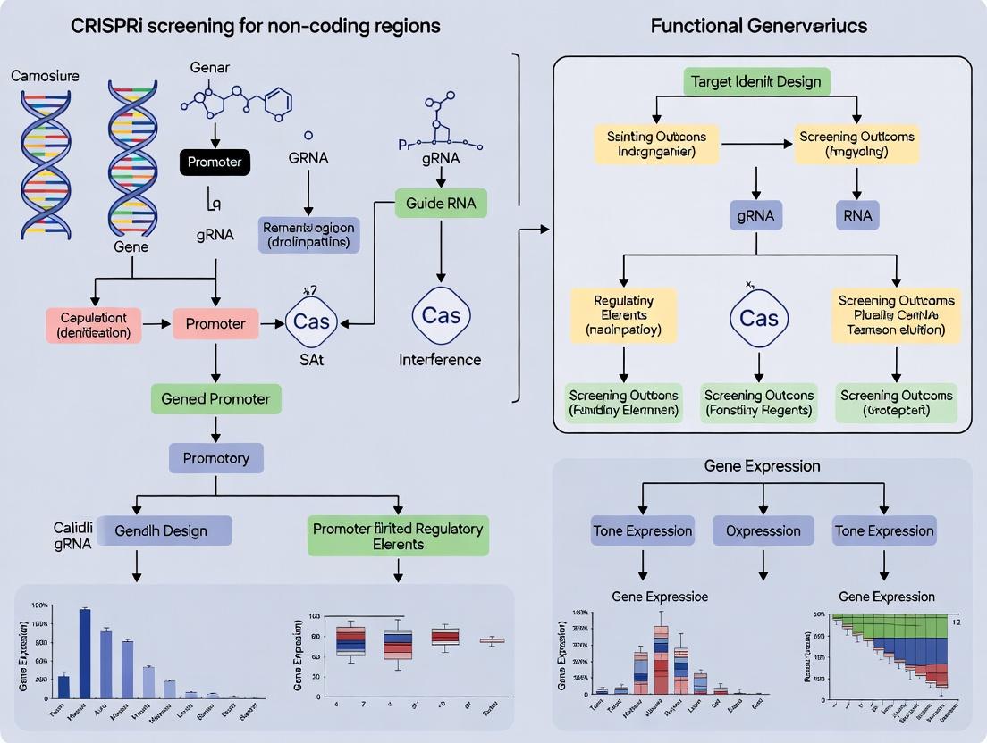
Visualizing Workflows and Molecular Relationships
Title: CRISPRi Screening Workflow for Non-Coding Elements
Title: CRISPRi dCas9-KRAB Repression Mechanism
The Scientist's Toolkit: Key Research Reagent Solutions
Table 3: Essential Reagents for CRISPRi Screening of Non-Coding Regions
| Reagent Name | Vendor (Example) | Catalog Number | Primary Function in Protocol |
|---|---|---|---|
| lentiGuide-Puro | Addgene | #52963 | Backbone vector for sgRNA expression and puromycin selection. |
| psPAX2 | Addgene | #12260 | Lentiviral packaging plasmid (gag/pol/rev). |
| pMD2.G | Addgene | #12259 | Lentiviral envelope plasmid (VSV-G). |
| dCas9-BFP-KRAB | Addgene | #46911 | Source plasmid for generating stable dCas9-KRAB cell lines. |
| Polybrene | Sigma-Aldrich | #H9268 | Increases lentiviral transduction efficiency. |
| Lenti-X Concentrator | Takara Bio | #631231 | Quickly concentrates lentiviral particles without ultracentrifugation. |
| Endura Electrocompetent Cells | Lucigen | #60242-2 | High-efficiency cells for large, complex library transformation. |
| BsmBI-v2 | New England Biolabs | #R0739 | Type IIS restriction enzyme for Golden Gate assembly of sgRNA library. |
| Herculase II Fusion DNA Polymerase | Agilent | #600675 | High-fidelity PCR for NGS library preparation from gDNA. |
| QIAamp DNA Blood Maxi Kit | Qiagen | #51194 | Scalable gDNA isolation from millions of cultured cells. |
| MAGeCK Software | N/A | Open Source | Essential computational tool for analyzing CRISPR screen NGS data. |
The development of CRISPR interference (CRISPRi) from the foundational CRISPR-Cas9 system represents a pivotal advancement for functional genomics, particularly for interrogating non-coding genomic regions. Unlike CRISPR-Cas9, which creates double-strand breaks, CRISPRi utilizes a catalytically "dead" Cas9 (dCas9) to sterically block transcription or recruit transcriptional repressors, enabling reversible, specific gene silencing without altering the DNA sequence. This application note details the engineering of dCas9 repressors and provides protocols for their use in large-scale CRISPRi screens aimed at identifying functional elements in non-coding regions, such as enhancers, promoters, and silencers—a core methodology for modern drug target discovery.
Engineering dCas9: From Nuclease to Repressor
The creation of dCas9 involves site-directed mutagenesis of two catalytic residues in the RuvC (D10) and HNH (H840) domains of Streptococcus pyogenes Cas9. This renders the protein incapable of cleaving DNA while maintaining its ability to bind DNA guided by a single-guide RNA (sgRNA).
Key Research Reagent Solutions:
| Reagent/Catalog # | Supplier | Function in CRISPRi |
|---|---|---|
| dCas9 (pLV-dCas9-KRAB) | Addgene (#71236) | Lentiviral expression vector for mammalian cell delivery of the dCas9-KRAB fusion repressor. |
| sgRNA Cloning Vector (pU6-sgRNA) | Addgene (#53188) | Backbone for cloning and expressing target-specific single-guide RNAs. |
| K562 dCas9-KRAB Cell Line | ATCC/Sigma | Ready-to-use chronic myelogenous leukemia cell line stably expressing dCas9-KRAB for screening. |
| MPP8/HP1 KRAB Fusion | Broad Institute GPP | Alternative repressor domain fusion for enhanced silencing, especially in heterochromatin. |
| Lentiviral Packaging Mix (psPAX2, pMD2.G) | Addgene (#12260, #12259) | Essential plasmids for producing replication-incompetent lentiviral particles. |
| Puromycin/Doxycycline | Thermo Fisher | Selection antibiotics for stable cell line generation and inducible system control. |
Table 1: Comparison of Key CRISPR-Cas9 and CRISPRi System Parameters
| Parameter | CRISPR-Cas9 (Wild-Type) | CRISPRi (dCas9-KRAB) |
|---|---|---|
| DNA Cleavage | Yes (DSBs) | No |
| Primary Mechanism | NHEJ/HDR | Steric Block & Chromatin Modification |
| Typical Knockdown Efficiency | N/A (Knockout) | 70-95% (Transcriptional) |
| Off-Target Effects (DNA) | Moderate (Sequence-Dependent) | Lower (No Cleavage) |
| Reversibility | No (Permanent) | Yes (Transient) |
| Ideal Targeting Region | Early Exons | TSS (-50 to +300 bp) or Enhancers |
| Common Delivery Method | Lentivirus, RNP | Lentivirus, Stable Cell Lines |
Core Protocol: Setting Up a CRISPRi Screen for Non-Coding Regions
This protocol outlines steps for a pooled lentiviral CRISPRi screen targeting putative regulatory regions.
Part A: Design and Cloning of a Non-Coding sgRNA Library
- Target Identification: Using databases like ENCODE or FANTOM5, identify candidate cis-regulatory elements (e.g., DNase I hypersensitive sites, H3K27ac peaks) linked to your gene of interest.
- sgRNA Design: Design 3-6 sgRNAs per target region (5-10 per positive control gene). For promoter targeting, design guides spanning -50 to +300 bp relative to the TSS.
- Tool: Use the Broad Institute's GPP sgRNA Designer (https://portals.broadinstitute.org/gpp/public/analysis-tools/sgrna-design).
- Controls: Include non-targeting control sgRNAs (≥ 100) and essential gene-targeting sgRNAs (for positive selection screens).
- Library Cloning: Perform pooled oligo synthesis of the sgRNA library, then clone into a lentiviral sgRNA expression backbone (e.g., pU6-sgRNA-EF1α-Puro) via BsmBI restriction sites.
- Quality Control: Sequence the pooled plasmid library to confirm representation and absence of bias. Ensure transformation yields >200x coverage of the library.
Part B: Generation of a Stable dCas9-Repressor Cell Line
- Lentiviral Production: Co-transfect HEK293T cells with the dCas9-KRAB expression plasmid (e.g., pLV-dCas9-KRAB) and packaging plasmids (psPAX2, pMD2.G) using a transfection reagent like PEI.
- Transduction & Selection: Transduce your target cell line (e.g., K562) with the dCas9-KRAB lentivirus. Select with appropriate antibiotic (e.g., Blasticidin, 5-10 µg/mL) for 7-10 days.
- Validation: Confirm dCas9-KRAB expression via western blot (anti-FLAG or anti-Cas9 antibody) and functional testing by targeting a known gene's promoter and measuring mRNA knockdown (qRT-PCR).
Part C: Pooled Screen Transduction and Analysis
- sgRNA Library Lentivirus Production: Produce lentivirus from the pooled sgRNA plasmid library as in Part B, Step 1. Titrate the virus on the dCas9-KRAB cell line.
- Transduction at Low MOI: Transduce the dCas9-KRAB cells at an MOI of ~0.3 to ensure most cells receive only one sgRNA. Maintain >500x library coverage.
- Selection and Harvest: Apply puromycin selection (1-2 µg/mL) for 7 days. Harvest cells at the initial timepoint (T0) and at the experimental endpoint (T_end, e.g., after 14-21 days or a selection pressure).
- Genomic DNA Extraction & NGS Prep: Isolate gDNA from ≥1e7 cells per sample. Amplify the integrated sgRNA sequences via PCR using indexing primers for next-generation sequencing (NGS).
- Data Analysis: Sequence samples and quantify sgRNA abundance. Use analysis pipelines (e.g., MAGeCK or pinAPL-Py) to identify significantly enriched or depleted sgRNAs comparing T_end to T0, highlighting critical non-coding regions.
Title: CRISPRi Screening Workflow for Non-Coding Regions
Advanced Application: Tiling Enhancer Regions
For fine-mapping functional elements within large putative enhancers, a tiling screen is essential.
Protocol: High-Density Tiling CRISPRi
- Design: Design sgRNAs tiling across the entire genomic region (e.g., every 50-100 bp).
- Sub-Library Cloning: Clone the tiling sgRNAs as a sub-library following Part A.
- Focused Screen: Conduct the screen as in Part C, but with deeper sequencing coverage (>1000x).
- Analysis: Plot sgRNA log2(fold-change) or p-values across genomic coordinates to identify specific "base-pair resolution" silencer or enhancer modules.
Title: dCas9-KRAB CRISPRi Mechanism
Critical Optimization and Troubleshooting
- Repressor Domain Choice: The KRAB domain is standard, but SID4x or MECP2 may offer stronger repression in certain cell types.
- Delivery Optimization: For difficult-to-transduce cells, consider using Cas9-ribonucleoprotein (RNP) complexes with purified dCas9-KRAB protein and in vitro transcribed sgRNA for transient, high-efficiency silencing.
- Multiplexing: For studying complex genetic interactions between non-coding regions, use libraries with multiple sgRNAs per cell (e.g., dual-guide libraries).
- Off-Target Validation: Always validate hits from a primary screen using individual sgRNAs and orthogonal assays (e.g., siRNA, reporter assays).
Table 2: Quantitative Outcomes from a Representative CRISPRi Enhancer Screen
| Metric | Value in K562 dCas9-KRAB Cells | Notes |
|---|---|---|
| Library Size | 50,000 sgRNAs | Targeting 5,000 putative enhancers |
| Screen Coverage | 750x | Cells per sgRNA at T0 |
| Transduction Efficiency | 40% | MOI = 0.3, puromycin selected |
| Positive Control Knokdown | 92% ± 3% (mRNA) | Essential gene (POLR2D) promoter targeting |
| Hit Rate (FDR < 10%) | 4.2% of targeted regions | 210 functional enhancers/silencers identified |
| Optimal sgRNAs per Region | 5 | Increased validation rate vs. 3 sgRNAs |
Application Notes
This application note, framed within a broader thesis on CRISPRi screening for functional non-coding element discovery, details the key advantages of CRISPR interference (CRISPRi) over conventional knockout (KO) screens for interrogating regulatory genomes. The following data, derived from current literature and benchmark studies, quantitatively summarizes these core advantages.
Table 1: Quantitative Comparison of CRISPRi vs. KO Screen Performance in Non-Coding Regions
| Performance Metric | CRISPRi (dCas9-KRAB) | CRISPR Knockout (Cas9 Nuclease) | Implication for Non-Coding Screens |
|---|---|---|---|
| Indel Spectrum at Target | No DNA cleavage; reversible transcriptional repression. | Complex mix of frameshift indels, in-frame deletions, large deletions, and translocations. | CRISPRi ensures phenotypic stability—the targeted locus remains genetically intact, preventing confounding synthetic effects from DNA damage response and genomic rearrangements. |
| Hypomorph Generation | Highly effective. Repression efficiency typically 70-95%, creating a tunable allelic series. | Inefficient and stochastic. Requires biallelic in-frame mutations, which are rare (~11% of events). | CRISPRi excels at modeling hypomorphic (partial loss-of-function) states, crucial for studying essential genes and dosage-sensitive regulatory elements where full KO is lethal or misleading. |
| Tiling Screening Density | High. Guides can be spaced as close as 50-100 bp for dense saturation mutagenesis. | Low. Requires guides near the Cas9 cut site (~3-4 bp upstream of PAM), limiting resolution. | Superior tiling capability allows precise mapping of functional sub-regions within enhancers, promoters, and non-coding RNAs, pinpointing key regulatory motifs. |
| Off-Target Transcriptional Effects | Minimal. Off-target binding of dCas9-KRAB rarely leads to significant gene repression. | High. Off-target DNA cleavage can cause mutagenic and p53-mediated transcriptional responses. | Reduces false positives/negatives from cellular stress pathways, enhancing screen accuracy. |
| Screen Dynamic Range (Fitness Screens) | Consistently high (Z'-factor > 0.5). Phenotypes are consistent and reproducible. | Can be variable. Lethal hits from essential gene KO can dominate, masking subtler regulatory phenotypes. | Enables discovery of subtle, biologically relevant phenotypes from modulating non-coding elements without being overshadowed by extreme essential gene signals. |
Table 2: Representative Outcomes from Published Non-Coding Screens
| Study Focus | CRISPRi Result | KO Screen Challenge | Key Advantage Demonstrated |
|---|---|---|---|
| Enhancer Mapping | Identified discrete 100-200 bp functional "cores" within super-enhancers linked to drug resistance. | Generated large, multi-kilobase deletions, unable to resolve functional sub-units. | Tiling |
| Essential Gene Regulation | Discovered non-essential, regulatory "dependency factors" via hypomorphic repression of essential gene promoters. | KO of same genes was lethal, removing them from the hit list entirely. | Hypomorphs |
| Long Non-Coding RNAs | Distinguished functional roles of specific transcript isoforms via promoter repression. | KO caused truncations or frameshifts in overlapping sense/antisense transcripts, creating complex, uninterpretable genotypes. | Phenotypic Stability |
Experimental Protocols
Protocol 1: Design and Cloning of a Saturated CRISPRi Guide Library for a Non-Coding Region
Objective: To construct a high-density tiling guide library targeting a candidate enhancer region of 50 kb.
Materials (Research Reagent Solutions):
- Synthesis Platform: Custom oligo pool synthesis (e.g., Twist Bioscience, Agilent). Contains thousands of unique sgRNA sequences tiling the target.
- Backbone Vector: lentiguide-puro (Addgene #52963) or similar lentiviral sgRNA expression plasmid.
- Enzymes: BsmBI-v2 (NEB #R0739), T4 DNA Ligase (NEB #M0202), Plasmid-Safe ATP-Dependent DNase (Lucigen).
- Cloning Strain: EndA- competent E. coli (e.g., Stbl3, NEB Stable).
- PCR Reagents: KAPA HiFi HotStart ReadyMix (Roche), Gel extraction kit (Qiagen), DNA clean-up beads (SPRI).
Methodology:
- Guide Design: Using a script (e.g.,
flashfryorCRISPick), design all possible sgRNAs (20-nt spacer) targeting both strands of the 50 kb region with an NGG PAM. Filter for on-target specificity and minimize off-target potential. Set a tiling density of 1 guide per 50-100 bp. Include 500 non-targeting control guides. - Oligo Pool Design: Add flanking BsmBI cloning sequences (5'- [ACACTCTTTCCCTACACGACGCTCTTCCGATCTNNN]-spacer-[GTTTA]-3') to each guide sequence. Order as a single-stranded oligo pool.
- Pool Amplification: Perform limited-cycle PCR (5 cycles) to generate double-stranded oligo DNA. Purify using SPRI beads.
- Vector Digestion: Digest 20 µg of lentiguide-puro vector with BsmBI at 55°C for 2 hours. Gel-purify the linearized backbone.
- Golden Gate Assembly: Set up a 50 µL Golden Gate reaction with 100 ng digested backbone, 20 ng amplified oligo pool, 10 U BsmBI-v2, and 400 U T4 DNA Ligase. Cycle: 30x (37°C for 5 min, 16°C for 5 min), then 55°C for 5 min, 80°C for 10 min.
- DNase Treatment: Add Plasmid-Safe DNase to digest residual linear DNA for 30 min at 37°C.
- Electroporation: Desalt the assembly reaction and electroporate into 500 µL EndA- competent cells. Plate on large 245 x 245 mm LB-ampicillin plates. Incubate overnight.
- Library Harvesting: Scrape all colonies, maxi-prep plasmid DNA. Validate library complexity by next-generation sequencing of the guide region.
Protocol 2: Conducting a CRISPRi Fitness Screen with a dCas9-KRAB Cell Line
Objective: To perform a negative selection (drop-out) screen to identify non-coding regulatory elements essential for cell proliferation.
Materials (Research Reagent Solutions):
- Cell Line: HEK293T or relevant cancer cell line stably expressing dCas9-KRAB (e.g., from lentiviral pLV hU6-sgRNA hUbC-dCas9-KRAB, Addgene #71237).
- Viral Packaging: psPAX2 (Addgene #12260), pMD2.G (Addgene #12259), Polyethylenimine (PEI) transfection reagent.
- Cell Culture: Appropriate medium, Puromycin, Polybrene (8 µg/mL).
- Genomic DNA Extraction: Quick-DNA Midiprep Kit (Zymo Research).
- Sequencing Library Prep: 2x KAPA HiFi HotStart Mix, P5/P7 indexing primers with flow cell adapters.
Methodology:
- Lentivirus Production: Co-transfect 70% confluent HEK293T cells in a 15-cm dish with 18 µg library plasmid, 12 µg psPAX2, and 6 µg pMD2.G using PEI. Harvest supernatant at 48 and 72 hours, concentrate via centrifugation or PEG-it virus precipitation.
- Library Transduction & Selection: Transduce dCas9-KRAB cells at a low MOI (~0.3) with polybrene to ensure >95% of cells receive ≤1 guide. 24 hours post-transduction, add puromycin (1-2 µg/mL) for 5-7 days to select transduced cells.
- Passaging & Harvesting: Maintain the selected cell population (minimum 500 cells per guide in the library) for 14-21 population doublings. Passage cells every 3-4 days, keeping detailed cell counts. Harvest 5e6 cells at the initial timepoint (T0) and at the final timepoint (Tfinal) for genomic DNA extraction.
- sgRNA Amplification & Sequencing: Amplify the integrated sgRNA cassette from 5-10 µg gDNA per sample in 50 µL PCR reactions (20-22 cycles) using primers adding Illumina adapters and sample indexes. Pool PCR products, quantify, and sequence on an Illumina NextSeq (75 bp single-end).
- Data Analysis: Align sequencing reads to the guide library reference. Count guide abundances in T0 and Tfinal samples. Use a robust statistical pipeline (e.g.,
MAGeCKorCRISPRcloud) to calculate guide depletion scores (log2 fold-change) and rank significantly depleted genomic regions.
Visualizations
Diagram 1: Mechanistic Comparison of CRISPRi vs KO
Diagram 2: CRISPRi Tiling Screen Workflow
The Scientist's Toolkit
Table 3: Essential Research Reagents for CRISPRi Screens in Non-Coding Regions
| Reagent / Material | Supplier Example (Catalog #) | Function in Experiment |
|---|---|---|
| dCas9-KRAB Expression Plasmid | Addgene (#71237) | Stable expression of the dead Cas9 fused to the KRAB transcriptional repressor domain. Forms the foundational protein for CRISPRi. |
| lentiguide-puro sgRNA Backbone | Addgene (#52963) | Lentiviral vector for cloning and expressing single guide RNA (sgRNA) libraries. Contains puromycin resistance for selection. |
| BsmBI-v2 Restriction Enzyme | New England Biolabs (R0739S) | Type IIS enzyme used in Golden Gate assembly to efficiently clone oligo-synthesized sgRNA libraries into the backbone vector. |
| Ultracompetent E. coli (EndA-) | NEB (C3040H) | High-efficiency cloning strain for library transformation, essential for maintaining complex sgRNA library diversity without recombination. |
| Polyethylenimine (PEI), Linear | Polysciences (23966-1) | High-efficiency, low-cost transfection reagent for producing lentivirus in HEK293T packaging cells. |
| psPAX2 Packaging Plasmid | Addgene (#12260) | Provides gag, pol, and rev genes for lentiviral particle production. |
| pMD2.G Envelope Plasmid | Addgene (#12259) | Expresses VSV-G glycoprotein, enabling broad tropism pseudotyping of lentiviral particles. |
| Polybrene | Sigma-Aldrich (TR-1003-G) | A cationic polymer that enhances lentiviral transduction efficiency by neutralizing charge repulsion between virus and cell membrane. |
| Puromycin Dihydrochloride | Gibco (A1113803) | Selection antibiotic for cells successfully transduced with the lentiviral sgRNA library (conferred by the lentiguide plasmid). |
| KAPA HiFi HotStart ReadyMix | Roche (07958846001) | High-fidelity PCR master mix for accurate amplification of sgRNA sequences from genomic DNA during sequencing library preparation. |
In the context of CRISPR interference (CRISPRi) screening for non-coding regulatory elements, success is critically dependent on rigorous pre-experimental planning. The choice of biological question and its corresponding phenotypic readout dictates screen design, library selection, and downstream validation strategies. This application note provides a framework for defining these foundational elements.
Defining the Biological Question
The biological question must be precise, testable, and tailored to the non-coding region of interest. Generic questions yield uninterpretable data.
Key Considerations:
- Specificity: Target a defined genomic locus class (e.g., enhancers of a specific gene, promoter-proximal regions, lncRNA loci).
- Function: Link the non-coding region to a specific cellular process (e.g., "Identify enhancers regulating resistance to drug X").
- Perturbation Outcome: Specify the expected transcriptional outcome (e.g., repression via CRISPRi) and the hypothesized phenotypic consequence.
Table 1: Refining the Biological Question for Non-Coding Screens
| Question Aspect | Vague Example | Precise, Screen-Ready Example |
|---|---|---|
| Genomic Target | "Study enhancers in cancer." | "Identify functional enhancers within the 1.6 Mb locus control region of gene Y in cell type Z." |
| Phenotypic Link | "Find regions affecting cell growth." | "Discover non-coding regulatory elements whose repression sensitizes cells to therapeutic agent X (IC25 dose)." |
| Transcriptional Output | "See how gene expression changes." | "Quantify changes in mRNA expression of gene Y (and its known paralogs) via RT-qPCR as a primary validation readout." |
Selecting a Phenotypic Readout
The readout must be robust, scalable, and quantitatively linked to the perturbation of non-coding function.
Table 2: Common Phenotypic Readouts for CRISPRi Non-Coding Screens
| Readout Type | Measurement | Throughput | Key Application | Primary Advantage | Primary Limitation |
|---|---|---|---|---|---|
| Viability / Proliferation | Cell count, ATP content, confluence. | Very High | Essential enhancers, drug-gene interactions. | Simple, low-cost, scalable. | Confounded by multiple indirect effects; slow. |
| Fluorescence-Activated Cell Sorting (FACS) | Fluorescent protein reporter, surface markers, dyes. | High | Enhancer-reporter assays, cell state transitions. | Multiplexable, rich quantitative data. | Requires flow cytometer; cell dissociation can introduce noise. |
| Massively Parallel Reporter Assays (MPRA) | DNA barcode abundance via sequencing. | Very High | Direct validation of enhancer activity. | Directly links regulatory element to output. | Typically uses episomal constructs, not native chromatin context. |
| Single-Cell RNA Sequencing (scRNA-seq) | Transcriptome-wide gene expression. | Medium | Unbiased discovery of regulatory networks & states. | Rich, multidimensional data. | Expensive; complex analysis; lower sgRNA recovery. |
Protocol: Framework for Coupling a FACS-Based Readout to a CRISPRi Screen
This protocol outlines steps for a screen identifying enhancers regulating a cell surface marker.
I. Experimental Design & Construct Assembly
- Cell Line Engineering: Generate a stable cell line expressing dCas9-KRAB (CRISPRi machinery) via lentiviral transduction and antibiotic selection.
- Reporter Validation: Confirm that the chosen cell surface marker (e.g., CD34) can be reliably detected via FACS with high signal-to-noise.
- Library Selection: Choose a tiled, non-coding CRISPRi sgRNA library targeting genomic regions of interest (e.g., all putative enhancers marked by H3K27ac ChIP-seq within a locus). Include non-targeting and positive control sgRNAs.
II. Screening Workflow
- Library Transduction: Transduce the CRISPRi cell line with the sgRNA library at a low MOI (<0.3) to ensure most cells receive one sgRNA. Maintain >500x library representation.
- Selection & Phenotyping: After 7-10 days for transcriptional repression:
- Harvest cells and stain for the target surface marker (e.g., anti-CD34-APC).
- Perform FACS to isolate the top and bottom 10-20% of cells based on marker fluorescence.
- Collect genomic DNA from each population and the unsorted input pool.
- sgRNA Recovery & Analysis:
- Amplify integrated sgRNA sequences via PCR using indexed primers.
- Sequence on an Illumina platform.
- Align reads to the library reference and count sgRNA abundances.
- Use statistical packages (MAGeCK, pinAPL-Py) to identify sgRNAs significantly enriched or depleted in the sorted populations versus input.
III. Hit Validation
- Individual sgRNA Reconstitution: Clone top-hit sgRNAs into lentiviral vectors.
- Phenotypic Confirmation: Transduce naive CRISPRi cells with individual sgRNAs and re-measure the surface marker via FACS.
- Orthogonal Validation: Measure expression of the putative target gene via RT-qPCR and assess changes in enhancer chromatin marks (e.g., H3K27ac) via ChIP-qPCR.
The Scientist's Toolkit: Key Research Reagent Solutions
| Reagent / Material | Function & Critical Consideration |
|---|---|
| dCas9-KRAB Expression System | Lentiviral construct for stable, inducible, or constitutive expression. Must be optimized for target cell type. KRAB domain ensures robust transcriptional repression. |
| Tiled Non-Coding sgRNA Library | Designed to densely tile putative regulatory regions (e.g., 2-5 sgRNAs/kb). Controls for sequence-specific artifacts. Essential for coverage of AT-rich regions. |
| High-Titer Lentiviral Particles | For efficient sgRNA library delivery. Functional titer must be determined on the CRISPRi cell line to achieve correct MOI. |
| Viability-Impermeable DNA Stain (e.g., 7-AAD) | Used during FACS to exclude dead cells, crucial for clean phenotypic sorting and reducing noise. |
| Antibodies for FACS (Conjugated) | High-quality, titrated antibodies for the target phenotypic marker. Fluorochrome choice must match cytometer configuration. |
| gDNA Extraction Kit (Scalable) | For efficient recovery of genomic DNA from 1e6 to 1e8 cells. Must yield high-molecular-weight DNA for PCR. |
| High-Fidelity PCR Kit | For specific, low-bias amplification of integrated sgRNA cassettes from genomic DNA. Critical for maintaining library representation. |
| Dual-Indexed Sequencing Primers | Allow multiplexing of multiple samples (Input, High, Low populations) in one sequencing run. Reduces batch effects and cost. |
| MAGeCK or pinAPL-Py Software | Open-source computational tools specifically designed for the statistical analysis of CRISPR screen count data to identify enriched/depleted sgRNAs/genes. |
This document provides detailed Application Notes and Protocols for the integrative use of major public genomic databases to prioritize non-coding genomic regions for functional validation. The protocols are framed within a broader thesis employing CRISPR interference (CRISPRi) screening to investigate gene regulation through non-coding elements. The systematic identification of putative functional regions from population-scale and epigenomic data is a critical first step in designing focused, high-yield CRISPRi libraries.
The following databases provide complementary data types essential for target nomination.
Table 1: Core Public Resources for Non-Coding Region Selection
| Database | Primary Data Type | Key Metrics | Use in Target Selection |
|---|---|---|---|
| ENCODE | Epigenomic profiles (ChIP-seq, ATAC-seq, Hi-C) | ~1,000,000 assays; 6,000+ experiments; 1,200+ cell/tissue types. | Identifies candidate cis-regulatory elements (cCREs) via chromatin accessibility, histone marks (H3K27ac, H3K4me3), and transcription factor binding. |
| SCREEN (ENCODE Registry) | Curated, annotated cCREs | 1,006,251 human cCREs (V4); 313,661 mouse cCREs. | Provides pre-defined, high-confidence regulatory elements (promoters, enhancers, CTCF-only sites) for direct candidate extraction. |
| GWAS Catalog | Disease/trait-associated genetic variants | 843,185 variant-trait associations (v1.0); 6,604 publications. | Maps phenotypic associations to genomic loci; prioritizes variants in non-coding regions for functional follow-up. |
| UCSC Genome Browser | Visualization & Data Integration | Hosts >1,000 public track hubs. | Central platform for visually overlaying ENCODE, SCREEN, and GWAS data with conservation and genome annotation. |
Application Note: Integrative Workflow for Target Prioritization
This protocol describes a stepwise approach to integrate data from ENCODE, SCREEN, and the GWAS Catalog to generate a ranked list of non-coding target regions for CRISPRi screening.
Phase 1: Disease/Trait Locus Definition via GWAS
Objective: Identify non-coding loci associated with a phenotype of interest.
- Access the NHGRI-EBI GWAS Catalog (https://www.ebi.ac.uk/gwas/).
- Perform a search using relevant trait/disease terms (e.g., "coronary artery disease").
- Download all significant variant-trait associations (p-value < 5x10^-8). Key columns:
CHR_ID,CHR_POS,SNPS,MAPPED_TRAIT,PVALUE_MLOG. - Define genomic intervals for each locus. A common method is to take a region spanning ± 500 kb from the lead variant, or use LD-based clumping from a reference population to define independent loci.
Phase 2: Intersection with Regulatory Annotations from SCREEN & ENCODE
Objective: Filter GWAS loci for presence of high-confidence regulatory elements.
- Access the SCREEN candidate cis-Regulatory Elements (https://screen.encodeproject.org/).
- Use the "Search by Genomic Region" tool to extract all cCREs (e.g., enhancer-like, promoter-like) overlapping the GWAS-defined intervals from Phase 1.
- For higher-resolution data, access the ENCODE portal (https://www.encodeproject.org/) to download cell-type-relevant epigenomic tracks (e.g., H3K27ac ChIP-seq, ATAC-seq) for your model system.
- Use
bedtools intersectto overlap GWAS variant coordinates (or their LD proxies) with cCRE regions and cell-type-specific epigenomic peaks.
Phase 3: Prioritization and Ranking
Objective: Rank overlapping cCREs to select top candidates for screening.
- Create a Prioritization Table: Compile the following metrics for each cCRE that overlaps a GWAS signal.
Table 2: Candidate Region Prioritization Metrics
Candidate Region (chr:start-end) Overlapping GWAS Trait(s) cCRE Class (SCREEN) Cell-Type-Specific Epigenetic Signal Conservation (phastCons) Final Priority Score chr6:123456-124000 Coronary Artery Disease Enhancer-like H3K27ac+, ATAC+ in HepG2 0.85 High chr6:124500-125100 LDL Cholesterol Promoter-like H3K4me3+ in HepG2 0.45 Medium - Scoring: Assign a qualitative score (High/Medium/Low) or a quantitative score based on:
- Number of overlapping GWAS traits/variants.
- Strength of epigenomic signals (peak intensity).
- Evolutionary conservation.
- Relevance of the cCRE's linked gene(s) to the disease biology.
Protocol: From Target List to CRISPRi sgRNA Design
Objective: Design and clone sgRNAs targeting prioritized non-coding regions.
Materials: Research Reagent Solutions
Table 3: Essential Reagents for CRISPRi Screening Preparation
| Reagent/Material | Function | Example Product/Catalog |
|---|---|---|
| dCas9-KRAB Expression Vector | CRISPRi effector; silences transcription via chromatin modification. | lenti dCas9-KRAB-blast (Addgene #125134) |
| sgRNA Cloning Backbone | Lentiviral vector for sgRNA expression with selection marker. | lentiGuide-Puro (Addgene #52963) |
| BsmBI-v2 Restriction Enzyme | Type IIS enzyme for Golden Gate assembly of sgRNA oligos. | BsmBI-v2 (NEB #R0739S) |
| T7 Endonuclease I or Surveyor Nuclease | For validation of genomic edits (if testing nuclease activity). | T7E1 (NEB #M0302S) |
| Next-Generation Sequencing Library Prep Kit | For quantifying sgRNA abundance pre- and post-screen. | Illumina Nextera XT DNA Library Prep Kit |
| Cell Line-Specific Culture Reagents | For maintenance and transduction of target cells. | Dependent on model system (e.g., HepG2, K562). |
Experimental Protocol:
Part A: sgRNA Design
- For each prioritized genomic region (e.g., a 500 bp enhancer), use design tools like CHOPCHOP or CRISPRitz to identify all potential 20bp sgRNA sequences conforming to an
NGGPAM. - Filter sgRNAs for:
- On-target efficiency: Use predictive scores (e.g., Doench '16 score).
- Off-target minimization: BLAST against the reference genome; discard sgRNAs with >2 mismatches elsewhere.
- Uniqueness: Ensure the sequence is unique in the genome.
- Select 3-5 sgRNAs per target region and include non-targeting control sgRNAs.
Part B: sgRNA Library Cloning (Golden Gate Assembly)
- Oligo Preparation: Order forward and reverse oligonucleotides for each sgRNA with overhangs compatible with BsmBI-digested vector:
- Forward:
5'-CACCG[20bp guide sequence]-3' - Reverse:
5'-AAAC[reverse complement of 20bp guide sequence]C-3'
- Forward:
- Annealing: Phosphorylate and anneal oligos in a thermocycler.
- Digestion & Ligation: Perform a one-pot Golden Gate reaction:
- Transformation & Validation: Transform into competent E. coli, pool colonies, and prepare plasmid DNA. Validate library representation by sequencing.
Visualization of Workflows and Relationships
Diagram 1: Integrative Target Prioritization and Screening Workflow
Diagram 2: Logical Relationship from GWAS Variant to Functional Target
Blueprint for Success: A Step-by-Step Protocol for CRISPRi Screening of Regulatory Elements
Within the broader thesis on CRISPR interference (CRISPRi) screening for non-coding regulatory element discovery, the design of single guide RNA (sgRNA) libraries is paramount. Two primary strategies exist for targeting expansive, non-coding regions: Tiling across broad genomic intervals to empirically map functional elements, and Saturation mutagenesis of specific motifs (e.g., transcription factor binding sites) to dissect functional nucleotides. This application note details the design principles, protocols, and practical considerations for implementing these strategies in CRISPRi screens.
Library Design Strategies: Quantitative Comparison
Table 1: Key Parameters for Tiling vs. Saturation Library Strategies
| Parameter | Tiling Strategy (Broad Regions) | Saturation Strategy (Motifs) |
|---|---|---|
| Primary Goal | Empirical discovery of functional elements within large (e.g., >50 kb) genomic loci. | Functional dissection of known short motifs or putative binding sites. |
| Design Basis | Genomic coordinates; agnostic to sequence features. | Specific DNA sequence motif(s). |
| sgRNA Density | Regular interval (e.g., 1 sgRNA every 100-200 bp). | All possible sgRNAs targeting every base or n-mer within motif. |
| Typical Library Size | 500 - 5,000 sgRNAs per locus. | 100 - 2,000 sgRNAs per motif. |
| Control sgRNAs | Essential: non-targeting controls; targeting inactive genomic regions. | Essential: non-targeting controls; scrambled motif sgRNAs. |
| Analysis Outcome | Functional "hits" defined by genomic clusters of sgRNAs affecting phenotype. | Nucleotide-resolution functional map of the motif. |
| Best For | Discovery of novel enhancers, repressors, or structural elements. | Validating and characterizing suspected regulatory elements. |
Table 2: Recommended Design Specifications for CRISPRi sgRNAs
| Design Rule | Specification | Rationale |
|---|---|---|
| Target Sequence | 20-nt guide sequence immediately 5' of NGG (PAM) for S. pyogenes dCas9. | CRISPRi requires PAM recognition but not cleavage. |
| Genomic Uniqueness | ≤3 mismatches to any other genomic locus (BLASTN). | Minimizes off-target repression. |
| On-Target Efficiency Predictors | Use Rule Set 2 (Doench et al., 2016) or CRISPRi-specific models (Horlbeck et al., 2016). | Predicts sgRNA binding/repression efficacy. |
| Target Strand | Prefer template strand for CRISPRi. | dCas9 fused to repressors (KRAB) more effective on template strand. |
| Avoidance Regions | Exclude sequences with homopolymer runs (>4), high GC (>70%) or low GC (<30%). | Can affect sgRNA stability or activity. |
Detailed Experimental Protocols
Protocol 1: Design and Construction of a Tiling sgRNA Library Objective: Generate a tiling sgRNA library to screen a 100-kb candidate region for regulatory elements affecting a reporter or endogenous gene of interest.
- Define Target Region: Using UCSC Genome Browser or Ensembl, specify chromosomal coordinates (e.g., chrX:10,000,000-10,100,000).
- Generate Guide Candidates: Use a script (e.g., Python) or tool (e.g.,
CRISPRitz) to extract all 20-nt sequences followed by a 5'-NGG-3' PAM on either strand within the region. - Apply Filtering & Scoring: Filter candidates for uniqueness (allowing ≤3 mismatches). Score remaining guides using an on-target efficiency predictor (see Table 2). Select the top-scoring guide at regular intervals (e.g., every 150 bp).
- Balance Library: Include a minimum of 1,000 non-targeting control sgRNAs (designed to not target the genome) and 500 safe-targeting controls (targeting inert genomic loci like intergenic deserts).
- Oligonucleotide Pool Synthesis: Order the final list of sgRNA sequences (scaffold constant) as an oligo pool. Include flanking cloning sequences (e.g., for BsmBI sites compatible with lentiviral sgRNA expression vectors like lentiGuide-Puro).
- Library Cloning: a. Amplify the oligo pool by PCR. b. Digest the PCR product and the recipient lentiviral vector with BsmBI. c. Ligate and transform into electrocompetent E. coli (e.g., Endura ElectroCompetent Cells). d. Harvest a library of colonies representing at least 200x coverage of the sgRNA library for plasmid DNA preparation. Verify complexity by NGS.
Protocol 2: Saturation Mutagenesis of a Transcription Factor Motif Objective: Saturate a 10-bp known motif to determine the functional importance of each nucleotide position.
- Define Core Motif & Flanking Region: Identify the exact genomic coordinates of the motif. Extend the target window by ~10 bp on each side to allow for PAM positioning.
- Generate All Possible sgRNAs: Programmatically generate every unique 20-nt guide sequence targeting the extended window that is adjacent to an NGG PAM. This will produce guides that tile the motif from all angles.
- Filter for Specificity: As in Protocol 1, filter all guides for genomic uniqueness. This step is critical for short, repetitive motifs.
- Include Controls: Design negative control sgRNAs targeting sequences with scrambled motif sequences but similar GC content.
- Synthesis, Cloning & Validation: Follow steps 5-6 from Protocol 1. Sequence the final plasmid library to confirm representation of all designed variant guides.
Visualization of Workflows & Concepts
Title: sgRNA Library Design Strategy Selection
Title: CRISPRi Screening Experimental Workflow
The Scientist's Toolkit: Key Research Reagent Solutions
Table 3: Essential Reagents & Materials for CRISPRi Library Screening
| Item | Function & Specification | Example Product/Catalog |
|---|---|---|
| dCas9-KRAB Expression Cell Line | Stable cell line expressing the CRISPRi machinery. Essential for screening. | Custom-made or available from repositories (e.g., hCRISPRi-v2, Addgene #154469). |
| Lentiviral sgRNA Backbone Vector | Vector for cloning sgRNA library and viral production. | lentiGuide-Puro (Addgene #52963) or similar. |
| High-Efficiency Electrocompetent E. coli | For efficient transformation of the ligated sgRNA library to maintain complexity. | Endura ElectroCompetent Cells (Lucigen #60242-2). |
| Lentiviral Packaging Plasmids | For production of VSV-G pseudotyped lentivirus. | psPAX2 (Addgene #12260) and pMD2.G (Addgene #12259). |
| Transfection Reagent | For HEK293T cell transfection during virus production. | Polyethylenimine (PEI Max) or Lipofectamine 3000. |
| Selection Antibiotic | To select for successfully transduced cells. | Puromycin dihydrochloride (for lentiGuide-Puro). |
| Genomic DNA Isolation Kit | For high-yield, high-quality gDNA from millions of cells. | QIAamp DNA Blood Maxi Kit (Qiagen #51194). |
| High-Fidelity PCR Mix | For accurate amplification of sgRNA inserts from gDNA for NGS. | KAPA HiFi HotStart ReadyMix (Roche #7958935001). |
| NGS Platform & Reagents | For deep sequencing of sgRNA abundance. | Illumina NextSeq 500/550, P5/P7 indexing primers. |
| Analysis Software | For statistical analysis of sgRNA depletion/enrichment. | MAGeCK (Li et al., 2014) or pinAPL (Spahn et al., 2017). |
Application Notes
Context in Non-Coding Region Research
Within the broader thesis on CRISPRi screening for functional annotation of non-coding genomes, the selection between commercial and custom-synthesized libraries is a foundational decision. This choice impacts screen design, cost, timeline, and the biological questions addressable. CRISPR interference (CRISPRi) is particularly suited for non-coding region perturbation due to its high specificity and reversible gene repression, enabling the systematic interrogation of enhancers, promoters, and other regulatory elements without DNA cleavage.
Commercial libraries offer pre-designed, validated, and ready-to-use reagents for genome-wide or focused non-coding screens. Vendors such as Addgene, Horizon Discovery, and Synthego provide libraries targeting predicted regulatory regions (e.g., ENCODE cCREs) or tiling regions of interest.
Advantages:
- Standardization: Consistent design and synthesis quality reduce batch effects.
- Validation: Often come with performance data (e.g., sgRNA activity scores).
- Time-Saving: Eliminate months of design, cloning, and QC.
- Support: Access to technical protocols and bioinformatic pipelines.
Limitations:
- Design Rigidity: May not cover novel or organism-specific regions of interest.
- Cost at Scale: For repeated or large-scale use, per-experiment costs can be high.
- Update Lag: May not incorporate the most recent genomic annotations.
Custom libraries are designed de novo by the researcher to target specific genomic loci, such as disease-associated haplotypes or evolutionary conserved regions identified in a thesis project.
Advantages:
- Flexibility: Enables tiling of any region, including novel variants or non-model organism genomes.
- Cost-Effectiveness for Focused Screens: Lower long-term cost for targeted, repeated screens.
- Integration: Can be combined with other custom elements (e.g., molecular barcodes, specific promoters).
Limitations:
- Development Time: Requires extensive in silico design, cloning, and quality control.
- Technical Hurdle: Demands expertise in library synthesis and normalization.
- Validation Burden: Performance of each sgRNA must be empirically assessed.
Quantitative Comparison
Table 1: Strategic Comparison of Library Types
| Parameter | Commercial Library | Custom-Synthesized Library |
|---|---|---|
| Lead Time | 1-4 weeks (shipping) | 3-6 months (design to QC) |
| Typical Cost (USD) | $5,000 - $15,000 per screen | $10,000 - $30,000 (initial design/synthesis) |
| Design Flexibility | Low to Moderate (pre-set designs) | Very High (fully user-defined) |
| Ideal Use Case | Genome-wide discovery, initial pilot screens | Focused, hypothesis-driven screens, novel loci |
| QC Provided | Extensive (NGS validation, titer) | Researcher-responsible |
| Scalability (Re-screening) | Cost scales linearly | High; marginal cost is low after initial investment |
Table 2: Example Library Specifications for Enhancer Screening
| Specification | Commercial Example (Horizon, "ENCODE cCRE v1") | Custom Design Example |
|---|---|---|
| Target Regions | ~330,000 candidate cis-regulatory elements (cCREs) from ENCODE | User-defined 2 Mb locus around a GWAS hit |
| sgRNA Density | 5 sgRNAs per cCRE | 10 sgRNAs per 500 bp tile |
| Library Size | ~1,650,000 sgRNAs | ~40,000 sgRNAs |
| Control Guides | Included (non-targeting, essential genes) | Must be designed separately |
| Backbone | lentiGuide-Puro (Addgene) | Custom lentiviral backbone with SFFV promoter |
| Delivery Format | High-titer lentiviral supernatant | Plasmid pool, requires virus production |
Experimental Protocols
Protocol 1: Screening with a Commercial CRISPRi Library
Objective: Perform a positive selection screen for essential regulatory elements in a cancer cell line using a commercial non-coding CRISPRi library.
Materials: See "The Scientist's Toolkit" below.
Procedure:
- Cell Line Preparation: Engineer your cell line of interest to stably express dCas9-KRAB (e.g., via lentiviral transduction and blasticidin selection). Confirm repression efficiency via qPCR of a known gene.
- Library Transduction:
- Seed 2 x 10^8 cells at a density ensuring >50x coverage of the library (e.g., for a 100k sgRNA library, seed at least 5 million cells per replicate).
- Thaw commercial lentiviral supernatant on ice. Transduce cells at an MOI of ~0.3-0.4 in the presence of 8 µg/mL polybrene. Include a non-transduced control for puromycin killing kinetics.
- Selection and Harvest:
- 24 hours post-transduction, replace medium with fresh medium containing puromycin (concentration determined by kill curve). Select for 5-7 days.
- At day 7 post-transduction (T0), harvest 5 x 10^7 cells (>=500x coverage) by centrifugation. Pellet, wash with PBS, and store at -80°C for genomic DNA (gDNA) extraction.
- Continue culturing the remaining cells for 14-21 population doublings, maintaining >500x coverage at all times. Harvest the final population (Tend) similarly.
- gDNA Extraction & sgRNA Amplification:
- Extract gDNA from T0 and Tend pellets using a mass-scale kit (e.g., Qiagen Maxi Prep). Quantify accurately.
- Perform a two-step PCR to amplify the sgRNA cassette from 100-200 µg of gDNA per sample. Use primers that add Illumina adapters and sample barcodes.
- Purify PCR products, quantify by qPCR, and pool equimolar amounts for sequencing on an Illumina NextSeq (75bp single-end run is sufficient).
- Data Analysis:
- Demultiplex sequences and map reads to the library manifest file using a tool like
MAGeCKorCRISPResso2. - Calculate sgRNA depletion/enrichment between T0 and Tend. Identify significantly depleted regions (putative essential enhancers) using robust statistical rank tests within the software.
- Demultiplex sequences and map reads to the library manifest file using a tool like
Protocol 2: Design and Validation of a Custom CRISPRi Library
Objective: Design and clone a custom sgRNA library to tile a 1 Mb non-coding region associated with disease.
Materials: See "The Scientist's Toolkit" below.
Procedure:
- sgRNA Design:
- Define the target genomic coordinates (e.g., chr6:25,000,000-26,000,000, hg38).
- Use a design tool (e.g.,
CRISPRi-v2design rules) to identify all 20bp sgRNAs with a 5' G (for U6 promoter) targeting the non-template strand within the region. - Filter for on-target specificity (minimize off-targets with <=3 mismatches) and exclude guides with homopolymers or poor sequence complexity.
- Design 10 sgRNAs per 500 bp tile. Include 500 non-targeting control guides and 500 positive control guides targeting essential gene promoters.
- Library Synthesis and Cloning:
- Submit the final list of ~22,000 oligo sequences (including flanking cloning sequences) for array-based oligo synthesis (e.g., Twist Bioscience).
- Receive the oligo pool, amplify by PCR, and purify the product.
- Digest the amplified pool and your chosen lentiviral sgRNA backbone (e.g., plentiGuide-Puro) with BsmBI.
- Ligate the insert pool into the vector backbone using a high-efficiency ligation master mix. Transform the ligation into Endura electrocompetent cells using a large-scale electroporation protocol (≥ 10 reactions) to ensure >200x library representation.
- Pool all transformations, grow in a single large-volume liquid culture, and maxiprep the plasmid DNA to create the library plasmid pool.
- Quality Control:
- Sequence Verification: Amplify the sgRNA insert region from the plasmid pool and submit for NGS (MiSeq). Analyze to confirm even representation and absence of major dropouts.
- Functional Validation: Produce lentivirus from the plasmid pool. Transduce your dCas9-KRAB cell line at low MOI and select with puromycin. After 7 days, extract gDNA and sequence the sgRNA pool. Compare to the plasmid pool NGS profile to check for no severe biases introduced by virus production and transduction.
Visualizations
Title: CRISPRi Library Selection Decision Tree
Title: Custom vs Commercial Library Workflow
The Scientist's Toolkit
Table 3: Essential Research Reagent Solutions for CRISPRi Screens
| Item | Function & Specification | Example Vendor/Product |
|---|---|---|
| dCas9-KRAB Cell Line | Stably expresses the CRISPRi effector protein (nuclease-dead Cas9 fused to the KRAB repressor domain). Required for all screens. | Generated in-house; or pre-made lines from ATCC. |
| Commercial CRISPRi Library | Pre-cloned, validated pool of sgRNAs targeting non-coding regions. Saves 3-6 months of development time. | Horizon Discovery (Dolcetto), Addgene (Human CRISPRi-v2 non-coding). |
| Array-Synthesized Oligo Pool | For custom libraries. A single tube containing thousands of unique oligo sequences encoding sgRNAs. | Twist Bioscience, Agilent. |
| High-Capacity Cloning Vector | Lentiviral backbone for sgRNA expression (U6 promoter) with selection marker (e.g., Puromycin N-acetyl-transferase). | plentiGuide-Puro (Addgene #52963). |
| Ultracompetent E. coli | Electrocompetent cells for high-efficiency transformation of the ligated library to maintain diversity. | Endura ElectroCompetent Cells (Lucigen). |
| Lentiviral Packaging Mix | Plasmids (psPAX2, pMD2.G) or system for producing the 3rd generation lentivirus from your sgRNA library pool. | psPAX2 & pMD2.G (Addgene #12260, #12259). |
| Polybrene / Hexadimethrine Bromide | A cationic polymer that enhances viral transduction efficiency by neutralizing charge repulsion. | Sigma-Aldrich, TR-1003. |
| Puromycin Dihydrochloride | Selection antibiotic for cells that have successfully integrated the sgRNA vector. Concentration must be determined via kill curve. | Thermo Fisher, A1113803. |
| Mass gDNA Extraction Kit | For isolating high-quality, high-quantity genomic DNA from millions of pooled screening cells. | Qiagen Blood & Cell Culture DNA Maxi Kit. |
| NGS Library Prep Kit for sgRNAs | Optimized kits for the two-step PCR amplification and barcoding of sgRNAs from gDNA. | NEBNext Ultra II Q5 (NEB). |
| Analysis Software | Computational tool for quantifying sgRNA abundance and identifying hit regions from NGS data. | MAGeCK, CRISPResso2, PinAPL-Py. |
Within the broader thesis exploring CRISPR interference (CRISPRi) screens for functional annotation of non-coding genomic regions, establishing a robust, stable cellular model is paramount. This application note details a delivery protocol using third-generation lentiviral vectors to stably integrate two core components: a dCas9-KRAB transcriptional repressor and a library of single guide RNAs (sgRNAs) targeting putative regulatory elements. Stable integration ensures consistent, long-term repression during prolonged screening assays, enabling the systematic identification of non-coding regions essential for cellular phenotypes, drug resistance, or disease pathways—a critical step for target discovery in pharmaceutical development.
Key Research Reagent Solutions
| Reagent / Material | Function in Experiment |
|---|---|
| Lentiviral Transfer Plasmid (pLV-dCas9-KRAB) | Expresses the nuclease-dead Cas9 (dCas9) fused to the KRAB repression domain. Contains a puromycin resistance gene for selection. |
| Lentiviral sgRNA Library Plasmid (pLKOsg) | Contains the U6-driven sgRNA expression cassette and a blasticidin resistance gene. Library targets thousands of non-coding genomic sites. |
| 3rd Gen Packaging Plasmids (psPAX2, pMD2.G) | psPAX2 provides Gag/Pol/Rev; pMD2.G provides VSV-G envelope protein for pseudotyping and broad tropism. |
| HEK293T Cells | Highly transfectable cell line used for lentivirus production due to high transfection efficiency and robust virus yield. |
| Polybrene (Hexadimethrine bromide) | A cationic polymer that enhances viral transduction efficiency by neutralizing charge repulsion between virions and cell membrane. |
| Puromycin & Blasticidin S | Selection antibiotics for stable pools expressing dCas9-KRAB and the sgRNA library, respectively. |
| Lenti-X Concentrator | PEG-based solution for gentle, high-efficiency precipitation and concentration of lentiviral particles. |
| Target Cell Line (e.g., K562, HeLa, iPSC) | The cell line of interest for the CRISPRi screen, requiring defined culture and transduction conditions. |
Table 1: Typical Lentiviral Production & Transduction Metrics
| Parameter | Typical Value/Range | Notes / Impact |
|---|---|---|
| HEK293T Transfection Efficiency | >80% (by GFP control) | Critical for high-titer virus production. |
| Viral Titer (Functional, TU/mL) | 1 x 10^7 - 1 x 10^8 | Measured via qPCR (physical titer) or functional assay on reporter cells. |
| MOI (Multiplicity of Infection) for dCas9-KRAB | 0.3 - 0.5 | Aim for low MOI to ensure single-copy integration per cell and prevent toxicity. |
| Transduction Efficiency (Target Cells) | 60-90% (by reporter) | Assessed by flow cytometry if virus encodes a fluorescent marker. |
| Puromycin Selection Duration | 5-7 days | Until all un-transduced control cells are dead. |
| sgRNA Library Coverage | >500x | Minimum representation to maintain library complexity during screening. |
| Cell Viability Post-Double Selection | 70-85% | Indicator of acceptable CRISPRi system burden. |
Table 2: CRISPRi Repression Efficiency Benchmarks
| Target Region Type | Expected Repression (mRNA Reduction) | Time Point for Assay |
|---|---|---|
| Strong Promoter (e.g., EF1α) | 70-90% | 7 days post-sgRNA transduction |
| Enhancer Region | 40-70% | 10-14 days post-transduction |
| Non-Targeting Control sgRNA | 0-10% | N/A (Baseline control) |
Detailed Protocols
Protocol 4.1: Production of Lentivirus for dCas9-KRAB
Objective: Generate high-titer lentivirus encoding dCas9-KRAB-puro in HEK293T cells.
- Day 0: Seed HEK293T cells in 10 cm poly-L-lysine coated dishes at 3x10^6 cells/dish in DMEM+10% FBS (no antibiotics). Aim for 70-80% confluency the next day.
- Day 1 (Morning): For each dish, prepare transfection mix in two tubes:
- Tube A (DNA): 1.5 mL Opti-MEM + 9 µg pLV-dCas9-KRAB, 6.75 µg psPAX2, 2.25 µg pMD2.G.
- Tube B (Reagent): 1.5 mL Opti-MEM + 54 µL of 1 mg/mL PEI Max (Polyethylenimine).
- Incubate Tube A and B separately for 5 min, then combine. Vortex briefly and incubate 20 min at RT.
- Add the 3 mL DNA-PEI complex dropwise to the dish. Gently swirl.
- Day 2 (Morning): Replace medium with 8 mL fresh, pre-warmed complete DMEM.
- Day 3 & 4 (48h & 72h post-transfection): Harvest viral supernatant, filter through a 0.45 µm PES filter. Pool harvests. Concentrate using Lenti-X Concentrator (1:3 reagent:supernatant ratio) per manufacturer’s instructions. Aliquot and store at -80°C.
Protocol 4.2: Generation of Stable dCas9-KRAB Cell Line
Objective: Transduce target cells and select a stable, polyclonal population.
- Day 0: Seed target cells (e.g., K562) at 2x10^5 cells/mL in appropriate medium.
- Day 1: In a 24-well plate, mix fresh medium, viral supernatant (MOI~0.5), and 8 µg/mL polybrene. Final volume 500 µL/well. Add 1x10^5 cells/well. Include a no-virus control.
- Day 2: Replace medium with 1 mL fresh growth medium.
- Day 3: Begin puromycin selection. Determine killing curve beforehand; use the minimal effective concentration (e.g., 1-5 µg/mL for K562). Maintain selection for 5-7 days.
- Day 10: Validate dCas9-KRAB expression via Western blot (anti-Cas9 antibody) and functional test with a control sgRNA targeting a known gene promoter.
Protocol 4.3: sgRNA Library Lentivirus Production & Transduction
Objective: Produce and titrate the sgRNA library virus, then transduce the stable dCas9-KRAB cells at optimized MOI to ensure single sgRNA integration.
- Virus Production: Repeat Protocol 4.1, substituting the dCas9 plasmid with the pooled sgRNA library plasmid (pLKOsg-blast).
- Functional Titering: Perform a pilot transduction on the stable dCas9-KRAB cells with serial dilutions of the library virus in the presence of 8 µg/mL polybrene. Begin blasticidin selection (e.g., 10 µg/mL) 48h later. Count surviving colonies or use cell viability to calculate the functional titer (TU/mL).
- Large-Scale Library Transduction: Scale up to transduce >500x10^6 dCas9-KRAB cells at an MOI of ~0.3 to ensure most cells receive one sgRNA. Use the calculated virus volume and polybrene.
- Selection: 48h post-transduction, begin dual selection with puromycin and blasticidin. Maintain for 7 days until control cells are dead. This creates the final screening pool.
- Harvest & Screening: Harvest a pre-selection genomic DNA sample (Day 0) and then proceed with the screening experiment (e.g., apply a drug pressure or sort based on phenotype). Harvest post-selection samples for NGS-based sgRNA abundance analysis.
Visualizations
Workflow for Stable CRISPRi Cell Line Generation
Mechanism of KRAB-Mediated Transcriptional Repression
Application Notes
Within CRISPRi screens targeting non-coding regulatory regions (e.g., enhancers, promoters), phenotypic assays are critical for linking genetic perturbations to functional outcomes. These assays move beyond simple fitness readouts to capture complex cellular states.
- FACS-Based Sorting: Enables high-throughput quantification of discrete phenotypic changes resulting from non-coding perturbations. Common readouts include fluorescent protein reporters of pathway activity, cell surface markers, and measures of cell health (e.g., Annexin V for apoptosis). It provides a robust, quantitative snapshot but is limited to pre-defined markers.
- Pooled Survival (Proliferation) Assays: The cornerstone of identifying essential non-coding elements. Cells carrying a library of sgRNAs are passaged over multiple generations, and sgRNA abundance is quantified by next-generation sequencing (NGS) at start and end points. Depletion or enrichment of specific sgRNAs reveals non-coding regions essential for cell growth or survival under selective conditions.
- Perturb-seq (CRISPR-seq): Represents a transformative advance. It combines pooled CRISPRi perturbations with single-cell RNA sequencing (scRNA-seq) as a readout. This allows for the simultaneous assessment of the transcriptional consequence of thousands of non-coding perturbations in a single experiment, revealing gene regulatory networks and cell state transitions at single-cell resolution.
Table 1: Comparison of Phenotypic Assay Modalities in Non-Coding CRISPRi Screens
| Assay Type | Readout | Throughput | Phenotypic Resolution | Key Application in Non-Coding Screens | Primary Data Output |
|---|---|---|---|---|---|
| FACS-Based Sorting | Fluorescence intensity of markers | High (10^7-10^8 cells) | Low (1-4 parameters) | Isolating cells based on specific reporter activity or marker expression changes. | Sorted cell fractions for NGS; FACS plots. |
| Pooled Survival | sgRNA abundance over time | Very High (pooled) | Population-average fitness | Identifying non-coding regions essential for proliferation/survival under baseline or selective pressures. | Fold-change in sgRNA abundance. |
| Perturb-seq | Whole-transcriptome (scRNA-seq) | High (10^4-10^5 cells) | Very High (thousands of genes/cell) | Directly linking non-coding perturbations to transcriptional outcomes and inferring gene regulatory networks. | Single-cell gene expression matrix with sgRNA barcodes. |
Detailed Protocols
Protocol 1: FACS-Based Sorting for a Fluorescent Reporter Phenotype Goal: Enrich cells where CRISPRi repression of a target non-coding region alters a specific pathway, reported by a fluorescent protein.
- Cell Line Preparation: Generate a stably expressing cell line containing the dCas9-KRAB (CRISPRi) machinery and a fluorescent reporter gene (e.g., GFP) driven by a pathway-responsive element.
- Transduction & Selection: Transduce cells with a pooled sgRNA library targeting non-coding regions of interest at a low MOI (<0.3) to ensure single integrations. Select with puromycin for 3-5 days.
- Phenotype Development: Culture cells for a sufficient period (e.g., 7-14 days) to allow for gene repression and downstream reporter expression changes.
- Cell Harvesting: Wash cells with PBS, dissociate, and resuspend in FACS buffer (PBS + 2% FBS + 1mM EDTA). Filter through a 35-μm cell strainer.
- FACS Sorting: Using a sorter capable of 4-way sorting, gate on live, single cells. Collect the top and bottom 10-20% of the GFP signal distribution into separate tubes.
- Genomic DNA Extraction & NGS: Extract gDNA from sorted populations and the unsorted control pool. Amplify the sgRNA region via PCR and submit for NGS. Calculate enrichment/depletion of sgRNAs in each bin.
Protocol 2: Pooled Survival Screen for Essential Non-Coding Regions Goal: Identify non-coding regions required for cellular proliferation.
- Library Transduction & Baseline Harvest: Transduce your CRISPRi cell line with the sgRNA library at ~500x coverage. After selection, harvest at least 50x coverage of cells as the "T0" baseline. Extract gDNA (or use a direct PCR protocol).
- Proliferation Phase: Passage the remaining cells, maintaining >500x library coverage at all times to prevent bottlenecking. Culture for ~14 population doublings.
- Endpoint Harvest: Harvest at least 50x coverage of cells at the final ("TEnd") time point. Extract gDNA.
- sgRNA Amplification & Sequencing: Perform a two-step PCR. Step 1: Amplify the sgRNA cassette from gDNA using primers containing partial Illumina adapters. Step 2: Add full Illumina adapters and sample barcodes. Pool and sequence.
- Analysis: Align reads to the sgRNA library. Count reads per sgRNA in T0 and TEnd samples. Normalize counts and calculate log2(fold-change) for each sgRNA. Use statistical models (e.g., MAGeCK, CERES) to rank essential non-coding regions.
Protocol 3: Perturb-seq Workflow for Transcriptional Phenotyping Goal: Obtain single-cell transcriptomic profiles for cells carrying individual sgRNA perturbations.
- Pooled Perturbation: Transduce CRISPRi cells with a Perturb-seq library (sgRNAs barcoded with transcript-compatible UMIs) at an MOI <0.3. Select and expand.
- Single-Cell Suspension Preparation: Harvest cells, ensuring high viability (>90%). Titrate enzyme and time to achieve a truly single-cell suspension with minimal stress.
- Single-Cell Library Preparation: Use a droplet-based (e.g., 10x Genomics) or nanowell-based platform. The protocol captures poly-adenylated RNA and the sgRNA from the same cell in separate libraries.
- Dual Library Sequencing: Sequence the Gene Expression Library deeply to quantify endogenous transcripts. Sequence the CRISPR Guide Capture Library to associate each cell with its sgRNA.
- Data Processing & Analysis:
- Align gene expression reads to the transcriptome (e.g., STARsolo).
- Extract and count sgRNA barcodes.
- Assign cells to sgRNAs based on barcode detection.
- Perform quality control, normalization, and clustering on the gene expression matrix.
- Compare differential expression between cells carrying a target sgRNA vs. non-targeting controls within each cell cluster.
Visualizations
Title: FACS-Based Sorting Assay Workflow
Title: Perturb-seq Core Experimental Pipeline
The Scientist's Toolkit: Research Reagent Solutions
Table 2: Essential Materials for Featured Assays
| Item | Function & Application | Example/Notes |
|---|---|---|
| dCas9-KRAB Stable Cell Line | Provides the repressive machinery for CRISPRi screens. Essential for all protocols. | Often generated via lentiviral integration and antibiotic selection. |
| Focused Non-Coding sgRNA Library | Targets putative regulatory elements (enhancers, promoters). The perturbation tool. | Designed using algorithms like CRISPRi/a sgRNA design rules, avoiding off-targets. |
| Fluorescent Reporter Construct | Visualizes the activity of a pathway or element of interest in FACS assays. | Plasmid with minimal promoter linked to element of interest driving GFP. |
| Perturb-seq sgRNA Library | Contains sgRNAs with embedded UMI barcodes compatible with scRNA-seq platforms. | Enables direct linking of sgRNA to cell transcriptome. Commercial kits available. |
| Droplet-Based scRNA-seq Kit | Partitions single cells, lyses them, and barcodes RNA. Required for Perturb-seq. | 10x Genomics Chromium Single Cell 3' Kit. |
| Cell Dissociation Reagent | Generates high-viability single-cell suspensions for FACS and Perturb-seq. | TrypLE, Accutase, or enzyme-free buffers. |
| Next-Generation Sequencer | For quantifying sgRNA abundance (pooled assays) and single-cell transcriptomes. | Illumina NextSeq or NovaSeq systems. |
| sgRNA Read Counting Software | Analyzes NGS data to quantify sgRNA representation from pooled screens. | MAGeCK, CRISPResso2, custom pipelines. |
| Single-Cell Analysis Pipeline | Processes scRNA-seq data, performs QC, clustering, and differential expression. | Cell Ranger (10x), Seurat, Scanpy. |
| Pooled Screen Analysis Tool | Statistically models sgRNA fold-changes to identify hit regions. | MAGeCK-RRA, CERES, PIN. |
This protocol details the critical steps from post-CRISPR screen sequencing to primary bioinformatics analysis. Within the broader thesis on CRISPRi screening for functional non-coding region discovery, this workflow transforms raw sequencing data into validated hit lists of regulatory elements. The focus is on robust, quantitative comparison of guide RNA abundances between initial (T0) and final (post-selection) populations to identify non-coding regions whose genetic perturbation confers a selective advantage or disadvantage.
Application Notes: Core Principles for Non-Coding Screens
- CRISPRi Specificity: For non-coding screens utilizing CRISPR interference (CRISPRi), library design targets regions within accessible chromatin (e.g., via ATAC-seq or DNase-seq data). Typically, multiple guides tile across putative regulatory elements (enhancers, promoters, silencers).
- Control Guides: Essential for normalization. Libraries must include:
- Non-targeting controls (NTCs): Guides with no genomic target.
- Targeting controls (Essential Genes): Guides targeting core essential genes (positive controls for negative selection).
- Targeting controls (Non-essential Genes): Guides targeting safe-harbor or non-essential genes (negative controls).
- Sequencing Depth: Minimum depth is critical. For a library of 100,000 guides, aim for >500 reads per guide at T0, requiring ~50 million reads per sample. Deeper sequencing (~1000x coverage) increases statistical power for detecting subtle phenotypes.
Protocols
NGS Library Preparation from Amplified sgRNA Plasmid Pools
Objective: Prepare Illumina-compatible sequencing libraries from PCR-amplified sgRNA inserts derived from genomic DNA of screened cells.
Materials:
- Input: Purified PCR product containing sgRNA cassette (e.g., ~200-300 bp amplicon).
- Enzymes: NEBNext Ultra II FS DNA Library Prep Kit or equivalent.
- Primers: Custom P5 and P7 primers containing Illumina adapters, sample index (i7), and a short constant sequence complementary to the sgRNA amplicon backbone.
- Clean-up: AMPure XP beads.
- QC: Agilent Bioanalyzer or TapeStation (High Sensitivity DNA assay).
Detailed Protocol:
- Fragmentation & End-Prep (Optional): If the initial PCR product is long (>350 bp), perform a controlled fragmentation and end-repair/dA-tailing step. For short amplicons (~200-300 bp), this step is often omitted to avoid losing the insert.
- Adapter Ligation: Dilute the custom P5/P7 primers to act as "forked" adapters. Ligate them to the blunt-ended, dA-tailed PCR amplicons using T4 DNA Ligase. Use a 15:1 molar excess of adapter to insert.
- Clean-up: Purify the ligation reaction with 1.0x bead volume of AMPure XP beads. Elute in 15-25 µL of 10 mM Tris-HCl, pH 8.0.
- Indexing PCR: Amplify the adapter-ligated product using a universal forward primer and an index-specific reverse primer (or vice-versa) to introduce the full P5/P7 flow cell binding sites and dual indices. Limit cycles (8-12) to minimize skewing.
- Cycling Conditions: 98°C for 30s; [98°C for 10s, 65°C for 30s, 72°C for 30s] x 10 cycles; 72°C for 5 min.
- Final Clean-up: Purify the PCR product with 0.9x bead volume of AMPure XP beads. Perform a second 0.9x clean-up to remove primer dimers thoroughly.
- Quality Control & Quantification: Analyze 1 µL on a High Sensitivity DNA Bioanalyzer chip. The library should appear as a single sharp peak ~50-100 bp larger than the original sgRNA amplicon. Quantify via qPCR (Kapa Library Quant Kit) for accurate pooling.
High-Throughput Sequencing
Objective: Generate balanced, high-quality FASTQ files for all samples in the screen (e.g., T0, Tfinalreplicate1, Tfinalreplicate2, etc.).
Parameters:
- Platform: Illumina NextSeq 550/2000 or NovaSeq 6000.
- Run Type: Single-Read (SR) or Paired-End (PE). SR 75 bp is often sufficient as the sgRNA sequence is short (~20 bp).
- Custom Primer: Use a custom sequencing primer that binds immediately upstream of the sgRNA spacer sequence to maximize base quality for the critical guide region.
- Loading Balance: Pool libraries equimolarly based on qPCR quantification. Aim for balanced representation to avoid under-sampling any sample.
Primary Bioinformatics Analysis with MAGeCK and BAGEL2
Objective: Quantify sgRNA depletion/enrichment and identify significantly hit non-coding regions.
Prerequisites:
- Computing Environment: Linux server or high-performance computing cluster.
- Software Installed: MAGeCK (version 0.5.9+), BAGEL2, Python 3, R.
- Input Files:
library.txt: A file mapping sgRNA IDs to their genomic target and gene symbol.count.txt: A table of raw read counts per sgRNA for each sample (generated via MAGeCKcount).sample_metadata.txt: A file defining experimental groups (e.g., T0 vs Tfinal).
Protocol A: Phenotype Scoring with MAGeCK (RRA)
- Demultiplex & Count: Use
mageck countto process FASTQ files. - Normalize & Test: Use
mageck test(Robust Rank Aggregation - RRA) to compare conditions. - Output: Generates
screen01.rra.gene_summary.txtwith p-values, FDR, and log2 fold-change for each targeted region.
Protocol B: Bayesian Classification with BAGEL2
- Prepare Input: Convert MAGeCK count output to BAGEL format (
BAGEL.py convert). - Run BAGEL2: Use a reference set of essential (from your library) and non-essential genes.
- Output: Generates a Bayes Factor (BF) for each targeted region. BF > 10 is strong evidence for essentiality (negative selection). BAGEL2 is particularly robust for classifying hits in negative selection screens common in non-coding loss-of-function (CRISPRi) studies.
Data Presentation
Table 1: Comparison of Core Bioinformatics Tools for CRISPR Screen Analysis
| Tool | Algorithm | Primary Use Case | Key Inputs | Key Outputs | Strengths for Non-Coding Screens |
|---|---|---|---|---|---|
| MAGeCK | Robust Rank Aggregation (RRA), MLE | Genome-wide screening (positive & negative selection) | Read counts, Library file, Sample groups | Gene/Region p-value, FDR, log2FC | Handles multiple guides per region; good for both strong and weak phenotypes. |
| BAGEL2 | Bayesian Framework | Negative selection screens (essentiality) | Read counts, Essential/Non-essential reference sets | Bayes Factor (BF), Probability of essentiality | Superior precision in classifying essential hits; less sensitive to outlier guides. |
Table 2: Recommended QC Metrics for NGS Libraries from CRISPR Screens
| Metric | Target | Method of Assessment | Implication of Deviation |
|---|---|---|---|
| Library Size | Sharp peak, expected size (±20 bp) | Bioanalyzer/TapeStation | Adapter dimers or incorrect PCR product. |
| Guide Representation | >99% guides detected at >30 reads | MAGeCK count output |
Insufficient sequencing depth or PCR bias. |
| Pearson Correlation (Reps) | R² > 0.9 between replicates | MAGeCK output | Poor screen reproducibility. |
| Gini Index | < 0.2 (post-normalization) | MAGeCK output | High inequality in guide counts; potential bottleneck. |
| Control Separation | Clear log2FC separation | MAGeCK RRA output | Screen worked (essential vs. non-essential controls differ). |
Mandatory Visualization
Title: Workflow from Screen DNA to Hit Identification
Title: Analytical Logic for Non-Coding CRISPRi Screens
The Scientist's Toolkit
Table 3: Essential Research Reagent Solutions for CRISPR Screen NGS & Analysis
| Item | Function & Description | Example/Provider |
|---|---|---|
| NEBNext Ultra II FS DNA Library Prep Kit | All-in-one kit for fragmentation, end-prep, adapter ligation, and PCR. Optimized for low-input amplicons. | New England Biolabs (E7805) |
| Custom "Forked" Adapter Primers | Primers containing the Illumina P5/P7 sequences, sample index, and a 20-25 bp overlap specific to your sgRNA amplicon backbone. | Integrated DNA Technologies (IDT) |
| Custom Sequencing Primer | Primer that binds the constant region 5' to the sgRNA spacer, ensuring high-quality base calls for the variable guide sequence. | IDT or Illumina |
| AMPure XP Beads | Solid-phase reversible immobilization (SPRI) beads for precise size selection and clean-up of NGS libraries. | Beckman Coulter (A63881) |
| Kapa Library Quantification Kit | qPCR-based kit for accurate molar quantification of adapter-ligated libraries prior to pooling and sequencing. | Roche (07960140001) |
| MAGeCK Software Suite | Command-line tool package for counting reads from FASTQ and performing robust statistical analysis (RRA, MLE). | Source on GitHub |
| BAGEL2 Software | Python tool that uses a Bayesian framework to classify genes/regions as essential or non-essential based on reference sets. | Source on GitHub |
| CRISPR Non-Targeting Control sgRNA Library | A set of validated sgRNAs with no perfect matches to the reference genome, crucial for normalization and background estimation. | Addgene (Set of plasmids) |
Solving the Puzzle: Troubleshooting Common Issues in Non-Coding CRISPRi Screens
I. Introduction & Context within Non-Coding Region CRISPRi Screening
This document provides application notes and protocols for ensuring high specificity in CRISPR interference (CRISPRi) screens targeting non-coding genomic regions. Within a thesis focused on identifying functional regulatory elements, off-target repression can lead to false-positive or false-negative hits, confounding the validation of enhancers or silencers. These guidelines focus on the two pillars of specificity: computational guide design and empirical validation of dCas9-repressor (e.g., KRAB) function.
II. Guide RNA Design Rules: A Quantitative Summary
The following table summarizes key parameters for designing specific sgRNAs targeting non-coding regions, derived from recent literature and design tools (CHOPCHOP, CRISPick).
Table 1: Quantitative Parameters for Specific CRISPRi Guide Design
| Parameter | Optimal Value/Rule | Rationale |
|---|---|---|
| On-Target Efficiency Score | >0.6 (Tool-specific, e.g., Rule Set 2 score) | Predicts strong on-target binding and repression. |
| Off-Target Mismatch Tolerance | ≤3 mismatches in seed region (PAM-proximal 8-12 nt) | Mismatches in the seed region drastically reduce off-target binding. |
| Genomic Off-Target Count | ≤5 sites with ≤3 mismatches genome-wide | Limits potential for widespread off-target repression. |
| Target Region | Within 50-100 bp downstream of TSS for promoter-proximal elements; within putative enhancer footprint. | Maximizes interference with transcriptional initiation or enhancer loop formation. |
| GC Content | 40-60% | Balances stability and specificity. |
| Poly-T & Homopolymers | Avoid >4 consecutive T's or other homopolymers | Prevents premature RNA Pol III termination. |
| SNP Overlap | Check for common SNPs (MAF >0.1%) in guide target site | Avoids allele-specific failure in heterogeneous populations. |
III. Key Experimental Protocol: Validating Repressor Specificity via RNA-seq
Protocol Title: Genome-Wide Transcriptome Profiling for Off-Target Assessment
1. Objective: To empirically determine the off-target transcriptional effects of a given CRISPRi sgRNA by comparing it to a non-targeting control (NTC).
2. Materials & Reagents:
- Stable cell line expressing dCas9-KRAB or equivalent repressor.
- Lentiviral vectors for sgRNA (target and NTC) delivery with a selection marker (e.g., puromycin).
- Puromycin dihydrochloride.
- TRIzol Reagent or equivalent RNA isolation kit.
- DNase I, RNase-free.
- High-capacity cDNA Reverse Transcription Kit.
- Strand-specific RNA-seq library preparation kit.
- Qubit Fluorometer and TapeStation/Bioanalyzer for QC.
3. Procedure:
Day 1-3: Cell Line Generation & Infection.
- Seed dCas9-expressing cells in 6-well plates.
- Transduce cells with lentivirus containing the target sgRNA or NTC sgRNA at an MOI of ~0.3-0.5 to ensure single copy integration. Include an uninfected control.
- 24 hours post-infection, replace medium with fresh medium.
Day 4-7: Selection & Expansion.
- Begin puromycin selection (concentration determined by kill curve). Maintain selection for at least 72-96 hours until uninfected control cells are dead.
- Harvest a small sample for genomic DNA to confirm sgRNA integration (by PCR) and for RNA to check on-target gene knockdown by RT-qPCR.
- Expand the selected polyclonal populations.
Day 8: RNA Harvest.
- Harvest 1-2 million cells per replicate (minimum n=3 biological replicates per condition) in TRIzol. Isolate total RNA according to manufacturer's protocol.
- Treat with DNase I. Quantity and assess integrity (RIN >9.5 recommended).
Day 9-12: Library Prep & Sequencing.
- Using 500-1000 ng of total RNA, prepare strand-specific RNA-seq libraries per kit instructions.
- Perform QC on final libraries (fragment size, concentration).
- Sequence on an Illumina platform to a minimum depth of 20-30 million paired-end reads per sample.
4. Data Analysis:
- Align reads to the reference genome (e.g., STAR aligner).
- Quantify gene-level counts (e.g., featureCounts).
- Perform differential expression analysis (e.g., DESeq2) comparing target sgRNA vs. NTC sgRNA cells.
- Key Specificity Metrics: The number of significantly differentially expressed genes (DEGs) besides the intended on-target. A high-fidelity system should show minimal off-target DEGs (e.g., <10-20 at FDR < 0.1).
IV. The Scientist's Toolkit: Research Reagent Solutions
Table 2: Essential Materials for Specific CRISPRi Screening
| Item | Function & Specification |
|---|---|
| dCas9-KRAB Expression System | Stable cell line or inducible vector. KRAB domain confers potent, chromatin-mediated repression. |
| Lentiviral sgRNA Backbone | Contains Pol III promoter (U6) for guide expression and selection marker (e.g., puromycin N-acetyl-transferase). |
| Next-Generation Sequencing Platform | For RNA-seq based off-target validation and, ultimately, sequencing of pooled screening libraries. |
| CHOPCHOP or CRISPick Web Tool | For designing and ranking sgRNAs with integrated off-target scoring. |
| CRISPOR Web Tool | For exhaustive off-target site prediction using multiple algorithms. |
| Bowtie2 or BWA Aligner | For rapid alignment of guide sequencing libraries or off-target prediction. |
| DESeq2 R Package | Statistical analysis of differential expression from RNA-seq count data. |
| Puromycin Dihydrochloride | Selection antibiotic for enriching sgRNA-transduced cells. |
| Strand-Specific RNA-seq Kit | Ensures accurate transcription strand assignment, crucial for non-coding RNA analysis. |
V. Visualizations
Title: CRISPRi Specificity Validation Workflow in a Screening Thesis
Title: CRISPRi dCas9-KRAB Repression Mechanism at Target
Context Within CRISPRi screening for non-coding regulatory elements, a major challenge is the high rate of false negatives and false positives, often stemming from low signal-to-noise ratios. This is particularly problematic when targeting repressive or heterochromatic regions, where dCas9-KRAB efficiency is highly variable. This protocol details systematic optimizations to enhance screening fidelity by controlling dCas9-KRAB expression and accounting for the local epigenetic landscape.
1. Quantitative Summary of Key Optimization Parameters Table 1: Impact of dCas9-KRAB Expression Levels on Screening Metrics
| Expression Vector | Promoter | Approx. Protein Copies/Cell | Knockdown Efficiency (% of Target Gene mRNA) | Off-Target Noise (Fold Change vs. Control) | Optimal Use Case |
|---|---|---|---|---|---|
| Lentiviral, constitutive | EF1α | 50,000 - 100,000 | 80-95% | 1.8 - 2.5 | Robust cell lines, primary screens |
| Lentiviral, inducible | TRE3G (Doxycycline) | 5,000 - 20,000 (uninduced) 50,000+ (induced) | <5% (uninduced), 70-90% (induced) | ~1.2 (uninduced), ~1.8 (induced) | Validation, sensitive cell types |
| PiggyBac transposon | CAG | 150,000+ | 90-98% | 3.0 - 5.0 | High-expression requirement, pooled screens with careful controls |
| mRNA Transfection | N/A | Transient peak | 60-80% | ~1.5 | Primary cells, short-term assays |
Table 2: Epigenetic Features Correlating with dCas9-KRAB Efficacy
| Epigenetic Mark (Assayed by CUT&Tag/ChIP-seq) | High Efficacy Context (Fold Enrichment) | Low Efficacy Context (Fold Enrichment) | Recommended gRNA Design Adjustment |
|---|---|---|---|
| H3K4me3 (Active Promoter) | 1.0 (Baseline) | 0.8 | Standard design |
| H3K27ac (Active Enhancer) | 1.2 | 0.5 | Prioritize regions within ±150bp of peak summit |
| H3K9me3 (Heterochromatin) | 0.3 | 0.1 | Avoid or use >5 gRNAs per target; consider synergistic repression |
| H3K27me3 (Facultative Heterochromatin) | 0.6 | 0.2 | Use high-expression dCas9-KRAB system |
| DNA Methylation (WGBS) | 0.7 (Low CpG) | 0.15 (High CpG Methylation) | Select gRNAs targeting CpG-poor sequences within the region |
| ATAC-seq (Open Chromatin) | 1.5 (High Signal) | 0.4 (Low Signal) | Design gRNAs centered on ATAC-seq peak |
2. Detailed Experimental Protocols
Protocol 2.1: Titration of dCas9-KRAB Expression via Inducible Systems Objective: To establish the minimal sufficient expression level for maximal on-target repression with minimal noise. Materials:
- HEK293T or relevant cell line
- Doxycycline-inducible dCas9-KRAB lentiviral vector (e.g., pLV-tTR-KRAB-dCas9-P2A-BFP)
- Lentiviral packaging plasmids (psPAX2, pMD2.G)
- Polybrene (8 µg/mL)
- Doxycycline hyclate (1 mg/mL stock)
- Flow cytometer for BFP sorting/analysis Method:
- Generate lentivirus and transduce target cells at a low MOI (~0.3) to ensure single-copy integration. Select with puromycin (1-2 µg/mL) for 5 days.
- Split cells into 5 cultures. Induce with doxycycline at: 0 ng/mL (control), 10 ng/mL, 50 ng/mL, 200 ng/mL, and 1000 ng/mL.
- After 72 hours, analyze BFP fluorescence via flow cytometry. Isolate cell populations with distinct expression levels (Low, Medium, High) using FACS.
- Transduce each sorted population with a lentiviral sgRNA library targeting a set of validated essential gene promoters and non-targeting controls.
- After 7 days, harvest cells, extract genomic DNA, and amplify sgRNA regions for sequencing.
- Calculate robust Z-scores for each sgRNA. The optimal expression level is the lowest doxycycline concentration that yields maximal Z-scores for positive controls while minimizing the spread of negative control scores.
Protocol 2.2: Epigenetic Context Assessment for gRNA Prioritization Objective: To pre-filter gRNAs based on local chromatin features to improve screen signal. Materials:
- Reference epigenomic datasets (H3K9me3, H3K27ac, ATAC-seq, etc.) for your cell type from public repositories (Cistrome, ENCODE) or generated in-house.
- gRNA design software (e.g., CRISPick, CHOPCHOP)
- Bedtools suite Method:
- Design a candidate list of gRNAs (e.g., 10 per genomic target) using standard tools.
- Convert gRNA genomic coordinates to a BED file.
- Using
bedtools intersect, overlap gRNA positions with BED files of chromatin marks. - Assign an "Epi-Score" to each gRNA: e.g., +1 for overlap with H3K27ac, -1 for overlap with H3K9me3 or H3K27me3, +0.5 for overlap with open chromatin (ATAC-seq).
- Prioritize gRNAs with the highest Epi-Scores for cloning. For regions exclusively marked by repressive marks (H3K9me3), design gRNAs tiling a 500bp window and consider using a synergistic repression system (e.g., dCas9-KRAB-MeCP2).
3. Visualizations
Diagram Title: Optimized CRISPRi Screening Workflow
Diagram Title: dCas9-KRAB Mechanism and Chromatin Influence
4. The Scientist's Toolkit: Key Research Reagent Solutions Table 3: Essential Reagents for Optimized CRISPRi Screens
| Reagent / Material | Function / Purpose | Example Product/Catalog |
|---|---|---|
| Inducible dCas9-KRAB System | Allows precise titration of dCas9-KRAB expression to balance efficacy and noise. | pLV-tTR-KRAB-dCas9-P2A-BFP (Addgene #122469) |
| Epigenome-Modulating Small Molecules | Used to perturb chromatin state and test dCas9-KRAB resilience (e.g., HDAC inhibitors, BET inhibitors). | Trichostatin A (TSA, HDACi), JQ1 (BETi) |
| Validated Positive Control sgRNAs | Targeting essential gene promoters to benchmark repression efficiency across conditions. | e.g., sgRNAs targeting POLR2A or RPL21 promoters |
| High-Complexity sgRNA Library | Contains gRNAs pre-filtered for epigenetic context and sequence quality. | Custom-designed from Twist Bioscience or Synthego |
| KAP1/TRIM28 Antibody | For ChIP-qPCR validation of dCas9-KRAB recruitment and local complex formation. | Anti-TRIM28 antibody (Abcam ab22553) |
| H3K9me3-Specific Antibody | Gold-standard validation of successful epigenetic silencing at target locus. | Anti-H3K9me3 antibody (Cell Signaling #13969) |
| Next-Gen Sequencing Kit | For deep sequencing of sgRNA representation from pooled screens. | Illumina Nextera XT DNA Library Prep Kit |
| Chromatin Analysis Software | To overlap gRNA targets with public/private epigenomic datasets. | Bedtools, HOMER, UCSC Genome Browser |
Application Notes
Variable penetrance—the phenomenon where a genetic perturbation fails to produce the expected phenotypic effect in all cells—poses a significant challenge in CRISPR interference (CRISPRi) screens targeting non-coding regulatory elements. This inconsistency can arise from epigenetic context, sgRNA inefficiency, redundancy in regulatory elements, and sequence heterogeneity. To overcome this, multi-sgRNA tiling and combinatorial targeting strategies are employed to ensure robust and complete suppression of transcriptional activity at target loci, thereby increasing phenotypic penetrance and screen confidence.
Key Quantitative Summary
Table 1: Comparison of Single-sgRNA vs. Multi-sgRNA Tiling Strategies
| Parameter | Single-sgRNA Targeting | Multi-sgRNA Tiling |
|---|---|---|
| Typical Penetrance Range | 30-70% | 75-95% |
| Recommended sgRNAs per Target | 1-2 | 4-10 |
| Primary Design Strategy | Proximal to TSS (dCas9) | Tile across element (50-500bp) |
| Library Size Factor | 1x | 5-10x |
| Key Advantage | Simplicity, lower library cost | Redundancy, higher on-target efficacy |
| Major Limitation | High false-negative rate from poor guides | Increased off-target potential, larger libraries |
Table 2: Combinatorial Targeting Strategies for Redundant Elements
| Strategy | Description | Target Scenario | Expected Synergy |
|---|---|---|---|
| Intra-element Tiling | Multiple sgRNAs against a single enhancer. | Large or poorly defined regulatory element. | Additive/Redundant. |
| Inter-element Targeting | sgRNAs against multiple enhancers of the same gene. | Redundant or shadow enhancers. | Additive/Synergistic. |
| Multi-gene Module | sgRNAs against non-coding elements of multiple genes in a pathway. | Genetic pathways or protein complexes. | Synergistic (strong phenotype). |
Detailed Protocols
Protocol 1: Design and Cloning of a Tiled sgRNA Library for a Non-Coding Region
Objective: To synthesize a pooled lentiviral library of tiled sgRNAs targeting a set of candidate cis-regulatory elements.
Materials: See "Research Reagent Solutions" below.
Procedure:
- Target Definition: For each candidate enhancer or silencer, define a genomic region of interest (e.g., DNaseI hypersensitive site ±250bp).
- sgRNA Design: Using a tool like CHOPCHOP or CRISPRscan, design 6-10 sgRNAs tiling across the defined region. Set constraints: NGG PAM, minimal off-target scores, and guide sequence uniqueness.
- Oligo Library Synthesis: Order a pooled oligonucleotide library where each sgRNA sequence (20mer) is flanked by constant cloning sequences (e.g., BsmBI sites compatible with lentiviral sgRNA backbone, lentiGuide-puro or similar).
- Golden Gate Cloning: a. Digest the lentiviral sgRNA backbone vector with BsmBI restriction enzyme. b. Perform a Golden Gate assembly reaction: Mix 50 ng digested backbone, 1 µL of the pooled oligo library (diluted to 10 ng/µL), T4 DNA Ligase buffer, BsmBI, and T7 DNA Ligase. c. Cycle: (37°C for 5 min, 16°C for 5 min) x 25 cycles, then 50°C for 5 min, 80°C for 10 min.
- Transformation & Plasmid Prep: Transform the assembly reaction into Endura electrocompetent cells via electroporation. Plate on large LB-ampicillin plates to ensure >200x library coverage. Harvest all colonies for maxiprep plasmid DNA.
- Library Validation: Sequence the pooled plasmid library via NGS (MiSeq) to confirm even representation of all designed sgRNAs.
Protocol 2: Combinatorial CRISPRi Screening with Paired sgRNAs
Objective: To perform a screen where each cell expresses two sgRNAs to target redundant non-coding elements.
Materials: See "Research Reagent Solutions" below.
Procedure:
- Dual-Vector System Lentivirus Production: Produce separate lentiviral preparations for (a) the dCas9-KRAB repressor (e.g., lenti-dCas9-KRAB-blast) and (b) the pooled tiled sgRNA library (from Protocol 1, e.g., lentiGuide-puro).
- Stable Cell Line Generation: Infect target cells (e.g., K562) with the dCas9-KRAB virus at low MOI (<0.3). Select with blasticidin (5-10 µg/mL) for 7 days.
- Combinatorial Infection & Screening: a. Infect the stable dCas9-KRAB cells with the sgRNA library virus at a low MOI (~0.3) to ensure most cells receive only one sgRNA. Include a non-targeting control sgRNA pool (≥10% of library). b. 24 hours post-infection, begin selection with puromycin (1-2 µg/mL) for 5-7 days. Maintain cells at a minimum coverage of 500x per sgRNA. c. Passage cells for the duration of the screen (e.g., 14-21 days). Harvest genomic DNA from ~50 million cells at the initial (T0) and final (Tf) time points.
- sgRNA Amplification & Sequencing: a. Amplify the integrated sgRNA sequences from genomic DNA using a two-step PCR protocol. Use indexing primers for multiplexing. b. Purify PCR products and sequence on a NextSeq or HiSeq platform to obtain >500 reads per sgRNA.
- Analysis: Align reads to the reference sgRNA library. Calculate depletion/enrichment scores (e.g., MAGeCK MLE algorithm) for individual sgRNAs and for the combined signal of all sgRNAs tiling a specific genomic region.
Visualizations
Title: Tiled sgRNA Library Screen Workflow
Title: Logic of Multi-sgRNA Strategies
Research Reagent Solutions
Table 3: Essential Reagents for Tiled CRISPRi Screens
| Reagent / Material | Function / Purpose | Example Product/Catalog |
|---|---|---|
| dCas9-KRAB Expression Vector | Stable expression of the transcriptional repressor fusion protein. | lenti-dCas9-KRAB-blast (Addgene #89567) |
| sgRNA Cloning Backbone | Lentiviral vector for expression of individual sgRNAs, contains selection marker. | lentiGuide-Puro (Addgene #52963) |
| BsmBI Restriction Enzyme | Type IIS enzyme for Golden Gate assembly of sgRNA oligo libraries. | NEB BsmBI-v2 (R0739S) |
| T7 DNA Ligase | High-efficiency ligase for Golden Gate assembly reactions. | NEB T7 DNA Ligase (M0318S) |
| Endura Electrocompetent Cells | High-efficiency bacteria for transformation of large, complex plasmid libraries. | Lucigen Endura ElectroCompetent Cells (60242-2) |
| Next-Generation Sequencing Kit | For validation and deconvolution of sgRNA library representation. | Illumina MiSeq Reagent Kit v3 (150-cycle) |
| MAGeCK Software | Computational tool for analyzing CRISPR screen data, essential for combinatorial analysis. | MAGeCK (https://sourceforge.net/p/mageck) |
Within the context of a CRISPRi screening thesis focusing on non-coding regulatory elements, maintaining high library representation is paramount. Screens targeting non-coding regions are exceptionally sensitive to dropout and biased representation, as effective gRNAs must hit precise, often singular, functional nucleotides within enhancers or repressors. This document provides application notes and protocols for systematic quality control (QC) to diagnose and mitigate poor representation from library transduction through to cell harvest.
Table 1: Common Causes and Diagnostic Metrics for Library Dropout
| Stage | Key Metric | Target Value (Benchmark) | Indicator of Problem |
|---|---|---|---|
| Viral Production | Viral Titer (TU/mL) | >1x10^8 | Low titer causes bottleneck. |
| Ratio of Infectious to Physical Particles | >1:1000 | High non-infectious particles reduce efficiency. | |
| Transduction | Transduction Efficiency (%)* | 30-50% (MOI~0.3-0.5) | <20%: Low coverage; >70%: high multiplicity. |
| Post-Transduction Cell Viability | >80% | High cytotoxicity indicates viral toxicity. | |
| Selection & Expansion | Post-Selection Library Coverage | >500x per gRNA | <200x risks stochastic loss. |
| Population Doubling Time | Consistent with control | Prolonged doubling suggests fitness effects. | |
| Harvest | Final Library Coverage for Sequencing | >200x per gRNA | <100x compromises statistical power. |
| gRNA Detection Rate (% of library) | >95% | <90% indicates significant dropout. |
*For CRISPRi, measured via surface marker (e.g., LRT) or PCR pre/post selection.
Table 2: Troubleshooting Actions Based on QC Failures
| Failed Metric | Primary Suspect | Corrective Protocol |
|---|---|---|
| Low Viral Titer | Plasmid quality, transfection efficiency | Implement large-scale maxiprep, verify 260/280 ratio, use fresh transfection reagents. |
| Low Transduction Efficiency | Cell line susceptibility, polybrene concentration | Titrate polybrene (e.g., 4-8 µg/mL), consider spinfection, use fresh virus aliquot. |
| High Multiplicity (MOI>1) | Over-estimated titer, high virus volume | Re-titer virus, reduce virus volume, aim for lower MOI (0.3). |
| Low Post-Selection Coverage | Insufficient starting cells, harsh selection | Scale up transduction, titrate selection antibiotic (e.g., puromycin: 0.5-2 µg/mL). |
| Biased gRNA Distribution Post-Expansion | gRNA fitness effects, rapid cell division | Shorten expansion time, harvest at consistent confluence, use early-passage cells. |
Experimental Protocols
Protocol 3.1: Accurate Determination of Functional Viral Titer (Lentivirus for CRISPRi)
Materials: Target cells (e.g., K562, HEK293T), polybrene, puromycin, qPCR reagents, serial dilutions of viral supernatant.
- Day 0: Seed 1x10^5 target cells per well in a 12-well plate.
- Day 1: Add serial dilutions (e.g., 10 µL to 100 µL) of viral supernatant to cells in the presence of polybrene (8 µg/mL). Include a no-virus control.
- Day 2: Replace medium with fresh growth medium.
- Day 3: Begin puromycin selection. Use the pre-determined kill curve concentration (e.g., 1 µg/mL for K562).
- Day 7: Count viable, puromycin-resistant cells in each well.
- Calculation: Titer (TU/mL) = (Number of colonies * Dilution Factor) / Volume of virus (mL). Use the well with 20-200 colonies for accuracy.
Protocol 3.2: Monitoring Library Representation via gRNA Amplification & Sequencing
Materials: Genomic DNA extraction kit, Herculase II fusion polymerase, indexing primers for Illumina, SPRI beads.
- gDNA Extraction: Harvest 1x10^7 cells (ensuring >200x coverage). Extract gDNA using a column-based method. Quantify via fluorometry.
- Primary PCR (Amplify gRNA cassette):
- Set up 100 µL reactions per sample: 2 µg gDNA, 2x Herculase II buffer, 0.5 µM forward/reverse primers (library-specific).
- Cycle: 95°C 2min; [95°C 20s, 60°C 20s, 72°C 30s] x 18 cycles; 72°C 3min.
- Clean-up: Purify PCR product using 1.8x SPRI bead ratio.
- Secondary PCR (Add Illumina adaptors & indices):
- Use 2 µL of purified primary PCR as template. 0.5 µM indexing primers.
- Cycle: 95°C 2min; [95°C 20s, 65°C 20s, 72°C 30s] x 12 cycles; 72°C 3min.
- Clean-up & Pool: Purify with 1x SPRI beads. Quantify by bioanalyzer/qPCR, and pool equimolar amounts for sequencing (MiSeq or NextSeq, single-end 75bp).
Protocol 3.3: In-Process QC for Transduction & Expansion
- Transduction Efficiency (Flow Cytometry): If using a lentiviral vector with a reporter (e.g., LRT: GFP linked to puromycin resistance), analyze GFP+ cells by flow cytometry 72h post-transduction (pre-selection). Target 30-50% GFP+.
- Coverage Check: Before selection and at harvest, count total cells. Calculate coverage: (Number of cells) / (Number of gRNAs in library). Immediately scale up culture if coverage drops below 500x.
Diagrams
Diagram 1: CRISPRi Screen QC Workflow
Diagram 2: Major Causes of gRNA Dropout
The Scientist's Toolkit: Research Reagent Solutions
Table 3: Essential Reagents for CRISPRi Screen QC
| Reagent / Material | Function | Key Consideration |
|---|---|---|
| High-Quality Lentiviral Plasmid Maxiprep Kit | Provides pure, endotoxin-free plasmid for high-titer virus production. | Ensure high concentration (>1 µg/µL) and 260/280 ratio ~1.8. |
| Polybrene (Hexadimethrine Bromide) | Enhances viral transduction by neutralizing charge repulsion. | Titrate for each cell line; typical range 4-10 µg/mL. |
| Puromycin Dihydrochloride | Selects for successfully transduced cells expressing resistance gene. | Determine kill curve (0.5-5 µg/mL) for each cell line batch. |
| Fluorometric DNA Quantification Kit | Accurately measures gDNA concentration pre-PCR; critical for equal representation. | Prefer dsDNA-specific dyes (e.g., PicoGreen) over absorbance. |
| High-Fidelity Polymerase (e.g., Herculase II) | Amplifies gRNA region from gDNA with minimal bias during library prep. | Low error rate and robust amplification from complex gDNA. |
| SPRIselect Beads | Size-selects and purifies PCR amplicons, removing primer dimers and contaminants. | Allows precise ratio-based clean-up (e.g., 0.8x for size selection). |
| Pooled CRISPRi Library | Targeted library focusing on non-coding regions (e.g., tiling enhancers). | Design includes non-targeting control gRNAs (≥100). |
| Cell Line with High dCas9-KRAB Expression | Stably expresses the CRISPRi machinery. | Validate repression efficiency at known target before screening. |
Within the broader thesis on CRISPR interference (CRISPRi) screening for functional non-coding genomic elements, a central challenge is the variable efficiency of guide RNA (gRNA) activity across diverse chromatin landscapes. The genome is partitioned into regions of open, transcriptionally active euchromatin and closed, repressive heterochromatin. These contexts profoundly influence the recruitment and function of the dCas9-repressor complex. This application note details experimental strategies and protocols to optimize CRISPRi screening specifically for these distinct environments, ensuring robust and interpretable results across the entire non-coding genome.
Chromatin Context & CRISPRi Efficiency: Quantitative Analysis
Table 1: Comparative CRISPRi Efficiency in Open vs. Closed Chromatin
| Metric | Open Chromatin (e.g., Active Enhancer/Promoter) | Closed Chromatin (e.g., Heterochromatic Region) | Measurement Method |
|---|---|---|---|
| Typical Repression Efficiency | 70-95% (High) | 20-50% (Low to Moderate) | RNA-seq, RT-qPCR of target gene |
| dCas9 Binding Affinity (Relative) | High | Low | ChIP-seq for dCas9 |
| Effective Screening Window (Fold-Change) | 2-10x | 1.5-3x | Pooled screen log2 fold-change |
| Optimal gRNA Length | 20-nt standard | 21-22 nt (extended) | Functional screening data |
| Key Limiting Factor | Competition with endogenous TFs | Histone methylation, nucleosome occupancy | Epigenetic profiling (ChIP-seq) |
Core Protocols
Protocol 3.1: Pre-Screen Chromatin Profiling for Guide RNA Design
Objective: To map open and closed chromatin regions in the target cell line prior to gRNA library design.
- Harness ATAC-seq (Assay for Transposase-Accessible Chromatin):
- Culture 50,000-100,000 target cells.
- Harvest cells, wash with PBS, and lyse using ice-cold lysis buffer (10mM Tris-Cl pH7.4, 10mM NaCl, 3mM MgCl2, 0.1% IGEPAL CA-630).
- Immediately pellet nuclei and resuspend in transposase reaction mix (Illumina Tagmentase TDE1 in TD Buffer).
- Incubate at 37°C for 30 minutes. Purify DNA using a MinElute PCR Purification Kit.
- Amplify library with 10-12 cycles of PCR, index, and sequence (Illumina platforms).
- Integrate with Histone Mark Data: If available, align gRNA targets with public (ENCODE) or in-house H3K27ac (active) and H3K9me3/H3K27me3 (repressive) ChIP-seq datasets.
- Binning for Library Design: Categorize target genomic regions into "Open" (high ATAC-seq signal, H3K27ac+) and "Closed" (low ATAC-seq signal, repressive mark+) for stratified gRNA design.
Protocol 3.2: Stratified CRISPRi gRNA Library Design
Objective: To create a screening library with context-optimized gRNAs.
- For Open Chromatin Regions:
- Design 5-10 gRNAs per target region using standard 20-nt spacer sequences.
- Prioritize proximity to TF binding motifs or transcription start sites of associated genes.
- For Closed Chromatin Regions:
- Extend spacer length to 21-22 nt to increase binding lifetime.
- Design a higher density of gRNAs (10-15 per target) to overcome low individual efficiency.
- Avoid nucleosome-dense centers; target linker regions using nucleosome positioning data.
- Incorporate Control Guides: Include intergenic negative controls and essential gene-targeting positive controls within both chromatin contexts.
- Library Synthesis: Order as an oligo pool, clone into a lentiviral CRISPRi vector (e.g., pLV hU6-sgRNA hUbC-dCas9-KRAB-MeCP2).
Protocol 3.3: CRISPRi Screening with Chromatin Context-Specific Analysis
Objective: To perform a pooled screen and analyze results with chromatin-aware statistical models.
- Viral Production & Cell Transduction:
- Produce lentivirus for the pooled gRNA library in HEK293T cells.
- Transduce target cells at an MOI of ~0.3 to ensure majority single copy integration.
- Select with puromycin (2-5 μg/mL, 5-7 days) to generate a stable knockout pool.
- Screen Execution:
- Passage cells for 14-21 population doublings under the relevant phenotypic pressure (e.g., cell proliferation, drug selection, FACS sorting).
- Harvest genomic DNA from a minimum of 1000x library coverage at T0 and Tfinal timepoints.
- Sequencing & Enrichment Analysis:
- Amplify gRNA cassettes via PCR and sequence on a NextSeq 500/550.
- Align reads to the reference library. Count gRNA abundances.
- Perform Stratified Analysis: Calculate log2 fold-changes and statistical significance (MAGeCK, BAGEL2) separately for gRNA sets derived from "Open" and "Closed" chromatin bins.
- Compare essential gene hit rates between bins to assess context-specific screening power.
Visualizations
Title: CRISPRi Strategy for Open Chromatin Regions
Title: CRISPRi Strategy for Closed Chromatin Regions
Title: Chromatin-Optimized CRISPRi Screening Workflow
The Scientist's Toolkit
Table 2: Key Research Reagent Solutions for Chromatin-Optimized CRISPRi
| Reagent/Material | Function in Context | Example Product/Catalog |
|---|---|---|
| Hyperactive Tn5 Transposase | For ATAC-seq library prep to map open chromatin. | Illumina Tagmentase TDE1 (20034197) |
| Lentiviral dCas9-KRAB-MeCP2 Vector | Enhanced repression in closed chromatin; MeCP2 binds methylated DNA. | Addgene #122209 (pLV hU6-sgRNA hUbC-dCas9-KRAB-MeCP2) |
| dCas9-KRAB-SWI/SNF Fusion | Recruits chromatin remodelers to displace nucleosomes. | Addgene #131267 (dCas9-KRAB-BRDM) |
| Pooled gRNA Library Synthesis | Custom oligo pool for stratified library design. | Twist Bioscience Custom Oligo Pools |
| Next-Generation Sequencing Kit | For high-throughput gRNA abundance quantification. | Illumina NextSeq 500/550 High Output Kit v2.5 (20024906) |
| MAGeCK or BAGEL2 Software | Computational analysis of screen data with statistical modeling. | Open-source from Bioconductor/GitHub |
| Nucleosome Positioning Dataset | In-silico design to avoid nucleosome cores. | MNase-seq data from target cell line (ENCODE) |
Beyond the Hit List: Validating Regulatory Function and Comparing CRISPRi to Alternative Platforms
Application Notes
Within the broader thesis on CRISPRi screening for functional non-coding regions, primary hit validation is the critical step that distinguishes true regulatory elements from screening artifacts. Following a primary pooled screen targeting putative enhancers or silencers, candidate hits require rigorous confirmation using orthogonal, targeted perturbation methods. The use of orthogonal CRISPRi reagents—specifically, independent sgRNAs targeting the same genomic locus and alternative dCas9 repressor constructs (e.g., dCas9-MeCP2 vs. dCas9-KRAB)—mitigates false positives arising from off-target effects, sgRNA-specific idiosyncrasies, or construct-specific biases.
Validation employs a shift from pooled, selection-based screening to arrayed, multi-parametric assays. The core strategy involves transducing candidate hits with individual, validated sgRNAs and dCas9 repressors in a low-throughput format, followed by high-resolution phenotypic measurement (e.g., qRT-PCR of a linked gene, reporter assay, or targeted proteomics). Quantitative data from these confirmatory experiments should demonstrate a consistent, dose-responsive phenotypic effect across multiple independent sgRNAs and dCas9 repressor systems, strongly supporting the identification of a bona fide regulatory element.
Table 1: Orthogonal Validation Results for a Candidate Enhancer (Example)
| Validation Method | Target Locus | Perturbation Construct | sgRNA ID | Gene Expression (Relative to Non-Target) | P-value | Effect Consistency |
|---|---|---|---|---|---|---|
| CRISPRi (dCas9-KRAB) | chr6:123456-123900 | LVsgRNA1 | EnhAsg1 | 0.35 ± 0.05 | 1.2E-06 | High |
| CRISPRi (dCas9-KRAB) | chr6:123456-123900 | LVsgRNA2 | EnhAsg2 | 0.41 ± 0.07 | 3.5E-05 | High |
| CRISPRi (dCas9-MeCP2) | chr6:123456-123900 | LVsgRNA3 | EnhAsg3 | 0.52 ± 0.06 | 7.8E-04 | Moderate |
| CRISPRi (dCas9-MeCP2) | chr6:123456-123900 | LVsgRNA4 | EnhAsg4 | 0.48 ± 0.09 | 2.1E-03 | Moderate |
| Control: Non-targeting | N/A | LVsgRNANT | NT_sg1 | 1.02 ± 0.08 | N/A | N/A |
Experimental Protocols
Protocol 1: Arrayed Validation of Hits Using Orthogonal sgRNAs and dCas9 Constructs
Objective: To validate primary screen hits by individually testing 2-4 independent sgRNAs per locus with at least two distinct dCas9 repressor architectures in an arrayed format.
Materials:
- Validated candidate genomic coordinates from primary screen.
- Cloned sgRNA sequences (in lentiviral transfer plasmids, e.g., lentiGuide-puro) for 2-4 distinct target sites per candidate region and non-targeting controls.
- Lentiviral plasmids for dCas9-repressor constructs: pLV-dCas9-KRAB and pLV-dCas9-MeCP2 (or equivalent).
- HEK293T cells for lentivirus production.
- Target cell line (relevant for screen phenotype).
- Polybrene, Puromycin, Blasticidin.
- qPCR reagents, antibodies for phenotypic readout.
Methodology:
- Virus Production: Produce separate lentiviral supernatants for each sgRNA and dCas9-repressor construct using standard HEK293T transfection protocols.
- Cell Line Generation: a. Stably express the dCas9 repressor in your target cell line by transduction with dCas9-KRAB or dCas9-MeCP2 lentivirus and selection with appropriate antibiotic (e.g., Blasticidin, 5-10 µg/mL) for 7-10 days. b. Validate dCas9 expression via Western blot or fluorescence if tagged.
- Arrayed sgRNA Transduction: a. Seed dCas9-expressing cells in a 96-well plate. b. Transduce cells in triplicate with individual sgRNA viruses (including non-targeting controls) at a low MOI (<0.3) in the presence of polybrene (8 µg/mL). c. 24 hours post-transduction, replace media with selection media containing Puromycin (1-3 µg/mL, titrated). Select for 3-5 days.
- Phenotypic Analysis: a. 7 days post-transduction, harvest cells for RNA extraction and subsequent qRT-PCR analysis of the gene putatively regulated by the non-coding candidate region. b. Normalize expression to housekeeping genes and compare to non-targeting sgRNA controls.
- Data Interpretation: A valid hit shows significant repression (>20-30%, p<0.05) with at least two independent sgRNAs for a given dCas9 construct. Orthogonal validation is achieved when this effect is reproduced with a second, distinct dCas9 repressor (e.g., both KRAB and MeCP2).
Protocol 2: Multiplexed Reporter Assay for Enhancer Validation
Objective: To functionally test the candidate regulatory DNA sequence by cloning it into a reporter plasmid and assessing its activity after CRISPRi-mediated repression.
Materials:
- Reporter plasmid (e.g., minimal promoter driving luciferase or GFP).
- Molecular cloning reagents (restriction enzymes, Gibson assembly mix).
- Co-transfection reagents (e.g., Lipofectamine 3000).
Methodology:
- Cloning: Clone the candidate genomic region (200-500 bp) into the reporter plasmid upstream of a minimal promoter.
- Co-transfection: In cells stably expressing dCas9-KRAB, co-transfect the reporter plasmid with plasmids expressing individual validation sgRNAs (targeting the endogenous genomic locus, not the reporter).
- Measurement: 48-72 hours post-transfection, measure reporter signal (luminescence/fluorescence) and normalize to a transfection control. The sgRNAs targeting the endogenous enhancer should reduce reporter activity specifically from the plasmid containing the enhancer sequence, confirming its cis-regulatory function.
Visualizations
Title: CRISPRi Hit Validation Workflow
Title: Orthogonal dCas9 Repressor Mechanisms
The Scientist's Toolkit: Key Research Reagent Solutions
Table 2: Essential Materials for Orthogonal CRISPRi Validation
| Reagent/Resource | Function & Role in Validation | Example Vendor/Catalog |
|---|---|---|
| dCas9-KRAB Expression Plasmid | Provides the foundational repressor; KRAB domain recruits heterochromatin machinery via KAP1/HP1, establishing H3K9me3. | Addgene #71237 (pLV hU6-sgRNA hUbC-dCas9-KRAB-T2A-Puro) |
| dCas9-MeCP2 Expression Plasmid | Orthogonal repressor; MeCP2 binds methylated DNA and compacts chromatin via interaction with SIN3A/HDAC and other complexes, offering a distinct repression mechanism. | Addgene #110821 (dCas9-MeCP2-p2a-BFP) |
| Lentiviral sgRNA Cloning Backbone | Enables cloning of multiple, sequence-verified sgRNAs targeting the same locus for independent validation of on-target effect. | Addgene #52963 (lentiGuide-Puro) |
| Validated Non-Targeting sgRNA Controls | Critical negative controls to establish baseline phenotype and account for non-specific effects of transduction and dCas9 binding. | Broad Institute GPP (e.g., Non-Targeting Human sgRNA #1) |
| Next-Generation Sequencing (NGS) Services/Kits | Required for quantifying sgRNA abundance in primary screens and can be used for targeted amplicon sequencing to verify on-target editing/perturbation in validation. | Illumina, IDT xGen Amplicon Panels |
| qRT-PCR Assays | The standard for quantitative measurement of gene expression changes from putative enhancer/silencer repression during arrayed validation. | Thermo Fisher TaqMan Assays, Bio-Rad PrimePCR |
| Arrayed Lentiviral Transduction Reagents | Facilitating efficient, low-MOI delivery of individual sgRNAs in a multi-well format (e.g., 96-well). | Takara Bio Lenti-X Packaging System, Polybrene |
| Cell Line-Specific Culture Media & Selection Antibiotics | For maintaining and selecting transduced cells (e.g., Puromycin for sgRNA, Blasticidin for dCas9). | Thermo Fisher, Sigma-Aldrich |
Application Notes
In the context of CRISPRi screening for non-coding regulatory elements, primary hits require orthogonal functional validation to confirm their role in gene regulation and prioritize candidates for therapeutic development. This involves three complementary approaches: direct epigenetic silencing, measurement of enhancer activity, and assessment of higher-order chromatin interactions.
1. CRISPRoff for Epigenetic Silencing Validation CRISPRoff (dCas9-KRAB-MeCP2 fusion) enables stable, heritable DNA methylation and heterochromatin formation without altering the DNA sequence. It is used to validate hits from CRISPRi screens by mimicking long-term repression and confirming phenotype persistence across cell divisions.
Table 1: Quantitative Outcomes of CRISPRoff-Mediated Silencing
| Metric | Typical Result Range | Measurement Method |
|---|---|---|
| DNA Methylation Increase at Target | 40-80% (CpG context) | Bisulfite Sequencing |
| H3K9me3 Enrichment | 5- to 15-fold over control | ChIP-qPCR |
| Target Gene Repression | 60-90% reduction in mRNA | RT-qPCR |
| Phenotype Stability (without dCas9 expression) | >50 cell divisions | Longitudinal assay |
2. STARR-seq for Direct Enhancer Activity Quantification STARR-seq (Self-Transcribing Active Regulatory Region Sequencing) quantitatively measures the intrinsic enhancer activity of DNA sequences. Genomic regions from screen hits are cloned into a reporter plasmid downstream of a minimal promoter. Active enhancers transcribe themselves, producing measurable RNA output.
Table 2: STARR-seq Output Metrics for Validated Enhancers
| Metric | Typical Value/Result | Interpretation |
|---|---|---|
| Enhancer Activity (Fold-Change over Input) | 2 - 100x | Higher fold indicates stronger enhancer |
| Library Size / Complexity | >10^7 independent clones | Ensures sufficient coverage |
| Replicate Correlation (Pearson's R) | >0.9 | Indicates high reproducibility |
3. Hi-C for 3D Chromatin Conformation Analysis Hi-C identifies long-range chromatin interactions, placing candidate non-coding elements into physical contact maps with target gene promoters. Validation involves confirming that CRISPRi-mediated silencing of an element disrupts specific chromatin loops and correlates with reduced gene expression.
Table 3: Hi-C Interaction Metrics Pre- and Post-Perturbation
| Interaction Metric | Pre-Perturbation (Wild-type) | Post-CRISPRi/KO Perturbation |
|---|---|---|
| Interaction Frequency (IF) at Loop | Significantly enriched (e.g., IF > 10) | Significant decrease (e.g., >50% reduction) |
| Loop Statistical Significance (-log10(p-value)) | >2 (e.g., by Fit-Hi-C) | Not significant (p > 0.05) |
| Compartment Shift (A/B Eigenvalue) | Element in active (A) compartment | Shift towards inactive (B) compartment |
Experimental Protocols
Protocol 1: CRISPRoff Validation for a Candidate Enhancer Objective: Stably silence a candidate regulatory element and measure downstream gene expression and phenotypic effects.
- gRNA Design & Cloning: Design two gRNAs flanking the candidate element (300-500bp). Clone into a lentiviral sgRNA expression vector (e.g., pLV-sgRNA).
- Lentivirus Production: Co-transfect HEK293T cells with the sgRNA vector, CRISPRoff expression plasmid (dCas9-KRAB-MeCP2), and packaging plasmids (psPAX2, pMD2.G). Harvest virus-containing supernatant at 48h and 72h.
- Cell Transduction & Selection: Transduce target cells with virus + polybrene (8µg/mL). Select with appropriate antibiotics (e.g., puromycin for sgRNA, blasticidin for CRISPRoff) for 7 days.
- Validation & Phenotyping:
- DNA Methylation Analysis: Perform targeted bisulfite sequencing on genomic DNA from pooled cells 14 days post-transduction.
- Gene Expression: Isolate RNA (21 days post-transduction) for RT-qPCR of putative target gene(s).
- Phenotype Assay: Perform relevant cell-based assay (e.g., proliferation, differentiation).
Protocol 2: STARR-seq Enhancer Assay for Candidate Regions Objective: Quantify the intrinsic transcriptional enhancer activity of genomic regions identified in a screen.
- Oligonucleotide Library Design: Synthesize 200-300bp oligonucleotides tiling the candidate genomic regions, with flanking adapters for cloning.
- Library Cloning: Clone the pooled oligonucleotide library into the STARR-seq reporter plasmid (e.g., pSTARR-seq_human) downstream of a minimal promoter, using high-efficiency Gibson Assembly. Transform into ultracompetent E. coli and harvest plasmid DNA.
- Cell Transfection & RNA Harvest: Transfect the plasmid library into relevant cell lines (e.g., K562, HEK293) in biological triplicate using a scalable method (e.g., nucleofection). Harvest total RNA 24-48h post-transfection.
- Library Preparation & Sequencing: Isolate polyadenylated RNA. Generate cDNA, and PCR-amplify inserts originating from enhancer-derived transcripts (not plasmid background). Sequence on an Illumina platform (≥20 million reads per replicate).
- Data Analysis: Map reads to the reference insert library. Calculate enhancer activity as the ratio of RNA output reads to DNA input reads for each insert. Identify significantly active fragments (FDR < 0.05).
Protocol 3: In-situ Hi-C for 3D Conformation Validation Objective: Map chromatin interactions in control and engineered cells (with CRISPRi knockout of a candidate element).
- Cell Fixation & Crosslinking: Crosslink 1-2 million cells per condition with 2% formaldehyde for 10 min at room temperature. Quench with glycine.
- Nuclei Isolation & Chromatin Digestion: Lyse cells, isolate nuclei, and digest chromatin with a 4-cutter restriction enzyme (e.g., MboI or DpnII) overnight.
- Marking Digested Ends & Proximity Ligation: Fill in restriction fragment ends with biotinylated nucleotides. Perform proximity ligation under dilute conditions to favor intra-molecular ligation.
- Reverse Crosslinking & DNA Purification: Reverse crosslinks with Proteinase K, purify DNA, and shear to ~350bp.
- Pull-down of Biotinylated Ligation Junctions: Capture biotinylated ligation junctions using streptavidin beads.
- Library Preparation & Sequencing: Prepare sequencing libraries on-bead. Perform paired-end sequencing (e.g., Illumina NovaSeq, ~500 million reads per sample).
- Data Analysis: Process reads using a standard pipeline (e.g., HiC-Pro, Juicer). Generate contact matrices. Call loops with tools like Fit-Hi-C or HiCCUPS. Compare interaction frequencies at specific candidate region-promoter loops between control and perturbed samples.
The Scientist's Toolkit: Research Reagent Solutions
| Item | Function & Application |
|---|---|
| dCas9-KRAB-MeCP2 (CRISPRoff) Plasmid | All-in-one construct for targeted DNA methylation and transcriptional repression via histone methylation and DNA methyltransferase recruitment. |
| pSTARR-seq_human Plasmid | Reporter vector where cloned inserts are transcribed as part of the mRNA, allowing direct quantification of enhancer strength via RNA-seq. |
| Ultracompetent E. coli (NEB Stable) | Essential for high-efficiency transformation of large, complex oligonucleotide libraries (e.g., for STARR-seq) without rearrangement. |
| Biotin-14-dATP | Used in Hi-C to biotin-label digested chromatin ends, enabling specific pull-down of successful ligation junctions. |
| Streptavidin C-1 Beads | Magnetic beads used to isolate biotinylated Hi-C ligation products, enriching for valid chimeric fragments. |
| Validated CRISPRi sgRNA (Non-targeting Control) | Critical negative control for all CRISPR-based experiments to rule out off-target or sequence-independent effects. |
| High-Sensitivity DNA/RNA Assay Kits (e.g., Qubit) | Accurate quantification of low-concentration nucleic acid samples (e.g., Hi-C libraries, STARR-seq input) is crucial for sequencing success. |
| PCR Enzymes for High-Fidelity Amplification | Essential for error-free amplification of library constructs (STARR-seq, sgRNA libraries) to maintain sequence diversity and integrity. |
Visualization Diagrams
Title: Orthogonal Validation Workflow for CRISPRi Hits
Title: CRISPRoff Mechanism for Epigenetic Silencing
Title: STARR-seq Principle for Enhancer Activity
Within a broader thesis on CRISPRi screening for non-coding regions, understanding when to deploy transcriptional repression versus activation is fundamental. This document provides application notes and detailed protocols for these complementary technologies in the study of non-coding genomic elements.
Application Notes: Strategic Selection for Non-Coding Regions
The choice between CRISPR interference (CRISPRi) and CRISPR activation (CRISPRa) depends on the biological question, the nature of the non-coding element, and the desired phenotypic readout.
CRISPRi (Repression): Utilizes a catalytically dead Cas9 (dCas9) fused to a transcriptional repressor domain (e.g., KRAB). It is ideal for loss-of-function studies, mimicking the effect of deletions or disruptive SNPs. Use CRISPRi to:
- Identify essential enhancers or regulatory elements that sustain gene expression and cell viability.
- Study the function of promoters by blocking transcription initiation.
- Model the effects of disease-associated variants that reduce enhancer/promoter activity.
- Perform high-confidence, specific knockdown of lncRNA transcription with minimal off-target effects compared to RNAi.
CRISPRa (Activation): Utilizes dCas9 fused to transcriptional activator domains (e.g., VPR, SAM). It is ideal for gain-of-function studies, mimicking the effect of copy number gains or activating SNPs. Use CRISPRa to:
- Identify latent or low-activity enhancers with the potential to drive gene expression.
- Activate the expression of silent genes or lncRNAs to assess their functional impact.
- Rescue gene expression in disease models by targeting endogenous promoters or enhancers.
- Screen for non-coding elements that, when activated, confer a therapeutic or resistance phenotype.
Comparative Quantitative Data
Table 1: Key Operational Characteristics of CRISPRi and CRISPRa
| Parameter | CRISPRi (dCas9-KRAB) | CRISPRa (dCas9-VPR/SAM) |
|---|---|---|
| Typical Repression/Activation Range | 70-99% knockdown | 5-50x activation (varies by locus) |
| Effective Targeting Distance from TSS | -50 to +300 bp (for promoters) | -400 to -50 bp (for promoters) |
| Effect on Enhancers | Highly effective; guides target enhancer core. | Effective; can upregulate enhancer activity. |
| Multiplexing Capacity | High (pooled libraries) | High (pooled libraries) |
| Key Advantage | High specificity, strong repression. | Can interrogate silent/poised loci. |
| Primary Limitation | May not fully ablate very strong enhancers. | Activation level is context-dependent. |
Table 2: Recommended Use Cases for Non-Coding Studies
| Target Element | Primary Choice | Rationale |
|---|---|---|
| Active Promoter | CRISPRi | Directly blocks transcription initiation at the source. |
| Poised/Silent Promoter | CRISPRa | Tests potential for transcriptional reactivation. |
| Active Enhancer | CRISPRi | Best to determine essentiality and function. |
| Weak/Shadow Enhancer | CRISPRa | Can reveal hidden functional capacity. |
| lncRNA Locus | CRISPRi (for knockdown) | Preferable over RNAi; avoids nuclear/cytoplasmic confusion. |
| Insulator/Boundary Element | CRISPRi | To disrupt function and assess impact on domain topology. |
Detailed Experimental Protocols
Protocol 1: Pooled CRISPRi/a Screen for Non-Coding Regulatory Elements
- Library Design & Cloning: Design sgRNAs (20bp) targeting putative regulatory regions (e.g., DNaseI peaks, H3K27ac marks). For CRISPRi, tile guides across the region. For CRISPRa, focus guides within 200-400 bp upstream of candidate TSSs. Clone into a lentiviral sgRNA expression backbone (e.g., lentiGuide-Puro).
- Virus Production: Produce lentivirus in HEK293T cells by co-transfecting the sgRNA library, psPAX2, and pMD2.G packaging plasmids. Harvest supernatant at 48h and 72h post-transfection.
- Cell Transduction & Selection: Transduce target cells (e.g., K562, iPSCs) at a low MOI (~0.3) to ensure single integration. Maintain at >500x library representation. Select with puromycin (1-3 µg/mL) for 5-7 days.
- Phenotypic Selection: For positive selection (e.g., drug resistance), apply selective pressure. For negative selection (e.g., essential enhancers), passage cells for 14-21 population doublings. Harvest genomic DNA from pre-selection and post-selection populations.
- NGS & Analysis: Amplify the integrated sgRNA cassette via PCR and sequence on a HiSeq platform. Quantify sgRNA abundance changes using MAGeCK or PinAPL-Py to identify significantly depleted (CRISPRi) or enriched (CRISPRa) hits.
Protocol 2: Validation of a Candidate Enhancer via CRISPRi/a (Flow Cytometry)
- Stable Cell Line Generation: Generate a cell line stably expressing dCas9-KRAB (for i) or dCas9-VPR (for a) via lentiviral transduction and blasticidin selection.
- sgRNA Transfection: Transfect 3-5 sgRNAs targeting the candidate enhancer and a non-targeting control (NTC) sgRNA into the stable cell line.
- Phenotype Measurement: 72h post-transfection, measure the expression of the putative target gene (regulated by the enhancer) via RT-qPCR or, ideally, by flow cytometry using an antibody against the target protein.
- Analysis: Normalize the median fluorescence intensity (MFI) of sgRNA-treated cells to the NTC control. For CRISPRi, expect a decrease in MFI; for CRISPRa, expect an increase.
Visualizations
Title: Decision Workflow for CRISPRi vs CRISPRa Selection
Title: Mechanism of Action for CRISPRi and CRISPRa
The Scientist's Toolkit
Table 3: Essential Research Reagents for CRISPRi/a Screening
| Reagent / Material | Function in Experiment | Key Considerations |
|---|---|---|
| dCas9-KRAB Expression Vector (e.g., pLV hU6-sgRNA hUbC-dCas9-KRAB-T2A-Puro) | Stable expression of the CRISPRi effector protein. | Ensure compatibility with sgRNA backbone; includes selectable marker. |
| dCas9-VPR/SAM Expression Vector (e.g., pHAGE EF1α dCas9-VPR) | Stable expression of the CRISPRa effector protein. | SAM system requires additional MS2-P65-HSF1 component. |
| Pooled sgRNA Library (e.g., custom-designed for enhancers) | Targets thousands of non-coding regions in parallel. | Maintain >500x coverage; include non-targeting and positive control sgRNAs. |
| Lentiviral Packaging Plasmids (psPAX2, pMD2.G) | Produces VSV-G pseudotyped lentivirus for efficient transduction. | Use 2nd/3rd generation systems for biosafety. |
| Puromycin / Blasticidin / Other Selection Agents | Selects for cells successfully transduced with sgRNA or dCas9 vectors. | Titrate to determine minimal effective concentration for your cell line. |
| Next-Generation Sequencing (NGS) Platform (e.g., Illumina HiSeq) | Quantifies sgRNA abundance pre- and post-selection. | Requires primers to amplify the sgRNA cassette from genomic DNA. |
| Analysis Software (MAGeCK, PinAPL-Py) | Identifies significantly enriched or depleted sgRNAs/genes from screen data. | Correct for multiple testing; use robust ranking algorithms (RRA). |
Functional interrogation of non-coding genomic regions is a central challenge in modern genetics. CRISPR interference (CRISPRi) screening has emerged as a powerful method for systematically assessing the function of regulatory regions by repressing transcription and measuring phenotypic consequences. This approach provides a regional function readout—identifying enhancers, silencers, and other regulatory elements critical for a given cellular state or phenotype.
In parallel, saturation base editing enables the precise, high-throughput introduction of single-nucleotide variants (SNVs) within a target region to directly measure variant effect. This application note contrasts these two complementary approaches, framing them within a unified thesis on decoding non-coding genome function. While CRISPRi reveals which regions are important, base editing reveals which specific nucleotides within those regions are functionally consequential.
Comparative Framework: Key Metrics and Applications
The following table summarizes the core distinctions between the two methodologies in the context of non-coding screens.
Table 1: Comparison of Saturation Base Editing and CRISPRi for Non-Coding Region Screening
| Aspect | Saturation Base Editing | CRISPRi Screening |
|---|---|---|
| Primary Output | Functional consequence of single-nucleotide variants (Variant Effect) | Functional necessity of a genomic region (Regional Function) |
| Genetic Perturbation | Introduction of precise SNVs (C•G to T•A, A•T to G•C, etc.) | Epigenetic repression via dCas9-KRAB fusion |
| Resolution | Single-nucleotide | ~200-500 bp (defined by sgRNA targeting) |
| Phenotype Link | Directly links a specific nucleotide change to phenotype | Links a regulatory region's activity to phenotype |
| Best For | Identifying causal variants, fine-mapping regulatory elements, characterizing variant mechanisms | Discovering novel regulatory elements, mapping functional boundaries, pathway analysis |
| Major Limitation | Restricted to editable bases within a window (~5nt PAM-proximal); limited to specific transition mutations. | Does not assess endogenous sequence variation; repressive mark may spread beyond target. |
| Readout | Deep sequencing of variant alleles coupled to phenotypic selection or sorting. | Sequencing of sgRNA abundance pre- and post-selection. |
| Typical Scale | ~100s to 1,000s of variants per locus. | ~10,000s to 100,000s of genomic loci per screen. |
Table 2: Quantitative Output Examples from Recent Studies (2023-2024)
| Study Type (PMID/Ref) | Region Targeted | Scale (Variants/Loci Tested) | Key Quantitative Finding |
|---|---|---|---|
| Base Editing Screen (36312540) | TERT promoter | ~2,500 SNVs generated | Identified 53 impactful SNVs; defined a 5-bp core motif with mutation effect size >5-fold change in expression. |
| CRISPRi Screen (36526899) | Genome-wide putative enhancers (H3K27ac+) | ~300,000 sgRNAs targeting ~40,000 regions | 971 non-coding regions essential for cell growth (FDR<0.01); median essential region size: 350 bp. |
| Integrated Approach (37036630) | MYC enhancer cluster | Base Edit: 120 SNVs; CRISPRi: 8 sgRNAs | CRISPRi knocked down MYC by 70%; top impactful SNV from base editing reduced MYC by 45%, pinpointing a key TF binding site. |
Detailed Experimental Protocols
Protocol A: CRISPRi Screen for Regional Function in Non-Coding Regions
Objective: To identify non-coding regulatory regions essential for cell viability.
Key Research Reagent Solutions:
- dCas9-KRAB Expression Vector: Lentiviral construct for stable expression. KRAB domain recruits repressive chromatin modifiers.
- Genome-wide CRISPRi sgRNA Library: e.g., Dolcetto or SAM sgRNA libraries targeting regions marked by H3K27ac, ATAC-seq peaks, or tiled across loci of interest.
- Lentiviral Packaging Mix: 2nd/3rd generation systems (psPAX2, pMD2.G).
- Puromycin or Blasticidin: For selection of stably transduced cells.
- Next-Generation Sequencing (NGS) Kit: For sgRNA amplification and library preparation (e.g., Illumina Nextera).
- Cell Titer or ATP-based Viability Assay: For phenotypic validation post-screen.
Methodology:
- Library Production: Generate high-titer lentivirus of the sgRNA library in HEK293T cells.
- Cell Line Engineering: Stably transduce your target cell line with the dCas9-KRAB expression construct and select with appropriate antibiotic.
- Screen Transduction: Transduce the dCas9-KRAB cell line with the sgRNA library at a low MOI (~0.3) to ensure single integration. Maintain a representation of >500 cells per sgRNA.
- Selection & Passaging: Select transduced cells with puromycin (for sgRNA vector). Passage cells for ~14-21 population doublings to allow phenotypic manifestation.
- Harvest & Sequencing: Harvest genomic DNA from the final cell population and an initial plasmid library reference. Amplify sgRNA cassettes via PCR and prepare for NGS.
- Analysis: Align sequences to the reference library. Use statistical tools (MAGeCK, CRISPResso2) to compare sgRNA abundance between initial and final time points. Regions targeted by significantly depleted sgRNAs are candidate essential regulatory elements.
Protocol B: Saturation Base Editing for Variant Effect Mapping
Objective: To assess the functional impact of all possible single-nucleotide changes within a target non-coding region.
Key Research Reagent Solutions:
- Base Editor Expression Vector: e.g., BE4max (C•G to T•A) or ABE8e (A•T to G•C) delivered via lentivirus or mRNA.
- Saturation sgRNA Library: A pool of sgRNAs tiling the target region, each designed with an NNN (or defined) sequence at the target base within the editing window.
- Fluorescence-Activated Cell Sorting (FACS) Reagents: For sorting based on a phenotypic reporter (e.g., GFP under control of the edited region).
- High-Fidelity PCR & NGS Library Prep Kit: For accurate amplification of the edited target locus.
- Variant Effect Analysis Software: Enrich2, DeepSequence, or custom pipelines for calculating fitness scores per variant.
Methodology:
- Library Design: Synthesize an oligo pool containing sgRNAs targeting the region of interest. For each position, include guides that place every possible nucleotide substitution within the base editor's activity window (~positions 4-8, counting the PAM as 21-23).
- Delivery & Editing: Co-deliver the base editor (via stable expression or transient transfection of mRNA/protein) and the sgRNA library into the target cell line.
- Phenotypic Selection: Apply a selective pressure (e.g., drug treatment, FACS based on a reporter) or collect samples at multiple time points for a proliferation-based assay.
- Targeted Amplicon Sequencing: Harvest genomic DNA, PCR-amplify the edited target region from all populations (pre-selection and post-selection), and prepare for deep sequencing (>500x coverage per variant).
- Variant Effect Calculation: Align sequences to the reference. For each intended variant, calculate its frequency change between the pre- and post-selection populations. Fit a statistical model to compute a functional score (e.g., beta score) for each variant, correcting for guide efficiency and initial abundance.
Visualizations of Workflows and Conceptual Relationships
Diagram 1: CRISPRi Screen for Regional Function
Diagram 2: Saturation Base Editing for Variant Effect
Diagram 3: Integrating Regional Function and Variant Effect
The Scientist's Toolkit: Essential Research Reagents
Table 3: Key Reagents for Non-Coding Region Functional Screening
| Reagent Category | Specific Example/Product | Function in Experiment |
|---|---|---|
| CRISPRi Effector | dCas9-KRAB lentiviral construct (Addgene #71237) | Provides stable, inducible transcriptional repression when co-expressed with an sgRNA. |
| Base Editor | BE4max plasmid (Addgene #112093) or ABE8e mRNA | Catalyzes precise C•G to T•A (or A•T to G•C) conversion without double-strand breaks. |
| sgRNA Library | Custom oligo pool synthesis (e.g., Twist Bioscience) | Defines the genomic targets for perturbation at scale. |
| Lentiviral Packaging | psPAX2 & pMD2.G (Addgene #12260, #12259) | Essential for producing high-titer, infectious lentiviral particles for delivery. |
| Selection Antibiotics | Puromycin, Blasticidin S | Selects for cells that have stably integrated the CRISPR effector or sgRNA vector. |
| NGS Library Prep | Illumina Nextera XT DNA Library Prep Kit | Prepares amplified sgRNA or target amplicon sequences for high-throughput sequencing. |
| Analysis Software | MAGeCK (for CRISPRi), Enrich2 (for Base Editing) | Open-source computational tools for statistical analysis of screen data and variant effect calculation. |
Within the broader thesis of employing CRISPR interference (CRISPRi) screens to interrogate non-coding genomic regions, a critical challenge lies in distinguishing functional regulatory elements from background noise. This protocol details an integrative multi-modal analysis framework designed to validate and contextualize primary CRISPRi screening hits. By systematically overlaying hit loci with expression quantitative trait loci (eQTL) data and assay for transposase-accessible chromatin (ATAC-seq) profiles, researchers can prioritize variants and elements with higher confidence for their role in gene regulation. This approach is essential for downstream applications in target identification and drug development, linking non-coding genetic variation to phenotypic outcomes through a mechanistic chain of evidence.
Experimental Protocols
Protocol 2.1: Primary CRISPRi Screen for Non-Coding Regions
Objective: Identify genomic regions whose repression influences a phenotype of interest (e.g., gene expression, cell growth).
- Design and Library Cloning: Design sgRNAs (20-nt guide sequence) targeting non-coding regions (e.g., putative enhancers, promoters) within a 100-200 kb locus of interest. Include non-targeting control sgRNAs. Clone pooled sgRNA library into a lentiviral CRISPRi vector (e.g., pLV hU6-sgRNA hUbC-dCas9-KRAB-MeGFP).
- Lentivirus Production: Produce lentivirus in HEK293T cells using standard transfection protocols (psPAX2, pMD2.G packaging plasmids). Titer virus.
- Cell Line Engineering & Transduction: Stably express dCas9-KRAB in your target cell line. Transduce cells with the sgRNA library at a low MOI (0.3-0.5) to ensure single integration, maintaining ≥500x library coverage.
- Selection and Phenotyping: Apply puromycin selection (2 µg/mL, 7 days). Split cells and apply phenotype selection (e.g., drug treatment, FACS sorting) for the relevant experimental and control arms for 14-21 population doublings.
- Sequencing and Hit Analysis: Extract genomic DNA. Amplify sgRNA cassettes via PCR and sequence on an Illumina platform. Use MAGeCK or CRISPResso2 to calculate sgRNA depletion/enrichment. Define hits as genomic regions where multiple independent sgRNAs show a significant phenotype (FDR < 0.05).
Protocol 2.2: Chromatin Accessibility Profiling (ATAC-seq)
Objective: Map open chromatin regions in the cell type/model used for screening.
- Nuclei Preparation: Harvest 50,000-100,000 viable cells. Lyse cells in cold lysis buffer (10 mM Tris-Cl pH 7.4, 10 mM NaCl, 3 mM MgCl2, 0.1% IGEPAL CA-630). Immediately pellet nuclei (500 rcf, 10 min, 4°C).
- Tagmentation: Resuspend nuclei in transposition reaction mix (25 µL 2x TD Buffer, 2.5 µL Tn5 Transposase, 22.5 µL nuclease-free water). Incubate at 37°C for 30 min. Purify DNA using a MinElute PCR Purification Kit.
- Library Amplification & Sequencing: Amplify tagmented DNA with 10-12 cycles of PCR using indexed primers. Clean up library with SPRI beads. Quality check via Bioanalyzer and sequence on Illumina (paired-end, 42 bp).
- Data Analysis: Align reads to reference genome (hg38) using Bowtie2. Call peaks using MACS2. Generate bigWig files for visualization.
Protocol 2.3: Integration with Public eQTL Datasets
Objective: Correlate CRISPRi hit coordinates with genetic variants known to affect gene expression.
- Data Acquisition: Download relevant tissue/cell-type-specific eQTL summary statistics from repositories like GTEx, eQTL Catalogue, or dbGaP.
- Coordinate Lifting and Intersection: Ensure all datasets use the same genome build (liftOver if necessary). Use BEDTools (
intersectBed) to find physical overlaps between CRISPRi hit regions (BED file) and eQTL variant coordinates (or their linkage disequilibrium blocks). - Statistical Colocalization Analysis: For overlapping loci, perform formal colocalization analysis (e.g., using
colocR package) to assess if the CRISPRi phenotype and eQTL signal share a common causal variant. A posterior probability (PP4) > 0.8 suggests strong evidence.
Data Presentation
Table 1: Example Results from Integrative Analysis of a CRISPRi Screen for Enhancer Regions
| Genomic Locus (hg38) | CRISPRi Log2 Fold Change | MAGeCK FDR | Overlapping ATAC-seq Peak (Y/N) | Colocalized eQTL Gene (PP4) | Prioritization Tier |
|---|---|---|---|---|---|
| chr2:120,450,100-120,450,800 | -1.85 | 1.2e-05 | Y | MYT1L (0.92) | Tier 1 (High) |
| chr2:120,512,300-120,513,100 | -1.42 | 0.003 | Y | PXN (0.45) | Tier 2 (Medium) |
| chr2:120,608,900-120,609,500 | -1.91 | 4.5e-06 | N | None | Tier 3 (Requires Validation) |
Table 2: Key Research Reagent Solutions
| Item | Function | Example Product/Catalog Number |
|---|---|---|
| Lentiviral CRISPRi Vector | Delivers dCas9-KRAB and sgRNA for stable, inducible gene repression. | Addgene #71236 (pLV hU6-sgRNA hUbC-dCas9-KRAB-MeGFP) |
| Tn5 Transposase | Enzyme that simultaneously fragments DNA and adds sequencing adapters for ATAC-seq. | Illumina Tagment DNA TDE1 Enzyme (20034197) |
| eQTL Summary Statistics | Dataset linking genetic variants to gene expression levels. | GTEx Portal V8, eQTL Catalogue |
| MAGeCK Software | Statistical tool for analyzing CRISPR screen knockout/activation data. | https://sourceforge.net/p/mageck/wiki/Home/ |
| Coloc R Package | Performs Bayesian colocalization analysis to test for shared causal variants. | https://cran.r-project.org/web/packages/coloc/ |
Visualizations
Title: Integrative Multi-Modal Analysis Workflow
Title: Mechanistic Link from Variant to Phenotype
Conclusion
CRISPRi screening has emerged as a powerful, systematic platform for moving beyond the exome to functionally map the vast regulatory landscape of the non-coding genome. By combining a solid foundational understanding with robust methodological execution, careful troubleshooting, and rigorous multi-modal validation, researchers can confidently assign function to enhancers, silencers, and other elusive elements. This approach is accelerating the interpretation of disease-associated genetic variants found in non-coding regions, revealing new layers of biological regulation and identifying novel, potentially druggable nodes within gene networks. The future lies in integrating CRISPRi data with single-cell multi-omics and high-resolution epigenetic mapping, paving the way for comprehensive functional annotatioin of entire genomes and the development of next-generation therapeutics targeting gene regulation.