Unlocking the Regulatory Code: A Comprehensive Guide to ATAC-seq Footprinting Analysis for Transcription Factor Discovery
This article provides a detailed, current guide to ATAC-seq footprinting analysis, a powerful technique for mapping transcription factor (TF) binding sites genome-wide in native chromatin.
Unlocking the Regulatory Code: A Comprehensive Guide to ATAC-seq Footprinting Analysis for Transcription Factor Discovery
Abstract
This article provides a detailed, current guide to ATAC-seq footprinting analysis, a powerful technique for mapping transcription factor (TF) binding sites genome-wide in native chromatin. Catering to researchers, scientists, and drug discovery professionals, we cover the foundational concepts of open chromatin and TF footprints, outline essential methodologies from data preprocessing to footprint calling, address common troubleshooting and optimization challenges, and critically evaluate validation strategies and computational tools. By synthesizing these four core intents, this guide equips readers to implement robust footprinting analyses, advancing research in gene regulation, disease mechanisms, and therapeutic target identification.
Decoding the Chromatin Landscape: The Foundation of ATAC-seq Footprinting Analysis
Introduction to Open Chromatin and the Principle of Nuclease Accessibility
Understanding open chromatin architecture is foundational to a thesis on ATAC-seq footprinting for transcription factor (TF) research. Open chromatin regions, characterized by nucleosome-depleted, accessible DNA, are the primary sites for TF binding and regulatory activity. The principle of nuclease accessibility—whereby enzymes like transposases or nucleases preferentially cut or tag accessible DNA—is the core mechanism enabling technologies like the Assay for Transposase-Accessible Chromatin with high-throughput sequencing (ATAC-seq). This application note details the principles, quantitative data, and protocols for studying open chromatin, serving as the essential methodological groundwork for subsequent ATAC-seq footprinting analysis aimed at identifying precise TF binding sites and inferring regulatory networks in drug discovery.
Core Principles and Quantitative Data
Open chromatin is not uniformly distributed. Its landscape varies by cell type, state, and disease condition. Key quantitative features are summarized below.
Table 1: Key Metrics of Open Chromatin Across Cell Types
| Metric | Typical Range in Mammalian Cells | Notes / Relevance to Footprinting |
|---|---|---|
| Fraction of Genome in Accessible Regions | 1-3% | Footprinting focuses on this small, functional subset. |
| Number of Accessible Peaks per Cell (ATAC-seq) | 50,000 - 150,000 | Provides the candidate regions for detailed TF binding analysis. |
| Size of Individual Accessible Regions | 100 - 2000 bp | Footprinting requires high-resolution sequencing within these peaks. |
| Nucleosome Repeat Length | ~200 bp | Positions of nucleosomes flanking accessible sites create protected regions. |
| TF Footprint Size | 6 - 12 bp | Corresponds to the physical binding site protected from transposase cleavage. |
Table 2: Nuclease Sensitivity Assays Comparison
| Assay | Enzyme Used | Principle | Key Output for Footprinting |
|---|---|---|---|
| DNase-seq | DNase I | Cleaves accessible DNA; fragments are sequenced. | DNase I hypersensitive sites (DHS); fine mapping of TF footprints. |
| MNase-seq | Micrococcal Nuclease | Digests linker DNA; protects nucleosome-bound DNA. | Maps nucleosome positions flanking TF sites; indirect footprinting. |
| ATAC-seq | Tn5 Transposase | Inserts sequencing adapters into accessible DNA. | Directly maps open chromatin + yields cleavage patterns for in-situ footprinting. |
| FAIRE-seq | (Chemical) | Isols nucleosome-depleted DNA via phenol-chloroform extraction. | Maps open regions; less precise for footprinting than enzyme-based methods. |
Detailed Experimental Protocols
Protocol 1: ATAC-seq Library Preparation (Omni-ATAC Protocol)
This optimized protocol reduces mitochondrial reads and improves signal-to-noise, critical for subsequent footprinting analysis.
A. Reagents & Equipment:
- Nuclei Isolation Buffer (NIB-250): 250 mM Sucrose, 25 mM KCl, 5 mM MgCl2, 10 mM Tris-HCl pH 7.5, 0.1% NP-40, 0.1 mM PMSF, 1x Protease Inhibitor.
- ATAC-seq Resuspension Buffer (RSB): 10 mM Tris-HCl pH 7.5, 10 mM NaCl, 3 mM MgCl2.
- Tagmentation Buffer (TD Buffer): Provided in Illumina Tagment DNA TDE1 Kit.
- Tagment DNA Enzyme (Tn5): Provided in Illumina Tagment DNA TDE1 Kit.
- Detergent (Digitonin or NP-40).
- Magnetic beads for DNA cleanup (e.g., SPRIselect).
- Thermomixer, centrifuge, magnetic rack, qPCR machine.
B. Procedure:
- Cell Lysis & Nuclei Isolation: Pellet 50,000-100,000 viable cells. Resuspend in 50 µL cold NIB-250 with 0.1% NP-40. Incubate 3 min on ice. Add 1 mL cold NIB-250 (no detergent), spin (500 rcf, 10 min, 4°C). Discard supernatant.
- Tagmentation: Resuspend pellet in 50 µL tagmentation mix: 25 µL TD Buffer, 22.5 µL nuclease-free water, 2.5 µL Tn5, and 0.1% Digitonin (final). Mix gently, incubate at 37°C for 30 min in a thermomixer (1000 rpm).
- Cleanup & PCR: Immediately purify tagmented DNA using a 2X SPRI bead cleanup. Elute in 21 µL elution buffer.
- Library Amplification: Perform a 50 µL PCR reaction: 21 µL tagmented DNA, 2.5 µL 25 µM i5 primer, 2.5 µL 25 µM i7 primer, 25 µL NEBNext High-Fidelity 2X PCR Master Mix. Use qPCR to determine optimal cycle number (N):
- Cycle 1: 72°C for 5 min.
- Cycle 2: 98°C for 30 sec.
- Cycles 3-N (test from 5-12 cycles): 98°C for 10 sec, 63°C for 30 sec.
- Final Cleanup: Purify amplified library with 1X SPRI beads. Size selection (0.5X to 0.8X bead ratios) can be used to remove large fragments and primer dimers. Quantify by Qubit and profile by Bioanalyzer/TapeStation.
Protocol 2: Computational Detection of Open Chromatin Peaks (Pre-processing for Footprinting)
- Sequencing & Alignment: Sequence paired-end (PE) libraries (e.g., 2x75 bp). Align reads to reference genome (e.g., hg38) using aligners like BWA-MEM or Bowtie2, with parameters to account for the 9bp duplication created by Tn5 insertion.
- Filtering: Remove mitochondrial reads, PCR duplicates, and low-quality/unmapped reads. Shift reads +4 bp (forward strand) and -5 bp (reverse strand) to account for Tn5 binding offset.
- Peak Calling: Call broad regions of accessibility using peak callers like MACS2 (
macs2 callpeak -f BED --nomodel --shift -100 --extsize 200 --broad). - Footprinting Analysis (Next Step): The resulting BAM (aligned reads) and BED (peak regions) files serve as input for specialized footprinting tools (e.g., HINT-ATAC, TOBIAS) which scan for systematic dips in cleavage coverage within peaks, indicating TF binding.
Visualization of Workflows and Principles
Diagram Title: Nuclease Principle & ATAC-seq Workflow to Footprinting
The Scientist's Toolkit: Research Reagent Solutions
Table 3: Essential Reagents for Open Chromatin Analysis (ATAC-seq Focus)
| Item | Function in Experiment | Key Consideration for Footprinting |
|---|---|---|
| Tn5 Transposase (Tagmentase) | Engineered transposase that simultaneously fragments and tags accessible DNA with sequencing adapters. | Commercial pre-loaded "loaded" Tn5 ensures consistent activity. Batch-to-batch variation affects cleavage bias. |
| Digitonin | Mild detergent used to permeabilize nuclear membranes for Tn5 entry without disrupting chromatin structure. | Critical for Omni-ATAC; concentration must be optimized for cell type to ensure efficient tagmentation. |
| SPRIselect Magnetic Beads | Solid-phase reversible immobilization beads for size selection and purification of DNA libraries. | Precise bead-to-sample ratios are crucial for removing primer dimers and selecting optimal fragment sizes. |
| Dual-Size DNA Ladder | For accurate sizing of tagmented libraries on bioanalyzers (e.g., Agilent High Sensitivity DNA Kit). | Verifies successful tagmentation (should show nucleosomal periodicity ~200 bp) prior to sequencing. |
| Indexed PCR Primers (i5 & i7) | Amplify tagmented DNA and add unique dual indices for sample multiplexing. | Unique dual indexing is essential to prevent index hopping in pooled sequencing runs. |
| Cell Viability Stain | (e.g., Trypan Blue, DAPI). | Only viable cells yield high-quality chromatin; dead cells contribute high background. Essential pre-step. |
| Nuclei Counter | (e.g., Automated cell counter or hemocytometer). | Precise nuclei count (50K-100K) is the single most important factor for optimizing tagmentation reaction saturation. |
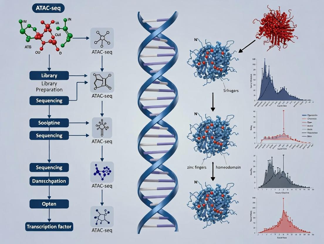
What is a Transcription Factor Footprint? Defining the Characteristic 'Dip' in ATAC-seq Data.
Within the broader thesis on ATAC-seq footprinting analysis for transcription factor (TF) research, this application note defines the core concept of a TF footprint. ATAC-seq (Assay for Transposase-Accessible Chromatin using sequencing) leverages a hyperactive Tn5 transposase to insert sequencing adapters into open chromatin regions. When a TF is bound to DNA, it physically occludes the Tn5 enzyme from cleaving and inserting adapters at that specific location. This protection results in a characteristic depletion or "dip" in sequencing read coverage at the TF binding site, flanked by enriched reads from adjacent accessible regions. This pattern is the Transcription Factor Footprint.
Defining the Characteristic 'Dip': Quantitative Signatures
The footprint "dip" is not merely an absence of signal but has quantifiable features derived from aggregated data across multiple binding sites. The table below summarizes the key quantitative parameters that define a confident footprint.
Table 1: Quantitative Parameters of a Characteristic TF Footprint 'Dip' in ATAC-seq Data
| Parameter | Typical Value/Range | Description & Interpretation |
|---|---|---|
| Footprint Depth | 20-50% reduction | The magnitude of read depletion at the center relative to flanking peaks. Deeper dips indicate stronger protection. |
| Footprint Width | 6-12 bp | The width of the protected region, corresponding closely to the physical binding site size of the TF. |
| Flank-to-Center Ratio | 1.5 - 3.0 | The ratio of read density in the flanking regions (e.g., +/- 50 bp) to the center. Higher ratios indicate a clearer footprint. |
| Statistical Significance (p-value) | < 0.01 | P-value from footprint detection algorithms (e.g., TOBIAS, HINT-BC, Wellington) assessing the likelihood the dip occurs by chance. |
| Cleavage Profile Skew | ≥ 2.0 bias | The ratio of forward vs. reverse Tn5 cleavage events at the footprint boundaries, indicating precise steric hindrance. |
Core Protocol: Detecting TF Footprints from ATAC-seq Data
This protocol details the computational detection of TF footprints using the TOBIAS (Transcription factor Occupancy prediction By Investigation of ATAC-seq Signal) suite, a current and widely adopted method.
Protocol: TF Footprint Analysis with TOBIAS
I. Prerequisites & Input Data
- Aligned ATAC-seq BAM files: From your experimental or public dataset (e.g., GEO accession GSE123456).
- Reference genome FASTA file: Corresponding to the alignment genome (e.g., hg38.fa).
- TF Motif Database: Position Weight Matrices (PWMs) in JASPAR or TRANSFAC format.
- Software: TOBIAS installed via conda (
conda install -c bioconda tobias).
II. Step-by-Step Methodology
Correct Tn5 Bias (TOBIAS ATACorrect):
- Purpose: Adjusts for the innate sequence bias of the Tn5 transposase, which favors cleavage at certain dinucleotides.
Command:
Output: Corrected, bias-free BED files of insertions.
Calculate Footprint Scores (TOBIAS FootprintScores):
- Purpose: Computes the footprinting score (FPS) across all peaks. The FPS quantifies the depletion at each base pair.
Command:
Output: BigWig file of per-base footprint scores.
Detect Significant Footprints & Bound TFs (TOBIAS BINDetect):
- Purpose: Identifies statistically significant footprints within peaks and predicts which specific TF motifs are bound based on the footprint signature.
Command:
Output: Directory containing:
*_bound_factors.bed: Genomic locations of bound TFs.*_footprints.bed: Genomic locations of significant footprint "dips".*_scores.pdf: Visualization of aggregate footprint profiles per TF.
III. Expected Results & Validation
- Successful execution yields a list of TF footprints with associated p-values and bound TFs. Validation should include:
- Comparison with ChIP-seq data for the same TF (if available).
- Inspection of aggregate footprint plots for clear "dips" at the motif location.
- Correlation of footprint depth with TF expression or activity from orthogonal assays.
Visualizing the Workflow and Signal
ATAC-seq Footprinting Analysis Workflow
TF Footprint Dip in ATAC-seq Insertion Profile
The Scientist's Toolkit: Research Reagent Solutions
Table 2: Essential Materials for ATAC-seq Footprinting Experiments
| Item | Function in Footprinting Analysis | Example Product/Catalog |
|---|---|---|
| Hyperactive Tn5 Transposase | Enzyme for simultaneous fragmentation and tagging of accessible DNA. The core reagent generating the footprint signal. | Illumina Tagmentase TDE1 (20034197) |
| Nextera-style Adapters | Oligonucleotides loaded onto Tn5, containing sequencing primer sites and sample barcodes. | Illumina Unique Dual Indexes (20027213) |
| Magnetic Beads (SPRI) | For size selection post-tagmentation to isolate nucleosomal fragments (e.g., < 300 bp for mononucleosomes). | Beckman Coulter AMPure XP (A63881) |
| High-Fidelity PCR Mix | To amplify library fragments with minimal bias, preserving the true footprint depth. | Kapa HiFi HotStart ReadyMix (KK2602) |
| Digital PCR or qPCR Kit | For accurate quantification of final library concentration prior to sequencing. | Qubit dsDNA HS Assay Kit (Q32851) |
| TF Motif Database | Curated Position Weight Matrices (PWMs) used to scan footprints for TF identity. | JASPAR2024 CORE vertebrates, HOCOMOCO v12 |
| Footprinting Software | Computational suite to correct bias, score, and detect significant footprints. | TOBIAS, HINT-ATAC, Wellington |
ATAC-seq (Assay for Transposase-Accessible Chromatin using sequencing) has revolutionized the study of chromatin accessibility. Its application for transcription factor (TF) footprinting—the detection of protein-bound DNA sequences from patterns of cleavage protection—offers a unique combination of sensitivity, scalability, and single-cell compatibility. This note details protocols and considerations for leveraging ATAC-seq in TF footprinting analysis as part of a thesis on regulatory genomics in drug discovery.
Key Advantages in Footprinting Analysis
Sensitivity
ATAC-seq requires far fewer cells than traditional DNase-seq or FAIRE-seq, detecting open chromatin from as few as 500-50,000 cells. This sensitivity is critical for rare cell populations and clinical samples.
Scalability & Single-Cell Potential
The protocol is rapid (<4 hours) and can be scaled from bulk to single-cell assays (scATAC-seq), enabling the profiling of TF binding heterogeneity within complex tissues—a key asset for developmental biology and oncology research.
Integrated Epigenomic Profiling
Beyond footprinting, ATAC-seq provides concurrent data on nucleosome positioning and broad chromatin accessibility from the same library.
Quantitative Comparison of Footprinting Assays
Table 1: Comparative Metrics of Chromatin Accessibility & Footprinting Assays
| Assay | Typical Cell Input | Time to Library | Key Footprinting Strength | Primary Limitation |
|---|---|---|---|---|
| DNase-seq | 1x10^6 - 50x10^6 | 3-4 days | High resolution, gold standard footprint depth | High cell input, technically challenging |
| ATAC-seq | 500 - 50,000 | 3-4 hours | Speed, low input, single-cell compatible | Sequence bias of Tn5, mitochondrial reads |
| MNase-seq | 1x10^6 - 10x10^6 | 2-3 days | Excellent nucleosome positioning | Poor for footprinting low-affinity TFs |
| scATAC-seq | 1 (per cell) | 1-2 days (post-sorting) | Cellular heterogeneity of TF binding | Sparse data per cell, complex analysis |
Table 2: Example ATAC-seq Footprinting Data Yield (Simulated Experiment)
| Condition | Cells Sequenced | Total Reads | TSS Enrichment | Footprints Detected (FDR<0.05) | Key TFs Identified |
|---|---|---|---|---|---|
| Healthy Donor PBMCs (Bulk) | 50,000 | 50 Million | 15 | ~1200 | PU.1, RUNX1, CTCF |
| Cancer Cell Line (Bulk) | 5,000 | 30 Million | 12 | ~900 | MYC, NF-κB, AP-1 |
| Mixed Tissue (scATAC-seq) | 10,000 cells | 200 Million (aggregate) | 10 (aggregate) | ~800 (aggregate) | Cell-type specific TF activ. |
Detailed Experimental Protocols
Protocol 1: Standard Bulk ATAC-seq for Footprinting
A. Cell Lysis and Tagmentation
- Cell Preparation: Wash 50,000 viable, nucleated cells once with 1x PBS. Do not fix cells.
- Lysis: Resuspend cell pellet in 50 µL of chilled Lysis Buffer (10 mM Tris-HCl pH 7.4, 10 mM NaCl, 3 mM MgCl2, 0.1% IGE PAL CA-630). Incubate on ice for 3 minutes.
- Immediate Nuclei Wash: Immediately add 1 mL of Wash Buffer (1x PBS, 0.1% BSA, 1 mM DTT) and invert gently. Pellet nuclei at 500 rcf for 10 minutes at 4°C. Carefully aspirate supernatant.
- Tagmentation Reaction: Prepare the Tagmentation Mix: 25 µL 2x TD Buffer, 2.5 µL Tn5 Transposase (Illumina), and 22.5 µL nuclease-free water. Resuspend the nuclei pellet in the 50 µL Tagmentation Mix by pipetting gently. Incubate at 37°C for 30 minutes in a thermomixer with gentle shaking (300 rpm).
- Clean-up: Purify tagmented DNA immediately using a MinElute PCR Purification Kit (Qiagen). Elute in 21 µL Elution Buffer.
B. Library Amplification and Sequencing
- PCR Setup: To the 21 µL eluate, add 2.5 µL of a uniquely barcoded Primer Ad1, 2.5 µL of a uniquely barcoded Primer Ad2, and 25 µL of NEBNext High-Fidelity 2x PCR Master Mix.
- Amplify with Limited Cycles: Run PCR: 72°C for 5 min; 98°C for 30 sec; then 5-12 cycles of (98°C for 10 sec, 63°C for 30 sec, 72°C for 1 min). Determine optimal cycle number via qPCR side reaction to avoid over-amplification.
- Final Purification: Clean the PCR product using a 1.2x ratio of SPRIselect beads (Beckman Coulter). Elute in 20 µL. Assess library quality on a Bioanalyzer (broad smear ~100-1000 bp).
- Sequencing: Sequence on an Illumina platform using paired-end sequencing (PE 2x50 bp or 2x75 bp). Aim for 50-100 million reads for robust footprinting.
Protocol 2: Computational Pipeline for ATAC-seq Footprinting Analysis
A. Preprocessing & Alignment
- Adapter Trimming & QC: Use
cutadaptorTrim Galore!to remove adapter sequences. Assess quality withFastQC. - Alignment & Filtering: Align reads to a reference genome (e.g., hg38) using
Bowtie2orBWAwith parameters-X 2000to allow large fragments. Remove duplicates usingPicard. Remove reads mapping to mitochondria and blacklisted regions. - Nucleosome Positioning & Accessibility: Generate fragment length distribution plots to identify nucleosome-free (<100 bp) and mono-/di-nucleosome fragments. Shift + strand reads by +4 bp and - strand reads by -5 bp to account for Tn5 offset when generating the BAM file for peak calling.
B. Footprint Detection & TF Inference
- Generate Coverage Tracks: Use
deepToolsto create Tn5 insertion site (cut site) bigWig tracks from the shifted BAM file. - Call Footprints: Run a footprinting algorithm. HINT-ATAC or TOBIAS are specifically designed for ATAC-seq data and correct for Tn5 sequence bias.
- Example TOBIAS command:
TOBIAS ATACorrect --reads ./alignments.bam --genome ./hg38.fa --peaks ./atac_peaks.bed --outdir ./corrected - Follow with:
TOBIAS FootprintScores --signal ./corrected/corrected.bw --regions ./atac_peaks.bed --output ./footprints.bw - Finally:
TOBIAS BINDetect --footprints ./footprints.bw --regions ./atac_peaks.bed --motifs ./JASPAR2020_CORE_vertebrates.meme --output ./TF_activities
- Example TOBIAS command:
- Integrate with TF Motifs: Match footprint locations to known TF binding motifs (from databases like JASPAR, CIS-BP) to infer bound TFs.
Visualizations
Bulk ATAC-seq Experimental Workflow
ATAC-seq Footprinting Computational Pipeline
Idealized ATAC-seq Footprint Signature
The Scientist's Toolkit: Essential Research Reagent Solutions
Table 3: Key Reagents and Materials for ATAC-seq Footprinting
| Item | Function & Importance | Example Product/Catalog # |
|---|---|---|
| Tn5 Transposase | Enzyme that simultaneously fragments and tags accessible DNA with sequencing adapters. Core reagent. | Illumina Tagment DNA TDE1 Enzyme (20034197) |
| Nuclei Isolation & Lysis Buffer | Gently lyses plasma membrane while keeping nuclear membrane intact for clean tagmentation. | 10x Nuclei Isolation Buffer (10x Genomics, 1000493) or homemade (see protocol). |
| SPRIselect Beads | For size selection and purification of tagmented DNA/PCR libraries. Critical for removing primer dimers. | Beckman Coulter SPRIselect (B23318) |
| High-Fidelity PCR Master Mix | For limited-cycle amplification of tagmented DNA with high fidelity to minimize biases. | NEBNext High-Fidelity 2x PCR Master Mix (NEB, M0541) |
| Dual-Indexed PCR Primers | Unique barcodes for multiplexing samples. Essential for scATAC-seq and pooling bulk samples. | Nextera Index Kit (Illumina) or custom ordered. |
| Cell Viability Stain | Distinguish live/dead cells prior to assay. Dead cells cause high background. | Trypan Blue, DAPI, or Propidium Iodide. |
| Motif Database | Curated collection of TF binding motifs for footprint annotation. | JASPAR, CIS-BP, HOCOMOCO |
| Footprinting Software | Corrects Tn5 bias and detects protected regions. | TOBIAS, HINT-ATAC, or pyDNase. |
Application Notes
Within the broader thesis on ATAC-seq footprinting analysis for transcription factor (TF) research, this methodology serves as a critical tool for deciphering the regulatory genome. Footprinting leverages the principle that a protein bound to DNA protects that region from enzymatic cleavage, creating a "footprint" of inaccessibility in sequencing data. This allows researchers to move beyond mere chromatin accessibility maps (provided by ATAC-seq) to infer precise protein-DNA interactions and the combinatorial logic of regulatory elements.
Key Questions Addressed:
- TF Occupancy and Binding Site Identification: Where do specific TFs bind in the genome under defined cellular conditions? Footprinting reveals protected sequences within open chromatin, pinpointing putative binding sites at base-pair resolution, even for TFs without available ChIP-grade antibodies.
- Differential TF Activity Across Conditions: How does TF binding change during differentiation, disease progression, or in response to a drug? Comparative footprinting analysis between samples can identify gains or losses of specific TF footprints, linking transcriptional regulators to phenotypic shifts.
- Deciphering cis-Regulatory Logic: How do TFs cooperate within enhancers or promoters? The co-localization of multiple TF footprints within a single accessible region reveals potential cooperative interactions and helps define the "regulatory grammar" of cis-regulatory modules.
- Linking Non-Coding Variants to Function: How do genetic variants in regulatory regions alter gene expression? Single-nucleotide polymorphisms (SNPs) or mutations that disrupt or create a TF footprint provide a mechanistic explanation for disease-associated non-coding variants identified in GWAS.
Quantitative Metrics in Footprinting Analysis: The following table summarizes core quantitative outputs derived from footprinting analysis.
Table 1: Key Quantitative Metrics from ATAC-seq Footprinting Analysis
| Metric | Description | Typical Value/Range | Biological Interpretation |
|---|---|---|---|
| Footprint Depth | The normalized reduction in cleavage (Tn5 insertion) signal at the protected site. | 2-10 fold depletion | Proportional to binding affinity and occupancy. Deeper footprints suggest stronger or more stable binding. |
| Footprint Score (e.g., TOBIAS) | A composite statistical score integrating cleavage depletion and flanking enrichment. | Z-scores or p-values | Confidence metric for a true TF binding event versus background noise. |
| Motif Disruption Score | Quantifies the impact of a genetic variant on the predicted TF binding motif (e.g., change in PWM score). | ∆PWM Score | Predicts the functional consequence of a non-coding variant on TF binding. |
| Differential Footprint Score | Statistical measure of change in footprint strength between two conditions (e.g., Wald statistic). | Log2 Fold Change, p-value | Identifies TFs with significantly altered genome-wide binding between experimental states. |
| Footprint Occupancy Correlation | Correlation coefficient between footprint strength and target gene expression across samples. | Pearson's r (-1 to 1) | Suggests activating (positive) or repressive (negative) regulatory relationships. |
Protocols
Protocol 1: ATAC-seq Library Preparation for Optimal Footprinting
Adapted from Buenrostro et al. (2013, 2015) with modifications for footprinting sensitivity.
Objective: Generate high-quality ATAC-seq libraries from nuclei with sufficient sequencing depth to detect cleavage patterns.
Materials:
- Cells of interest (50,000 - 100,000 viable cells per reaction)
- ATAC-seq Buffer Set (Resuspension, Lysis, Wash Buffers)
- Tn5 Transposase (Loaded) (Commercial kit, e.g., Illumina Tagmentase)
- DNA Cleanup Beads (SPRIselect beads)
- Indexing PCR Primers
- Qubit dsDNA HS Assay Kit
- Bioanalyzer/TapeStation High Sensitivity DNA Assay
Procedure:
- Cell Lysis & Transposition: Pellet cells. Lyse in cold lysis buffer (10 mM Tris-HCl pH 7.4, 10 mM NaCl, 3 mM MgCl2, 0.1% IGEPAL CA-630) for 3 min on ice. Immediately pellet nuclei and resuspend in transposition mix (25 μL TD Buffer, 2.5 μL Tn5, 22.5 μL nuclease-free water). Incubate at 37°C for 30 min.
- DNA Purification: Clean up transposed DNA using SPRIselect beads at a 1:1 beads-to-sample ratio. Elute in 20 μL EB buffer.
- Library Amplification: Amplify purified DNA using indexed primers (5-12 cycles, depending on input). Use qPCR to determine optimal cycle number to avoid over-amplification.
- Final Clean-up & QC: Perform a double-sided SPRI bead cleanup (e.g., 0.5x then 1.5x ratios) to remove primers and select for properly sized fragments. Quantify library concentration (Qubit) and assess size distribution (Bioanalyzer; expect a periodicity of ~200 bp). Sequence on Illumina platform (minimum 100M paired-end reads for human/mouse footprinting).
Protocol 2: Computational Footprinting Analysis with TOBIAS
Based on Bentsen et al. (Nature Communications, 2020).
Objective: Identify and quantify transcription factor footprints from ATAC-seq data.
Prerequisites: Installed TOBIAS suite, aligned ATAC-seq BAM files, and reference genome.
Procedure:
- Data Preprocessing: This step corrects for Tn5 insertion bias, creating bias-corrected BigWig files.
Footprint Identification:
Calculates footprint scores across all accessible regions.
TF Binding Inference:
Integrates footprint scores with known TF motif positions to infer bound/unbound sites and calculate binding scores per TF.
Differential Analysis (for two conditions):
Outputs statistics on TFs with significantly differential binding between conditions.
The Scientist's Toolkit: Research Reagent Solutions
Table 2: Essential Reagents and Materials for ATAC-seq Footprinting
| Item | Function in Experiment | Example/Notes |
|---|---|---|
| Loaded Tn5 Transposase | Simultaneously fragments open chromatin and adds sequencing adapters. Critical for library generation. | Illumina Tagmentase TDE1, or custom-loaded "homebrew" Tn5. |
| SPRIselect Beads | Size-selective purification of DNA fragments. Used to clean up transposition reactions and final libraries. | Beckman Coulter SPRIselect. Essential for removing short fragments and adapter dimers. |
| High-Sensitivity DNA Assay | Accurate quantification and size profiling of final sequencing libraries. | Agilent Bioanalyzer High Sensitivity DNA chip or equivalent. Confirms nucleosomal patterning. |
| Cell Permeabilization Detergent | Gently lyses the plasma membrane while keeping nuclei intact for transposition. | IGEPAL CA-630 (Nonidet P-40). Concentration and timing are critical. |
| Nuclei Counter | Ensures precise input of nucleus numbers into transposition reaction, a key variable for reproducibility. | Automated cell counter (e.g., Countess II) or hemocytometer. |
| PCR Library Amplification Kit | Amplifies transposed DNA with minimal bias. | KAPA HiFi HotStart ReadyMix or NEB Next Ultra II Q5. |
| TF Motif Database | Curated collection of position weight matrices (PWMs) for mapping predicted TF binding sites. | JASPAR, CIS-BP, HOCOMOCO. Required for BINDetect step. |
| Cluster Analysis Software | For visualizing footprint signals and cleavage patterns at specific genomic loci. | IGV (Integrative Genomics Viewer) or pyGenomeTracks. |
Visualizations
Title: ATAC-seq Footprinting Analysis Computational Workflow
Title: Principle of a TF Footprint in ATAC-seq Data
Title: cis-Regulatory Logic from Co-localized TF Footprints
Within the broader thesis investigating ATAC-seq footprinting for transcription factor (TF) binding dynamics in drug discovery, the foundational steps of paired-end sequencing and precise read alignment are critical. These prerequisites determine the resolution needed to detect the short (~10 bp), protected regions indicative of TF occupancy amidst open chromatin, directly impacting downstream analyses of gene regulation and potential therapeutic targets.
Core Concepts and Quantitative Data
Paired-End vs. Single-End Sequencing for Footprinting
Paired-end sequencing generates reads from both ends of each DNA fragment, providing superior alignment accuracy and fragment length determination—essential for footprinting.
Table 1: Comparative Metrics for Sequencing Strategies in ATAC-seq Footprinting
| Parameter | Paired-End Sequencing | Single-End Sequencing |
|---|---|---|
| Alignment Accuracy | High (precise mapping of both ends) | Moderate (reliance on one end) |
| Insert Size Estimation | Direct and accurate measurement | Indirect or inferred |
| Error Correction | Enables self-correction of alignment errors | Limited error correction |
| Footprint Signal | Clear, high-resolution protected regions | Noisy, lower resolution |
| Typical Read Length | 2 x 50-150 bp | 50-150 bp |
| Cost per Sample | Higher | Lower |
| Suitability for TFBS | Excellent (required for base-pair resolution) | Poor (insufficient for precise footprint detection) |
Alignment Quality Metrics Impacting Footprint Sensitivity
The quality of read alignment directly influences the signal-to-noise ratio in footprinting assays.
Table 2: Key Alignment Metrics and Their Impact on Footprinting Analysis
| Alignment Metric | Optimal Range for Footprinting | Impact on Footprint Detection |
|---|---|---|
| Overall Alignment Rate | > 80% | Low rates indicate poor library quality or contamination, obscuring true signal. |
| Uniquely Mapped Reads | > 70% of total reads | Multi-mapping reads create ambiguous signal, diluting footprint clarity. |
| Properly Paired Rate | > 90% of mapped pairs | Ensures accurate fragment size representation, crucial for identifying protected regions. |
| Mitochondrial Read % | < 20% (after depletio n strategies) | High mitochondrial alignment consumes sequencing depth without informative chromatin data. |
| Duplicate Rate | < 30% (post-filtering) | PCR duplicates over-amplify certain fragments, biasing accessibility quantification. |
Experimental Protocols
Protocol: Paired-End Sequencing Library Preparation from ATAC-seq Samples
This protocol follows the Omni-ATAC method with optimizations for footprinting-ready libraries.
Materials:
- Nextera DNA Library Prep Kit (Illumina)
- AMPure XP beads (Beckman Coulter)
- Qubit dsDNA HS Assay Kit
- Tapestation or Bioanalyzer (Agilent)
- PCR thermocycler
- Size-selection reagents (e.g., SPRIselect)
Procedure:
- Tagmentation: Use pre-loaded transposomes (from Omni-ATAC or similar) on 50,000 nuclei. Incubate at 37°C for 30 minutes. Immediately purify using MinElute PCR Purification Kit.
- PCR Amplification:
- Assemble PCR reaction: tagmented DNA, 1x Hi-Fi PCR Master Mix, custom Nextera index primers (i5 and i7).
- Cycle conditions: 72°C for 5 min; 98°C for 30 sec; then cycle (98°C for 10 sec, 63°C for 30 sec, 72°C for 1 min) for 8-12 cycles (determined by qPCR side reaction); final extension at 72°C for 5 min.
- Size Selection and Cleanup:
- Perform double-sided SPRI bead cleanup (e.g., 0.55x and 1.5x ratios) to isolate fragments primarily between 150-800 bp, removing short fragments (<100 bp) that hamper paired-end alignment.
- Elute in 20 µL EB buffer.
- Quality Control:
- Quantify using Qubit.
- Assess fragment size distribution using Tapestation D5000/High Sensitivity screentape.
- Validate library complexity (ensure minimal duplicate rate).
- Sequencing:
- Pool libraries appropriately.
- Sequence on Illumina platform with paired-end 75 bp or longer cycles. Aim for > 50 million unique, non-mitochondrial read pairs per sample for footprinting.
Protocol: Alignment of ATAC-seq Paired-End Reads for Footprinting
This protocol uses the Burrows-Wheeler Aligner (BWA-MEM2) and SAMtools for optimal mapping.
Materials:
- High-performance computing cluster or server
- Reference genome (e.g., GRCh38/hg38 primary assembly)
- BWA-MEM2 software
- SAMtools
- Picard Toolkit or sambamba
Procedure:
- Prepare Reference Genome:
- Download reference FASTA and corresponding
.gtfannotation. - Generate BWA index:
bwa-mem2 index GRCh38.primary_assembly.genome.fa - Generate FASTA index:
samtools faidx GRCh38.primary_assembly.genome.fa
- Download reference FASTA and corresponding
- Align Reads:
- Run BWA-MEM2 in paired-end mode:
bwa-mem2 mem -t 16 -M -R "@RG\tID:sample1\tSM:sample1" \ GRCh38.primary_assembly.genome.fa \ sample1_R1.fastq.gz sample1_R2.fastq.gz > sample1.sam(-Mmarks shorter split hits as secondary;-Radds read group).
- Run BWA-MEM2 in paired-end mode:
- Process SAM/BAM Files:
- Convert to BAM, sort, and index:
samtools view -@ 16 -b sample1.sam | samtools sort -@ 16 -o sample1_sorted.bamsamtools index sample1_sorted.bam
- Convert to BAM, sort, and index:
- Remove Duplicates:
- Use Picard:
java -jar picard.jar MarkDuplicates \ I=sample1_sorted.bam O=sample1_deduped.bam M=dup_metrics.txt - Index the deduplicated BAM.
- Use Picard:
- Filter Alignments:
- Retain properly paired, uniquely mapping, non-mitochondrial reads with mapping quality (MAPQ) ≥ 30:
samtools view -@ 16 -b -h -f 2 -F 1804 -q 30 sample1_deduped.bam \ | samtools idxstats - \ | cut -f 1 \ | grep -v '^chrM$\|^MT$' \ | xargs samtools view -b -o sample1_final.bam
- Retain properly paired, uniquely mapping, non-mitochondrial reads with mapping quality (MAPQ) ≥ 30:
- Generate Alignment Metrics:
- Use
samtools flagstatandsamtools idxstatsto generate metrics matching Table 2.
- Use
Visualization
Title: ATAC-seq Paired-End Data Generation & Processing Workflow
Title: Paired-End Reads Define TF Footprint
The Scientist's Toolkit: Research Reagent Solutions
Table 3: Essential Materials for Paired-End ATAC-seq Footprinting Studies
| Item Name | Supplier Examples | Function in Workflow |
|---|---|---|
| Nextera DNA Library Prep Kit | Illumina | Provides reagents for tagmentation, PCR amplification, and index addition for multiplexing. |
| AMPure/SPRIselect Beads | Beckman Coulter | For post-PCR cleanup and precise size selection to optimize fragment length distribution. |
| BWA-MEM2 Software | Open Source | Efficient and accurate alignment algorithm for paired-end sequencing data to a reference genome. |
| SAMtools/Picard Toolkit | Open Source/Broad Institute | For processing, filtering, sorting, and deduplicating alignment files; critical for data quality control. |
| D5000 High Sensitivity Tape | Agilent | Accurately assesses library fragment size distribution and quality before sequencing. |
| Qubit dsDNA HS Assay Kit | Thermo Fisher Scientific | Fluorometric quantification of library concentration, more accurate for diluted samples than spectrophotometry. |
| Custom Index Primers | IDT, Thermo Fisher | Unique dual-index barcodes for sample multiplexing, reducing index hopping and enabling large-scale studies. |
From FASTQ to Footprints: A Step-by-Step Pipeline for ATAC-seq Footprinting Analysis
Within a thesis on ATAC-seq footprinting analysis for transcription factor (TF) research, robust data preprocessing is the critical foundation. This phase directly impacts the detection of subtle, TF-protected footprints in chromatin accessibility data. This document details application notes and protocols for adapter trimming, quality control, and alignment, optimized for sensitive downstream footprinting analysis.
Application Notes
Adapter Trimming and Quality Control
ATAC-seq libraries contain transposase adapters. Incomplete tagmentation leaves adapter sequences in reads, which can interfere with alignment, especially at the ends of accessible regions where TF footprints reside. Quality control ensures data integrity.
Table 1: Recommended Tools for Pre-Alignment Processing
| Tool | Primary Function | Key Parameter for ATAC-seq | Rationale |
|---|---|---|---|
| cutadapt | Adapter Trimming | -a CTGTCTCTTATACACATCT... |
Removes Nextera transposase sequence. Prevents false mismatches. |
| FastQC | Quality Assessment | Per-sequence GC content | Flags biases from ATAC's periodicity. |
| Trimmomatic | Quality Trimming | SLIDINGWINDOW:4:20 |
Removes low-quality ends while preserving short inserts. |
| Picard Tools | Duplicate Marking | REMOVE_SEQUENCING_DUPLICATES=false |
ATAC duplicates are often biological; mark but don't remove. |
Alignment with BWA-MEM2
Precise alignment is paramount for footprinting. BWA-MEM2 offers speed and accuracy, critical for mapping the mixed-length (nuclear vs. mitochondrial) ATAC-seq reads.
Table 2: BWA-MEM2 Alignment Parameters for ATAC-seq Footprinting
| Parameter | Recommended Setting | Purpose in Footprinting Analysis |
|---|---|---|
-T (minimum score) |
30 | Increases mapping stringency, reducing spurious alignments that obscure footprint boundaries. |
-M |
Flagged | Marks shorter hits as secondary for compatibility with downstream tools. |
-B (mismatch penalty) |
4 | Standard setting; increasing can improve specificity but reduce sensitivity. |
-p |
Enabled | Signals interleaved paired-end FASTQ input. |
| Reference Genome | hg38 (primary assembly) | Use consistent genome build for TF motif matching. Include mitochondrial DNA. |
Experimental Protocol: End-to-End Preprocessing for ATAC-seq Footprinting
Protocol 1: Adapter Trimming and QC
- Quality Check (FastQC):
Adapter Trimming (cutadapt):
Post-Trimming QC: Run FastQC on trimmed files and compare reports.
Protocol 2: Alignment with BWA-MEM2
- Index Reference Genome (if not done):
Align Reads:
Convert, Sort, and Index (samtools):
Filter for Mapping Quality and Remove Mitochondrial Reads (typical):
Visualized Workflows
ATAC-seq Data Preprocessing Workflow for Footprinting
From Aligned Reads to TF Inference in ATAC-seq Footprinting
The Scientist's Toolkit: Research Reagent Solutions
Table 3: Essential Materials for ATAC-seq Library Prep & Analysis
| Item | Function in ATAC-seq/Footprinting |
|---|---|
| Tn5 Transposase (Loaded) | Enzyme that simultaneously fragments and tags accessible DNA with adapters. |
| NEBNext High-Fidelity 2X PCR Master Mix | Amplifies library post-tagmentation with minimal bias. |
| SPRIselect Beads | Size selection to enrich for nucleosome-free fragments (<100bp). |
| DNeasy Blood & Tissue Kit | Isolate high-quality nuclei from cells/tissues. |
| Bioanalyzer/TapeStation HS DNA Kit | Assess final library size distribution pre-sequencing. |
| BWA-MEM2 Software | High-speed aligner for accurate mapping of sequenced reads. |
| Picard Tools | Process aligned files (mark duplicates, collect metrics). |
| ATAC-seq Footprinting Software (e.g., HINT-ATAC, TOBIAS) | Specialized tools to detect footprints and infer TF binding. |
Application Notes
Within the thesis framework of ATAC-seq footprinting analysis for transcription factor (TF) research, post-alignment processing is a critical determinant of data quality and interpretability. This step transforms raw aligned sequencing reads into a clean, biologically relevant signal suitable for detecting the subtle, short depressions in cleavage profiles that constitute TF footprints. The three core procedures—duplicate marking, mitochondrial read filtering, and Tn5 shift correction—each address distinct artifacts that would otherwise obscure these footprints.
Duplicate Marking: PCR amplification during library preparation can generate multiple read pairs originating from a single original DNA fragment. These technical duplicates inflate coverage uniformity and can create false-positive peaks or mask genuine low-coverage footprints. Marking and subsequently removing these duplicates is essential for quantitative accuracy in downstream footprinting tools.
Mitochondrial Read Filtering: The ATAC-seq protocol preferentially targets accessible DNA due to mitochondrial membrane permeabilization, resulting in a high proportion (often 20-50%) of reads aligning to the mitochondrial genome. As mitochondrial DNA is not of interest for nuclear TF footprinting, these reads consume sequencing depth and computational resources. Their removal is mandatory to focus analysis on the nuclear genome and improve the signal-to-noise ratio.
Tn5 Shift Correction: The Tn5 transposase binds as a dimer and inserts adapters 9 bp apart on opposite DNA strands. Consequently, the exact cleavage sites are offset from the true accessible DNA boundaries. A simple alignment creates a 9 bp stagger in the read start positions. Applying a +4 bp/-5 bp shift (forward/reverse strand) aligns the read ends to represent the actual physical ends of the accessible region, yielding sharper peaks and more precise footprint boundaries.
Table 1: Impact of Post-Alignment Processing Steps on ATAC-seq Data for Footprinting Analysis
| Processing Step | Primary Artifact Addressed | Consequence if Omitted for Footprinting | Typical Quantitative Impact |
|---|---|---|---|
| Duplicate Marking | PCR amplification duplicates | Overestimation of coverage; false uniformity in signal; reduced ability to call faint footprints. | Duplicate rate typically 20-40% of aligned reads. |
| Mitochondrial Filtering | High mt-DNA alignment | Severe reduction in usable nuclear sequencing depth; increased computational overhead. | mt-DNA reads constitute 15-50% of total aligned reads. |
| Tn5 Shift Correction | 9 bp stagger from Tn5 dimer binding | "Double-peak" artifact; blurred peak and footprint boundaries; reduced precision in TF motif mapping. | Applies +4 bp shift to + strand reads, -5 bp shift to – strand reads. |
Experimental Protocols
Protocol 1: Duplicate Marking using picard MarkDuplicates
- Input: Coordinate-sorted BAM file from aligner (e.g., BWA, Bowtie2).
- Tool Execution: Run the following command:
- Parameters:
REMOVE_DUPLICATES=falseflags duplicates for downstream filtering.ASSUME_SORT_ORDERensures correct processing. - Output: A BAM file with duplicate reads flagged (bit 0x400). The accompanying metrics file details the number and percentage of duplicates.
- Downstream: Filter flagged reads in subsequent steps (e.g., using
samtools view -F 1024).
Protocol 2: Mitochondrial Read Filtering using samtools
- Reference: Identify the mitochondrial chromosome name in your reference genome (e.g.,
chrM,MT). - Filtering: Use
samtoolsto exclude reads aligning to this sequence and extract properly paired reads.
- Parameters:
-f 2requires reads be properly paired.-F 1024excludes marked duplicates. - Verification: Generate a new alignment statistics report (
samtools idxstats) to confirm mt-DNA depletion.
Protocol 3: Tn5 Shift Correction and BED File Generation
- Input: Filtered, deduplicated BAM file (
filtered_noMT.bam). - Shift Reads: Use a tool like
bedtoolsor a custom script to adjust read start positions. Example usingawkafter BED conversion:
Filter Fragments: Remove fragments unlikely to represent open chromatin (e.g., > 1000 bp).
Output: A BED file of shifted, size-selected DNA fragments, ready for peak calling and footprinting analysis.
Visualization
Title: ATAC-seq Post-Alignment Processing Workflow
Title: Tn5 Shift Correction Rationale
The Scientist's Toolkit: Essential Research Reagents & Tools
Table 2: Key Solutions and Tools for ATAC-seq Post-Alignment Processing
| Item | Function/Description | Example/Note |
|---|---|---|
| High-Quality Reference Genome | Sequence for aligning reads; must include mitochondrial DNA. | GRCh38, mm10. Includes chrM/MT. |
| Sequence Alignment Tool | Aligns sequenced reads to the reference genome. | BWA-MEM, Bowtie2. Optimized for short reads. |
| Picard Tools Suite | Java-based utilities for handling high-throughput sequencing data. | MarkDuplicates is the standard for duplicate marking. |
| SAMtools | Utilities for manipulating SAM/BAM files; filtering and statistics. | Critical for view, sort, index, and filter operations. |
| BEDTools | Swiss-army knife for genomic interval operations. | Used for shifting coordinates and fragment analysis. |
| Cluster/Cloud Computing | High-performance computing resources. | Necessary for processing large-scale ATAC-seq datasets. |
| Footprinting Analysis Software | Detects TF footprints from processed fragment data. | TOBIAS, HINT-ATAC, Wellington. |
| Programming Environment | For custom scripting and pipeline integration. | Python/R, bash scripting. |
Within the broader thesis on ATAC-seq footprinting analysis for transcription factor (TF) research, a critical methodological choice is the selection of a footprint detection algorithm. These algorithms identify regions of protected chromatin, indicative of TF binding, from ATAC-seq data. This application note details two dominant computational paradigms: site-centric (e.g., HINT, Wellington) and window-centric (e.g., TOBIAS) approaches, providing protocols and comparative analysis for researchers and drug development professionals.
Core Algorithm Paradigms and Quantitative Comparison
- Site-Centric (HINT, Wellington): These methods first identify candidate TF binding sites, typically from a position weight matrix (PWM) scan, and then evaluate the cleavage profile (read distribution) specifically at those discrete genomic locations to confirm a footprint.
- Window-Centric (TOBIAS): This approach performs a genome-wide scan using sliding windows to identify regions with a significant depletion of cleavage events (footprints) without prior knowledge of candidate sites, later correlating these regions with TF motifs.
Quantitative Comparison Table
Table 1: Comparative Summary of Footprint Detection Algorithms
| Feature | Site-Centric (HINT) | Site-Centric (Wellington) | Window-Centric (TOBIAS) |
|---|---|---|---|
| Primary Strategy | Statistical evaluation of cleavage patterns at predefined candidate sites. | Permutation-based significance testing at candidate sites. | Genome-wide correction of Tn5 bias followed by sliding-window footprint scoring. |
| Input Requirement | ATAC-seq reads, candidate regions (BED), PWM models. | ATAC-seq reads (BAM), candidate sites (BED). | ATAC-seq reads (BAM/FASTQ), reference genome, optional PWM models. |
| Key Output | Footprint scores & significance per candidate site. | Footprint p-value per candidate site. | Corrected chromatin accessibility track and footprint scores across the genome. |
| Strengths | High specificity at known motifs; robust to local noise. | Simple, direct statistical test; part of Suite. | Comprehensive; corrects sequence bias; identifies novel sites. |
| Limitations | Blind to sites not pre-defined by PWM. | Performance sensitive to cleavage profile quality. | Computationally intensive; may require deeper sequencing. |
| Typical Runtime* | ~30 min per sample (human, 50k sites) | ~15 min per sample (human, 50k sites) | ~2 hours per sample (human genome) |
*Runtime estimates are approximate and depend on data size and computational resources.
Detailed Experimental Protocols
Protocol 1: Site-Centric Footprinting with HINT
Objective: Identify significant footprints at known TF motif locations.
- Prerequisite Data: Aligned ATAC-seq reads (BAM format), genome reference (FASTA), TF PWMs (JASPAR/ENCODE motif databases).
- Candidate Site Identification:
- Use
fimo(MEME Suite) to scan the genome with PWMs (p-value < 1e-5). Output candidate sites in BED format.
- Use
- Run HINT Footprinting:
- Command:
rgt-hint footprinting --atac-seq --organism=hg38 --output-location=./hint_results --output-prefix=sample1 sample1.bam candidate_sites.bed
- Command:
- Post-processing & Analysis:
- Filter footprints by HINT's statistical score (e.g., footprint score > 0.5).
- Annotate footprints with gene features using
rgt-hint annotation.
Protocol 2: Window-Centric Footprinting with TOBIAS
Objective: Perform genome-wide unbiased footprint detection and correct for Tn5 sequence bias.
- Prerequisite Data: ATAC-seq reads (BAM or FASTQ), reference genome (FASTA).
- Bias Correction & Footprint Calling:
- Correct Tn5 insertion bias:
TOBIAS ATACorrect --bam sample1.bam --genome hg38.fa --peaks sample1_peaks.bed --outdir ./corrected - Calculate footprint scores across genome:
TOBIAS FootprintScores --signal ./corrected/sample1_corrected.bw --regions sample1_peaks.bed --output ./footprints/sample1_footprints.bw
- Correct Tn5 insertion bias:
- Identify Significant Footprints & TFs:
TOBIAS BINDetect --motifs motifs.jaspar --signals ./footprints/sample1_footprints.bw --genome hg38.fa --peaks sample1_peaks.bed --outdir ./bindetect_results
Visualizing the Analysis Workflows
Workflow: Site-Centric Footprint Analysis
Workflow: TOBIAS Window-Centric Analysis
The Scientist's Toolkit: Key Research Reagent Solutions
Table 2: Essential Computational Tools and Resources for ATAC-seq Footprinting
| Item | Function | Example/Format |
|---|---|---|
| Aligned ATAC-seq Reads | Primary input data containing genomic locations of Tn5 insertions. | BAM file (coordinate-sorted, indexed). |
| Transcription Factor Motifs | Digital representations of TF binding specificity for site prediction. | PWM files (JASPAR, HOCOMOCO, CIS-BP formats). |
| Reference Genome | Genomic sequence for mapping, motif scanning, and annotation. | FASTA file with index (e.g., hg38.fa, mm10.fa). |
| Genomic Annotation File | For correlating footprints with genomic features (promoters, enhancers). | GTF or GFF3 format. |
| Bias Correction Tool | Corrects inherent sequence preference of Tn5 transposase, critical for accuracy. | TOBIAS ATACorrect, pyDNase. |
| Footprint Calling Software | Core algorithm suite for detection. | HINT-ATAC, Wellington, TOBIAS, PIQ. |
| Motif Scanning Software | Identifies candidate binding sites from PWMs. | FIMO (MEME Suite), TFBSTools. |
| Visualization Browser | Enables manual inspection of cleavage profiles and footprints. | IGV, UCSC Genome Browser. |
This protocol, framed within a broader thesis on ATAC-seq footprinting analysis for transcription factor (TF) research, details an integrative bioinformatics pipeline. The core aim is to move from identifying regions of protected chromatin (footprints) to predicting the specific transcription factors bound at those sites. This is achieved by combining digital genomic footprints from ATAC-seq data with in vitro and in vivo TF binding motifs from curated databases like JASPAR and CIS-BP.
Application Notes
Rationale and Utility
Footprinting analysis of ATAC-seq data identifies putative protein-DNA binding sites based on a characteristic pattern of reduced cleavage (protected region) flanked by peaks of cleavage. However, a footprint alone does not reveal TF identity. By scanning the nucleotide sequence underlying a footprint against a library of known position weight matrices (PWMs), one can infer which TFs are likely bound. This integrative analysis is crucial for:
- Hypothesis Generation: Predicting which TFs drive regulatory programs in specific cell states or disease conditions.
- Mechanistic Insight: Linking open chromatin regions to specific transcriptional regulators.
- Drug Development: Identifying novel, targetable TFs in pathways of interest.
Key Databases for Motif Matching
Two primary databases are used for motif scanning. Their key characteristics are summarized in Table 1.
Table 1: Comparison of Primary Motif Databases
| Database | Full Name | Primary Source | Key Features | Typical Use Case |
|---|---|---|---|---|
| JASPAR | JASPAR CORE | Curated, non-redundant set of PWMs from published experiments. | High-quality, minimal redundancy, open access. | Standard, high-confidence TF prediction. |
| CIS-BP | Catalog of Inferred Sequence Binding Preferences | Mix of curated motifs and motifs inferred from protein sequences via DAP-seq, PBM, etc. | Extremely comprehensive, includes predicted motifs for many TFs. | When seeking motifs for less-studied TFs or isoforms. |
Quantitative Performance Metrics
The accuracy of TF identity prediction is assessed using benchmarking data from published studies (e.g., ENCODE ChIP-seq validation). Table 2 summarizes typical performance metrics when footprint-motif integration is performed under optimal conditions.
Table 2: Typical Performance Metrics for Prediction Accuracy
| Metric | Description | Typical Range (Optimal Conditions) |
|---|---|---|
| Precision (PPV) | % of predicted TF bindings that are validated by ChIP-seq. | 60-75% |
| Recall (Sensitivity) | % of ChIP-seq peaks correctly predicted by footprint+motif. | 50-65% |
| Area Under Curve (AUC) | Overall performance of classifier (motif score threshold). | 0.80-0.90 |
Experimental Protocols
Protocol: Integrative Footprint & Motif Analysis Workflow
I. Prerequisites & Input Data Preparation
- Input 1: A BED file of consensus footprint locations (e.g., from
TOBIAS,HINT-ATAC, orPyAtac). - Input 2: Reference genome FASTA file (hg38/mm10).
- Input 3: PWM files from JASPAR/CIS-BP (in MEME or TRANSFAC format).
II. Step-by-Step Procedure
Step 1: Extract Genomic Sequences Underlying Footprints
Step 2: Scan Footprint Sequences for TF Motifs
- Critical Parameter:
--threshsets p-value threshold. A stringent threshold (1e-4 to 1e-5) is recommended to minimize false positives.
Step 3: Integrate and Annotate Results
- Parse
fimo_output.txtto associate each significant motif hit (column 2:motif_id) with its genomic footprint location. - Map
motif_idto standard TF name using the database's metadata file. - Aggregate results: Count motif occurrences per TF across all footprints.
Step 4: Validation & Prioritization (Optional but Recommended)
- Filter by Chromatin Accessibility: Retain only motifs found within the central region of the footprint (greatest protection).
- Integrate with Expression Data: Prioritize TFs with cognate mRNA expression (from RNA-seq) in the sample.
- Compare to Public ChIP-seq Data: Use resources like CistromeDB or ENCODE to validate predictions.
Visualizations
Workflow Diagram
Title: Workflow for ATAC-seq Footprint & Motif Integration
Footprint-Motif Matching Logic
Title: Decision Logic for TF Prediction at a Single Footprint
The Scientist's Toolkit
Table 3: Essential Research Reagent Solutions & Tools
| Item / Software | Category | Function / Purpose | Example / Version |
|---|---|---|---|
| TOBIAS | Bioinformatics Tool | Suite for ATAC-seq footprinting; corrects for Tn5 bias, calls footprints. | TOBIAS v0.15.0 |
| MEME Suite | Bioinformatics Toolkit | Contains FIMO for motif scanning; converts motif formats. | MEME Suite v5.5.2 |
| JASPAR CORE | Database | Curated, non-redundant collection of TF binding profiles (PWMs). | JASPAR 2024 |
| CIS-BP | Database | Comprehensive catalog of TF motifs, including inferred models. | CIS-BP v2.0 |
| bedtools | Bioinformatics Utility | Extracts DNA sequences from genomic intervals (BED to FASTA). | bedtools v2.30.0 |
| UCSC Genome Browser | Visualization & Data Mining | Visualizes footprints alongside motif hits and public ChIP-seq data. | hg38 browser |
| Cistrome DB | Data Repository | Validates predictions using public ChIP-seq and ATAC-seq datasets. | Cistrome DB Toolkit |
| R/Bioconductor (ChIPseeker, motifmatchr) | Analysis Environment | For downstream annotation, enrichment, and motif analysis in R. | Bioconductor 3.18 |
Within the broader thesis on ATAC-seq footprinting analysis for transcription factor (TF) research, this document details advanced protocols for scaling footprinting to single-cell resolution and integrating it with matched single-cell RNA-seq (scRNA-seq). This integration moves beyond mere chromatin accessibility to directly infer the regulatory impact of TF binding on target gene expression, enabling the construction of cell-type-specific gene regulatory networks (GRNs) critical for understanding development, disease, and drug response.
Current State and Key Quantitative Data
Recent advancements in joint profiling assays and computational tools have enabled simultaneous measurement of chromatin accessibility and gene expression from the same single cell. The table below summarizes key quantitative metrics from recent studies and benchmark tools.
Table 1: Performance Metrics of Single-Cell Multiome Assays & Footprinting Tools
| Metric / Tool | Typical Output/Value | Description & Implication |
|---|---|---|
| 10x Genomics Multiome ATAC + Gene Exp. | ~5,000 - 15,000 cells per run; ~10,000 median fragments/cell in ATAC; ~1,000-5,000 genes detected/cell in RNA. | Industry-standard kit for paired scATAC-seq and scRNA-seq from the same nucleus. Enables direct linkage. |
| ArchR / Signac (Peak Calling) | ~50,000 - 200,000 peaks identified per experiment. | Standard pipelines for scATAC-seq processing. Provide the feature matrix for downstream footprinting. |
| TOBIAS (Footprinting Score) | ATI (Accessibility Track Index) Score per TF per cell group. Scores >0 indicate binding. | Computes footprinting scores corrected for accessibility bias. Can be applied to single-cell clusters. |
| ArchR GeneScore | Correlation (Pearson's r) with matched scRNA-seq expression typically r = 0.2 - 0.5. | Predicts gene activity from chromatin accessibility. Used for integration with expression data. |
| Cicero (Co-accessibility) | Connection scores range 0-1. Scores >0.8 indicate high-confidence cis-regulatory links. | Predicts enhancer-promoter connections from scATAC-seq data, informing TF target genes. |
| SCENIC+ (GRN Inference) | AUC (Area Under Curve) for regulon activity. Benchmarked recovery of known motifs >80%. | Integrates motifs, footprinting, and expression to infer active TF regulons per cell state. |
Detailed Application Notes & Protocols
Protocol A: Generating Paired Single-Cell Multiome Data
Objective: To generate nuclei preparations suitable for simultaneous profiling of chromatin accessibility and gene expression using the 10x Genomics Chromium Next GEM Single Cell Multiome ATAC + Gene Expression kit.
Materials & Reagents:
- Fresh or frozen tissue sample or cultured cells.
- Nuclei Isolation Kit (e.g., 10x Genomics Nuclei Isolation Kit, Covaris truChIP).
- 10x Chromium Next GEM Single Cell Multiome ATAC + Gene Expression Kit.
- Tn5 Transposase (loaded within kit): Fragments accessible DNA and adds adapters.
- Template Switch Oligo (TSO) reagents (within kit): For cDNA synthesis and amplification during RNA-seq library prep.
- Dual Index Kit TT Set A.
- SPRIselect or AMPure XP beads for size selection and cleanup.
- Bioanalyzer/TapeStation and Qubit for QC.
Procedure:
- Nuclei Isolation & QC: Isolate intact nuclei according to tissue/cell type-specific best practices. Filter through a 40μm flowmi cell strainer. Count using a hemocytometer with Trypan Blue or AO/PI staining. Aim for >50% viability and target recovery of ~20,000 nuclei for loading.
- Tagmentation & GEM Generation: Combine nuclei with loaded Tn5 transposase and partition them with Gel Beads containing barcoded oligos into GEMs (Gel Bead-in-emulsions) on the Chromium Controller. The Tn5 simultaneously fragments accessible DNA and adds sequencing adapters within each droplet.
- Post-GEM Incubation & Cleanup: Break emulsions, pool the barcoded products, and perform a post-tagmentation cleanup with silane magnetic beads.
- Library Construction (Split):
- ATAC Library: Amplify the tagmented DNA with index primers via PCR (cycles determined by sample input). Purify with SPRIselect beads.
- RNA Library: Perform reverse transcription, cDNA amplification, and fragmentation followed by end-repair, A-tailing, and adapter ligation. Perform a final index PCR.
- Library QC & Sequencing: Assess library fragment size distribution (ATAC: major peak < 1kb; RNA: broad peak ~500bp). Quantify by qPCR or Qubit. Pool libraries at appropriate molar ratios and sequence on an Illumina platform:
- ATAC-seq: Paired-end 50bp (or longer) sequencing. Recommended depth: 25,000-50,000 read pairs per nucleus.
- RNA-seq: Paired-end 50bp sequencing. Recommended depth: 20,000-50,000 reads per nucleus.
Protocol B: Computational Integration and Footprinting Analysis
Objective: To process paired multiome data, perform TF footprinting on scATAC-seq clusters, and integrate results with matched scRNA-seq to infer active TF regulons.
Software Toolkit: Snakemake/Nextflow, Cell Ranger ARC, ArchR/Signac, MOFA2, TOBIAS, SCENIC+.
Procedure:
- Primary Processing & Alignment:
- Use
cellranger-arc count(10x) with default parameters to align ATAC reads (to reference genome) and RNA reads (to transcriptome), call cells, and generate peak-by-cell and gene-by-cell matrices.
- Use
- Dimensionality Reduction & Clustering (ArchR/Signac):
- Create an Arrow/Seurat object. Filter cells (min. fragments, TSS enrichment, RNA complexity).
- Perform iterative LSI (Latent Semantic Indexing) on ATAC data and PCA on RNA data.
- Use Harmony or Weighted Nearest Neighbor (WNN) integration to align ATAC and RNA modalities in a shared low-dimensional space.
- Cluster cells based on the integrated embeddings to define cell states.
- Cell-State-Specific TF Footprinting with TOBIAS:
- Input: Merged scATAC-seq fragments file and cell cluster assignments from Step 2.
- Calculate per-cluster aggregate ATAC tracks:
TOBIAS ATACorrect --reads --genome --peaks --outdir(Corrects for Tn5 sequence bias). - Run Footprinting:
TOBIAS ScoreBigwig --signal --regions --output(regions are motif positions from JASPAR/ CIS-BP). - Output: A footprint score (e.g., ATI) per TF motif per cell cluster, indicating bound (protected) vs. unbound (accessible) status.
- Integrative Gene Regulatory Network Inference with SCENIC+:
- Input: Peak-by-cell and gene-by-cell matrices, cell clusters, and TF footprint scores (from TOBIAS).
- Step 1 - Region-to-gene linking: Use the multiome data to empirically link candidate cis-regulatory elements (cCREs, e.g., peaks) to target genes based on correlation between accessibility and expression.
- Step 2 - Regulon inference: For each TF, identify target genes where the TF's motif is present in a linked cCRE and shows a footprint (bound signal) and the TF's own expression (from RNA) correlates with target gene expression.
- Step 3 - Cellular regulatory activity: Calculate an AUCell score per cell for each TF regulon, representing the activity of that TF's regulatory program in each individual cell.
Table 2: Key Research Reagent Solutions for scMultiome Footprinting
| Item | Function in Experiment | Example Product/Provider |
|---|---|---|
| Nuclei Isolation Buffer | Lyse cytoplasmic membrane while preserving nuclear integrity for clean ATAC and RNA capture. | 10x Genomics Nuclei Isolation Kit, Covaris truChIP Lysis Buffer |
| Loaded Tn5 Transposase | Enzyme that simultaneously fragments accessible DNA and adds sequencing adapters ("tagmentation"). Core of ATAC-seq. | Illumina Tagment DNA TDE1 Enzyme, provided in 10x Multiome Kit |
| Template Switch Reverse Transcriptase | Synthesizes cDNA from poly-A RNA and adds a universal adapter sequence via template switching for RNA-seq library prep. | Maxima H Minus Reverse Transcriptase (used in 10x kit) |
| Dual Indexed PCR Primers | Uniquely barcode each library during amplification for multiplexed sequencing. | 10x Dual Index Kit TT Set A, Illumina IDT for Illumina |
| SPRIselect Beads | Solid-phase reversible immobilization beads for precise size selection and cleanup of DNA libraries. | Beckman Coulter SPRIselect, Thermo Fisher AMPure XP |
| Chromium Chip K | Microfluidic chip used to generate single-cell GEMs on the Chromium Controller. | 10x Genomics Chromium Chip K (Single Cell Multiome) |
| JASPAR/CIS-BP Database | Curated collections of TF binding motifs (position weight matrices) required for footprinting analysis. | Publicly available databases (jaspar.genereg.net, cisbp.ccbr.utoronto.ca) |
Visualized Workflows and Pathways
Title: Single-Cell Multiome Footprinting & Integration Workflow
Title: Multiomic Data Integration for Regulon Inference
Overcoming Common Pitfalls: Optimization and Troubleshooting in Footprinting Experiments
Within the broader thesis on ATAC-seq footprinting analysis for transcription factor (TF) research, a central technical challenge is determining the minimum sequencing depth required to reliably detect TF footprints. Insufficient depth leads to high false-negative rates, obscuring the regulatory landscape. This application note synthesizes current data and provides protocols to establish coverage requirements for robust footprinting analysis.
Quantitative Coverage Requirements
The required depth is influenced by genome size, chromatin openness, TF binding characteristics, and the specific footprint detection algorithm. Below is a synthesis of current recommendations.
Table 1: Recommended Sequencing Depth for ATAC-seq Footprinting
| Experimental Goal | Minimum Recommended Depth (Nuclear Fragments) | Key Rationale and Considerations |
|---|---|---|
| Pilot Study / Major TF Motifs | 50 - 100 million | Sufficient for detecting footprints of high-abundance TFs with strong, canonical motifs in accessible regions. |
| Comprehensive Footprinting | 200 - 300 million | Required for reliable detection of a broad range of TFs, including those with lower abundance or weaker binding sites. |
| High-Resolution or Complex Samples | 500 million - 1 billion+ | Essential for heterogeneous samples (e.g., primary tissue), differential footprinting, or detecting very low-occupancy sites. |
Table 2: Impact of Sequencing Depth on Detection Metrics
| Sequencing Depth | Estimated Footprint Recovery | Typical Use Case |
|---|---|---|
| 50M fragments | ~40-60% of high-confidence sites | Focused analysis on strong, canonical TF motifs. |
| 100M fragments | ~60-75% of high-confidence sites | Standard for many published studies on cell lines. |
| 200M fragments | ~80-90% of high-confidence sites | Robust, reproducible mapping for most TFs. |
| 500M+ fragments | >95% of high-confidence sites | Benchmarking, discovering novel/weak sites, complex tissues. |
Protocol: Empirical Determination of Sufficient Depth
This protocol describes a downsampling analysis to assess if achieved sequencing depth is adequate for a given sample.
Materials & Equipment:
- Processed ATAC-seq alignment file (BAM format).
- High-performance computing cluster or server.
- Footprinting software (e.g., HINT-ATAC, TOBIAS, PIQ).
- BEDTools and SAMtools.
Procedure:
- Library Preparation: Generate a standard ATAC-seq library from your target cells using a validated protocol (e.g., Buenrostro et al., 2013, 2015).
- High-Depth Sequencing: Sequence the library to a very high depth (target ≥500 million passed-filter fragments) to create a "gold standard" dataset.
- Downsampling:
a. Use
samtools view -sto randomly subsample your high-depth BAM file at incremental depths (e.g., 10M, 25M, 50M, 100M, 200M fragments). b. For each subsampled BAM, call accessible chromatin peaks (using MACS2 or Genrich) and subsequently identify TF footprints with your chosen tool (see Protocol below). - Saturation Analysis: a. Calculate the total number of unique, high-confidence footprints detected at each depth. b. Plot footprint count vs. sequencing depth. The point where the curve plateaus indicates sufficient depth. c. Alternatively, measure the overlap (e.g., Jaccard index) of footprints from each subsample with the "gold standard" set.
Protocol: Standardized ATAC-seq Footprinting Workflow
A detailed methodology for footprint detection from a sequenced library.
Step 1: Data Preprocessing & Alignment
- Adapter Trimming: Use Trimmomatic or Cutadapt to remove Nextera adapters.
- Alignment: Align reads to the reference genome (e.g., hg38) using Bowtie2 with
-X 2000parameter to allow large fragments. Retain only properly paired, non-mitochondrial, non-duplicate reads. - Fragment Size Selection: Filter the BAM file to keep fragments less than ~120 bp (nucleosome-free) for footprinting. Use
samtools viewandawk. - Track Generation: Generate a Tn5-corrected, smoothed insertion track in BigWig format using software like
deeptools bamCoveragewith--normalizeUsing RPKM --binSize 1 --smoothLength 5 --offset 1and then--offset -1, averaging the two.
Step 2: Footprint Detection with HINT-ATAC
- Installation: Install HINT-ATAC via Conda (
conda install -c bioconda rgt-hint). - Run Footprinting: Execute the following command:
peaks.bedis the file of accessibility peaks called from the same data.
- Binding Estimation: To estimate TF binding scores from footprints, run:
Step 3: Differential Footprinting (Optional) For comparing conditions (e.g., drug-treated vs. control):
Visualizations
Title: Downsampling Workflow for Depth Assessment
Title: ATAC-seq Footprinting Analysis Pipeline
The Scientist's Toolkit
Table 3: Essential Research Reagent Solutions for ATAC-seq Footprinting
| Item | Function | Example/Notes |
|---|---|---|
| Tn5 Transposase | Simultaneously fragments chromatin and inserts sequencing adapters. Core enzyme for library prep. | Illumina Tagmentase TDE1, or homemade purified Tn5. |
| AMPure XP Beads | Size selection and clean-up of libraries. Critical for removing small fragments and adapter dimers. | Beckman Coulter, A63881. |
| Qubit dsDNA HS Assay Kit | Accurate quantification of low-concentration ATAC-seq libraries prior to sequencing. | Thermo Fisher Scientific, Q32851. |
| Next-Generation Sequencing Kit | High-output, paired-end sequencing to achieve the required depth. | Illumina NovaSeq 6000 S4 Reagent Kit (300-400M read pairs). |
| RGT (Regulatory Genomics Toolbox) | Software suite containing HINT-ATAC for footprint detection and differential analysis. | Essential computational tool. |
| JASPAR/CIS-BP Database | Curated TF motif position weight matrices (PWMs). Used to assign identity to detected footprints. | Required for motif enrichment analysis within footprints. |
Within the broader thesis on ATAC-seq footprinting analysis for transcription factor (TF) research, addressing technical artifacts is paramount. The assay for transposase-accessible chromatin with sequencing (ATAC-seq) is powerful for identifying open chromatin regions and inferring TF occupancy via footprinting. However, the accuracy of footprint calls is critically undermined by two major technical artifacts: the sequence insertion bias of the Tn5 transposase and the inflation of signal from PCR duplicates. This document details their impacts, quantitative assessments, and protocols for mitigation to ensure robust biological interpretation in drug discovery and mechanistic studies.
The Impact of Tn5 Sequence Bias
The hyperactive Tn5 transposase exhibits a pronounced sequence preference during integration, preferentially cutting and inserting adapters at specific DNA motifs. This creates non-uniform coverage not reflective of true chromatin accessibility, generating false-positive or false-negative footprint signals.
Table 1: Quantitative Impact of Tn5 Sequence Bias on Simulated Footprint Calls
| Bias Correction Method | False Positive Rate (Change) | False Negative Rate (Change) | Footprint Prediction Precision (%) |
|---|---|---|---|
| Uncorrected Data | Baseline (0%) | Baseline (0%) | 62.4 |
| In Silico Bias Modeling & Subtraction | -38% | -12% | 78.9 |
| Using Stabilized Tn5 Variants* | -41% | -15% | 81.2 |
| Paired-end Signal Correlation Filter | -22% | -5% | 70.5 |
*Theoretical data based on published characterizations of E54T/L372P Tn5.
The Impact of PCR Duplicates
During library amplification, over-amplification of identical DNA fragments creates PCR duplicates. These artificially inflate read counts at specific loci, distorting accessibility quantitation and obscuring the subtle, protected regions indicative of TF footprints.
Table 2: Effect of PCR Duplicate Removal on Footprint Sensitivity
| Duplicate Handling Strategy | Mean Reads per Nucleus* | Unique Fragments for Footprinting | Footprint Detection Sensitivity (vs. ChIP-seq) |
|---|---|---|---|
| No Removal (All Reads) | 85,000 | 52,000 (61%) | 65% |
| Standard Deduplication | 52,000 | 52,000 (100%) | 88% |
| UMI-Based Deduplication | 55,000 | 54,500 (99%) | 92% |
*Example data from a typical bulk ATAC-seq experiment (50,000 nuclei).
Application Notes & Protocols
Protocol 1: Experimental Mitigation of Tn5 Bias Using Stabilized Enzyme Preparations
Objective: To reduce sequence-specific integration bias by using a stabilized Tn5 transposase pre-loaded with adapters (a "loaded Tn5 complex"). Materials: See "The Scientist's Toolkit" below. Procedure:
- Complex Preparation: Incubate purified Tn5 transposase (E54T/L372P mutant) with a 5-fold molar excess of annealed mosaic-end (ME) adapters in 1x Dialysis Buffer (50 mM HEPES pH 7.2, 0.1M NaCl, 0.1mM EDTA, 1mM DTT, 0.1% Triton X-100, 50% glycerol) for 1 hour at room temperature.
- Purification: Remove excess free adapters using a size-exclusion spin column (e.g., Illustra MicroSpin G-25).
- Quality Control: Assess adapter loading via native PAGE (4-20% gradient gel) stained with SYBR Gold. The shifted band indicates successful complex formation.
- Tagmentation: For nuclei tagmentation, replace standard Tn5 with the pre-loaded complex from Step 2. Use 2 µL of prepared complex per 50,000 nuclei in 1x Tagmentation Buffer (10 mM Tris-acetate pH 7.6, 5 mM Mg-acetate, 10% Dimethylformamide). Incubate at 37°C for 30 minutes.
- Clean-up: Immediately purify DNA using a MinElute PCR Purification Kit. Elute in 10 µL EB buffer.
- Library Amplification: Proceed with limited-cycle PCR (5-12 cycles) using indexing primers.
Protocol 2: Computational Correction of Tn5 Bias
Objective: To model and subtract Tn5 insertion bias in silico from sequencing data. Procedure:
- Generate a Bias Model: Use the
TOBIASsuite orBiasFiltertool.- Input: Your ATAC-seq BAM file and a reference genome.
- Run:
TOBIAS ATACorrect --bam <input.bam> --genome <genome.fa> --peaks <peaks.narrowPeak> --out <corrected_output>. - The tool calculates a genome-wide bias score based on sequence context around cut sites.
- Correct Footprint Scores: Apply the bias model to footprinting scores (e.g., from
HINT-ATACorTOBIAS ScoreBigwig). - Visualization: Compare footprint depth profiles before and after correction at known TF binding sites (from ENCODE ChIP-seq) to validate reduction in sequence-driven noise.
Protocol 3: UMI-Based Deduplication for Accurate Fragment Counting
Objective: To accurately identify and remove PCR duplicates using Unique Molecular Identifiers (UMIs). Procedure:
- Library Preparation with UMIs: Use custom mosaic-end adapters that contain a random 8-10bp UMI sequence adjacent to the genomic insertion point during tagmentation.
- Sequencing: Perform paired-end sequencing (e.g., 2x50 bp) ensuring the UMI is read in the first cycles of read 1.
- Preprocessing (using
fgbio):fgbio ExtractUmisFromBam -i input.bam -o umi_extracted.bam -r 12M_8S+T -t ZA
- Deduplication (using
picardorumi_tools):umi_tools dedup --stdin=umi_extracted.bam --stdout=deduplicated.bam --method=unique
- Verification: Compare fragment size distributions and enrichment at positive control regions (e.g., promoter open chromatin) before and after deduplication.
Diagrams
Diagram 1: ATAC-seq Footprinting Workflow with Artifact Mitigation
Diagram 2: How Artifacts Obscure True Footprint Signals
The Scientist's Toolkit
Table 3: Essential Reagents and Solutions for Artifact Mitigation
| Item | Function/Description | Example Product/Catalog |
|---|---|---|
| Stabilized Tn5 Transposase (E54T/L372P) | Reduced sequence bias variant for more uniform tagmentation. | Illumina Tagmentase TDE1 (custom mutant expression required). |
| Mosaic-End (ME) Adapters with UMIs | Adapters containing random Unique Molecular Identifiers for true duplicate removal. | Custom synthesized oligos (e.g., IDT, Twist Bioscience). |
| Dialysis & Storage Buffer (50% Glycerol) | For stabilizing pre-loaded Tn5 complexes during preparation and storage. | 50 mM HEPES pH 7.2, 0.1M NaCl, 0.1mM EDTA, 1mM DTT, 0.1% Triton X-100, 50% glycerol. |
| Size-Exclusion Spin Columns | Rapid purification of loaded Tn5 complexes from free adapters. | Illustra MicroSpin G-25 Columns (Cytiva). |
| High-Sensitivity DNA Assay Kit | Accurate quantification of low-yield post-tagmentation DNA for optimal PCR cycles. | Qubit dsDNA HS Assay Kit (Thermo Fisher). |
| Bias Correction Software Suite | In silico modeling and subtraction of Tn5 insertion bias. | TOBIAS (https://github.com/loosolab/TOBIAS). |
| UMI-Aware Deduplication Tools | Software for processing UMIs and removing PCR duplicates. | fgbio (Fulcrum Genomics), umi_tools. |
Within the broader thesis on ATAC-seq footprinting analysis for transcription factor (TF) research, the initial wet-lab steps of nuclei isolation and transposition are paramount. These steps directly determine the signal-to-noise ratio, library complexity, and ultimately, the ability to resolve TF footprinting patterns. This application note details optimized protocols and critical considerations for these procedures to ensure high-quality data suitable for digital genomic footprinting analysis.
ATAC-seq (Assay for Transposase-Accessible Chromatin with high-throughput sequencing) has become a cornerstone for profiling chromatin accessibility. When performed with high sequencing depth and quality, it enables the detection of transcription factor binding sites through the characteristic "footprints" they leave—small regions of protection from transposase cleavage. The resolution of these subtle patterns is exquisitely sensitive to the quality of the initial biochemical steps: the isolation of intact, clean nuclei and the controlled, efficient reaction of the engineered Tn5 transposase.
Critical Parameters & Quantitative Benchmarks
The success of footprinting analysis hinges on key quantitative metrics from the initial experimental phases. The following table summarizes optimal targets and common pitfalls.
Table 1: Key Quality Control Metrics for Nuclei Isolation and Tagmentation
| Parameter | Optimal Target / Value | Impact on Footprinting | Common Pitfall |
|---|---|---|---|
| Nuclei Integrity | >90% intact by microscopy (DAPI) | Fragmented nuclei release genomic DNA, causing high-molecular-weight contamination and background. | Over-zealous homogenization or lysis. |
| Nuclei Count Input | 50,000 - 100,000 for standard protocol | Underloading reduces library complexity; overloading causes inefficient tagmentation and transposase "star" activity. | Inaccurate counting (hemocytometer/automated). |
| Tagmentation Time | 30 min at 37°C (varies by cell type) | Over-digestion reduces fragment size, erasing footprint signals; under-digestion yields low library complexity. | Inconsistent temperature or timing. |
| Transposase Concentration | Follow mfgr. specs (e.g., 2.5 µL TD buffer per 50K nuclei) | Excessive transposase leads to very short fragments; insufficient leads to poor accessibility representation. | Improper dilution or mixing. |
| Post-Tagmentation DNA Size | Major peak ~200-600 bp (Bioanalyzer/Fragment Analyzer) | A skewed size distribution (e.g., predominance of <100 bp) indicates over-tagmentation or nuclei degradation. | Inadequate QC before sequencing. |
| Mitochondrial DNA Reads | <20% of total reads (aim for <10%) | High mt-DNA consumes sequencing depth, reducing usable coverage for nuclear footprinting analysis. | Incomplete nuclei purification/lysis. |
Detailed Protocols
Protocol 3.1: Optimized Nuclei Isolation from Cultured Cells (Cold Lysis Method)
This protocol minimizes mechanical shear to preserve nuclei integrity.
Materials:
- Research Reagent Solutions:
- Hypotonic Lysis Buffer (HLB): 10 mM Tris-HCl (pH 7.4), 10 mM NaCl, 3 mM MgCl₂, 0.1% IGEPAL CA-630, 1% BSA, 1 mM DTT (fresh). Function: Gentle, detergent-based plasma membrane lysis while stabilizing nuclear membrane.
- Nuclei Wash Buffer (NWB): 1x PBS, 1% BSA, 0.1% Tween-20. Function: Removes cytoplasmic debris and dilutes detergent without pelleting nuclei harshly.
- Sucrose Cushion: 24% sucrose in 1x PBS. Function: Provides a dense layer for gentle pelleting of nuclei, separating from lighter cellular debris.
Method:
- Harvest & Wash: Collect 50,000-100,000 cells. Wash once with 1x cold PBS.
- Lysis: Resuspend cell pellet thoroughly in 50 µL of ice-cold HLB by gentle pipetting (10 times). Incubate on ice for 5 minutes.
- Quench & Layer: Immediately add 150 µL of NWB to quench lysis. Gently layer this 200 µL suspension over a 300 µL cushion of 24% sucrose in a 1.5 mL tube.
- Pellet Nuclei: Centrifuge at 500 x g for 5 minutes at 4°C. The nuclei will form a soft pellet; debris remains at the interface.
- Wash: Carefully aspirate the supernatant without disturbing the pellet. Gently resuspend nuclei in 50 µL of NWB. Count using a hemocytometer with DAPI (1:1000 dilution). Adjust concentration to ~1000 nuclei/µL in Tagmentation Buffer (provided in kit).
- Proceed immediately to tagmentation or flash-freeze in liquid N₂.
Protocol 3.2: Controlled In-Nucleus Tagmentation for Footprinting
This protocol emphasizes precision in reaction conditions to avoid over-digestion.
Materials:
- Research Reagent Solutions:
- Commercially Available Tagmentation DNA Buffer (TDB): (e.g., from Illumina Tagment DNA TDE1 Kit). Function: Provides optimal ionic conditions (Mg²⁺) for Tn5 transposase activity.
- Engineered Tn5 Transposase: Loaded with sequencing adapters. Function: Simultaneously fragments accessible DNA and ligates sequencing adapters.
- Stop & Clean-Up Reagents: SDS, Proteinase K, SPRI beads. Function: Halts reaction and removes transposase and other contaminants.
Method:
- Assemble Reaction: In a 0.2 mL PCR tube, combine:
- 10 µL nuclei suspension (~10,000 nuclei)
- 10 µL TDB
- 2.5 µL engineered Tn5 transposase (commercial kit).
- Mix & Incubate: Mix gently by pipetting 5 times. Immediately place in a pre-heated thermal cycler at 37°C for 30 minutes. Critical: Do not exceed 30 min for most cell types.
- Stop Reaction: Add 2.5 µL of 10% SDS and 5 µL of Proteinase K (20 mg/mL). Mix thoroughly. Incubate at 55°C for 30 minutes to digest transposase and nuclear proteins.
- DNA Purification: Add 50 µL of AMPure XP or equivalent SPRI beads (1:1 ratio) to the 30 µL reaction. Follow standard bead-based cleanup protocol. Elute in 20 µL 10 mM Tris-HCl (pH 8.0).
- QC: Analyze 1 µL on a High Sensitivity DNA Bioanalyzer/Fragment Analyzer. Expect a nucleosomal ladder with a major peak between 200-600 bp.
Diagrams of Workflows & Pathways
Title: Optimized Nuclei Isolation Workflow
Title: Tagmentation Conditions Determine Data Quality
The Scientist's Toolkit: Essential Reagents & Materials
Table 2: Key Research Reagent Solutions for ATAC-seq Footprinting
| Item | Example Product/Chemical | Critical Function |
|---|---|---|
| Cell Lysis Detergent | IGEPAL CA-630 (NP-40 alternative) | Non-ionic detergent that solubilizes plasma membrane while leaving nuclear envelope intact. |
| Nuclei Stabilizer | Bovine Serum Albumin (BSA) | Reduces non-specific adhesion and aggregation of nuclei during isolation steps. |
| Transposase Enzyme | Illumina Tn5 (Tagment DNA TDE1), Diagenome | Engineered hyperactive Tn5 that simultaneously fragments DNA and ligates sequencing adapters. |
| Size Selection Beads | AMPure XP SPRI beads | Magnetic beads for precise size selection and cleanup of tagmented DNA, crucial for removing primers and short fragments. |
| Nucleic Acid QC System | Agilent Bioanalyzer High Sensitivity DNA Kit | Provides precise electrophoregram of fragment size distribution, essential QC before sequencing. |
| DNase/RNase-free Water | Invitrogen UltraPure Water | Prevents nucleic acid degradation during all reaction setups. |
| Protease | Proteinase K | Efficiently digests and inactivates Tn5 transposase after tagmentation, stopping the reaction. |
For researchers pursuing ATAC-seq footprinting analysis to map transcription factor dynamics, meticulous attention to nuclei isolation and transposition is non-negotiable. The protocols and benchmarks outlined here provide a framework to generate libraries with the high complexity, appropriate fragment size distribution, and low mitochondrial contamination required to resolve the subtle, yet biologically critical, patterns of TF footprints. Consistency in these wet-lab steps forms the bedrock upon which all subsequent bioinformatic footprinting analysis rests.
Application Notes & Protocols
Within a broader thesis investigating transcription factor (TF) binding dynamics via ATAC-seq footprinting analysis, optimal parameter tuning of computational tools is paramount. Footprinting tools infer TF occupancy from patterns of cleaved (footprint) and protected (signal) regions in chromatin accessibility data. Suboptimal parameter selection can lead to either high false-negative rates (low sensitivity, missing true TF binding events) or high false-positive rates (low specificity, assigning biological significance to artifactual signals). This document provides protocols for systematically tuning critical parameters in a standard ATAC-seq footprinting workflow to maximize both sensitivity and specificity for downstream validation and drug target identification.
Core Parameter Landscape for ATAC-seq Footprinting
The performance of footprinting tools (e.g., TOBIAS, HINT-ATAC, PyAtac) hinges on several interdependent parameters. The table below summarizes the primary tunable parameters, their impact on sensitivity/specificity, and recommended starting values based on current literature (2024 benchmarks).
Table 1: Critical Parameters for ATAC-seq Footprinting Tools
| Parameter Category | Example Parameter (Tool) | Effect on Sensitivity | Effect on Specificity | Default/Starting Value | Tuning Recommendation |
|---|---|---|---|---|---|
| Read Processing | Minimum mapping quality (All) | ↓ if set too high | ↑ | Q30 | Tune (Q20-Q40) based on data quality. |
| Footprint Detection | Footprint window size (HINT-ATAC) | ↑ with larger window | ↓ with larger window | 100 bp | Optimize (80-150 bp) using known positive sites. |
| Footprint Detection | p-value cutoff (TOBIAS) | ↓ with stricter cutoff | ↑ with stricter cutoff | 0.05 | Adjust (1e-2 to 1e-5) via ROC curve analysis. |
| TF Motif Integration | Motif p-value threshold (All) | ↑ with less strict cutoff | ↓ with less strict cutoff | 1e-4 | Calibrate (1e-3 to 1e-8) with ChIP-seq validation set. |
| Bias Correction | Smoothing factor (PyAtac) | Can recover true signals ↑ | Reduces technical artifacts ↑ | Tool-specific | Essential for DNase/ATAC-seq bias; keep enabled. |
| Peak Prerequisite | ATAC-seq peak caller & stringency | Fundamental upstream driver | Fundamental upstream driver | MACS2, q<0.05 | Use consistent, high-quality peaks as input. |
Experimental Protocol: A Systematic Tuning Workflow
Protocol Title: Grid Search Parameter Optimization with Hold-Out Validation Set for ATAC-seq Footprinting.
Objective: To empirically determine the parameter set that yields the optimal balance between sensitivity and specificity for a given TF of interest (e.g., JUN).
Duration: 3-5 days (computational time).
I. Prerequisite Data Preparation
- ATAC-seq Data: Process paired-end reads (alignment, duplicate marking, mitochondrial read filtering) to generate BAM files for experimental and control conditions.
- Peak Calling: Call broad, reproducible peaks (e.g., using MACS2 with
--broadflag) from the pooled ATAC-seq samples to define the universe of candidate regulatory regions. - Validation Gold Standard: Compile a high-confidence, condition-relevant set of positive (e.g., JUN ChIP-seq peaks) and negative (non-bound, accessible regions) genomic regions. Hold out 20% of this set for final validation.
II. Parameter Grid Definition
- Define a grid for 2-3 most critical parameters (e.g.,
footprint_window_size: [80, 100, 120, 140] bp;motif_pvalue: [1e-3, 1e-4, 1e-5, 1e-6]). - Fix all other parameters to standard defaults.
III. Iterative Footprinting & Evaluation
- For each parameter combination in the grid, run the footprinting tool (e.g., TOBIAS).
- For each run, calculate performance metrics against the training portion (80%) of the gold standard:
- True Positives (TP): Footprints overlapping positive sites.
- False Positives (FP): Footprints overlapping negative sites.
- Sensitivity (Recall): TP / (TP + FN).
- Precision: TP / (TP + FP).
- Record results in a structured table.
IV. Optimal Parameter Selection
- Identify the parameter set that maximizes the F1-score (harmonic mean of precision and sensitivity) or the area under the Precision-Recall curve (AUPRC) on the training set.
- Apply this optimal parameter set to the held-out validation set to report final, unbiased performance metrics.
V. Downstream Analysis
- Run the full dataset with optimized parameters.
- Perform differential footprinting analysis between conditions to identify TF binding changes relevant to the thesis hypothesis.
Visualizations
Title: Parameter Tuning and Validation Workflow
Title: Sensitivity vs. Specificity Trade-Off in Parameter Tuning
The Scientist's Toolkit: Research Reagent Solutions
Table 2: Essential Materials for ATAC-seq Footprinting Analysis
| Item/Category | Example Product/Software | Function in Experiment |
|---|---|---|
| Nuclei Isolation Kit | 10x Genomics Nuclei Isolation Kit | Ensures clean, intact nuclei preparation for ATAC-seq, critical for signal-to-noise ratio. |
| Tagmentase Enzyme | Illumina Tagmentase TDE1 (Tn5) | Enzymatically inserts sequencing adapters into open chromatin regions. Core reagent. |
| High-Fidelity PCR Mix | NEBNext High-Fidelity 2X PCR Master Mix | Amplifies tagmented DNA with minimal bias for library preparation. |
| Sequencing Platform | Illumina NovaSeq 6000 | Generates high-depth (>50M non-mt pairs/sample), paired-end sequencing data. |
| Alignment Software | BWA-MEM2, Bowtie2 | Aligns sequenced reads to the reference genome with high accuracy. |
| Peak Caller | MACS2 | Identifies regions of significant chromatin accessibility from aligned reads. |
| Footprinting Suite | TOBIAS, HINT-ATAC, PyAtac | Core computational tool for detecting footprint signals and inferring TF binding. |
| Motif Database | JASPAR, CIS-BP | Provides position weight matrices (PWMs) for TF motif scanning within footprints. |
| Validation Reagent | Anti-JUN Antibody (ChIP-seq grade) | Used to generate orthogonal ChIP-seq data for gold standard creation and validation. |
| High-Performance Computing | Linux cluster (>=32GB RAM/core) | Essential for processing large datasets and running intensive grid search computations. |
Distinguishing True Footprints from Nucleosome-Driven Patterns and Other Confounding Signals
Application Notes
ATAC-seq footprinting analysis promises genome-wide mapping of transcription factor (TF) binding sites at single-nucleotide resolution. However, the reliable identification of true TF footprints is confounded by multiple factors. These application notes detail the primary confounding signals and provide protocols to mitigate them.
Core Confounding Factors & Quantitative Summary
Table 1: Major Confounds in ATAC-seq Footprinting Analysis
| Confounding Factor | Underlying Cause | Typical Genomic Signature | Impact on Tn5 Cut Frequency |
|---|---|---|---|
| Nucleosome Phasing | Regular spacing of nucleosomes downstream of TSS/stable binding events. | Periodic peaks & troughs every ~180-200 bp. | Creates artificial, periodic "troughs" mimic footprints. |
| TF Motif Sequence Bias | Intrinsic sequence preference of the Tn5 transposase itself. | Depletion at short, specific sequences (e.g., ~4-6 bp YCGR/AG motifs). | Creates cuts at motif centers, erasing or distorting true TF footprints. |
| Multi-TF Competition/Co-binding | Dense, overlapping binding of multiple TFs in regulatory hubs. | Broad, complex regions of depletion. | Obscures clean, single-TF footprint patterns. |
| Chromatin Accessibility Variance | Global differences in open chromatin signal between cell types/conditions. | Widely varying baseline insertion rates. | Reduces power for differential footprinting. |
Table 2: Key Metrics for Footprint Caller Performance (Representative Data)
| Footprint Calling Tool/Method | Strategy to Mitigate Confounds | Precision (vs. ChIP-seq) | Recall (vs. ChIP-seq) | Key Limitation |
|---|---|---|---|---|
| Traditional Window-based (e.g., HINT-ATAC) | Statistical model of cut distribution. | ~0.45 | ~0.60 | Sensitive to nucleosome phasing & coverage. |
| Motif-aware (e.g., TOBIAS) | Corrects for Tn5 bias; integrates motif information. | ~0.65 | ~0.55 | Dependent on motif database accuracy. |
| Deep Learning (e.g., BPNet, Basenji2) | Learns complex sequence & accessibility patterns. | ~0.70 | ~0.65 | Requires very high coverage & extensive training data. |
Experimental Protocols
Protocol 1: Systematic Assessment of Tn5 Sequence Bias Purpose: To generate a cell-type-specific Tn5 bias model for footprint correction. Materials: Purified genomic DNA (gDNA) from cell line of interest, Tn5 transposase (commercial or homebrew), PCR reagents, NGS library prep kit. Procedure:
- Tn5 Digestion of gDNA: Incubate 100 ng of purified, intact gDNA with Tn5 transposase (e.g., Illumina Tagment DNA Enzyme) in a 50 µL reaction for 30 min at 37°C. Use a range of enzyme concentrations (e.g., 0.5x, 1x, 2x) to assess saturation.
- Library Preparation: Stop reaction with SDS (0.1% final) and purify DNA using SPRI beads. Amplify with 12-15 PCR cycles using indexed primers.
- Sequencing & Analysis: Sequence to a depth of ~50 million paired-end reads on an NGS platform. Map reads to the reference genome. Use tools like
TOBIAS BINDetectorHINT-ATAC's bias modeling function to calculate the insertion frequency for every k-mer (typically 4-6 bp). This profile is used to correct subsequent ATAC-seq data.
Protocol 2: Nucleosome-Phasing-Aware Footprint Calling Purpose: To distinguish TF footprints from troughs caused by nucleosome positioning. Materials: High-quality ATAC-seq data (>50 million non-mitochondrial, deduplicated reads). Procedure:
- Nucleosome Positioning Analysis: Use
Danpos3orNucleoATACto call nucleosome positions from the ATAC-seq fragment length distribution. - Phasing Analysis: Calculate the autocorrelation of insertions downstream of transcription start sites (TSS) to confirm nucleosome phasing periodicity.
- Integrated Footprint Calling: Employ a footprint caller that explicitly models nucleosome signal. For example, run
HINT-ATACwith the--histoneflag, which uses a multi-scale decomposition to separate the nucleosome, footprint, and accessibility signals before calling footprints.
Protocol 3: Orthogonal Validation via Cleavage Under Targets and Release Using Nuclease (CUT&RUN) Purpose: To validate high-confidence footprint predictions with low-background TF binding data. Materials: Cells (> 100,000), target TF antibody, CUT&RUN assay kit (e.g., EpiCypher), Protein A/G-MNase, low-salt buffers. Procedure:
- Cell Preparation: Bind cells to concanavalin A-coated magnetic beads.
- Antibody Binding: Permeabilize cells with Digitonin and incubate with target TF antibody (e.g., anti-PU.1) overnight at 4°C.
- MNase Cleavage: Incubate with Protein A/G-MNase fusion protein for 1 hr at 4°C. Activate MNase by adding CaCl₂ (2 mM final) for 30 min on ice.
- DNA Recovery: Stop reaction with EGTA, release fragments, purify DNA, and prepare sequencing library.
- Comparison: Overlap high-scoring ATAC-seq footprints from bias- and nucleosome-corrected analysis with CUT&RUN peak calls. A significant overlap (Fisher's exact test, p < 1e-10) validates the specificity of the footprinting pipeline.
Visualizations
Workflow for Confound-Robust ATAC-seq Footprinting
Deconvolving ATAC-seq Signal Components
The Scientist's Toolkit
Table 3: Essential Research Reagent Solutions for Robust Footprinting
| Item | Function & Relevance to Mitigating Confounds |
|---|---|
| High-Activity Tn5 Transposase (Tagment DNA Enzyme) | Ensures uniform, high-efficiency tagmentation, reducing technical variability that obscures true footprints. Commercial versions offer batch consistency. |
| Tn5 Bias Correction Software (TOBIAS, HINT-ATAC) | Computational tools that apply sequence bias models (from gDNA controls) to correct ATAC-seq data, removing false-positive footprints. |
| Nucleosome Positioning Tool (NucleoATAC, Danpos3) | Identifies nucleosome locations and phasing, allowing subtraction of this signal to reveal underlying TF footprints. |
| Motif-Centric Footprint Caller (TOBIAS, PIQ) | Integrates known TF motif databases to prioritize footprint calls, increasing biological relevance and precision. |
| Orthogonal Validation Antibody (CUT&RUN validated) | High-quality, ChIP-seq/CUT&RUN grade antibody for the target TF is essential for validating predicted footprints. |
| gDNA Control for Bias Modeling | Purified genomic DNA from the same cell line used to generate an empirical Tn5 sequence bias model. Critical for Protocol 1. |
| High-Sensitivity DNA Library Prep Kit (e.g., NEBNext Ultra II) | For efficient library construction from low-input material like CUT&RUN eluates or gDNA tagmentation reactions. |
| High-Coverage NSequencing Service | True footprint deconvolution requires deep sequencing (>50M paired-end, non-mito reads) to resolve subtle depletion patterns. |
Benchmarking and Validation: Ensuring Confidence in Your ATAC-seq Footprint Predictions
Application Notes and Protocols
1. Introduction & Thesis Context Within the broader thesis on ATAC-seq footprinting analysis for transcription factor (TF) research, a critical challenge is the high false-positive rate of in silico footprinting algorithms. Footprint calls predict TF binding based on patterns of reduced cleavage in accessible chromatin but require orthogonal validation. This protocol details the gold-standard validation strategy of integrating ATAC-seq footprint calls with direct binding evidence from ChIP-seq (Chromatin Immunoprecipitation followed by sequencing). This integration confirms direct TF binding, refines footprint prediction models, and strengthens downstream mechanistic or drug-targeting conclusions.
2. Core Quantitative Data Summary
Table 1: Comparison of Key Validation Metrics for Integrated Footprint/ChIP-seq Analysis
| Metric | Description | Typical Benchmark (High-Quality Data) | Interpretation |
|---|---|---|---|
| Spatial Overlap (Jaccard Index) | Proportion of overlapping bases between footprint call and ChIP-seq peak. | > 0.3 | Indicates significant co-localization. |
| Precision (Positive Predictive Value) | % of footprint calls overlapping a ChIP-seq peak for the same TF. | 40-70% (algorithm-dependent) | Measures reliability of footprint predictions. |
| Recall (Sensitivity) | % of ChIP-seq peaks containing a central footprint call. | 20-50% | Measures completeness of footprint detection. |
| Peak-to-Footprint Distance | Median distance from ChIP-seq peak summit to nearest footprint center. | < 50 bp | Confirms precise spatial agreement. |
| Motif Enrichment (p-value) | Significance of known TF motif within overlapping sites vs. background. | < 1e-10 | Confirms sequence specificity of integrated sites. |
Table 2: Essential Research Reagent Solutions & Materials
| Item/Category | Function in Integrated Validation | Example Product/Kit |
|---|---|---|
| Chromatin Shearing Reagent | Fragments chromatin for both ATAC-seq and ChIP-seq libraries. | Covaris ME220 Focused-ultrasonicator; Micrococcal Nuclease (MNase) |
| Tn5 Transposase | Enzymatic tagmentation of open chromatin for ATAC-seq library prep. | Illumina Tagment DNA TDE1 Enzyme; DIY purified Tn5 |
| TF-Specific Antibody | Immunoprecipitation of TF-DNA complexes for ChIP-seq. | Validated ChIP-grade antibody (e.g., from Cell Signaling, Abcam, Diagenode) |
| Magnetic Protein A/G Beads | Capture antibody-TF-DNA complexes during ChIP. | Dynabeads Protein A/G |
| Library Prep Kit (Dual-Index) | Prepares sequencing libraries from immunoprecipitated or tagmented DNA. | KAPA HyperPrep Kit; NEBNext Ultra II DNA Library Prep Kit |
| High-Fidelity PCR Mix | Amplifies library fragments with minimal bias. | KAPA HiFi HotStart ReadyMix; Q5 High-Fidelity DNA Polymerase |
| Size Selection Beads | Cleanup and size selection of DNA fragments (e.g., 100-700 bp). | SPRIselect Beads (Beckman Coulter) |
| qPCR Primers (Positive/Negative Control Loci) | Validate ChIP enrichment efficiency prior to sequencing. | Primers for known binding sites and gene deserts. |
3. Detailed Experimental Protocols
Protocol 3.1: Paired ATAC-seq and ChIP-seq Sample Preparation Goal: Generate matched chromatin samples from the same cell population (≤ 2 passages apart).
- Cell Culture: Grow at least 1x10^6 cells per assay (ATAC-seq & ChIP-seq) under consistent conditions.
- ATAC-seq Sample (Fast Protocol): a. Harvest cells, wash with PBS, and lyse in cold lysis buffer (10 mM Tris-HCl pH 7.4, 10 mM NaCl, 3 mM MgCl2, 0.1% Igepal CA-630). b. Immediately pellet nuclei (500g, 10 min, 4°C). Do not freeze. c. Perform tagmentation reaction on nuclei using loaded Tn5 transposase (e.g., 37°C for 30 min). d. Purify DNA using a MinElute PCR Purification Kit. Proceed to library amplification.
- ChIP-seq Sample (Crosslinking Protocol): a. Crosslink proteins to DNA by adding 1% formaldehyde directly to culture media for 10 min at RT. b. Quench with 125 mM glycine for 5 min. Wash cells 2x with cold PBS. c. Lyse cells in ChIP lysis buffer (e.g., 50 mM HEPES-KOH pH 7.5, 140 mM NaCl, 1 mM EDTA, 1% Triton X-100, 0.1% Na-Deoxycholate) with protease inhibitors. d. Shear chromatin to 200-500 bp fragments via sonication (e.g., Covaris) or enzymatic digestion (MNase). Verify fragment size by agarose gel.
Protocol 3.2: ChIP-seq for Target TF
- Pre-clear & Immunoprecipitation: Incubate sheared chromatin with Protein A/G magnetic beads for 1 hour at 4°C to pre-clear. Incubate supernatant with 1-5 µg of target TF-specific antibody (or IgG control) overnight at 4°C with rotation.
- Bead Capture: Add fresh beads for 2 hours to capture immune complexes.
- Washes: Wash beads sequentially with: Low Salt Wash Buffer, High Salt Wash Buffer, LiCl Wash Buffer, and TE Buffer.
- Elution & Decrosslinking: Elute complexes in Elution Buffer (1% SDS, 100 mM NaHCO3). Add NaCl to 200 mM and reverse crosslinks at 65°C overnight.
- DNA Purification: Treat with RNase A and Proteinase K. Purify DNA using SPRI beads. Validate enrichment via qPCR at control loci.
Protocol 3.3: Bioinformatic Integration & Validation Analysis
- Data Processing: a. ATAC-seq: Align reads to reference genome (e.g., using BWA-MEM). Call footprints using tools like TOBIAS, HINT-ATAC, or PIQ. b. ChIP-seq: Align reads. Call peaks using MACS2 or SPP (FDR < 0.01).
- Spatial Overlap Analysis (Core Validation):
a. Use BEDTools
intersectto find footprints overlapping ChIP-seq peaks (e.g., requiring ≥1 bp overlap). b. Calculate Precision and Recall (see Table 1). c. Use BEDToolsclosestto compute peak-summit-to-footprint-center distances. - Motif & Functional Validation: a. Extract DNA sequences from overlapping regions. b. Perform de novo motif discovery (MEME-ChIP) and/or known motif scanning (HOMER) to confirm expected TF binding motif. c. Annotate integrated sites to nearest gene TSS for functional pathway analysis (e.g., with GREAT).
4. Mandatory Visualizations
Diagram 1: Workflow for Integrating ATAC-seq Footprints with ChIP-seq.
Diagram 2: Spatial Co-localization of ATAC-seq, Footprint, and ChIP-seq Signal.
This analysis is framed within a broader thesis investigating the utility of ATAC-seq footprinting for identifying transcription factor (TF) binding dynamics in disease models. Accurate footprinting is critical for inferring TF activity, mapping regulatory networks, and identifying potential therapeutic targets in drug development. This document provides a comparative application guide for leading computational tools.
Comparative Analysis Table
Table 1: Quantitative & Functional Comparison of Footprinting Tools
| Tool | Core Algorithm | Input Requirements | Key Outputs | Strengths | Limitations | Citation (Example) |
|---|---|---|---|---|---|---|
| HINT-ATAC | Multinomial model of cleavage statistics considering strand-specific signals. | ATAC-seq BAM, genome FASTA. | Footprint locations, TF binding scores, nucleosome positions. | Explicitly models Tn5 insertion bias, robust to noise. | Computationally intensive for large datasets. | (Li et al., 2019) |
| TOBIAS | Composite methodology: corrects Tn5 bias, calculates footprint scores, and performs differential binding. | ATAC-seq BAM (single or multiple). | Corrected signals, footprint scores, differential TF activity plots. | Comprehensive pipeline, integrated bias correction and differential analysis. | Requires matched chromatin accessibility for some corrections. | (Bentsen et al., 2020) |
| PIQ | Machine learning (PWMs + DNase I cleavage patterns) adapted for ATAC-seq. | ATAC-seq BAM, TF PWMs. | Probability of TF binding per site. | Can predict binding for many TFs simultaneously, good for low-quality data. | Older method; requires adaptation for ATAC-seq specifics. | (Sherwood et al., 2014) |
| Wellington | Statistical segmentation of cleavage profiles (protected vs. accessible). | ATAC-seq BED files (from BAM). | Footprint regions with p-values. | Simple, effective for clear, strong footprints. | Less sensitive to subtle or wide footprints. | (Piper et al., 2013) |
| MICS2 | Deep learning model trained on cleavage patterns. | Pre-processed ATAC-seq read count matrix. | Footprint probability scores. | High predictive accuracy, models complex patterns. | Requires specific input formatting, less interpretable. | (Baek et al., 2021) |
Experimental Protocols
Protocol 1: Standard ATAC-seq Library Preparation for Footprinting (Adapted from Buenrostro et al.)
- Cell Lysis: Isolate 50,000-100,000 viable cells. Pellet and lyse in cold lysis buffer (10 mM Tris-HCl pH 7.4, 10 mM NaCl, 3 mM MgCl2, 0.1% IGEPAL CA-630).
- Tagmentation: Immediately resuspend nuclei pellet in transposase reaction mix (25 µL 2x TD Buffer, 2.5 µL Tn5 Transposase, 22.5 µL nuclease-free water). Incubate at 37°C for 30 min with shaking.
- DNA Purification: Clean up tagmented DNA using a Qiagen MinElute PCR Purification Kit. Elute in 21 µL elution buffer.
- Library Amplification: Amplify using NEBNext High-Fidelity 2X PCR Master Mix with indexed primers (1-12 cycles, determined by qPCR side reaction).
- Size Selection & QC: Purify final library using double-sided SPRI bead selection (e.g., 0.5x left-side, 1.2x right-side) to retain fragments primarily < 600 bp. Quantity by Qubit and profile by Bioanalyzer/TapeStation.
Protocol 2: Footprinting Analysis with HINT-ATAC
- Data Preprocessing:
- Align reads to reference genome (e.g., hg38) using
bowtie2with-X 2000parameter. Remove mitochondrial reads and duplicates. - Sort and index BAM file using
samtools.
- Align reads to reference genome (e.g., hg38) using
- Footprint Calling:
- Run HINT-ATAC:
rgt-hint footprinting --atac-seq --paired-end --organism=hg38 --output-location=./output input.bam.
- Run HINT-ATAC:
- Transcription Factor Analysis:
- Match footprints to TF motifs:
rgt-hint matching --output-location=./match_output --organism=hg38 ./output/footprints.bed.
- Match footprints to TF motifs:
Protocol 3: Comprehensive Pipeline with TOBIAS
- Bias Correction:
TOBIAS ATACorrect --bam input.bam --genome hg38.fa --blacklist hg38_blacklist.bed --out corrected/
- Footprint Scoring:
TOBIAS FootprintScores --signal corrected/corrected.bw --regions accessible_regions.bed --output footprints.bw
- TF Binding Inference:
TOBIAS BINDetect --motifs JASPAR2020.pfm --signals footprints.bw --genome hg38.fa --peaks accessible_regions.bed --output bindetect_results/
Visualizations
Title: ATAC-seq Footprinting Analysis Workflow
Title: TF Footprint Signal in ATAC-seq Data
The Scientist's Toolkit
Table 2: Essential Research Reagent Solutions
| Item | Function in ATAC-seq Footprinting | Example/Notes |
|---|---|---|
| Tn5 Transposase | Enzyme that simultaneously fragments ("tags") DNA and adds sequencing adapters. Core of ATAC-seq. | Illumina Tagmentase TDE1, or homemade loaded Tn5. |
| SPRI Beads | Magnetic beads for size selection and clean-up. Critical for removing large fragments (>600 bp) to enrich for nucleosome-free regions. | AMPure XP, SpeedBeads. |
| High-Fidelity PCR Mix | Amplifies tagmented DNA library with minimal bias, essential for accurate representation of fragment abundance. | NEBNext Q5, KAPA HiFi. |
| Cell Permeabilization Buffer | Gently lyses the cytoplasmic membrane while keeping nuclei intact for tagmentation. | IGEPAL CA-630 (NP-40) based lysis buffer. |
| DNase-free RNase | Removes RNA that can contaminate the DNA library and interfere with sequencing. | Added during purification steps. |
| DNA Size Marker | Validates the final library size distribution (strong peak < 300 bp). | Agilent High Sensitivity DNA Kit, TapeStation D1000. |
| Reference Genome & Annotations | For read alignment and downstream annotation of footprint regions. | ENSEMBL/UCSC hg38, mm10. FASTA and GTF files. |
| Transcription Factor Motif Database | Collection of Position Weight Matrices (PWMs) to match footprints to potential TFs. | JASPAR, CIS-BP, HOCOMOCO. |
Within the context of a thesis on ATAC-seq footprinting analysis for transcription factor (TF) binding site prediction, the rigorous evaluation of computational tools is paramount. Accurate performance metrics are essential for benchmarking algorithms, comparing methodologies, and ultimately ensuring the biological validity of predicted TF binding sites that may inform downstream drug discovery efforts. This document details the core quantitative metrics—Precision, Recall, and Receiver Operating Characteristic (ROC) analysis—and their specific application in evaluating ATAC-seq footprinting tools.
Core Quantitative Metrics: Definitions and Applications
The performance of a binary classification system, such as a tool that predicts whether a genomic region is a TF binding site (Positive) or not (Negative), is quantified using a confusion matrix derived from comparison against a gold standard (e.g., ChIP-seq validated sites).
Table 1: The Confusion Matrix for TF Binding Site Prediction
| Actual Positive (ChIP-seq+) | Actual Negative (ChIP-seq-) | |
|---|---|---|
| Predicted Positive | True Positive (TP) | False Positive (FP) |
| Predicted Negative | False Negative (FN) | True Negative (TN) |
From this matrix, key metrics are calculated:
- Precision (Positive Predictive Value): The fraction of predicted binding sites that are true bindings.
- Formula: Precision = TP / (TP + FP)
- Interpretation: High precision indicates low false positive rates, crucial when experimental validation (e.g., electrophoretic mobility shift assay) is costly.
- Recall (Sensitivity, True Positive Rate - TPR): The fraction of all true binding sites that are successfully identified by the tool.
- Formula: Recall = TP / (TP + FN)
- Interpretation: High recall indicates a comprehensive capture of true sites, important for generating hypotheses for downstream functional assays.
- F1-score: The harmonic mean of Precision and Recall, providing a single balanced metric.
- Formula: F1 = 2 * (Precision * Recall) / (Precision + Recall)
- False Positive Rate (FPR): The fraction of true non-binding sites incorrectly predicted as binders.
- Formula: FPR = FP / (FP + TN)
ROC Analysis
Receiver Operating Characteristic (ROC) analysis evaluates a classifier's performance across all possible discrimination thresholds. By plotting the True Positive Rate (Recall) against the False Positive Rate at various thresholds, it provides a threshold-agnostic view of predictive power.
- ROC Curve: A plot of TPR (y-axis) vs. FPR (x-axis).
- Area Under the Curve (AUC): The integral under the ROC curve. An AUC of 1.0 represents perfect classification, while 0.5 represents performance no better than random chance.
- Application in ATAC-seq Footprinting: Footprinting tools often output a continuous score (e.g., cleavage score deviation). ROC analysis is used to determine the optimal score cutoff for calling footprints and to compare the inherent discriminative ability of different algorithms.
Table 2: Performance Metrics for Hypothetical ATAC-seq Footprinting Tools
| Tool | Precision | Recall | F1-Score | AUC-ROC | Optimal Use Case |
|---|---|---|---|---|---|
| Tool A | 0.85 | 0.60 | 0.70 | 0.88 | Prioritizing high-confidence sites for validation. |
| Tool B | 0.65 | 0.92 | 0.76 | 0.91 | Exploratory analysis to capture most potential sites. |
| Tool C | 0.78 | 0.81 | 0.79 | 0.95 | Balanced discovery and precision for large-scale studies. |
Experimental Protocol: Benchmarking an ATAC-seq Footprinting Tool
Objective: To evaluate the performance of a novel footprinting algorithm (Tool X) against a validated set of TF binding sites.
Materials: See "The Scientist's Toolkit" below. Gold Standard Dataset: A genome-wide set of high-confidence binding sites for a specific TF (e.g., CTCF) defined by overlapping ChIP-seq peaks from two independent consortia (e.g., ENCODE, CistromeDB).
Procedure:
- Data Alignment & Processing:
- Process raw ATAC-seq FASTQ files through a standard pipeline (e.g., Trimmomatic for adapter trimming, Bowtie2/BWA for alignment to reference genome, removal of duplicates, and alignment shift for Tn5 offset).
- Generate a BAM file of uniquely mapped, non-mitochondrial reads.
Footprint Prediction:
- Run Tool X on the processed BAM file using its default model/parameters.
- Output a BED file of predicted footprint regions, each with an associated prediction score.
Generate Binary Classification:
- For a range of prediction score thresholds (e.g., from 0 to 1 in increments of 0.05), convert the footprint BED file to a binary genome-wide track (1=predicted site, 0=not predicted).
- Overlap predictions with the gold standard ChIP-seq peak BED file using
bedtools intersect. A predicted site overlapping a ChIP-seq peak by ≥1 bp is counted as a True Positive (TP). Predictions outside ChIP-seq peaks are False Positives (FP). ChIP-seq peaks with no overlapping prediction are False Negatives (FN). All other genomic regions are True Negatives (TN).
Calculate Metrics & Plot:
- For each threshold, calculate Precision, Recall/TPR, and FPR.
- Plot the Precision-Recall curve.
- Plot the ROC curve (TPR vs. FPR) and calculate the AUC using the trapezoidal rule (e.g., with
sklearn.metrics.auc). - Identify the threshold that maximizes the F1-score or balances Precision/Recall as per research goals.
Title: Workflow for Benchmarking a Footprinting Tool
Table 3: Key Research Reagent Solutions for ATAC-seq Footprinting Evaluation
| Item | Function in Evaluation |
|---|---|
| Validated ChIP-seq Datasets (ENCODE/CistromeDB) | Provides the gold standard "ground truth" for true transcription factor binding sites required to calculate TP, FN, FP. |
| High-Quality ATAC-seq Library | The primary input data. Library quality (low mitochondrial read percentage, high fragment complexity) directly impacts footprint signal-to-noise. |
| Compute Cluster/Cloud Instance | Essential for running alignment, footprinting algorithms, and large-scale genomic overlaps (bedtools) across the whole genome. |
| Bedtools Suite | Core software for efficient genomic interval arithmetic (intersect, coverage) to compare prediction BED files with gold standard BED files. |
| R/Python with sci-kit learn, ggplot2/matplotlib | Programming environments and libraries for calculating metrics (Precision, Recall, AUC) and generating publication-quality ROC/Precision-Recall plots. |
| Footprinting Software (HINT, TOBIAS, PIQ, etc.) | The tools being evaluated. Often require specific dependencies (e.g., Python/R packages, genome index files). |
Title: Relationship Between Data, Tools, and Performance Metrics
Within the broader thesis on ATAC-seq footprinting analysis for transcription factor (TF) research, a critical challenge is the functional interpretation of identified footprints. Footprints signify TF binding, but binding alone does not confirm regulatory impact on gene expression. This application note details protocols for integrating footprinting data with orthogonal RNA-seq data to biologically validate putative regulatory TFs by correlating their binding signal with the differential expression of proximal genes, thereby distinguishing passive binders from active transcriptional regulators.
Application Notes: Rationale and Workflow
The core principle is to test the hypothesis that genes showing significant changes in expression (e.g., upon a treatment or in a disease state) are more likely to be directly regulated by TFs exhibiting changed footprint activity in their cis-regulatory elements. Orthogonal validation strengthens conclusions beyond sequence-based motif prediction.
Key Analytical Steps:
- Differential Footprint Analysis: Identify genomic regions with statistically significant changes in TF footprint depth (e.g., using tools like
TOBIAS,HINT-ATAC, orWellington). - Differential Gene Expression Analysis: Identify genes with statistically significant changes in expression (e.g., using
DESeq2,edgeR, orlimma-voom). - Integration & Correlation: Assign footprinted regions to target genes (typically nearest TSS or via chromatin interaction data) and correlate the magnitude/direction of footprint change with the magnitude/direction of gene expression change.
- Pathway Enrichment: Perform pathway analysis on genes linked to TFs with strong footprint-expression correlation to derive biological insight.
Detailed Experimental Protocols
Protocol 1: Differential ATAC-seq Footprinting with TOBIAS
Objective: To quantify changes in TF binding activity between two conditions (e.g., Control vs. Treated).
- Input: Replicate ATAC-seq BAM files (aligned, filtered for duplicates, and QC-passed) for two conditions.
- Footprint Calling: Run TOBIAS
ATACorrecton each BAM file to correct for Tn5 insertion bias, thenFootprintScoresto calculate footprint scores.
Differential Footprinting: Use TOBIAS
BINDetectto compare footprint scores across conditions, using accessible peaks as input regions.Output: A table of differentially bound footprints, including TF motif, genomic coordinates, footprint score difference, and p-value.
Protocol 2: Integrating Differential Footprints with RNA-seq Data
Objective: Correlate TF footprint changes with expression changes of associated genes.
- Input:
- Differential footprint results (from Protocol 1).
- Differential gene expression results (e.g., from DESeq2:
gene, log2FoldChange, padj). - Gene annotation (GTF file).
- Gene Assignment: Assign each differential footprint to the gene whose transcription start site (TSS) is nearest (within a defined window, e.g., 100 kb). Use
bedtools closest.
- Correlation Analysis: In R, for each TF, perform a statistical test (e.g., hypergeometric test) to determine if its target genes (with footprints) are enriched among differentially expressed genes (DEGs). Alternatively, calculate a correlation coefficient between the footprint score fold-change and the gene expression log2FoldChange for all assigned gene-footprint pairs.
Data Presentation
Table 1: Example Output of Integrated Footprint-Gene Expression Analysis for Key TFs
| Transcription Factor | # Diff. Footprints (FDR<0.05) | # Target Genes Overlapping DEGs (FDR<0.05) | Hypergeometric P-value | Enriched Pathway (FDR<0.05) | Proposed Regulatory Role |
|---|---|---|---|---|---|
| SPI1 (PU.1) | 145 | 78 | 2.5e-12 | Inflammatory Response | Activator in Disease |
| NR3C1 (Glucocorticoid Receptor) | 89 | 52 | 1.8e-07 | Apoptosis | Repressor upon Treatment |
| TCF7L2 | 120 | 15 | 0.34 | (None significant) | Passive Binder / Context-dependent |
Mandatory Visualization
Diagram Title: Orthogonal Validation Workflow for TF Footprints
Diagram Title: Logic of Footprint-Expression Correlation
The Scientist's Toolkit
Table 2: Essential Research Reagent Solutions for Integrated Footprint & Expression Analysis
| Item | Function in Protocol | Example Product/Resource |
|---|---|---|
| Tn5 Transposase | Enzymatic tagmentation of open chromatin for ATAC-seq library prep. | Illumina Tagment DNA TDE1, or homemade Tn5. |
| Dual-indexed PCR Primers | For amplification and multiplexing of ATAC-seq & RNA-seq libraries. | Illumina TruSeq indices, Nextera XT indexes. |
| Poly(A) or rRNA Depletion Beads | Selection of mRNA or removal of ribosomal RNA for RNA-seq. | NEBNext Poly(A) mRNA Magnetic Kit, Illumina Ribo-Zero. |
| High-Fidelity PCR Mix | Accurate amplification of ATAC-seq libraries post-tagmentation. | KAPA HiFi HotStart ReadyMix, NEB Next Ultra II Q5. |
| Chromatin-ready Cell Lysis Buffer | Gentle nuclei isolation preserving chromatin structure for ATAC-seq. | 10 mM Tris-HCl pH 7.4, 10 mM NaCl, 3 mM MgCl2, 0.1% IGEPAL. |
| RNase Inhibitor | Prevents RNA degradation during RNA-seq library preparation. | Recombinant RNasin, SUPERase•In. |
| SPRIselect Beads | Size selection and cleanup of DNA/RNA libraries (ATAC & RNA-seq). | Beckman Coulter SPRIselect, AMPure XP. |
| Reference Genome & Annotation | Essential for alignment and functional assignment in bioinformatics steps. | GENCODE human/mouse genome (FASTA) and annotation (GTF). |
| Curated TF Motif Database | For identifying TFs from footprint sequences. | JASPAR, CIS-BP, HOCOMOCO. |
Within the broader thesis on ATAC-seq footprinting analysis for transcription factor (TF) research, this document establishes the current state of computational footprinting. ATAC-seq reveals open chromatin regions via transposase insertion. The premise of footprinting is that a bound TF protects underlying DNA from transposase cleavage, creating a characteristic "footprint" dip in the insertion count profile. Accurate detection of these footprints is critical for inferring TF occupancy and regulatory networks, directly impacting target identification in drug development. This application note details the protocols, analytical frameworks, and reagent tools essential for robust footprinting analysis.
Current Methodologies: Strengths and Quantitative Limitations
Footprinting accuracy is benchmarked by the ability to predict validated TF binding sites (e.g., from ChIP-seq). Performance varies significantly by TF motif, chromatin context, and data depth.
Table 1: Comparative Performance of Leading Footprinting Tools (Summary of Recent Benchmarks)
| Tool (Algorithm Type) | Average Precision (Range across TFs) | Key Strength | Primary Limitation |
|---|---|---|---|
| TOBIAS (Bias-corrected) | 0.68 (0.42 - 0.88) | Corrects for Tn5 sequence bias; high specificity. | Requires high sequencing depth; performance drop in low-AT regions. |
| HINT-ATAC (DNase-based) | 0.62 (0.35 - 0.85) | Integrates cleavage bias & nucleosome maps; robust. | Less effective for TFs with very short residence times. |
| Wellington (DNase-based) | 0.55 (0.28 - 0.80) | Simple, effective F-statistic; good for clear footprints. | High false positive rate in noisy or shallow data. |
| ArchR (Machine Learning) | 0.71 (0.50 - 0.92)* | Integrates single-cell data & motif matches; powerful for complex cells. | Computationally intensive; requires large cell numbers. |
| BinDNase (SVM Classifier) | 0.60 (0.30 - 0.82) | Machine learning model trained on DNase features. | Model may not generalize across all cell types. |
*Estimated from integrated motif+footprint scores.
Detailed Experimental Protocols
Protocol 3.1: Standard ATAC-seq Library Preparation for Footprinting-Quality Data
Objective: Generate high-quality ATAC-seq libraries with sufficient coverage for footprinting analysis. Reagents: See "The Scientist's Toolkit" (Section 5). Procedure:
- Cell Lysis & Transposition: Isolate 50,000-100,000 viable, nuclei. Resuspend nuclei in 25 μL transposition mix (Tagmentase, Buffer). Incubate at 37°C for 30 min in a thermomixer with shaking (1000 rpm).
- DNA Purification: Immediately clean up reaction using a DNA Clean & Concentrator-5 column. Elute in 21 μL EB.
- Library Amplification: Amplify transposed DNA using 1x KAPA HiFi HotStart ReadyMix and custom-barcoded primers (Nextera Index Kit). Determine optimal cycle number via qPCR side reaction.
- Run 5 μL of purified DNA in a 25 μL qPCR with SYBR Green. Calculate cycles needed to reach 1/3 maximum fluorescence.
- PCR Amplification: Perform bulk PCR with remaining 16 μL of DNA using the calculated cycles.
- Size Selection & Cleanup: Purify PCR reaction with a 1.2x ratio of AMPure XP beads to select fragments primarily below 700 bp. Elute in 20 μL EB.
- Quality Control: Assess library profile using a Bioanalyzer (High Sensitivity DNA kit). Sequence on Illumina platform to a minimum depth of >100 million paired-end reads for footprinting.
Protocol 3.2: Computational Footprinting Analysis with TOBIAS
Objective: Detect transcription factor footprints from ATAC-seq alignment files. Software: TOBIAS (Suite of tools: ATACorrect, FootprintScores, BINDetect). Input: BAM file (aligned, duplicate-marked), reference genome FASTA, TF motif database (JASPAR/ENCODE). Procedure:
- Bias Correction:
TOBIAS ATACorrect --bam <aligned.bam> --genome <genome.fa> --pe- This step generates a corrected BAM file accounting for Tn5 sequence insertion bias.
- Calculate Footprint Scores:
TOBIAS FootprintScores --signal <corrected.bam> --output <footprints.bw> --sequence <genome.fa>- Creates a genome-wide track of footprint scores (negative dips indicate protection).
- Detect Bound TF Motifs:
TOBIAS BINDetect --motifs <jaspar_motifs.txt> --signals <footprints.bw> --genome <genome.fa> --peaks <atac_peaks.narrowPeak> --outdir <results/>- Scores all motif occurrences within ATAC-seq peaks for footprint evidence, outputting a table of bound/unbound motifs.
Visualization of Key Concepts and Workflows
Diagram 1: ATAC-seq Footprinting Principle & Analysis Pipeline
Diagram 2: Factors Influencing Footprinting Accuracy
The Scientist's Toolkit: Research Reagent Solutions
Table 2: Essential Materials for ATAC-seq Footprinting Studies
| Item | Function & Relevance to Footprinting | Example Product |
|---|---|---|
| Tagmentase (Tn5 Transposase) | Engineered transposase that simultaneously fragments and tags open chromatin. Batch-to-batch consistency is critical for reproducible insertion bias. | Illumina Tagmentase TDE1, Diagenode Hyperactive Tn5 |
| Nuclei Isolation/Permeabilization Kit | Gentle lysis to preserve nuclear integrity without damaging DNA or TF binding. Critical for clean background signal. | 10x Genomics Nuclei Isolation Kit, CHAPS-based buffers |
| High-Fidelity PCR Master Mix | For limited-cycle amplification of transposed DNA. Minimizes PCR duplicates and bias, preserving quantitative footprint signals. | KAPA HiFi HotStart ReadyMix, NEB Next Ultra II Q5 |
| SPRIselect Beads | For precise size selection post-PCR. Removes large fragments (>700 bp) dominated by nucleosomal DNA, enriching for accessible regions. | Beckman Coulter AMPure XP |
| High-Sensitivity DNA QC Kit | Accurate quantification and size profiling of final libraries. Ensures proper fragment distribution before sequencing. | Agilent High Sensitivity DNA Kit, Fragment Analyzer |
| Validated TF ChIP-seq Positive Control | Cell line or tissue sample with well-characterized TF binding sites. Essential for benchmarking footprinting accuracy. | ENCODE cell lines (e.g., K562 for CTCF) |
Conclusion
ATAC-seq footprinting analysis has emerged as an indispensable, accessible method for inferring genome-wide transcription factor occupancy directly from chromatin accessibility data. By mastering the foundational principles, implementing robust methodological pipelines, proactively troubleshooting experimental and computational challenges, and rigorously validating predictions against orthogonal datasets, researchers can unlock profound insights into gene regulatory networks. As single-cell and multi-omics integrations advance, coupled with improved computational models, footprinting will play an increasingly critical role in deciphering the regulatory underpinnings of development, disease pathogenesis, and drug response. This positions it as a cornerstone technique for target discovery and mechanistic biology in the era of precision medicine.